Koringa College of Pharmacy, Korangi, Kakinada, Andhra Pradesh
Silver nanoparticles (AgNPs) have emerged as one of the most extensively studied nanomaterials due to their unique physicochemical properties and wide spectrum of biomedical and industrial applications. Their small size, high surface area-to-volume ratio, and tunable surface chemistry make them highly versatile for diverse therapeutic and diagnostic purposes. This review provides a comprehensive overview of AgNPs, beginning with their fundamental characteristics and diverse synthesis strategies, including chemical, physical, and biological approaches. Particular emphasis is placed on green synthesis methods, which are gaining attention for their eco-friendly and sustainable nature. The characterization techniques used to evaluate particle size, morphology, stability, and functional groups are also discussed in detail. Furthermore, the article highlights the multifaceted applications of AgNPs, such as their potent antibacterial, antifungal, antiviral, anticancer, and anti-inflammatory activities, alongside their roles in wound healing and dental care. Despite significant progress, challenges such as toxicity, stability, and large-scale production remain, which necessitate further research. Overall, AgNPs hold great promise as next-generation nanomaterials for biomedical and pharmaceutical applications, offering innovative solutions to pressing healthcare challenges.
A particle of matter with a diameter of one to hundred nanometers (nm) is commonly known as a nanoparticle. It is also called as ultrafine particle. Nanoparticles frequently exhibit distinctive size-dependent features, mostly due to their tiny size and colossal surface area.[1] The term nanometer is often used by Richard Adolf Zsigmondy in 1914. Richard Feynman, an American physicist and Nobel laureate, introduced the concept of nanotechnology in 1959 during a lecture "There's Plenty of Room at the Bottom" at the American Physical Society's annual meeting.[2] The prefix "nano" originates from the Latin word "nanus," meaning dwarf or tiny. In the International System of Units (SI), it denotes a factor of 10^-9, where 1 nanometer (nm) equals one billionth of a meter (10^-9 m). Nanoparticles (NPs) have complex structure. They are comprised of two or three layers, they are: a surface layer, the shell layer and the core material.[3] Earlier than nanotechnology, ancient civilizations unknowingly utilized nanosized objects and processes. Ancient Egyptians dyeing their hair using lead sulfide nanoparticles. Their burial sites revealed hair dyeing using a paste of lime, lead oxide, and water, which formed lead sulfide (PbS) nanoparticles, enabling even and steady dyeing through a reaction with sulfur in hair keratin. The Lycurgus Cup (4th century CE) featuring gold and silver nanoparticles that change color based on lighting. Evidence of nanotechnology use also found in Mesopotamia, Ancient India, and the Maya.[4] Nanoparticles exhibit unique physical, chemical, electrical, thermal, magnetic, mechanical, dielectric, optical, and biological properties that differ significantly from those of bulk materials. Metal nanoparticles offer several advantages over bulk materials, including unique optical properties such as
These properties make metal nanoparticles valuable for applications in sensing, imaging, and diagnostics.[5]Nanotechnology is revolutionizing life sciences and engineering, enhancing the quality of life for modern civilizations. In the 21st century, its impact is being felt across various fields, including information technology, biotechnology, and biomedicine. With rapid advancements and breakthroughs in physics, chemistry, biology, and medicine, nanotechnology is poised to transform industries and improve human life. It's likely to spark the next industrial revolution globally.[6]Nanotechnology involves the manipulation of matter at the atomic and molecular level, typically on the nanometer scale, to create innovative materials, devices, and systems. This cutting-edge field is increasingly being applied across various disciplines, including healthcare, where it's revolutionizing medical treatments and diagnostics. As nanotechnology continues to advance, it's gradually replacing traditional medicines with more targeted, efficient, and effective solutions.[7]Nanoparticles (NPs) exhibit distinct physical and chemical properties due to their high surface area and nanoscale dimensions. Notably, their optical properties are size-dependent, resulting in varying colors due to absorption in the visible spectrum. This unique characteristic enables nanoparticles to be tailored for specific applications, leveraging their size-tunable properties.[8]Metallic nanoparticles have emerged as a promising solution due to their potent antibacterial properties, attributed to their high surface area-to-volume ratio. With the growing concern of microbial resistance to antibiotics and the development of resistant strains, researchers are increasingly exploring the antimicrobial effects of metallic nanoparticles, offering a potential breakthrough in combating infectious diseases.[9]Nanoparticles are kinetically stable but thermodynamically driven to aggregate into bulk metal. To prevent this, stabilizing agents like polymers, surfactants, and ionic liquids (ILs) are used to provide electronic and steric protection, allowing NPs to maintain their dispersion and stability in solution. ILs, in particular, have gained significant attention as effective stabilizers for synthesizing and stabilizing metal and metal oxide nanoparticles.[10,11]The properties of nanoparticles are influenced by two key factors: the increased surface area-to-volume ratio and the dominance of quantum effects at the nanoscale. As particles shrink, the surface area-to-volume ratio grows, making surface atoms' behavior and interactions with other materials more significant. This shift enables unique properties and interactions that differ from bulk materials.[12]The structural arrangement of atoms and the length scale of materials are crucial parameters that, when tailored at the nanometer scale, can significantly alter material properties compared to their bulk counterparts. By precisely controlling these factors, unique properties can be achieved, enabling innovative applications and advancements in various fields.[13]. In the past decade, the advancement of high-resolution analytical techniques such as atomic force microscopy (AFM), scanning tunnelling microscopy (STM), and high-resolution transmission electron microscopy (HRTEM) has significantly enhanced our ability to observe, characterize, and manipulate nanoparticles at the atomic and molecular levels.[14] These tools have provided researchers with unprecedented insight into the structural, morphological, and surface properties of nanoparticles, leading to a more refined understanding of their behaviour and interactions.[15] Complementing these experimental techniques, theoretical frameworks particularly quantum mechanics and computational modelling have become indispensable in predicting and simulating nanoparticle properties, enabling scientists to fine-tune particles for specific functionalities before they are physically synthesized.[16] A remarkable shift has taken place in recent years, from merely observing nanoparticles to actively engineering them with precision. Researchers now focus on tailoring nanoparticle attributes such as size, shape, surface charge, and functional groups to meet scientific or technological objectives. This transition marks a new era of nanoengineering, where design and function go hand in hand. Moreover, the progress in this field underscores the importance of interdisciplinary collaboration. Breakthroughs in nanoparticle research increasingly rely on the integrated expertise of physicists, chemists, biologists, materials scientists, and computational researchers working in synergy to explore the full potential of nanoscale materials. Such a multidisciplinary approach has become essential for addressing complex challenges and for developing innovative solutions that harness the unique properties of nanoparticles in a controlled and purposeful manner.[17,18] Various types of support materials—such as polymers, organic ligands, mesoporous structures, activated carbon, and metal oxides—have been employed to stabilize metal nanoparticles. This opens significant opportunities for the development of supported metal and metal oxide nanoparticles, along with their potential applications across diverse scientific and industrial fields. [19] One of the most critical factors influencing the properties of nanoparticles is their high surface-to-volume ratio. In bulk materials, changes in size have minimal impact on their overall properties. However, in the case of nanoparticles, this ratio becomes significantly higher, leading to unique and enhanced properties. For instance, a nanoparticle with a size of just 1 nm comprising around 13 atoms can have nearly 92% of its atoms exposed on the surface, drastically affecting its reactivity and behavior.[20]
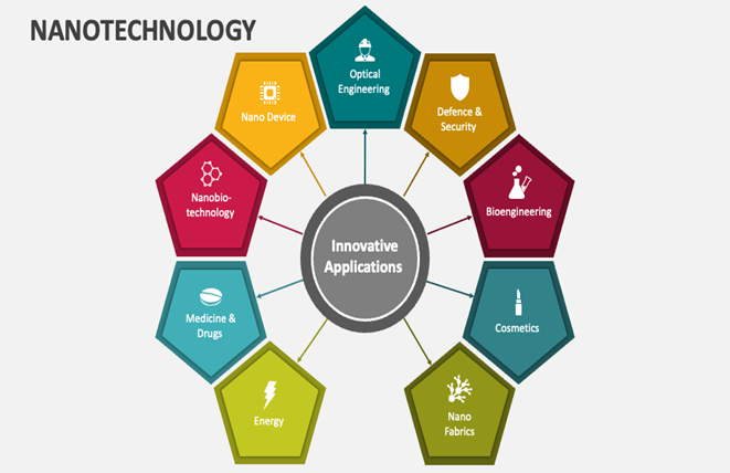
Figure 1: Nanotechnology applications.
Silver nanoparticles
Silver has been harnessed for its antibacterial properties for over 5000 years. As nanoparticles, silver is particularly valued for its ability to combat bacteria without harming human cells. This unique property makes it an ideal material for various applications, including antibacterial activity, industrial purpose, household, and healthcare products, as well as in consumer goods, optical sensors, cosmetics, pharmaceuticals, food industry, diagnostics, orthopedics, and drug delivery systems, medical devices, wound dressings, and even cancer treatment.[21,22]With its growing demand, silver nanoparticles are being increasingly used in pharmaceuticals, healthcare, food, cosmetics, and other industries. Their versatility stems from their ability to alter physical, chemical, and biological properties at the nanoscale, opening up diverse possibilities for exploitation.[23] The ancient Greeks and Romans utilized silver containers to preserve water, food, and wine. Hippocrates employed silver preparations to treat ulcers and aid wound healing. Silver nitrate was also applied for wound care and instrument disinfection. Notably, in 1852, Sims used fine silver wires to suture vesicovaginal fistulas, reducing infection risk and marking a significant medical advancement.[24]Metallic nanoparticles, especially those derived from plant extracts, exhibit remarkable medicinal properties. Silver nanoparticles (AgNPs) stand out due to their stability and low reactivity, making them ideal for biological applications. AgNPs can be synthesized via chemical, physical, or biological methods, each with its pros and cons. While chemical and physical methods require complex purification and high energy, biological approaches offer a sustainable alternative, despite being slower.[25] Silver nanoparticles (Ag NPs) synthesized using silk sericin (SS), a water-soluble protein extracted from silkworms at pH 11, exhibit unique properties due to the presence of hydrophilic proteins with highly polar functional groups. These groups, including hydroxyl, carboxyl, and amino groups, play a crucial role in the reduction of silver ions (Ag+) from AgNO3 to elemental silver (Ag0), forming nanoparticles.[26] The hydroxyl groups in silk sericin are believed to act as reducing agents, facilitating the formation of Ag NPs. Moreover, these groups are thought to form complexes with silver ions, preventing their aggregation or precipitation. This stabilizing effect ensures the formation of stable and dispersed Ag NPs. The work by Aramwit et al highlights the potential of silk sericin as a natural and effective stabilizing agent in the synthesis of Ag NPs, offering a promising approach for the development of biocompatible nanomaterials with diverse applications.[27,28]To comprehensively understand the environmental fate and transport of AgNPs, it's crucial to develop effective methods for preconcentration, separation, and speciation. Additionally, advanced analytical tools are needed to characterize and detect AgNPs in complex environmental samples. Furthermore, thorough monitoring of AgNP behavior under various environmentally relevant conditions is necessary to elucidate their transformation and interactions in the environment.[29] Modern analytical techniques for investigating Ag NPs and nanocomposites encompass a wide range of methods, including microscopic techniques (such as optical, TEM, SEM, STEM, and AFM), spectroscopic methods (like UV-Vis, IR, Raman, NMR, and XRD), spectrometric approaches (including MALDI-TOF MS, SIMS, and ICP-MS), and separation techniques (such as CE, FFF, and gel electrophoresis).[30] This review highlights the capabilities and limitations of these techniques, as well as potential artifacts. The comprehensive coverage of various techniques sets this review apart. Furthermore, the physicochemical and biological properties of Ag NPs are shown to be dependent on fundamental parameters like size, shape, and chemical composition, underscoring the importance of precise characterization.[31] Silver nanoparticles (AgNPs) demonstrate potent antibacterial activity against a broad spectrum of bacteria, including both Gram-negative and Gram-positive strains, as well as multidrug-resistant bacteria.[32] Their unique mechanism of action involves multiple simultaneous pathways, making it challenging for bacteria to develop resistance. When combined with other antibacterial agents or antibiotics, AgNPs exhibit a synergistic effect, enhancing their efficacy against pathogens like Escherichia coli and Staphylococcus aureus.[33] The characteristics of AgNPs make them an attractive option for medical and healthcare applications, where they can effectively treat or prevent infections. Given the growing need for new antibacterial agents, this review focuses on the factors influencing the antibacterial and cytotoxic effects of AgNPs.[34] It also highlights the benefits of using AgNPs in combination with antibiotics, which can reduce the required dosage and minimize associated side effects, offering a promising approach for combating bacterial infections.[35]

Figure 2: Silver nanoparticles.
Synthesis of Silver Nanoparticles:
Nanoparticle synthesis can be broadly classified into physical, chemical, and biological methods, each with distinct mechanisms and applications. Physical methods (top-down) reduce bulk materials to nanoscale dimensions using techniques such as laser ablation, vapor deposition, or ball milling, offering uniform particle size and solvent-free processing but requiring high energy. Chemical methods (bottom-up) build nanoparticles atom-by-atom or molecule-by-molecule through reactions like sol-gel, precipitation, or microemulsion, allowing precise control over size and composition but often involving toxic chemicals. Biological methods (green synthesis) utilize microorganisms or plant extracts as natural reducing and stabilizing agents, producing eco-friendly, biocompatible nanoparticles under mild conditions, making them increasingly attractive for biomedical and sustainable applications. [36,37]
Physical methods:
Physical methods for the synthesis of nanomaterials operate on the fundamental principle of utilizing external physical forces—such as thermal energy, radiation energy, and mechanical pressure—to induce processes like condensation, dissolution, evaporation, or abrasion of bulk materials, ultimately resulting in the formation of nanoparticles.[38] Unlike chemical synthesis routes, these methods are generally considered more environmentally friendly, as they avoid the use of hazardous chemical reagents or solvents. They also minimize pollution and can produce homogeneous nanoparticles with controlled size and morphology. [39]
Laser Ablation:
Laser ablation involves focusing a high-energy pulsed or continuous-wave laser on a silver target, often submerged in a liquid medium. The localized energy deposition leads to plasma formation, causing silver atoms to be ejected from the target surface. These atoms are rapidly cooled and nucleate to form nanoparticles. The choice of liquid medium (e.g., water, ethanol, or surfactant-containing solutions) influences particle stability and prevents aggregation. Parameters such as laser wavelength, pulse duration, and fluence directly impact particle size and morphology. This method is particularly valued for producing ligand-free, contamination-free AgNPs. However, low production rates and the need for high-cost laser systems limit its large-scale adoption.[40]
Thermal Evaporation–Condensation:
The thermal evaporation–condensation technique is a classical top-down approach widely used for the synthesis of metal and alloy nanoparticles, including silver. It is considered one of the earliest physical methods for nanoparticle production. The process involves the thermal evaporation of the target material in either electron beam evaporation devices or Joule-heated refractory crucibles, typically operated under a controlled inert atmosphere at pressures ranging from 1 to 50 mbar. During evaporation, atoms or clusters of the material enter the gas phase and subsequently undergo collisions, leading to cooling and nucleation. This nucleation is followed by condensation into ultrafine nanoparticles. The particle size and distribution are influenced by factors such as evaporation rate, ambient pressure, inert gas type, and temperature gradients within the chamber. [41, 42]
Mechanical Ball Milling:
Mechanical ball milling is a widely used top-down technique for the synthesis of metal nanoparticles, where high-energy collisions between milling media and the bulk material reduce particle size to the nanoscale. The basic principle involves repeated fracture and cold welding of particles through the application of mechanical energy. This method was first introduced by John Benjamin in 1970 to produce fine metal powders and later adapted for nanoparticle synthesis. By reducing particle size, the process also modifies surface properties such as surface energy, reactivity, and catalytic activity.[43] The mechanical milling process can be classified into low-energy milling and high-energy milling, depending on the magnitude of the mechanical energy applied. High-energy mechanical milling involves intense impact forces and is generally more effective for producing ultrafine nanoparticles. The outcome of the milling process depends on both the intrinsic properties of the powder material (e.g., hardness, ductility, brittleness) and process parameters (e.g., milling speed, time, atmosphere).[44]
In this method, the material to be converted into nanoparticles is placed in a sealed container along with solid milling balls—typically made of hardened steel, tungsten carbide, or zirconia—under an inert gas atmosphere (e.g., argon or nitrogen) to prevent oxidation.[45] The ball-to-powder weight ratio is often maintained around 2:1, although it can be adjusted to optimize particle size and yield. The container is rotated at high speed around its central axis, causing the powder particles to be repeatedly trapped and compressed between the milling balls and the container walls. This repeated impact and shear force leads to gradual size reduction and refinement of the particles. [46] Several types of milling equipment are employed, including planetary ball mills, attrition mills, vibrating mills, low-energy rolling mills, and high-energy ball mills. The rotation speed and milling duration are critical factors that directly affect the production rate, crystallite size, and morphology of the nanoparticles. Using optimized parameters, particle sizes in the range of 3–25 nm can be achieved.[47]
Physical Vapor Deposition (PVD):
Physical Vapor Deposition (PVD) is a versatile and environmentally friendly physical method for producing nanomaterials by converting a material from its solid or liquid state into vapor and condensing it onto a cooler substrate, forming thin films or nanoparticle coatings with thicknesses ranging from a few nanometers to several hundred nanometers. The process is typically carried out in a high-vacuum or controlled inert gas environment to prevent oxidation and contamination, ensuring the production of high-purity nanomaterials. Common PVD techniques include resistive heating (thermal evaporation), electron beam evaporation, and sputtering, each offering different advantages in terms of deposition rate, adhesion, and control over nanostructure formation. [48] The high precision and reproducibility of PVD make it particularly suitable for tailoring mechanical, optical, electronic, and catalytic properties of materials. By depositing nanoparticle-based coatings, PVD can significantly improve mechanical hardness, wear resistance, and corrosion resistance in metals, while also modifying their electrical conductivity and optical reflectivity. Control over deposition parameters—such as substrate temperature, vaporization rate, deposition angle, and ambient pressure—allows fine-tuning of particle size, crystallinity, and surface morphology. silver nanoparticles produced via this method are widely used in plasmonic devices, biosensors, and antimicrobial coatings [49]
Laser Pyrolysis:
Laser pyrolysis is an advanced physical–chemical method for synthesizing a wide range of nanoparticles, including magnetic nanoparticles. In this process, a high-power infrared carbon dioxide (CO?) laser irradiates gaseous or aerosolized precursors, causing rapid excitation, decomposition, and quenching to form nanoparticles. The reactants—either in gaseous form or derived from a sprayed salt solution—absorb the laser energy, creating localized high temperatures that drive precursor breakdown. The vaporized species then nucleate, grow, and are cooled to yield nanoparticles.[50] Sulfur hexafluoride (SF?) is often introduced as an inert photosensitizer because it strongly absorbs CO? laser radiation and transfers energy to the reactants, enhancing heating efficiency and particle yield. Critical process parameters, such as laser power, reactant concentration, carrier gas flow, and quenching rate, determine the resulting nanoparticle size, morphology, and crystallinity.
Apart from magnetic nanoparticles, laser pyrolysis has been used to produce molybdenum sulfide (MoS?), silicon carbide (SiC), pure silicon, various metal oxides (e.g., TiO?, Fe?O?), ceramics, ternary nanocomposites, and other composite nanoparticles. Its main advantages include rapid reaction rates, high product purity, and narrow particle size distributions. However, the method requires high-energy laser systems and precise operational control, which may limit large-scale application.[51]
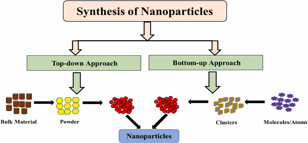
Figure 3: Synthesis of Nanoparticles
Chemical methods:
Microemulsion Techniques:
Microemulsion techniques are widely used for synthesizing uniform and size-controllable silver nanoparticles (AgNPs). In this method, a two-phase aqueous–organic system is employed where the metal precursor and reducing agent are separated in immiscible phases. The reaction takes place at the interface, mediated by a quaternary alkyl-ammonium salt that controls the transport of reactants and influences particle size. Once the reactants meet, silver clusters form and are stabilized by surfactant molecules in the nonpolar medium, preventing agglomeration and ensuring controlled growth. These stabilized nanoparticles are then transferred into the organic phase to form stable dispersions.[52]
A major limitation of this technique is the requirement for large amounts of surfactants and organic solvents, many of which are toxic and difficult to remove. To address this, less harmful solvents like dodecane have been tested to simplify purification. Despite such challenges, microemulsion-prepared nanoparticles offer advantages in practical applications.[53] They are highly stable, well-dispersed, and effective for conductive inks, where good surface wetting is required, as well as for catalytic applications in organic reactions. Thus, while the method provides excellent control over nanoparticle size and stability, further improvements are needed to reduce toxicity and enhance environmental compatibility for large-scale use.[54]
Microwave-Assisted Synthesis:
Microwave-assisted synthesis of silver nanoparticles (AgNPs) has emerged as a rapid and efficient technique since its introduction in the 1940s. This method uses microwave irradiation to provide uniform and instantaneous heating of the reaction medium, leading to faster nucleation and controlled growth of silver nanoparticles.[55] The localized heating effect significantly shortens reaction times compared to conventional heating methods, thereby enhancing the regulation of nucleation and growth phases. As a result, silver nanoparticles synthesized through microwave assistance typically exhibit high crystallinity, uniform morphology, and a narrow size distribution. [56]
Chemical Reduction:
Among the different methods for synthesizing silver nanoparticles (AgNPs), the chemical reduction method is considered the most straightforward, effective, and widely used approach for producing stable nanoparticles with minimal aggregation [57]. This process generally involves three essential components: a metal precursor (commonly silver salts), a reducing agent (to convert silver ions into metallic silver), and a stabilizer or capping agent (to prevent agglomeration and control particle growth). In the presence of a stabilizer, reducing agents efficiently convert silver precursors into silver atoms, which then nucleate and grow into nanoparticles.[58]
A variety of silver precursors such as AgNO?, silver ammonia (Tollens’ reagent) ,silver sulfate, and silver chlorate [59,60] are commonly used as continuous sources of silver ions. Reducing agents like sodium borohydride (NaBH?), hydrazine, trisodium citrate (TSC), ascorbic acid, ethylene glycol, polysaccharides, and formaldehyde are frequently employed depending on the required nanoparticle size and morphology. The final characteristics of AgNPs are strongly influenced by factors such as the type and ratio of reducing agents and precursors, the presence and concentration of stabilizers, as well as reaction conditions including pH, temperature, and ionic strength.[61] By fine-tuning these parameters, the chemical reduction method allows the synthesis of silver nanoparticles with diverse morphologies such as nanospheres, nanoprisms, nanoplates, nanowires, nanocubes, and nanorods, making it a versatile and controllable approach for nanomaterial production.[62]
Sol-Gel Method:
The sol–gel method is a simple way to prepare silver nanoparticles (AgNPs) in colloidal form. In this process, silver nitrate (AgNO?) is used as the silver precursor and glucose (C?H??O?) acts as the reducing agent. Both are dissolved separately in distilled water and then mixed under stirring. This reaction reduces silver ions (Ag?) to metallic silver (Ag?), forming a transparent or colorless colloidal solution of nanoparticles. Different concentrations of silver colloids (0.5–5 μg/ml) can be prepared, and the reaction is represented as:[63]
2AgNO3 + C6H12O6 + H2O → 2Ag0 + C6H12O7 + 2HNO3
To study the prepared solutions, their turbidity, pH, and viscosity were measured. Turbidity showed how stable the colloids were, while pH helped determine the solution’s acidic or basic nature. The viscosity was checked to understand the flow behavior of the sols. Finally, the size and shape of the silver nanoparticles were analyzed using Atomic Force Microscopy (AFM), which gives detailed images of particle surfaces.
Overall, the sol–gel method allows easy and controlled synthesis of silver nanoparticles with adjustable size and stability, making it useful for coatings, catalysis, and biomedical applications.[64]
Hydrothermal Synthesis:
The hydrothermal method is a widely used technique for synthesizing silver nanoparticles (AgNPs) under high temperature and pressure conditions. In this process, a silver precursor such as silver nitrate (AgNO?) is dissolved in water along with a suitable reducing agent and stabilizer.[65] The solution is then sealed in an autoclave (high-pressure reactor) and heated to temperatures typically between 100–250 °C. Under these conditions, silver ions are reduced to metallic silver, leading to the formation of nanoparticles.[66]
The advantages of the hydrothermal method include good control over particle size, shape, and crystallinity, as the reaction environment is uniform and stable. By adjusting parameters such as temperature, pressure, precursor concentration, pH, and reaction time, different morphologies of silver nanoparticles (nanospheres, nanocubes, nanorods, etc.) can be obtained. The method is also relatively eco-friendly, since water is often used as the solvent instead of harmful organic solvents.[67]
Polyol Synthesis:
The polyol method is a popular chemical approach for synthesizing silver nanoparticles (AgNPs). In this process, a silver precursor such as silver nitrate (AgNO?) is dissolved in a polyol solvent (commonly ethylene glycol or diethylene glycol). The polyol plays a dual role: it acts as both the solvent (medium for the reaction) and the reducing agent, converting Ag? ions into metallic silver (Ag?). To prevent particle aggregation and control growth, stabilizers, or capping agents such as polyvinylpyrrolidone (PVP) are often added.
This method offers excellent control over particle size, shape, and distribution, since parameters like reaction temperature, precursor concentration, type of polyol, and amount of stabilizer can be tuned. As a result, a variety of nanostructures such as nanospheres, nanorods, nanocubes, and nanowires can be produced. The polyol method is also advantageous because it yields highly uniform and stable nanoparticles, is relatively simple, and can be scaled up for large-scale synthesis.[68,69]
Biological methods:
The biological method (also called green synthesis) is an eco-friendly approach for producing silver nanoparticles (AgNPs) using natural biological agents instead of toxic chemicals. In this method, microorganisms such as bacteria, fungi, algae, or plant extracts act as both reducing agents (to convert Ag? ions into Ag? nanoparticles) and stabilizers (to prevent aggregation). Plant extracts are used taken from bark, stems, roots, leaves, flowers, oil, fruit peels, seeds, seaweed, citrus lemon zest and microorganisms like fungi, bacteria, and yeast are used as well. Plant-derived biomolecules like proteins, enzymes, polysaccharides, flavonoids, alkaloids, and phenolic compounds play a major role in reducing silver ions and controlling nanoparticle size. This method is considered advantageous because it is simple, cost-effective, non-toxic, and environmentally friendly compared to chemical and physical methods. Moreover, the biological approach can yield nanoparticles with good stability and diverse shapes, making them suitable for applications in medicine, antimicrobial coatings, drug delivery, catalysis, and biosensing. However, the main challenge lies in controlling the size, shape, and uniformity of nanoparticles, since biological systems are often complex.[70]
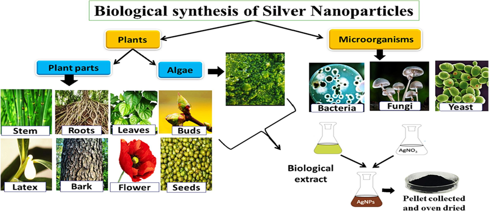
Figure 4: Biological synthesis of silver nanoparticles.
Synthesis of Silver Nanoparticles Using Microorganisms:
The synthesis of silver nanoparticles (AgNPs) using microorganisms has emerged as one of the most promising green nanotechnology approaches in recent years. Microorganisms such as bacteria, fungi, and algae serve as eco-friendly biofactories, producing nanoparticles in a sustainable and cost-effective manner. Microorganisms are naturally equipped with a wide range of biomolecules, including enzymes, proteins, peptides, alkaloids, terpenoids, polysaccharides, and secondary metabolites, which act as reducing and stabilizing agents.[71]
Bacteria-Mediated Synthesis:
Bacteria are widely recognized as efficient biofactories for silver nanoparticle (AgNP) synthesis due to their rapid growth, genetic diversity, and metabolic adaptability. The pioneering work of Klaus et al. (1999) first demonstrated that Pseudomonas stutzeri from a silver mine could reduce silver ions and form intracellular AgNP aggregates, and since then strains such as Lactobacillus acidophilus, Bacillus cereus, Streptomyces sp., Pseudoduganella eburnean, and Bacillus brevis have been reported for AgNP biosynthesis. [72] These nanoparticles typically display spherical or quasi-spherical morphologies with sizes ranging from 8–68 nm and are synthesized either intracellularly, through accumulation within bacterial cells, or extracellularly, via secreted enzymes and metabolites that reduce Ag? ions in the medium. The extracellular route is particularly advantageous for large-scale applications because it simplifies recovery and purification.[73]
Characterization of bacterially synthesized AgNPs is achieved using techniques such as UV–Visible spectroscopy (surface plasmon resonance), XRD (crystallinity), FTIR (capping biomolecules), TEM/SEM/AFM (morphology and surface), DLS (particle size distribution), and SAED (crystal structure). [74] Functionally, these AgNPs exhibit strong antioxidant, antibacterial, antifungal, and antiviral activities, with remarkable effectiveness against multidrug-resistant (MDR) pathogens including E. coli, S. aureus, P. aeruginosa, and K. pneumoniae. This highlights their potential as valuable nanomaterials for biomedical applications, particularly in antimicrobial therapy and nanomedicine.[75]
Fungi-Mediated Synthesis:
Fungi constitute an important group of microorganisms for silver nanoparticle (AgNP) synthesis owing to their large surface area, high tolerance to metals, and ability to secrete abundant extracellular enzymes and proteins. Several fungal species, including Talaromyces purpureogenus, Trichoderma harzianum, Penicillium verrucosum, Fusarium scirpi, and Aspergillus brunneoviolaceus, have been effectively utilized for extracellular AgNP production.[76] Bioactive metabolites such as phenols, flavonoids, alkaloids, and enzymes serve as reducing agents, while proteins and polysaccharides act as stabilizing and capping agents to maintain nanoparticle uniformity and prevent aggregation.
Fungal-mediated AgNPs are generally spherical or quasi-spherical, with particle sizes ranging from 2–90 nm. Unlike bacteria, fungi produce higher quantities of extracellular enzymes, making them particularly advantageous for large-scale nanoparticle biosynthesis with consistent morphology and stability. These nanoparticles display remarkable antibacterial, antifungal, antioxidative, and anticancer activities, underscoring their significance in biomedical and therapeutic applications.[77,78]
Algae-Mediated Synthesis:
Marine and freshwater algae have also emerged as efficient biological systems for the green synthesis of silver nanoparticles (AgNPs). Species such as Chaetomorpha ligustica, Chlorella vulgaris, Gelidium corneum, Noctiluca scintillans, and Botryococcus braunii have been widely reported for AgNP biosynthesis.[79] Algal phytochemicals, including polyphenols, pigments, polysaccharides, and proteins, act as natural reducing agents that convert Ag? to Ag?, while simultaneously stabilizing and capping the nanoparticles to prevent aggregation. Algae-derived AgNPs generally exhibit spherical, cubical, or crystalline morphologies with particle sizes ranging from 2–90 nm.[80] In addition to strong antimicrobial and antioxidant properties, these nanoparticles demonstrate significant photocatalytic and anticancer potential. Their photocatalytic efficiency has been effectively applied in environmental remediation, especially in the degradation of organic pollutants and synthetic dyes from wastewater.[81]
Synthesis of Silver Nanoparticles Using Plants and Plant Extracts
Plants and their extracts have emerged as robust biological systems for the eco-friendly synthesis of silver nanoparticles (AgNPs), owing to their rich reservoir of phytochemicals with reducing and stabilizing potential. The pioneering study by Gardea-Torresdey et al. (2003) demonstrated the ability of Medicago sativa (alfalfa) roots to uptake silver ions, transport them to the shoots, and subsequently assemble them into nanoparticles, thereby laying the foundation for plant-mediated nanotechnology.[82] Since then, diverse plant parts—including leaves, stems, roots, fruits, flowers, seeds, and even agricultural by-products—have been extensively explored for AgNP biosynthesis, making this approach cost-effective, sustainable, and highly scalable.[83]
Plant extracts are rich in phytochemicals such as terpenoids, polysaccharides, phenolics, alkaloids, flavonoids, amino acids, proteins, enzymes, vitamins, and pigments, which act simultaneously as reducing and stabilizing agents during nanoparticle formation.[84] Several reports highlight the efficiency of plant extracts in producing stable, monodispersed AgNPs. For example, Ahmad and Sharma (2012) synthesized spherical AgNPs (~12 nm) using Ananas comosus (pineapple juice) as both a reducing and stabilizing agent, while Singh et al. (2010) reported ~30 nm AgNPs using Argemone mexicana leaf extract.[85] Similarly, extracts of neem (Azadirachta indica), triphala, papaya (Carica papaya), and Ocimum sanctum have been effectively employed to produce stable, predominantly spherical AgNPs with sizes ranging between 5–60 nm.[86]
Characterization of plant-mediated AgNPs has been extensively carried out using a range of analytical techniques to confirm their structural, morphological, and functional attributes. UV–Visible spectroscopy is employed to detect the characteristic surface plasmon resonance (SPR) peaks, while X-ray diffraction (XRD) confirms crystallinity and often indicates a face-centered cubic geometry. Transmission and Scanning Electron Microscopy (TEM/SEM) provide insights into nanoparticle morphology and size distribution, whereas Fourier Transform Infrared Spectroscopy (FTIR) identifies functional groups from plant metabolites that participate in reduction and capping processes. Additional methods such as Energy-Dispersive X-ray Spectroscopy (EDX), Atomic Absorption Spectroscopy (AAS), and Nanoparticle Tracking Analysis (NTA) further validate elemental composition, stability, and dispersion. Collectively, these analyses demonstrate that plant-derived AgNPs are predominantly crystalline, spherical, and stable.[87,88,89]
A wide diversity of plant species has been successfully utilized for nanoparticle biosynthesis, including Ficus benghalensis, Jatropha curcas, Cassia auriculata, Polyalthia longifolia, Malus domestica, Chrysanthemum indicum, Rumex hymenosepalus, Parthenium hysterophorus, and Acalypha indica.[90] Extracts from these plants typically yield nanoparticles in the size range of 10–70 nm, although variations depend on crucial synthesis parameters such as extract concentration, pH, temperature, and incubation time (Mittal et al., 2013). Comparative studies indicate that plant-extract-mediated AgNPs exhibit potent antifungal, antibacterial, antioxidant, and anticancer activities—often demonstrating equal or superior efficacy to chemically synthesized nanoparticles.[91]
The major advantages of employing plant extracts lie in their abundance, eco-friendliness, and safety in handling, coupled with the inherent richness of phytochemicals that act as both reducing and stabilizing agents. Moreover, this synthesis pathway is rapid and often proceeds efficiently under ambient conditions, enabling nanoparticle formation within a short time span without the need for hazardous reagents. Overall, plant-mediated synthesis represents a simple, sustainable, and scalable platform for generating biocompatible nanomaterials with immense potential in medicine, agriculture, and environmental remediation.[92,93]
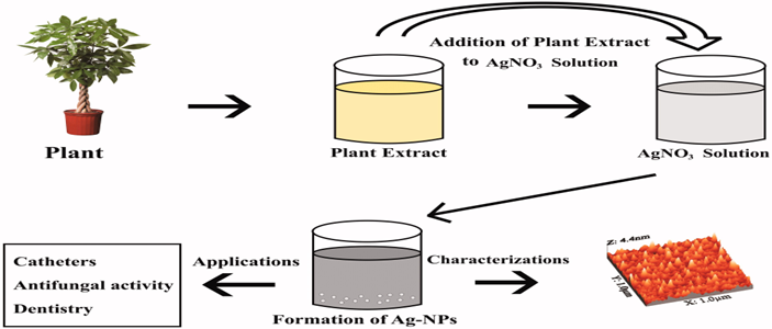
Figure 5: Schematic diagram for synthesis of Ag-NPs by using plant /plant extracts.
Characterization of Silver Nanoparticles
Characterization is a vital step in the green synthesis of silver nanoparticles (AgNPs) as it helps to determine their morphology, size distribution, surface chemistry, crystallinity, and stability. Proper characterization confirms the successful formation of nanoparticles and provides insights into their functional properties. Several analytical techniques, including UV–Visible spectrophotometry, X-ray diffraction (XRD), Fourier-transform infrared spectroscopy (FTIR), Transmission and Scanning Electron Microscopy (TEM/SEM), and Energy Dispersive X-ray Spectroscopy (EDX), are commonly employed to study the structural and optical features of AgNPs. These techniques together provide a reliable understanding of nanoparticle characteristics, ensuring their suitability for diverse applications.
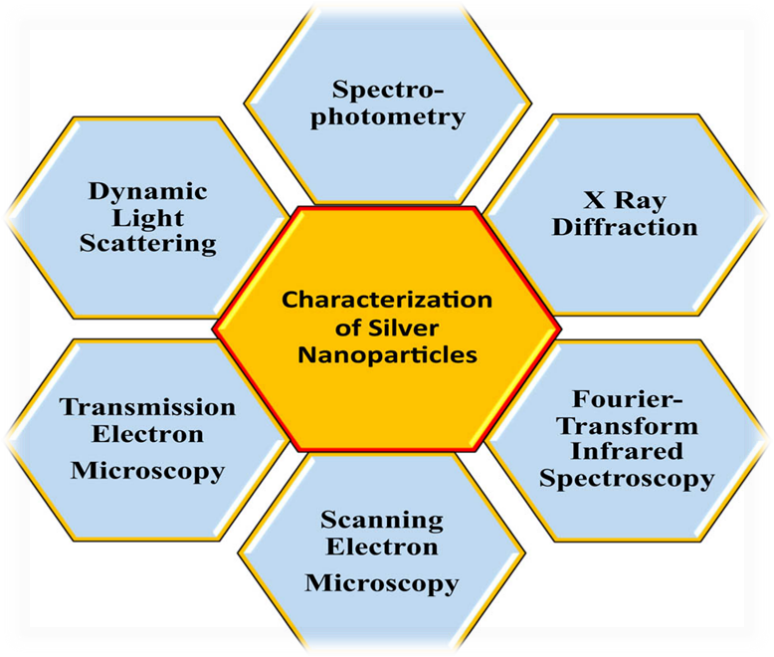
Figure 6: Various techniques used for characterization of silver nanoparticles.
UV–Visible Spectrophotometry
UV–Visible (UV–Vis) spectrophotometry is one of the most widely employed techniques for the characterization of metallic nanoparticles, particularly for monitoring their synthesis and stability. The reduction of metallic salts into nanoparticles generates a distinct absorption peak in the visible region, which serves as an important indicator of nanoparticle formation.[94] Typically, nanoparticles in the size range of 2–100 nm exhibit absorption bands between 200–800 nm, making this wavelength range most suitable for their characterization. In the case of silver nanoparticles (AgNPs), the proximity of valence and conduction bands allows free electron oscillation, giving rise to a characteristic surface plasmon resonance (SPR) band.[95] The position and intensity of the SPR peak are strongly influenced by particle size, dielectric medium, and surrounding chemical environment. Numerous studies have shown that the stability of biologically synthesized AgNPs can be reliably assessed through UV–Vis spectrophotometry, with stable samples retaining their SPR peak even after extended storage, sometimes up to 12 months.[96]
X-ray Diffraction Analysis (XRD)
X-ray diffraction (XRD) is a widely employed analytical technique for determining the crystalline structure of metallic nanoparticles. When X-rays penetrate deeply into the material, they produce a characteristic diffraction pattern that confirms nanoparticle formation and crystallinity. The particle size can be calculated from XRD data using the Debye–Scherrer equation: d = Kλ / β cos θ, where d is the particle size, K the Scherrer constant, λ the X-ray wavelength, β the full width at half maximum, and θ the diffraction angle. XRD not only provides information on nanoparticle size and geometry but is also extensively used to study structural properties of biomolecules, polymers, glasses, superconductors, and various nanomaterials, making it a powerful tool for nanotechnology research.[97,98]
Field Emission Scanning Electron Microscopy (FE-SEM)
Field Emission Scanning Electron Microscopy (FE-SEM) is a powerful technique used to analyze the surface morphology, size, and shape of silver nanoparticles with high resolution. Prior to analysis, samples are typically centrifuged, washed, and dried to remove impurities, then mounted on a conductive substrate (such as a platinum or palladium-coated mesh). FE-SEM provides detailed surface images by scanning the sample with a focused electron beam, while complementary techniques such as Energy Dispersive Spectroscopy (EDS) and elemental mapping further confirm the elemental composition and distribution of nanoparticles.[99]
Transmission Electron Microscopy (TEM)
Transmission Electron Microscopy (TEM) is a highly advanced technique widely employed in nanoscience for determining the size, morphology, and structural details of silver nanoparticles. In this method, a beam of high-energy electrons passes through an ultrathin sample, generating high-resolution images at the nanometer scale. For AgNP analysis, dried nanoparticle precipitates are typically dispersed in ethanol, sonicated to achieve uniform suspension, and a drop is placed on a carbon-coated copper grid. After drying, the thin transparent film allows electrons to pass through, producing images that reveal particle shape, size distribution, and crystalline features with exceptional clarity.[100]
Scanning Electron Microscopy (SEM)
Scanning Electron Microscopy (SEM) is a widely employed surface imaging technique that enables detailed investigation of nanoparticles at the micro- and nanoscale. By utilizing a highly energetic electron beam (1–30 keV), SEM provides valuable information on particle morphology, size, size distribution, and surface texture.[101] Modern high-resolution SEM instruments can even resolve nanoparticles smaller than 10 nm. The imaging is primarily based on backscattered electrons (BSEs), which offer insights into both surface features and nanoparticle interactions with supporting materials.[102]
SEM has been extensively applied to study silver nanoparticle (AgNP) deposition on different substrates, such as silica spheres, polyester fibers, and linen, providing clear evidence of their distribution and morphology. Advanced modifications, such as Focused Ion Beam (FIB)-SEM, allow cross-sectional imaging, while Environmental SEM (ESEM) reduces charging effects in non-conductive biological samples by maintaining water vapor inside the chamber, preventing dehydration and structural changes.[103]
Despite its wide utility, SEM has limitations such as charging issues in non-conductive samples, lower spatial resolution, and a reduced signal-to-noise ratio compared to Transmission Electron Microscopy (TEM). Nonetheless, SEM remains one of the most versatile and reliable techniques for analyzing nanoparticle morphology and surface characteristics in nanomaterials research.[104]
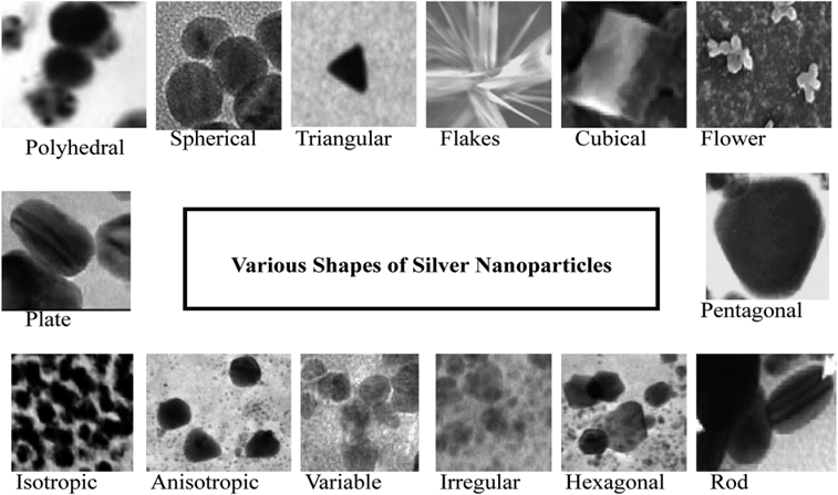
Figure 7: SEM images of AgNPs synthesized from different sources.
Dynamic Light Scattering (DLS)
Dynamic Light Scattering (DLS) is a widely used characterization technique to determine the hydrodynamic size distribution and polydispersity index (PDI) of nanoparticles in suspension. In the present study, the average size of silver nanoparticles (AgNPs) synthesized using trisodium citrate was found to be 58.3 nm, which lies within the typical nanoparticle size range of 1–100nm.[105] DLS works by analyzing fluctuations in the intensity of scattered light caused by the Brownian motion of particles in a colloidal medium, providing accurate insights into nanoparticle size and dispersion stability. The particle size is highly dependent on synthesis parameters, including precursor concentration and reducing agent ratio.Furthermore, the size distribution determined by DLS complements other microscopic techniques, confirming the heterogeneous nature of AgNPs and their stability in suspension.[106]
Applications of Silver Nanoparticles
Silver nanoparticles (AgNPs) are among the most widely studied nanomaterials due to their unique physical, chemical, and biological properties. Their nanoscale size imparts a high surface-to-volume ratio, enhanced reactivity, and strong surface plasmon resonance, which make them highly versatile. One of the most prominent applications of AgNPs is in the biomedical field, where they exhibit excellent antimicrobial, antiviral, antifungal, and anti-inflammatory properties. They are extensively used in wound dressings, coatings for medical devices, drug delivery systems, and diagnostic tools. Beyond medicine, AgNPs are applied in food packaging to prevent microbial contamination, in textiles for producing antibacterial fabrics, and in water treatment for their disinfection capabilities. Additionally, their unique optical and electrical properties make them valuable in sensors, catalysis, solar cells, and other electronic applications. This wide spectrum of applications highlights the importance of AgNPs as multifunctional nanomaterials in both healthcare and industry. [107]
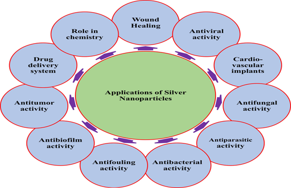
Figure 8: Different applications of silver nanoparticles
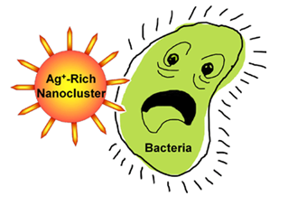
Antibacterial Properties
Silver nanoparticles (AgNPs) effectively inhibit a wide range of bacteria, fungi, and viruses, including Staphylococcus aureus, E. coli, Pseudomonas aeruginosa, dermatophytes, and HIV-1. They generally show stronger activity against Gram-negative bacteria due to thinner cell walls.[108] The antibacterial effect of AgNPs depends on size, shape, concentration, exposure time, and surface charge. Smaller particles (<10 nm) exhibit stronger activity, with spherical shapes showing the best performance due to larger surface-to-volume ratios. Surface charge also plays a role, as positively charged AgNPs adhere more easily to negatively charged bacterial membranes. Stabilizers like citrate and PVP further influence particle characteristics and antibacterial activity. [109]
Wound Healing
Silver nanoparticles (AgNPs) exhibit potent antibacterial properties, making them highly effective in preventing wound infections and promoting faster recovery. Their nanoscale size allows deeper penetration into the dermis, which not only accelerates tissue repair but also reduces the risk of scarring. Wound healing is a complex process involving sequential stages—inflammation, cell proliferation, extracellular matrix formation, and tissue remodeling—all of which can be positively influenced by AgNPs.[110] Studies report that AgNP-based dressings enhance burn wound healing and support fibroblast activity without inhibiting the proliferation of keratinocytes, which are essential for skin regeneration. In addition to their intrinsic antimicrobial effect, AgNPs demonstrate a synergistic action when combined with antibiotics such as tetracycline, leading to a greater reduction in bacterial load compared to either treatment alone. This combined therapy also helps address the challenge of antibiotic resistance. Furthermore, AgNPs have been shown to stimulate angiogenesis, collagen deposition, and growth factor release, all of which are crucial for effective tissue repair. These findings highlight the potential of AgNPs as innovative wound care agents, particularly in the management of chronic wounds, burns, and other infection-prone injuries.[111]
Dental Materials
The oral cavity is highly susceptible to microbial colonization, leading to contamination of dental materials and implants (Pokrowiecki, 2017). Silver-based nanostructures are widely incorporated in dentistry due to their strong antibacterial effects (Zhang, 2016). Their small size increases surface area, enhancing antimicrobial efficiency (Santos, 2013). AgNPs have been effectively added to adhesive resins, orthodontic cements, and dental composites, reducing biofilm formation and improving the durability of restorations. [112]
Antiviral properties
Silver nanoparticles (AgNPs) show promise as antiviral agents, inhibiting the growth and viability of various viruses, including hepatitis B virus (HBV), human immunodeficiency virus (HIV), and influenza virus. While the exact mechanism of their antiviral action remains unclear, research suggests that AgNPs may interfere with viral replication, particularly at early stages. Studies have demonstrated the effectiveness of AgNPs in reducing viral titers and preventing infection in various cell lines. Further research is needed to fully understand the antiviral mechanisms of AgNPs and explore their potential applications.[113]
Antifungal properties
Silver nanoparticles (AgNPs) demonstrate significant antifungal activity against a wide range of fungal species, including Candida albicans, Aspergillus fumigatus, and Fusarium oxysporum. The proposed mechanism of action involves disrupting the fungal cell membrane, causing damage and ultimately inhibiting growth. Notably, AgNPs have shown potent antifungal activity when combined with fluconazole, enhancing their efficacy. Green-synthesized AgNPs have also been found to suppress conidial germination and display potent antifungal activity against various fungal pathogens, including plant pathogens like Fusarium oxysporum. Furthermore, carbon nanoscrolls composed of AgNPs and graphene oxides exhibit intense antifungal activity, highlighting the potential of AgNPs as a promising solution for developing biocompatible, non-toxic, and environmentally friendly antifungal agents.[114]
Anti inflammatory properties
Silver nanoparticles (AgNPs) exhibit anti-inflammatory properties by reducing the production of pro-inflammatory cytokines and inflammatory markers. Studies have shown that AgNPs can significantly decrease colonic inflammation in rats and slow down the production of inflammatory markers during wound healing. Additionally, biologically synthesized AgNPs can inhibit UV-B-induced cytokine production and reduce edema and cytokine levels in tissues, highlighting their potential as an effective anti-inflammatory agent.[115]
Anti-cancer properties
Silver nanoparticles (AgNPs) show promise in cancer treatment by inducing apoptosis and sensitizing cancer cells. They induce cell death in a concentration-dependent manner, causing changes in cell morphology, reducing metabolic activity and cell viability, and accumulating reactive oxygen species (ROS). AgNPs can target specific cancer cells when coated with starch or delivered through carrier molecules like chitosan, exhibiting selective cell-specific toxicity in various cancer cell lines, including breast cancer, leukemia, and lung cancer cells. Biologically synthesized AgNPs, using plant extracts or bacterial/fungal extracts, also demonstrate significant cytotoxicity in cancer cells, highlighting their potential as a targeted cancer therapy.[116]
RESULTS
The literature confirms that silver nanoparticles (AgNPs) can be synthesized through physical, chemical, and biological methods, each influencing their size, morphology, and stability. Chemical reduction remains the most widely used method due to its simplicity and control over particle size (5–100 nm). Physical methods produce high-purity nanoparticles but require expensive instrumentation. Biological (green) synthesis using plant extracts and microorganisms offers an eco-friendly and biocompatible alternative.
Characterization techniques such as UV–Visible spectroscopy (SPR peak around 400–450 nm), XRD, FTIR, SEM, TEM, and DLS confirm the formation of crystalline, predominantly spherical AgNPs. Reported studies demonstrate strong antimicrobial activity against Gram-positive and Gram-negative bacteria, along with promising applications in wound healing, dental materials, and drug delivery systems.
DISCUSSION
The biological and pharmaceutical performance of AgNPs is strongly size-dependent, with smaller nanoparticles exhibiting enhanced antimicrobial activity due to increased surface area and reactive oxygen species (ROS) generation. The synthesis method significantly affects nanoparticle stability and cytotoxicity. While chemical methods provide better size control, green synthesis improves biocompatibility.
Despite their broad biomedical potential, concerns regarding toxicity, bioaccumulation, and long-term safety require further investigation. Standardization of synthesis protocols and comprehensive toxicological evaluation are essential for safe clinical and pharmaceutical applications.
CONCLUSION
Silver nanoparticles (AgNPs) have emerged as a promising nanomaterial with diverse applications in the biomedical field. The various synthesis methods, including physical, chemical, and biological approaches, offer a range of options for producing AgNPs with tailored properties. The characterization techniques, such as SEM, TEM, X-ray, FTIR, and spectrophotometry, provide valuable insights into the size, shape, and composition of AgNPs. The biological applications of AgNPs are vast and impressive, with significant potential in anticancer, antibacterial, anti-inflammatory, antifungal, antiviral, and wound healing therapies. The unique properties of AgNPs, including their high surface area and reactivity, make them an attractive option for developing novel therapeutic agents. Further research is needed to fully explore the potential of AgNPs and to overcome the challenges associated with their use in biomedical applications. Nevertheless, the existing evidence suggests that AgNPs are a promising area of research with significant potential for improving human health and well-being.
ACKNOWLEDGEMENT
The authors express their sincere gratitude to Koringa College of Pharmacy for providing the necessary academic support and facilities to carry out this review work. The authors are especially thankful to Mrs. K. Jyothsna for her valuable guidance, encouragement, and continuous support throughout the preparation of this manuscript. No external funding was received for this study.
REFERENCES


K. Nikitha, K. Sai Seetha Lakshmi Harika, K. Jyothsna, An Overview of the Silver Nanoparticles: Synthesis Methods, Characterization Techniques, and Biomedical Applications – A Comprehensive Review, Int. J. of Pharm. Sci., 2026, Vol 4, Issue 2, 3333-3358. https://doi.org/10.5281/zenodo.18716112
 10.5281/zenodo.18716112
