
We use cookies to ensure our website works properly and to personalise your experience. Cookies policy
Ashokrao Mane Institute of Pharmacy, Ambap, Maharashtra, India.
It has long been common practice to employ animal models in scientific study and medicine development. However, a quest for other strategies has been spurred by ethical issues, animal welfare, and the growing need for more precise and efficient testing techniques. Several possible substitutes for animal testing have surfaced in recent years, including as 3D bioprinting, organ-on-chip technologies, in vitro cell-based tests, and computer modeling. By minimizing the need for animal testing and increasing the relevance and repeatability of results, these alternatives have the potential to provide more accurate models of human physiology and illness. High-throughput drug screening is possible in vitro utilizing human-derived cell lines, while organ-on-chip technologies provide a more realistic representation of human reactions by simulating intricate tissue interactions. Furthermore, sophisticated computer models are able to forecast.
The utilization of animals for a multitude of purposes, including sustenance, transportation, companionship, sports, and recreation, has been a practice as ancient as humanity itself. Among these various applications, the employment of animals in research represents a significant extension of their use. A diverse array of species, such as mice, rats, hamsters, rabbits, various fish (including zebra fish and trout), birds (predominantly chickens), guinea pigs, amphibians (notably xenopus frogs), as well as primates, dogs, and cats, have been integral to research endeavors for an extensive period. (1) Each year, millions of these experimental animals are utilized globally.(2) In clinical research laboratories, animals are often separated from their social groups and utilized as experimental subjects, disregarding their inherent behaveiors. These experiments may involve the use of entire animals or specific organs and tissues. To facilitate this research, animals are typically euthanized using established protocols. Frequently, those animals that survive the testing phase are also euthanized afterward to prevent any potential suffering.(3) In certain experimental scenarios, such as LD 50 testing, animals may die as a direct consequence of the procedures. The suffering, distress, and mortality that animals endure during scientific research have sparked ongoing ethical debates. Critics argue that as sentient beings, animals possess rights that protect them from pain and suffering, leading to the conclusion that their use in experimentation is unethical and should be abolished.(4)Currently, numerous regulations and laws are in place globally to safeguard animals from cruelty and exploitation. Organizations such as the International Conference on Harmonization (ICH), the Committee for the Purpose of Control and Supervision of Experiments on Animals (CPCSEA), the National Institutes of Health (NIH), and the Organization for Economic Cooperation and Development (OECD) offer guidelines on animal care, including aspects like housing, breeding, feeding, transportation, and their application in scientific research.(4)Alternative approaches to animal research have been proposed as an effective strategy to circumvent unethical practices involving animals and to enhance the humane aspect of scientific experimentation.(5)In contemporary research, various alternative methods have emerged to supplant animal testing in many prevalent biomedical domains,(6-7) such as cell-based cytotoxicity assessments, genotoxicity evaluations, and biochemical assays. These non-animal strategies offer faster, more efficient, and cost-effective chemical safety evaluations, serving as viable substitutes for conventional animal experiments. Among the alternatives to animal testing are chemical-based assays, in vitro cell culture techniques, in silico computational biomodeling, and ex vivo tests utilizing tissues from deceased animals. (8)In contrast to ex vivo methodologies, tissue engineering presents a more ethical solution for potentially replacing animal models.(9)
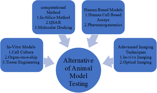
In this article, we explore a range of alternative approaches to animal model testing, including the 3R system, in silico methods, QSAR models, molecular docking, and cell-based techniques. Additionally, computational methods, in vitro studies, omics technologies, organ-on-a-chip innovations, high-throughput screening (HTS), and mathematical biology can all contribute valuable complementary insights.(10)
Three Rs: reduction, refinement and replacement
The 3Rs are a set of principles for the ethical use of animals in scientific research and product testing:
This framework encourages researchers to minimize the number of animals used in experiments, emphasizing 'reduction' in overall animal usage. It is essential to meticulously plan and 'refine' the use of animals to ensure that any pain and distress experienced during the experiments are kept to a minimum.(11,12)
ALTERNATIVE METHODS
Several approaches have been proposed to eliminate the use of animals in research. These alternatives offer different ways to conduct drug and chemical testing to a certain extent. The benefits of these methods include increased efficiency in time management, reduced manpower requirements, and cost savings. A detailed description of these methods is provided below.
IN VITRO MODELS
CELL BASED METHOD
Cell-based assays, referred to as in vitro methods, are widely employed to evaluate the safety and toxicity of pharmaceuticals and chemicals as alternatives to animal testing.(13)Cells and tissues from various organs such as the liver, kidney, brain, and skin are extracted from animals and can be maintained outside the body in appropriate growth media for periods ranging from a few days to several years. The in vitro culture of animal or human cells involves isolating them and cultivating them as a monolayer on the surfaces of culture plates or flasks. Additionally, cellular components such as membrane fragments and enzymes can also be utilized. Different culture types, including cell culture, callus culture, tissue culture, and organ culture, serve various research purposes. The advantages of these techniques include ease of use, reduced time requirements, and lower costs. These methodologies are commonly applied for the initial screening of potential drug candidates and chemicals to assess their toxicity and effectiveness.(14)
STEM CELL TECHNOLOGY
At present, cell-based evaluations are employed in the management of several significant conditions, including cardiovascular, neurological, ophthalmologic, skeletal, and autoimmune disorders, as well as for assessing the toxicity linked to these treatments.(14)Human embryonic stem cell (ESC) research was initially documented in 1998,(16) demonstrating that cells derived from pre-implantation embryos possess pluripotent characteristics, enabling them to differentiate into a variety of cell types. Consequently, human ESCs are considered a potential resource for advancing tissue engineering.(17) However, there are ongoing ethical and religious debates surrounding the use of human ESCs in toxicological research and experimental applications.(18-19)The ethical concerns primarily stem from the destruction of human embryos,(20)prompting researchers to explore alternative sources of stem cells to mitigate these ethical dilemmas.(21)
ORGAN-ON-CHIP
The most notable "animal alternative" to support pre-clinical drug development is the Organs on Chips (OOC) technology, which was developed by researchers from Harvard University and the University of Pennsylvania.(22)OOCs are sophisticated micro-engineered biomimetic systems that feature microfluidic channels lined with living human cells. These systems effectively replicate essential functional units of living organs, thereby reconstituting integrated human organ-level pathophysiology in vitro.(23)Even though they have linked 4-, 7-, and 10-organ cultures in a microfluidic system with different subcircuits and adjustable flow rates, they have only shown that individual organs can survive for a long time so far.(23)
Human-derived three-dimensional tissue models:
In vitro models for studying skin pathophysiology and conducting drug testing have been established for quite some time. Early innovations in testing human skin equivalents (HSE) featured EpiDerm(24) and full-thickness EpiDerm.(25) Currently, HSE models encompass a wide variety, serving purposes from illustrating basic physiological functions to investigating complex model diseases, including autoimmune conditions and cancers.(26-27)Dependent on standardization and quality, these models could surpass animal models. This advantage is partly due to the fact that the initial skin samples are derived from humans. Furthermore, these tissue models are cultivated in vitro within a biochemical and physiological environment that closely mimics human homeostatic conditions.Applying the principle of replacement proposed by Russel and Burch,(28) numerous human-derived three-dimensional models have been developed, evaluated, validated, and utilized.(29)
These three-dimensional in vitro models offer not only an ethical advantage but also a significant pathophysiological relevance due to their human tissue origin. This relevance is evident as the tissue samples are derived from humans and cultivated in an in vitro environment that closely mimics human biochemical and physiological conditions. Furthermore, animal models, such as commonly used inbred murine models, exhibit considerably fewer biological and genetic variations compared to the intricate genetic and biological diversity found in humans, thereby enhancing the physiological relevance of human tissues. The three-dimensional models derived from human tissues encompass various types, including oral epithelial,(30) gastrointestinal epithelia,(31) vaginal epithelia,(32)ocular tissue,(33)gingival tissue,(34)respiratory epithelia,(35) and dendritic antigen-presenting cells.(31)
TISSUE ENGINEERING
Tissue engineering is an interdisciplinary domain that integrates principles from engineering, materials science, medicine, and biology.(36) The objective of this approach is to effectively create optimal biopolymer-based three-dimensional organ structures suitable for nanoparticle toxicity evaluations, ultimately aiming to substitute animal testing.(37)
The construction of 3D organ structure models necessitates the incorporation of either synthetic or natural biological materials along with stem cells to promote cell growth and differentiation. This integration supports essential cell-to-cell interactions and establishes suitable signaling pathways, enabling multidirectional growth throughout the cell culture process.(38) Tissue engineering has been utilized to create 3D culture environments as a substitute for traditional animal testing methods in toxicity assessments.(39-40) These three-dimensional cell culture systems serve as a viable alternative to animal testing, offering a cost-effective and time-efficient approach to tissue development.(41)
COMPUTATIONAL METHOD
IN SILICO METHOD
In silico techniques encompass computer-based methodologies and simulations designed to model, forecast, and examine biological processes, behaviors, or impacts in both animals and humans. The constraints associated with animal research have led to the creation of these computational models. In silico techniques play a significant role in the realm of animal testing by providing alternative methods for evaluating biological responses. One prominent approach is computational modeling, which employs mathematical frameworks to replicate intricate biological systems, processes, or organs, enabling predictions regarding the behavior of various substances or diseases. In silico modeling represents an emerging field that integrates experimental methodologies, offering a robust technique for elucidating mechanisms at the atomic scale.(42-43) Computers play a crucial role in enhancing our understanding of fundamental biological principles. Specialized software for computer modeling is instrumental in the design of new pharmaceuticals. Simulations generated by computers can forecast the potential biological and toxicological effects of chemicals or drug candidates, thereby eliminating the need for animal testing in initial assessments. Only the most promising compounds identified through preliminary screening proceed to in vivo studies. For instance, determining the receptor binding site of a drug necessitates in vivo experimentation. Computer Aided Drug Design (CADD) software is employed to predict the binding sites for prospective drug molecules, effectively identifying likely interaction points and minimizing the testing of inactive compounds. Furthermore, these software tools enable the customization of new drugs for specific binding sites, with final animal testing conducted to validate the results.(44)
The strength of in silico models lies in their ability to generate predictions based solely on the chemical structures of the substances being studied. These models operate on the principle that a chemical's inherent properties, possible interactions, and eventual impacts are embedded within its molecular architecture. This understanding facilitates the creation of quantitative structure–activity relationship ((Q)SAR) and quantitative structure–property relationship ((Q)SPR) models. It is anticipated that chemicals with similar structures will produce comparable effects, allowing insights gained from one chemical or a related group to inform predictions about the characteristics of analogous substances.(45)Additionally, advanced computer modeling programs now enable the screening of pathophysiological simulations,(46)while toxicity assessments(47)and essential pharmacokinetic processes—such as intestinal absorption, protein binding, and passage through endothelial barriers—can be efficiently conducted in vitro, contingent upon the availability of specific in silico modeling tools.(48)Many models and numerous software tools for forecasting ADME characteristics and biological functions are currently accessible.
QSAR
QSAR-based methodologies can significantly minimize animal testing through three primary approaches: category formation (including read-across), endpoint prediction, and hypothesis generation to guide testing. Generally, chemicals that exhibit similar structural characteristics are likely to interact in comparable ways with biological systems, which justifies the categorization of structurally analogous chemicals.(49) The effectiveness of a QSAR model is influenced by several factors, including the quality of biological data, the selection of descriptors, and the statistical methods employed.
The potential drug candidate's carcinogenicity and mutagenicity can be effectively predicted using computer databases. Recent advancements in QSAR software have demonstrated improved accuracy in forecasting the carcinogenic potential of various molecules. The benefits of utilizing computer models over traditional animal testing include faster results and lower costs.(50)QSAR methodologies have found applications across numerous scientific fields. For example, the QSAR framework is commonly employed in risk assessments, toxicological studies, regulatory decision-making, and chemical safety evaluations. Consequently, in silico methods hold promise for replacing animal testing due to their capacity to statistically and reliably assess the potential hazards of chemical substances, simulating conditions that closely resemble human biological systems.(51)
Nonetheless, the quantification of QSAR limitations remains insufficiently addressed, posing a challenge for professionals in the field.The applicability limitations of a QSAR model can be categorized into three key areas: overall quality, applicability domain, and potential for chance correlation. (52)A common challenge in QSAR model development is the presence of outliers—compounds exhibiting unexpected biological activity that do not conform to the model. These outliers are crucial for understanding the boundaries within which compounds operate under a shared molecular mechanism, as well as for identifying the experimental constraints of the biological data.(53)
MOLECULAR DOCKING
Molecular docking studies represent a valuable method for computational simulations and assessing interactions between chemicals and biomolecules, leveraging three-dimensional structural data. The initial phase of a docking study involves generating all potential conformations and orientations of each ligand, tailored to fit the specific shape of the designated binding site within the protein structure. The subsequent phase employs scoring functions to estimate the likelihood of favorable interactions between the protein and the docked ligand, as well as to evaluate various orientations. Following the docking procedure, the scores derived from these functions are utilized to rank each ligand based on its fit within the binding site, ultimately identifying the ligand with the highest affinity for the target protein. A favorable docking score indicates that the molecule exhibits beneficial intermolecular interactions, such as hydrogen bonds, electrostatic forces, and hydrophobic interactions, suggesting it is a strong candidate as a binder. The docking process involves positioning rigid chemical compounds into the active site of the protein crystal structure, which is sourced from the RCSB protein databank. Typically, the precision and efficiency of the docking conformations are closely linked to the search algorithms employed, with each docking application relying on a specific conformational search algorithm, such as the genetic algorithm (GA).(54)
Molecular docking serves numerous purposes in the realm of drug discovery, encompassing structure–activity relationship studies, lead optimization, the identification of potential candidates through virtual screening, and the formulation of binding hypotheses to enhance predictions for mutagenesis research. It also plays a crucial role in aiding x-ray crystallography by assisting in the alignment of substrates and inhibitors with electron density, as well as contributing to studies on chemical mechanisms and the design of combinatorial libraries.(55)While docking technology has reached a level of maturity, it still has significant room for improvement. Most existing docking software can predict known binding poses with an average accuracy of approximately 1.5–2 Å, achieving success rates between 70% and 80%.However, a key challenge in molecular docking remains the accurate calculation of binding energies, which is closely linked to the various approximations made during the docking process, such as solvent treatment and molecular flexibility.(56)
HUMAN BASED MODELS
HUMAN CELL BASED ASSAYS
High-throughput screening (HTS) is a method that involves the rapid testing of a vast array of chemical compounds to efficiently identify small molecules that exhibit biological activity. This process aims to discover lead compounds that can advance through the drug discovery and development stages in therapeutic applications or serve as tool molecules for investigating biological phenomena in fundamental research.(57) The ability to rapidly and cost-effectively produce thousands of new compounds sparked significant enthusiasm regarding the future of drug discovery, which in turn propelled the advancement of high-throughput screening (HTS) technologies designed to assess the vast array of new compounds generated through combinatorial chemistry. Concurrently, significant strides in genomics unveiled numerous potential drug targets. However, the absence of well-documented "druggability" and structural data for many of these novel genomic targets(58)led to HTS emerging as the preferred approach for identifying small-molecule modulators from the expanding libraries of compounds within the pharmaceutical sector.(59-60)
It is essential to assess a wide array of experimental conditions to identify the optimal signal-to-background ratios. In the context of cell-based assays, optimization encompasses various factors, including the titration of cell density, assay reagents, and the ideal concentrations of modulators for screening, as well as the appropriate incubation duration with the compounds. An important early step in this process is miniaturization, which aims to preserve effective signal detection and satisfactory signal-to-background ratios while significantly reducing the volume of reactions, the quantity of reagents, and the number of cells used. Additionally, ensuring reproducibility is vital; variations across wells, plates, days, and batches (whether involving proteins or cells) must be evaluated using positive controls and DMSO-only controls when applicable.(61)
PHARMACOGENOMICS
Pharmacogenomics involves utilizing genome-wide techniques to identify genetic factors that influence drug responses and to create innovative therapies. By analyzing the gene expression profiles associated with specific disorders, targeted treatments can be developed, resulting in improved effectiveness and minimized side effects.(62)Recent progress in high-throughput biological data generation, along with advancements in computational capabilities and bioinformatics, has significantly changed the landscape of pharmacogenomic research. In situations where human studies are not feasible due to ethical or practical constraints, animal models serve a crucial role in these investigations.
ADVANCED IMAGING TECHNIQUES
IN VIVO IMAGING
The advancement of high-resolution in vivo imaging technologies presents a remarkable opportunity to investigate the biological processes of living organisms at the molecular level in real time. Cutting-edge small-animal imaging techniques offer non-invasive imaging that is abundant in quantitative anatomical and functional data, facilitating longitudinal studies that enable accurate tracking of disease progression and therapeutic responses across various disease models.(63)Among the various imaging techniques available for in vivo small-animal studies, the most suitable options include optical imaging (OI), ultrasonography (US), computed tomography (CT), magnetic resonance imaging (MRI), and the nuclear medicine techniques of positron emission tomography (PET) and single photon emission computed tomography (SPECT).(64)
OPTICAL IMAGING
Optical imaging in the field of medicine employs specific molecules that function either as inherent light sources through luminescence or can be externally stimulated to release photons via fluorescence. This capability significantly improves image contrast and facilitates the monitoring of molecular activities.(65) Bioluminescence is a phenomenon that arises from the action of luciferases, which are specialized enzymes produced by various organisms, including protists, fungi, insects, and bacteria.(66)These enzymes facilitate the oxidation of luciferins, their substrate, resulting in the formation of non-reactive oxyluciferins and the emission of light photons.(67)
The concept of Optical Imaging (OI) includes a range of techniques that utilize optical signals beyond mere luminescence or fluorescence. A prominent method is laser speckle imaging, which involves capturing and analyzing interference patterns, referred to as 'speckles,' that occur when coherent light is scattered by a disordered medium, typically using CCD cameras. The movement of scattering particles, such as red blood cells, causes phase shifts in the scattered light, leading to changes in the interference pattern. This property makes laser speckle imaging a valuable tool for evaluating tissue perfusion in areas such as the retina, skin, and brain. As a result, it has become a key technique in the research and treatment of various vascular conditions, including skin disorders, neurotrauma therapies, and preclinical studies.(68)
The application of various source-detector pairs enables the acquisition of reconstructed 3D images through diffuse optical tomography (DOT), which offers remarkable sensitivity and commendable spatial resolution. This capability surpasses that of planar imaging, facilitating precise quantification and volumetric localization.(69-70)The exceptional sensitivity of optical imaging (OI) makes it a widely employed technique in small-animal research, particularly in fields such as oncology, in vivo stem-cell investigations, and more recently, in the detection of vascular endothelial growth factor (VEGF), integrin, or matrix metalloprotease activity.(71-73)
The primary drawback of optical imaging (OI) lies in the absorption and scattering of photons within the tissue, which occurs over just a few millimeters. This phenomenon significantly limits the depth of penetration and results in images that lack quantitative accuracy and exhibit suboptimal effective resolution.(74)While light in the near-infrared spectrum offers slightly improved tissue penetration, this technique is mainly utilized in preclinical studies, with limited applications in clinical settings, such as in the imaging of breast cancer.(75)
Below table outlining experiments with alternative methods, reducing the reliance on animal testing:
|
Experimental Animal |
Experiment |
Alternative Animal Testing Method |
|
Mouse |
Cancer research |
In-vitro cell culture |
|
Rat |
Toxicity testing |
In silico modeling |
|
Rabbit |
Skin irritation test |
Reconstructed human epidermis models |
|
Dog |
Cardio vascular research |
Human organ-on-a-chip technology |
|
Monkey |
Neurological disorder |
Human induced pluripotent stem cell |
|
Fish |
Environmental toxicity |
In-vitro bioassay |
|
Guinea pig |
Respiratory research |
Human lung-on-a chip model |
|
Hamster |
Infectious disease research |
In-vitro 3-d cell culture model |
CHALLENGES AND FUTURE DIRECTION
The evaluation of safety and quality in pharmaceuticals and chemicals is a domain where alternatives to animal testing are gaining significant traction among both industry stakeholders and regulatory bodies. Numerous alternative methodologies have been effectively validated and received approval for use.(76)
Replacement techniques have been effectively validated and received approval. Three in vitro or synthetic methods have achieved regulatory acceptance for assessing chemical corrosivity: the rat skin assay, the human skin model assay, and the Corrositex model,(77)which have supplanted the use of severe in vivo tests on rabbits. Research into the mechanisms underlying the human fever response, combined with advancements in cell biology methodologies, has led to the creation of innovative in vitro pyrogenicity tests utilizing human blood cells. These new assays, which are founded on the activation of monocytes in reaction to pyrogens, have been fully validated by ECVAM. They effectively circumvent issues related to species specificity and offer enhanced sensitivity, accuracy, speed, and cost-effectiveness.(78)The implementation of cell-based methods in the production and quality assurance of vaccines and biological medicines has led to the preservation of hundreds of thousands of animals globally.(79) Additionally, physicochemical techniques, including colorimetric assays and high-performance liquid chromatography, have been adopted as quality control measures for biological medicines. Furthermore, chromatographic assays have supplanted animal bioassays for substances such as growth hormone, oxytocin, and lypressin. The idea that we can easily apply findings from one species to another needs to be questioned, especially considering the many drugs that work well in animals to lessen pain but don’t succeed in human trials. The current task is to figure out how to create new and safe pain relievers more effectively. (76) Animal behavior is usually studied by watching how they react or by measuring their nervous system responses when a harmful stimulus is applied, both before and after a physiological change is made. One major issue with understanding animal models is that we can only see their physical reactions, which makes it hard to connect those reactions to actual changes in how they feel pain. Because of this, animal pain models can only give us some hints about how pain might work in humans.The objective is to Utilize various types of information to create more effective therapies for pain management. We propose several strategies to accomplish this. Initially, it is essential to transition from traditional classifications of pain based on anatomy, disease, and duration to a more physiological understanding of clinical pain syndromes, utilizing physiological techniques like functional brain imaging. This can be realized through meticulous physiological and psychological assessments in patients experiencing different pain syndromes. For example, we (AJ) have recently discovered that patients with fibromyalgia, a form of widespread chronic pain, struggle to modify their focus on pain.(80)
Change is imminent. Recent legislative advancements, including the new chemical regulatory framework in the European Union and the revision of laws concerning animal testing, are likely to accelerate this transformation. The notion that research aimed at replacing animal testing is merely a specialized area is slowly being challenged, and it is hoped that the concept of replacement will soon be integrated into mainstream academic publications. In the United States, the Interagency Coordinating Committee on the Validation of Alternative Methods has recently formulated a five-year strategy aimed at the development and validation of alternatives to animal testing. Meanwhile, Japan took a significant step in 2005 by establishing a center dedicated to alternative methods. Significant financial backers, including the Wellcome Trust, are beginning to explore ways to support this area of research. Prominent institutions like the Royal Society(81) and the Nuffield Council on Bioethics are progressively recognizing the shortcomings of animal data in medical studies within their publications.(82) Transitioning away from animal experimentation presents a cultural challenge that demands adaptability and receptiveness to innovative concepts, as well as a scientific challenge that requires a novel, interdisciplinary strategy. The necessary research in this domain holds the promise of advancing scientific knowledge, enhancing medical development, and improving the safety of patients and consumers.(76)
CONCLUSION
In conclusion, even though animal models have long been essential to biomedical research, their usage is being increasingly recognized for its limits, ethical issues, and unpredictability. In vitro systems, organ-on-a-chip technologies, computer modeling, and human-derived cell cultures are some promising alternatives to animal testing that can lessen the need for animals, increase the applicability of study results, and more accurately depict human physiology. These substitutes provide fresh chances for more precise, compassionate, and effective scientific breakthroughs in addition to being consistent with the 3Rs (Replacement, Reduction, and Refinement). But in order for these substitutes to completely replace animal testing, more funding for technology, validation, and standardization is required, as well as cooperation from scientists, regulatory agencies, and the scientific community.
REFERENCES

Sanika Kanase, Anuja Nirwane, Dr. Nilesh Chougule, Mohini Jagoje, Shweta Gurav, Alternative Methods of Animal Models Testing, Int. J. of Pharm. Sci., 2025, Vol 3, Issue 9, 2854-2868. https://doi.org/10.5281/zenodo.17193497
 10.5281/zenodo.17193497