Dattakala College of Pharmacy, Swami Chincholi, Bhigwan, Daund, Pune, Maharashtra, 413130
One of the most revolutionary developments in contemporary science, nanotechnology holds enormous promise for the health, industrial, and environmental sectors. This paper provides a comprehensive overview of the design, formulation, assessment, classification, production methods, and applications of nanoparticles. Due to their surface features and nanoscale size, nanoparticles ranging from organic to composite and carbon-based systems exhibit distinct physicochemical properties. The concepts of nanoparticle engineering prioritize accurate control over surface functionalization, charge, size, and shape in order to maximize targeting efficiency, stability, and biocompatibility. Numerous synthesis methodologies, such as top-down, bottom-up, and green biological approaches, are examined with an emphasis on their benefits, drawbacks, and applicability for various materials. The evaluation of homogeneity, stability, and drug release kinetics requires the use of sophisticated characterization techniques such as DLS, zeta potential, and microscopy. Although toxicity, large-scale production, and regulatory uniformity continue to be obstacles, applications include medicine delivery, diagnostics, catalysis, and energy systems. As to the review's findings, the future generation of nanoparticle-based technologies will be defined by the integration of sustainable and intelligent responsive nanocarriers.
One of the most revolutionary developments in contemporary science is nanotechnology, which deals with the manipulation and use of materials at the nanoscale, usually between 1 and 100 nm (Feynman, 1960; Whitesides, 2003; Drexler, 1986). Because of their small size and distinct physicochemical components, nanoparticles are used in a variety of sectors, such as biotechnology, electronics, medicine, and environmental science (Salata, 2004; De Jong & Borm, 2008; Chatterjee & Das, 2020; Thomas & Thomas, 2017). Since they offer much higher reactivity, surface area, and functional adaptability than bulk materials, their development has been a major source of innovation (Rao et al., 2004; Laurent et al., 2008; Khan et al., 2019).
Throughout the 20th and 21st centuries, technological advancements in synthesis and characterization led to the evolution of nanoparticles, which started with basic findings in colloidal science (Zsigmondy, 1925; Hulla et al., 2015; Roco, 2011). Nanoparticles are now broadly categorized according to their origin (organic, inorganic, and hybrid), dimensions, and functionality, each of which is suited for a particular application (Mohanraj & Chen, 2006; Bhattacharyya et al., 2011; Couvreur & Vauthier, 2006).
A sensible approach to nanoparticle design pays close consideration to factors including stability, size, shape, and surface chemistry, all of which have a significant impact on a nanoparticle's behavior and applicability (Danaei et al., 2018; Barua & Mitragotri, 2014; Laurent et al., 2008). A variety of formulation techniques, from physical and chemical to biological procedures, have been created over time. Each has its own advantages and disadvantages and is tailored to a particular purpose (Fessi et al., 1989; Catarina et al., 2006; Iravani, 2011; Kharissova et al., 2019).
To guarantee uniformity, safety, and effectiveness, these nanosystems must be thoroughly characterized and assessed utilizing cutting-edge methods like dynamic light scattering and electron microscopy (Calvo et al., 1997; Singh & Lillard, 2009; Chen et al., 2016). Among many other uses, the wide range of applications includes environmental monitoring, targeted medicine administration, diagnostics, and catalytic processes (Chatterjee & Das, 2020; Yu et al., 2012; Jain, 2012; Sanvicens & Marco, 2008). Nonetheless, issues including nanoparticle formation, toxicity, and scalability are still being researched in this dynamic sector (Nel et al., 2006; Arora et al., 2012; Fadeel & Garcia-Bennett, 2010; Gupta & Xie, 2018).
To fully utilize the potential of nanoparticles and direct the innovations of the next generation, it is crucial to comprehend their history, design, tactics, formulation techniques, applications, and future outlook (Roco, 2011; Torchilin, 2014; Patra et al., 2018; Hulla et al., 2015).
Historical Background and Evolution
A dynamic evolution influenced by both scientific discovery and technological innovation can be seen in the historical background of nanoparticle formulation and evaluation (Hulla et al., 2015; Roco, 2011). Here is a quick summary that highlights significant turning points and major developments in synthesis and assessment techniques (Feynman, 1960; Zsigmondy, 1925; Drexler, 1986; Fessi et al., 1989; Leroux et al., 1993; Gupta & Gupta, 2005; Laurent et al., 2008; Mahmoudi et al., 2011).
Historical Background and Key Milestones
The use of nanoparticles has been known for centuries; as early as the fourth century AD, Roman glassware was adorned with gold nanoparticles (Hulla et al., 2015; Whitesides, 2003). By observing that colloidal gold solutions have different properties from bulk gold, Michael Faraday made a crucial contribution to modern chemistry in 1857. This observation laid the foundation for the current research on nanoparticles (Roco, 2011; Hulla et al., 2015). In 1897, Collargol, a nanosilver compound, was created as the first commercial nanoparticle and was utilized in medicine (Sharma et al., 2009; Rai et al., 2008). Similar to modern chemical reduction techniques, M. C. Lea described the synthesis of citrate-stabilized silver nanoparticles more than a century ago (Kumar & Yadav, 2009; Kharissova et al., 2013).
Different types of nanoparticles have been created over time:
Need for Work
The need for work in nanoparticle formulation is driven by the critical role these tiny particles play in advancing modern medicine and therapeutics (De Jong & Borm, 2008; Nikalje, 2015; Patra et al., 2018). By meticulously designing nanoparticles with specific sizes, shapes, and surfaces, it is possible to create drug delivery systems that enhance the absorption of medicines and allow for targeted treatment, reducing unwanted side effects while increasing the efficiency of therapies (Singh & Lillard, 2009; Torchilin, 2014; Wilczewska et al., 2012). Formulation work also focuses on ensuring the stability of nanoparticles so that they maintain their effectiveness during storage and after administration in the body (Danaei et al., 2018; Barua & Mitragotri, 2014). Additionally, modifying the surface of nanoparticles helps in targeting particular tissues or cells and avoiding detection by the immune system, which results in improved therapeutic results (Jain & Stylianopoulos, 2010; Yu et al., 2012; Mahmoudi et al., 2011). Overall, continued effort in nanoparticle formulation is needed to realize safer, more effective treatments and unlock new possibilities for innovative therapies such as gene delivery and cancer immunotherapy (Bhattacharyya et al., 2011; Chinnathambi et al., 2014; Torchilin, 2014).
The preparation of nanoparticles involves a series of well-structured steps that depend largely on the physicochemical properties of the materials involved, such as the polymers and drugs to be encapsulated (Calvo et al., 1997; Couvreur & Vauthier, 2006). The process typically begins with the careful selection of a suitable synthesis method, which could be based on emulsion–solvent evaporation, salting out, emulsion–diffusion, or other chemical and physical methods (Fessi et al., 1989; Catarina et al., 2006; Mohanraj & Chen, 2006). For example, in the emulsion–solvent evaporation method, the polymer solution is emulsified into an aqueous phase, followed by the removal of the solvent through evaporation. This causes the polymer to precipitate and form nanoparticles, which are then collected by ultracentrifugation, washed, and lyophilized for storage (Tamizhrasi et al., 2009). The choice of method and exact parameters, such as polymer concentration, stabilizer amount, and stirring rates, are crucial, as they influence the size, shape, and drug-loading efficiency of the nanoparticles (Danaei et al., 2018; Barua & Mitragotri, 2014; Mohanraj & Chen, 2006).
In addition to synthesis, the preparation work encompasses controlling environmental factors like pH, temperature, and mixing conditions to ensure the stability and reproducibility of nanoparticles (Laurent et al., 2008; Mahmoudi et al., 2011). Post-synthesis, purification steps such as centrifugation or dialysis are essential to remove unreacted substances, stabilizers, and free drugs (Fessi et al., 1989; Leroux et al., 1993). The process concludes with a comprehensive characterization of the nanoparticles for size distribution, surface charge, morphology, and drug encapsulation efficiency using techniques like dynamic light scattering and electron microscopy (Chen et al., 2016; Singh & Lillard, 2009). All steps require meticulous documentation and standard operating procedures to meet quality and regulatory standards, particularly in pharmaceutical applications where consistency and safety are paramount (Couvreur & Vauthier, 2006; Nikalje, 2015). Overall, the work involved in nanoparticle preparation is a careful interplay of formulation science, process engineering, and rigorous quality control to produce nanoparticles with the desired functional properties and efficacy (Patra et al., 2018; Torchilin, 2014).
Nanoparticle synthesis requires a variety of specialized laboratory equipment to ensure the precise formulation, processing, and characterization of nanoparticles (Chatterjee & Das, 2020; Chen et al., 2016). Common essential equipment includes magnetic hot plate stirrers for controlled mixing and heating, ultrasonic baths or homogenizers to disperse and break up particles, and centrifuges for separation and purification (Mohanraj & Chen, 2006; Fessi et al., 1989). Analytical balances are critical for accurate measurement of reagents, while programmable furnaces provide controlled heating environments for processes like calcination (Xu et al., 2006; Khan et al., 2019). For characterization, instruments such as dynamic light scattering (DLS) analyzers measure particle size and distribution, zeta potential analyzers evaluate surface charge, and electron microscopes (SEM and TEM) provide detailed images of nanoparticle morphology (Chen et al., 2016; Danaei et al., 2018).
Additional devices may include UV–visible spectrophotometers for assessing particle concentration and absorbance properties and gas chromatography systems for chemical analysis (Rao et al., 2004; Xu et al., 2006). The synthesis process may also require reactors with temperature and pH control and deionized water systems to maintain sample purity (Laurent et al., 2008; Mahmoudi et al., 2011). Freeze-dryers can be used to lyophilize nanoparticles for long-term storage without degradation (Tamizhrasi et al., 2009). The equipment list may also extend to surface coating machines, nanoindenters for mechanical testing, and spectrofluorometers, depending on the complexity of the research (Singh & Lillard, 2009; Torchilin, 2014).
In terms of cost, setting up a nanoparticle synthesis lab can range widely. Basic laboratory apparatus, like stirrers, centrifuges, and balances, might cost from a few hundred to several thousand dollars each. Mid-range instruments such as ultrasonication equipment and DLS analyzers typically fall in the $10,000 to $80,000 range. High-end equipment like electron microscopes or advanced spectrometers are a significant investment and can cost from hundreds of thousands to over a million dollars, depending on their capabilities and brand (Roco, 2011; Thomas & Thomas, 2017). Overall, establishing such a facility requires a considerable budget to acquire reliable and precise instruments essential for the preparation, purification, and comprehensive characterization of nanoparticles, making careful investment planning critical for research or production environments (Hulla et al., 2015; Chen et al., 2016; Patra et al., 2018).
Why Work on Nanoparticle Preparation?
Key Work Areas in Nanoparticle Preparation
Types of Nanoparticles
Organic Nanoparticles:
These include dendrimers, liposomes, and polymeric nanoparticles (such as PLGA, chitosan, and gelatin), which are widely employed in drug administration and are usually biodegradable (Soppimath et al., 2001; Alexis et al., 2008; Parveen et al., 2012; Torchilin, 2014; Jain et al., 2010).
Inorganic Nanoparticles:
These include metals (such as gold, silver, and iron oxide), metal oxides (such as TiO? and ZnO), and quantum dots. They exhibit unique optical, magnetic, and catalytic characteristics that make them useful for imaging, sensing, and medicine (Jain et al., 2008; Laurent et al., 2008; Daniel & Astruc, 2004; Kharissova et al., 2013; Rai et al., 2008)
Carbon-Based Nanoparticles:
Fullerenes, graphene, carbon nanotubes, and nanodiamonds are examples of materials that have special mechanical strength, electrical conductivity, and functional diversity, making them valuable in composites, electronics, and biomedical systems (Iijima, 1991; Rao et al., 2004; Geim & Novoselov, 2007; Kroto et al., 1985; Baughman et al., 2002).
Composite Nanoparticles:
These are created by fusing two or more distinct materials, either inorganic or organic, to provide hybrid capabilities that are suited to certain needs (such as increased stability or multifunctionality) (Li et al., 2008; Zhang et al., 2010; Parveen et al., 2012; Bakhshi et al., 2020; Roco, 2011).
Classification by Dimensionality
Nanoparticles are also classified according to how many dimensions are limited to the nanoscale (1–100 nm).
Table 1. Classification of Nanomaterials Based on Dimensionality
|
Dimensionality |
Example Types |
Features/ Applications |
|
0D |
Quantum dots, fullerenes, nanospheres-. 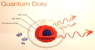
|
All dimensions below 100 nm; unique optical/electronic properties |
|
1D |
Nanotubes, nanofibers, nanorods, thin films 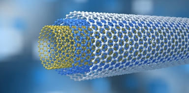
|
Two dimensions at the nanoscale can be metallic, ceramic, or polymeric and used in sensors and electronics. |
|
2D |
Nanosheets, graphenes, platelets, thin layers 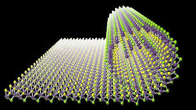
|
One dimension at the nanoscale: application in membrane technologies, coating, and energy storage |
|
3D |
Nanoparticle assemblies, bulk nanomaterials 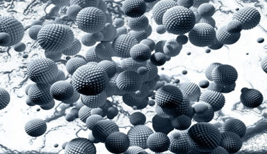
|
Incorporated nanostructures in all dimensions or as clusters; applied in composite materials, heterogeneous catalysis |
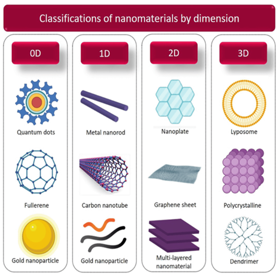
Figure 1: Classification of nanoparticles by dimension
Synthesis of nanoparticles
NPs can be synthesized using a variety of techniques; however, they can be broadly categorized into two classes: (1) bottom-up approaches and (2) top-down approaches. Depending on the operation, reaction state, and accepted protocols, these techniques are further subdivided into several subclasses (Rao et al., 2004; Li et al., 2008; Kharissova et al., 2013; Bakhshi et al., 2020).
1. Bottom-up Approach
In this approach, nanoparticles are constructed from smaller units such as atoms or molecules through controlled chemical reactions, condensation, or self-assembly. The technique emphasizes the “building-up” concept to achieve precise size and morphology control (Daniel & Astruc, 2004; Feynman, 1960; Li et al., 2008; Parveen et al., 2012; Kharissova et al., 2013).
Techniques such as sol-gel synthesis, chemical vapor deposition (CVD), hydrothermal processing, and biological synthesis are common examples of the bottom-up approach (Alexis et al., 2008; Zhang et al., 2010; Rai et al., 2008). For instance, the milling method has been effectively applied to the synthesis of coconut shell (CS) nanoparticles, where prolonged grinding using a planetary mill and ceramic balls reduced particle size significantly as shown by XRD and SEM analyses. The crystallite size was computed using the Scherrer equation, showing a consistent decrease with increased milling duration (Mahapatro & Singh, 2011; Hulla et al., 2015).
Similarly, spherical magnetite nanoparticles (Fe?O?) ranging from 20–50 nm were synthesized using a destructive top-down milling variant, demonstrating effective size control with oleic acid as a stabilizer. Colloidal carbon spherical nanoparticles have also been prepared through adsorption of polyoxometalates (POMs) onto the carbon interfacial surface, resulting in a narrow size distribution and high dispersion capacity. Sonication and grinding-assisted top-down synthesis have also yielded uniform transition-metal dichalcogenide nanodots (TMD-NDs) under 10 nm with excellent dispersibility (Laurent et al., 2008; Bakhshi et al., 2020).
Recently, laser fragmentation—a hybrid top-down method—has been used to produce highly photoactive Co?O? nanoparticles, achieving particle sizes between 5.8 and 1.1 nm with well-defined oxygen vacancies, enhancing photocatalytic behavior (Kharissova et al., 2013; Li et al., 2008).
2. Top-down Approach
Conversely, the top-down approach starts from bulk materials, which are subsequently broken down into nanoparticles using physical or chemical means (Roco, 2011; Hulla et al., 2015; Zhang et al., 2010). Common examples include grinding, lithography, laser ablation, sputtering, and etching.
This approach has been employed in the synthesis of TiO? anatase nanoparticles containing graphene domains, where titanium isopropoxide and alizarin precursors were used to fabricate photoactive composites for dye degradation applications. The XRD patterns confirmed the anatase phase, and SEM revealed temperature-dependent growth in nanoparticle size (Mogilevsky et al., 2018; Li et al., 2008).
In another study, monocrystalline gold (Au) nanospheres were prepared via laser irradiation, allowing precise morphological conversion from octahedral to spherical forms depending on irradiation time and intensity. SEM and TEM analyses confirmed highly uniform nanostructures with average diameters around 75 ± 2.6 nm (Liu et al., 2017; Daniel & Astruc, 2004).
Furthermore, Needham et al. (2019) developed low-density lipoprotein (LDL) nanoparticles for cancer drug delivery using a solvent-exchange method, highlighting the importance of hydrophobicity and size control in medical nanoparticle design.
Both bottom-up and top-down approaches have also been applied in the fabrication of bismuth (Bi) nanoparticles, with particle sizes ranging from 100 nm to 500 nm depending on whether chemical reduction or molten-metal emulsification was employed (Li et al., 2008; Zhang et al., 2010).
The growing focus on green and biogenic synthesis—a subclass of bottom-up methods—emphasizes environmental safety and cost-effectiveness. These methods employ biological systems such as plant extracts, bacteria, fungi, and yeasts to produce metallic nanoparticles through natural reduction processes. For instance, gold nanoparticles (Au NPs) have been synthesized using extracts from wheat, oats, tamarind, and aloe vera, producing stable and eco-friendly nanostructures suitable for biomedical use (Rai et al., 2008; Sharma et al., 2009; Kharissova et al., 2013; Hulla et al., 2015).
Method of Nanoparticle Preparation
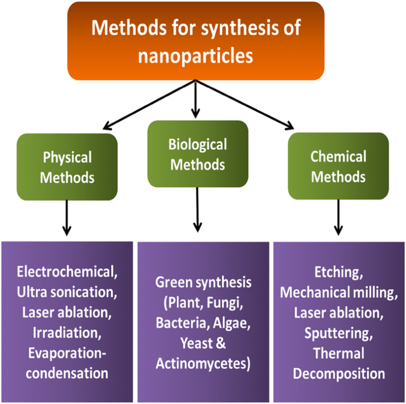
Figure 2: Typical synthesis methods of nanoparticles
1. Solvent Evaporation Method
The solvent evaporation method is one of the earliest and most widely employed techniques for the preparation of polymeric nanoparticles (Fessi et al., 1989; Couvreur et al., 1977; Li et al., 2008; Parveen et al., 2012). In this process, a nanoemulsion is first prepared by dissolving a biodegradable polymer (such as PLGA, ethyl cellulose, or polycaprolactone) in an organic solvent such as ethyl acetate, chloroform, or dichloromethane. The drug to be encapsulated is then dispersed within this organic phase.
An oil-in-water (O/W) emulsion is subsequently formed by emulsifying this organic mixture into an aqueous phase containing suitable surfactants or stabilizers, such as polysorbates, poloxamers, sodium dodecyl sulfate (SDS), polyvinyl alcohol (PVA), or gelatin, using mechanical stirring, sonication, or microfluidization under high pressure (Soppimath et al., 2001; Alexis et al., 2008; Mahapatro & Singh, 2011). After the emulsion is stabilized, the organic solvent is evaporated under reduced pressure and continuous stirring, leading to the precipitation of polymeric nanoparticles as the polymer solidifies around the drug.
This technique is valued for its simplicity, reproducibility, and suitability for hydrophobic drugs, producing nanoparticles with well-defined morphology and size control. However, optimization of parameters such as polymer concentration, emulsifier type, solvent selection, and stirring rate is crucial for achieving high encapsulation efficiency and narrow size distribution (Li et al., 2008; Parveen et al., 2012; Jain et al., 2010).
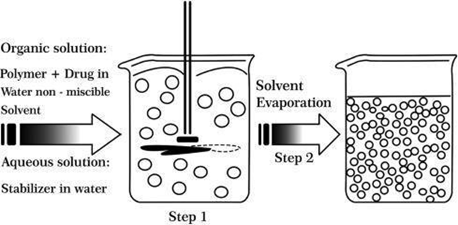
Figure 3: Representation of the solvent evaporation technique
2. Double Emulsification Method (W/O/W Method)
The double emulsification method (commonly known as water-in-oil-in-water, W/O/W) is primarily used for the encapsulation of hydrophilic drugs, which are often poorly trapped using conventional single emulsion or solvent evaporation methods (Barichello et al., 1999; Quintanar-Guerrero et al., 1998; Bilati et al., 2005; Li et al., 2008). In this process, the aqueous drug solution is first emulsified into an organic polymer solution (typically containing PLGA, ethyl cellulose, or polylactic acid) under continuous stirring or sonication to form a primary W/O emulsion.
This primary emulsion is then re-emulsified into a second aqueous phase containing surfactants (e.g., PVA or poloxamer) to form a double W/O/W emulsion. Finally, the organic solvent is removed by evaporation or extraction under reduced pressure, resulting in the solidification of nanoparticles and entrapment of the hydrophilic drug within the polymeric matrix (Sahoo et al., 2002; Budhian et al., 2008; Mundargi et al., 2008).
This technique allows for controlled encapsulation efficiency, sustained drug release, and size uniformity, although optimization is necessary to minimize drug leakage into the external aqueous phase and to enhance stability of the formed nanoparticles (Bilati et al., 2005; Li et al., 2008; Parveen et al., 2012).
3. Emulsion–Diffusion Method
The emulsion–diffusion method is a modified variant of the salting-out technique, originally patented by Leroux and colleagues, and is particularly known for its scalability and reproducibility (Leroux et al., 1999; Quintanar-Guerrero et al., 1998; Lamprecht et al., 2000). In this method, the polymer is dissolved in a partially water-miscible organic solvent such as benzyl alcohol, ethyl acetate, or propylene carbonate, which is saturated with water to ensure phase compatibility. This organic phase is then emulsified into an aqueous solution containing a stabilizer (e.g., PVA or gelatin) under mild stirring to form a uniform emulsion.
After the emulsion is formed, diffusion of the solvent into the aqueous phase occurs spontaneously, leading to precipitation of nanoparticles as the polymer solidifies. The organic solvent is then eliminated by evaporation or filtration.
This technique offers multiple advantages — narrow particle size distribution, high encapsulation efficiency (up to 70%), easy scale-up, and excellent batch-to-batch reproducibility — and requires no high-energy homogenization (Leroux et al., 1999; Lamprecht et al., 2000; Li et al., 2008; Mahapatro & Singh, 2011). However, water-soluble drugs may diffuse into the outer aqueous phase during emulsification, which can reduce entrapment efficiency if not optimized.
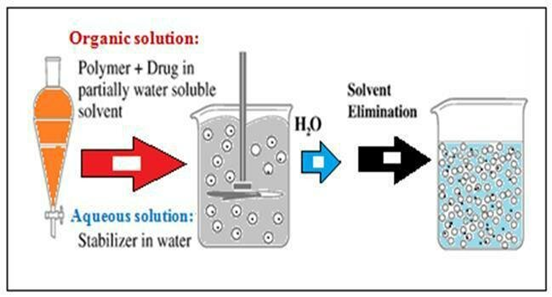
Figure 4: Representation of the emulsification-diffusion technique
4. Coacervation Method
The coacervation technique is widely employed for the formulation of biodegradable nanoparticles using hydrophilic polymers such as gelatin, sodium alginate, and chitosan (Calvo et al., 1997; Balthasar et al., 2005; Gupta & Gupta, 2005). This process typically involves phase separation in a polymer solution, leading to the formation of a coacervate phase that entraps the active drug molecules. The ionic gelation process, a specific type of coacervation, utilizes two aqueous phases — one containing a cationic polymer (e.g., chitosan or polyethylene oxide-propylene oxide copolymers) and another containing a polyanionic cross-linker such as sodium tripolyphosphate (TPP). Electrostatic interactions between these oppositely charged species induce the formation of nanocoacervates, which upon stabilization yield uniform nanoparticles suitable for drug encapsulation and controlled release (Calvo et al., 1997; Lin et al., 2012).
In a related study, Das et al. (2015) prepared albumin-based drug-loaded nanoparticles using the coacervation method. A 2% w/v drug-protein solution was first incubated at room temperature for one hour, and the pH was adjusted to 5.5 using 1M HCl. Ethanol was then added dropwise (1 mL/min) at a 2:1 v/v ratio, inducing coacervation. To achieve structural rigidity, protein cross-linking was performed using 25% glutaraldehyde (1.56 µg/mg of protein) for two hours. Following rotary vacuum evaporation to remove solvents, the nanoparticles were centrifuged at 4°C, resuspended in phosphate buffer (0.1 M, pH 7.4), and lyophilized with 2% w/v mannitol as a cryoprotectant for 24 hours at −48°C and 28 × 10?³ MBar pressure. The resulting nanoparticles displayed excellent uniformity, stability, and encapsulation efficiency, demonstrating the method’s potential for protein-based drug delivery systems (Das et al., 2015; Calvo et al., 1997; Lin et al., 2012).
5. Nanoprecipitation Method (Solvent Displacement Technique)
The nanoprecipitation method, also known as the solvent displacement technique, is one of the most widely employed and straightforward approaches for the synthesis of polymeric nanoparticles (Fessi et al., 1989; Quintanar-Guerrero et al., 1998; Bilati et al., 2005; Mahapatro & Singh, 2011). This technique was first developed by Fessi and co-workers, who described a spontaneous emulsification process in which a polymer and drug are dissolved in a miscible organic solvent (such as acetone, ethanol, or acetonitrile), followed by their diffusion into an aqueous medium containing a stabilizer or surfactant (e.g., PVA or poloxamer). The rapid diffusion of the solvent into water results in the precipitation of nanoparticles due to supersaturation of the polymer–drug solution, leading to instantaneous formation of nanoparticles with uniform size distribution.
This method offers multiple advantages — including simplicity, reproducibility, narrow particle size range, and suitability for hydrophobic drugs — while requiring no external energy input such as heat or high shear (Lamprecht et al., 2000; Li et al., 2008; Parveen et al., 2012). However, it is primarily limited to lipophilic compounds, as hydrophilic drugs tend to diffuse rapidly into the aqueous phase, reducing encapsulation efficiency.
In one application, Tamizhrasi et al. (2009) successfully prepared lamivudine-loaded nanoparticles using the nanoprecipitation method. The drug was first dissolved in water and combined with a cosolvent (acetone) to enhance homogeneity. A separate polymeric phase containing ethyl cellulose, Eudragit, and propylene glycol in chloroform was then added gradually to form a uniform dispersion. Upon mixing with 70% aqueous ethanol, the organic solvents were evaporated at 35°C under normal pressure, and nanoparticles were recovered via centrifugation at 10,000 rpm for 20 minutes, washed, and dried in a desiccator at room temperature. The resulting nanoparticles exhibited consistent morphology, controlled drug release, and good physical stability, confirming the efficiency of this method (Tamizhrasi et al., 2009; Mahapatro & Singh, 2011; Parveen et al., 2012).
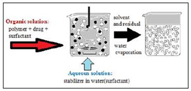
Figure 5: Representation of the nanoprecipitation technique
6. Salting-Out Method
The salting-out technique is a well-established method for the preparation of polymeric nanoparticles, conceptually similar to the emulsion–diffusion method, and was first described and patented by Ibrahim et al. (1992) and Bindschaedler et al. (1991). It relies on the salting-out phenomenon, where the water-miscible organic solvent (such as acetone or ethanol) is separated from the aqueous phase by the presence of salting-out agents (Catarina et al., 2006; Lamprecht et al., 2000). These agents may include electrolytes (e.g., magnesium chloride, calcium chloride) or non-electrolytes (e.g., sucrose).
In this process, a polymer and drug are first dissolved in the organic solvent and then emulsified into an aqueous phase containing a salting-out agent and stabilizers such as polyvinyl alcohol (PVA), poly(ethylene oxide) (PEO), or hydroxyethylcellulose. Upon subsequent dilution with water, the solvent diffuses into the aqueous phase, leading to precipitation of polymeric nanospheres. The absence of high shear energy or elevated temperature makes this method suitable for thermosensitive drugs and easily scalable for industrial production (Ibrahim et al., 1992; Catarina et al., 2006; Bilati et al., 2005).
Although the salting-out method provides advantages such as simple operation, low energy requirement, and high encapsulation efficiency for hydrophobic drugs, its major limitation lies in the lengthy washing steps required to remove residual salts and the reduced efficiency for hydrophilic drug encapsulation (Bilati et al., 2005; Lamprecht et al., 2000).
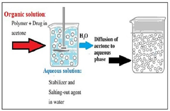
Figure 6: Representation of the salting out technique
7. Dialysis
Dialysis is a successful technique for creating nanoparticles. This process involves first dissolving the medication and polymer (such as poly(benzyl-l-glutamate)-b-poly(ethylene oxide) or poly(lactide)-b-poly(ethylene oxide)) in an organic solvent. Dialysis was conducted using this solution in a dialysis tube beside a non-solvent miscible. The increasing aggregation of the polymer as a result of a loss of solubility and the creation of uniform suspensions of nanoparticles occur when the solvent is displaced inside the membrane. The dialysis process for the creation of nanoparticles is not fully known at present. The mechanism behind it might be comparable to that of nanoprecipitation.
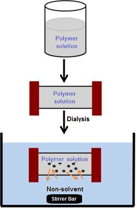
Figure 7: Representation of the osmosis-based method for the preparation of nanoparticles
8. Supercritical fluid technology
The supercritical fluid (SCF) technique represents a clean and sustainable alternative to conventional nanoparticle synthesis methods that rely heavily on organic solvents, which are often hazardous to both the environment and biological systems (Reverchon & Adami, 2006; Mishra et al., 2009). A supercritical fluid is defined as a substance maintained above its critical temperature and pressure, where it exhibits both liquid-like density and gas-like diffusivity, resulting in superior solvation power and rapid mass transfer (Subramaniam et al., 1997; Türk & Lietzow, 2004).
Among available SCFs, carbon dioxide (CO?) is the most widely employed due to its nontoxicity, nonflammability, low critical temperature (31.1 °C) and moderate critical pressure (73.8 bar), as well as its low cost and easy availability. CO? is also classified as a “green solvent”, making the technique particularly attractive for the production of pharmaceutical nanoparticles, biodegradable polymers, and drug delivery systems (Reverchon & De Marco, 2006; Türk, 2000).
Two primary SCF-based approaches are commonly used:
A) Supercritical Anti-Solvent (SAS) Method
In this process, the supercritical fluid acts as an anti-solvent, precipitating the solute from an organic solution. The liquid solvent (such as methanol, acetone, or ethanol) must be completely miscible with the supercritical CO?. When the supercritical fluid is introduced into the liquid solution, it extracts the solvent rapidly, leading to instant supersaturation and the formation of uniformly sized nanoparticles. The SAS process provides excellent control over particle morphology, size, and distribution, and is widely used for thermosensitive bioactives (Matson et al., 1987; Reverchon, 2002; Mishima, 2008).
B) Rapid Expansion of Supercritical Solutions (RESS)
In the RESS method, the solute is first dissolved in the supercritical fluid (typically CO?), and the resulting supercritical solution is rapidly expanded through a fine nozzle into a region of lower pressure (usually atmospheric). This rapid decompression results in supersaturation, homogeneous nucleation, and particle formation. The RESS method yields narrow particle size distributions and solvent-free nanoparticles, though it is limited by the solubility of many polymers in supercritical CO? (Tom & Debenedetti, 1991; York, 1999; Weidner, 2009).
Collectively, supercritical fluid technologies offer a versatile, scalable, and eco-friendly route for nanoparticle synthesis. Their ability to eliminate residual organic solvents and precisely tune process parameters makes them particularly advantageous for producing pharmaceutical nanoparticles, controlled-release formulations, and advanced nanocarriers suitable for biomedical use (Reverchon & Adami, 2006; Weidner, 2009).
Characterization and Evaluation of Nanoparticles
1. Particle Size
The particle size and distribution of the prepared nanoparticles were analyzed using Dynamic Light Scattering (DLS), a key technique that estimates hydrodynamic diameter and polydispersity index (PDI) from Brownian motion (Bhattacharjee, 2016). Samples were dispersed in distilled water and sonicated for 3–5 minutes to ensure uniform suspension and prevent agglomeration. Measurements were performed at 25°C with a detection angle of 90°, and the results were expressed as mean ± standard deviation (Malvern Instruments, 2011; Danaei et al., 2018).
2. Entrapment Efficiency and Drug Content
Entrapment efficiency (EE) and drug content determine how effectively the active drug is incorporated within the nanoparticle matrix (Govender et al., 1999; Bala et al., 2016). Approximately 10 mg of freeze-dried nanoparticles were dissolved in 10 mL of methanol, vortexed, and sonicated to extract the drug completely. The absorbance was measured at 238 nm (simvastatin λmax) using a UV–Visible spectrophotometer. The drug content (%) and entrapment efficiency (%) were calculated using standard equations (Sahoo et al., 2002; Abdelwahab et al., 2015).
3. Surface Charge (Zeta Potential)
The surface charge of nanoparticles, expressed as zeta potential, was measured using electrophoretic light scattering. This parameter predicts suspension stability, with values beyond ±30 mV indicating good electrostatic repulsion (Hunter, 1981; Honary & Zahir, 2013). Measurements were conducted at 25°C after appropriate dilution with deionized water filtered through a 0.22 µm membrane (Mohanraj & Chen, 2006).
4. Scanning Electron Microscopy (SEM)
SEM was used to examine nanoparticle morphology, surface texture, and aggregation tendencies (Goldstein et al., 2017). Samples were mounted on aluminum stubs using carbon tape, sputter-coated with gold/palladium, and imaged at ~5 kV. SEM provided detailed topographical information that complements DLS data (Singh & Lillard, 2009; Tiwari & Amiji, 2006).
5. Differential Scanning Calorimetry (DSC)
DSC assesses thermal properties and potential drug–polymer interactions, offering insight into crystallinity and miscibility (Craig, 1995; Sahoo et al., 2002). Samples (3–5 mg) were sealed in aluminum pans and heated from 20–160°C at 10°C/min under nitrogen. The thermograms of pure drug, polymer, and nanoparticles were compared for melting point shifts or disappearance of endothermic peaks, indicating successful encapsulation (Shah et al., 2006).
6. Solubility Study
Solubility enhancement of the drug post-nanoparticle formulation was determined using the shake-flask method in phosphate buffer (pH 6.8) for 48 h at 25°C (Higuchi & Connors, 1965; Kipp, 2004). The filtered supernatant was analyzed at 238 nm. Enhanced solubility compared to the pure drug suggested nanoscale dispersion and amorphization (Date & Nagarsenker, 2008; Singh & Kim, 2010).
7. In Vitro Drug Release
Drug release studies were carried out via the dialysis bag diffusion method in phosphate buffer (pH 6.8, 37 ± 0.5°C, 50 rpm). Samples withdrawn at specific intervals were replaced with fresh buffer, and drug concentration was measured spectrophotometrically at 238 nm. This method simulated controlled release behavior (Dash et al., 2010; Sahoo et al., 2002).
8. Kinetic Study
Release data were fitted to zero-order, first-order, and Higuchi models, and correlation coefficients (R²) were determined to evaluate the release mechanism. Additional modeling with Korsmeyer–Peppas was employed to confirm diffusion or erosion-driven kinetics (Costa & Sousa Lobo, 2001; Peppas, 1985).
9. Powder X-ray Diffraction (PXRD)
PXRD was performed to determine crystallinity and detect structural transitions within nanoparticles. The broadening of diffraction peaks indicated reduced crystallinity and nanoscale dimensions (Cullity & Stock, 2001; Patterson, 1939). PXRD was complemented by TEM and DSC for comprehensive phase analysis (Bunaciu et al., 2015).
10. Stability
Nanoparticle stability was monitored by assessing zeta potential, hydrodynamic diameter, and PDI over time under various storage conditions. Instability is often indicated when zeta potential falls below ±20 mV, leading to aggregation (Honary & Zahir, 2013; Abdelwahed et al., 2006). DLVO theory explains the balance between attractive van der Waals and repulsive electrostatic forces governing colloidal stability (Derjaguin & Landau, 1941; Verwey & Overbeek, 1948).
11. Transmission Electron Microscopy (TEM)
TEM provided high-resolution visualization of nanoparticles, allowing determination of size, shape, and internal structure (Wang et al., 2008; Reimer & Kohl, 2008). More than 1,000 particles were analyzed to ensure statistical significance in particle size distribution. TEM also confirmed crystallinity when coupled with electron diffraction (Niemeyer, 2001).
12. Morphology
Morphology governs diffusion, drug release, and cellular uptake characteristics of nanoparticles (Zhang et al., 2008; Fessi et al., 1989). Controlled morphology, such as spherical or rod-shaped structures, influences drug loading and biodistribution. Techniques like TEM and SEM aid in correlating morphology with performance (Singh & Lillard, 2009; He et al., 2010).
13. In Vivo Release
In vivo release studies evaluate pharmacokinetics and tissue distribution, often using animal models to simulate physiological conditions. Data are analyzed using kinetic models such as zero-order, first-order, Higuchi, Hixson–Crowell, and Korsmeyer–Peppas, revealing the release mechanism (Dash et al., 2010; Siepmann & Peppas, 2011). Sustained release behavior confirms effective nanoparticle-based delivery.
Applications of NPs
1. Applications of NPs in the Environment Industry
NPs are attractive for a variety of environmental applications because of their small size and unique physical and chemical properties. The characteristics and benefits of nanoparticles are well documented. Some potential applications of NPs in the environment include the following (Chatterjee & Das, 2020; Tiwari et al., 2008).
1.1. Bioremediation
Environmental contaminants, including organic pollutants from soil or heavy metals from water, can be eliminated using nanoparticles (NPs). For instance, organic colors and chemicals present in wastewater are among the contaminants that silver nanoparticles (AgNPs) efficiently break down (Rai et al., 2008; Sharma et al., 2009). Numerous nanomaterials, including metal oxides, carbon nanotubes and fibers, and nanoscale zeolites, have been investigated for cleanup applications (Chen et al., 2016; Mohanraj & Chen, 2006). When employed in cleanup, nanoscale particles can reach places that larger particles cannot. Before reacting with pollutants, they can be coated to aid in transportation and stop them from reacting with the surrounding soil matrices. Nanoscale zerovalent iron (nZVI) is a popular nanomaterial for cleanup. Chlorinated solvents that have contaminated groundwater have been cleaned up using it at a number of hazardous waste disposal sites (Chatterjee & Das, 2020). Because of their detrimental impact on the environment and human health, heavy metals, including mercury, lead, thallium, cadmium, and arsenic, have drawn a lot of interest when it comes to their removal from natural water. For this hazardous soft substance, superparamagnetic iron oxide nanoparticles (NPs) work well as a sorbent (Gupta & Gupta, 2005; Laurent et al., 2008). Therefore, because there are no analytical techniques that can assess the trace concentration of NPs, there have been no measurements of manufactured NPs in the environment (Gupta & Xie, 2018).
1.2. Sensors in the Environment
NPs and nanotechnology are already being employed to help with environmental cleanup efforts and to enhance the quality of water. Additionally, their potential as environmental sensors to track contaminants is starting to materialize (Chatterjee & Das, 2020; Thomas & Thomas, 2017). NPs can be employed as sensors to find out whether contaminants or heavy metals are present in the environment. In real-world applications, the nanosensors’ tiny size and broad detection range offer a great deal of versatility (Gupta & Xie, 2018). According to reports, biological substances, including toxins and microbial infections, can be detected in aquatic settings using nanoscale sensors (Bumb et al., 2011; Chen et al., 2016). In order to detect pollutants at low concentrations, NPs can be engineered to attach to particular types of contaminants selectively. For instance, gold nanoparticles (AuNPs) have been employed as mercury sensors in water (Huang & El-Sayed, 2010; Ankamwar et al., 2005).
1.3. Catalysts in the Environment
In chemical reactions, such as those involving the generation of biofuels or environmental remediation procedures, nanoparticles (NPs) operate as catalysts. They can also catalyze the conversion of biomass into fuels, such as ethanol or biodiesel (Bhattacharyya et al., 2011; Mohanraj & Chen, 2006). For instance, because of their capacity to accelerate the conversion of biomass into fuels, platinum nanoparticles (PtNPs) have been investigated for use in the generation of biofuels (Chen et al., 2016). PtNPs also demonstrated encouraging sensing capabilities; for instance, using PtNPs, Hg ions were measured in MilliQ, tap, and groundwater samples in the 50–500 nM range, with quantification limits of 16.9, 26, and 47.3 nM. Hg ion detection and quantification were successfully accomplished using the biogenic PtNPs-based probe (Sun et al., 2014; Bhattacharyya et al., 2011). All things considered, NPs are being actively studied for a range of applications and have a great deal of promise for usage in the environment (Chatterjee & Das, 2020).
2. Applications of NPs in the Medicine Industry
The tiny size of nanoparticles (NPs) gives them special physical and chemical characteristics that make them appealing for usage in a variety of fields, including medicine (Banik et al., 2016; Salata, 2004).
2.1. Drug Delivery
Because of their distinct optical characteristics, ease of synthesis, and chemical stability, AuNPs have attracted technological attention. Applications for the particles in biomedicine include medication administration, chemical sensing, biological imaging, and cancer treatment (Huang & El-Sayed, 2010; Chinnathambi et al., 2014). Both sustained (diffusion-controlled and erosion-controlled) and stimuli-responsive (pH-sensitive, enzyme-sensitive, thermoresponsive, and photosensitive) controlled release of medicines coupled with NPs have been discussed in detail (Sun et al., 2014; Torchilin, 2014). NPs work as therapeutic gene delivery vehicles to synthesize desired proteins in targeted cells and as targeted delivery of medications to treat cancer cells (Jain & Stylianopoulos, 2010; Yu et al., 2012). Because NPs can transport medications to specific body parts, treatment can be more focused and efficient (Wilczewska et al., 2012). For instance, AgNPs are stable and can accumulate in some malignant tumors, making them candidates for medication delivery (Rai et al., 2008; Singh & Lillard, 2009). ZnONPs can target cancer cells specifically, while CuNPs are investigated for antibacterial drug delivery (Patra et al., 2018). Wound dressings, bone cement, and implants have all been made with silver nanoparticles (AgNPs) (Banik et al., 2016; Calvo et al., 1997).
2.2. Diagnostics
NPs can be employed as imaging agents to aid in visualization of tissues and organs (Gupta & Gupta, 2005; Xu et al., 2006). Iron oxide nanoparticles (Fe?O? NPs) have been used as contrast agents in magnetic resonance imaging (MRI) (Laurent et al., 2008; Mahmoudi et al., 2011). AuNPs can aggregate in malignant tumors, and their optical, electrical, and catalytic properties are explored for diagnostics (Chen et al., 2016; Chinnathambi et al., 2014).
2.3. Tissue Engineering
NPs can aid in promoting tissue and organ growth and repair. Titanium dioxide nanoparticles (TiO? NPs) have demonstrated the potential to promote bone cell proliferation and are being investigated in tissue engineering (Mohanraj & Chen, 2006; Banik et al., 2016).
2.4. Antimicrobials
Some NPs, such as CuNPs and AgNPs, have potent antibacterial properties and are being explored for use in medical devices and wound dressings (Rai et al., 2008; Sharma et al., 2009; Banik et al., 2016). All things considered, NPs are being actively studied for a number of applications and hold a great deal of promise in medicine. Ensuring safe and responsible usage requires thorough assessment of their benefits and risks (Fadeel & Garcia-Bennett, 2010; Hulla et al., 2015).
3. Applications of NPs in the Agriculture Industry
NPs can significantly influence agriculture by improving efficiency, yield, and sustainability (Khan et al., 2019; Gupta & Xie, 2018).
3.1. Pesticides and Herbicides
Targeted delivery of insecticides and herbicides via NPs can reduce environmental contamination and chemical use (Patra et al., 2018; Banik et al., 2016). AgNPs and CuNPs show promise in managing crop diseases and pests. Metal NPs in pesticides and herbicides are still in early stages, and more research is needed to evaluate environmental and human health impacts (Gupta & Xie, 2018; Khan et al., 2019).
3.2. Fertilizers and Plant Growth
Nano fertilizers can improve plant mineral nutrition and controlled release of nutrients, enhancing uptake efficiency and reducing environmental nutrient loss (Patra et al., 2018; Xu et al., 2006). NPs based on Ag, Zn, Cu, Au, Al, and Fe have fertilizing and plant growth-promoting properties (Kumar & Yadav, 2009; Banik et al., 2016). Application in fertilizers is still in its infancy, requiring further investigation for safety and effectiveness (Kharissova et al., 2013; Kharissova et al., 2019).
3.3. Food Safety
NPs can detect and remove pathogens in food products, reducing the risk of foodborne illnesses (Patra et al., 2018; Chen et al., 2016).
3.4. Water Purification
NPs can purify irrigation water, reduce crop contamination, and improve agricultural productivity (Tiwari et al., 2008; Thomas & Thomas, 2017). They help increase yield, reduce environmental impact, and improve food safety and quality (Chatterjee & Das, 2020).
4. Applications of NPs in the Food Industry
NPs have a wide range of potential uses in the food industry due to their small size, high surface area, and unique properties (Banik et al., 2016; Salata, 2004).
4.1. Food Processing and Food Preservation/Food Packaging
NPs can improve food processing operations such as grinding, mixing, and drying. AgNPs are used as natural antimicrobial agents to inhibit bacterial growth and enhance food safety (Sharma et al., 2009; Rai et al., 2008). NPs also enhance the performance of food packaging materials by improving resistance to gases and moisture, thereby increasing shelf life (Sanvicens & Marco, 2008; Banik et al., 2016).
4.2. Food Fortification
NPs can deliver essential nutrients like vitamins and minerals more efficiently to food products. For example, Fe?O? and CuNPs have been used to fortify food with iron, an essential nutrient often deficient in diets, especially in underdeveloped countries (Patra et al., 2018; Banik et al., 2016).
4.3. Sensors
NPs enhance the sensitivity and specificity of food sensors, allowing better detection of contaminants or chemical signals (Catarina et al., 2006; Chatterjee & Das, 2020). This improves the nutritional quality, safety, and performance of food products and processing methods (Chen et al., 2016; Banik et al., 2016).
5. Applications of NPs in the Electronics and Automotive Industries
NPs can revolutionize electronics and automotive industries by improving performance, efficiency, and durability (Khan et al., 2019; Banik et al., 2016).
5.1. Display Technologies/Storage Devices
NPs can enhance displays such as LCD and OLED screens by improving brightness, color, and contrast. Silver and gold NPs have been explored to increase electrical conductivity in displays (Huang & El-Sayed, 2010; Banik et al., 2016). NPs also improve energy storage in batteries and supercapacitors by increasing energy density and charging speed. For example, ZnO NPs are investigated for their use in high-performance energy storage devices (Xu et al., 2006; Mahmoudi et al., 2011).
5.2. Data Storage
Magnetic NPs, such as iron oxide NPs, can store and retrieve data through magnetization and demagnetization. Magnetic metals like iron, cobalt, or nickel are commonly used (Laurent et al., 2008; Banik et al., 2016). Overall, NPs enhance the performance and efficiency of a variety of electronic devices (Whitesides, 2003; Banik et al., 2016).
5.3. Chemical Processing/Catalysis
NPs act as efficient catalysts for chemical reactions, enabling lower temperature operation and higher selectivity. For example, PtNPs are used in fuel cell reactions, hydrogenation, and oxidation reactions (Bhattacharyya et al., 2011; Banik et al., 2016). PdNPs, FeNPs, and NiNPs have been employed in hydrolysis, hydrogenation, and oxygen reduction reactions (Bumb et al., 2011; Banik et al., 2016).
5.4. Separation and Purification
NPs can separate and purify gases, liquids, and chemicals based on size and surface properties. Fe?O? NPs have been used for purification processes, while AgNPs and AuNPs are effective for removing viruses, bacteria, and heavy metals from water (Xu et al., 2006; Banik et al., 2016). AlNPs can remove impurities from fuels, oils, and water, demonstrating the broad industrial applicability of NPs in separation and purification (Banik et al., 2016; Santra & Wang, 2005).
6. Applications of NPs in the Defense Industry
NPs enhance the performance, durability, and efficiency of defense materials and systems (Khan et al., 2019; Banik et al., 2016).
6.1. Sensors
Defense system sensors that detect chemical, biological, or radiological threats benefit from NPs’ enhanced sensitivity and specificity (Catarina et al., 2006; Chatterjee & Das, 2020).
6.2. Protective Coatings
NPs improve protective coatings on defense equipment by increasing resistance to corrosion, wear, and chemical or biological agents. Al and Zn NPs enhance corrosion resistance, while Ni and Cr NPs improve wear resistance (Bumb et al., 2011; Banik et al., 2016).
6.3. Weapons
NPs are used in the development of armor, protective materials, and pathogen-targeting weapons. Incorporating metal or ceramic NPs into polymers improves mechanical properties and resilience (Calvo et al., 1997; Banik et al., 2016). NPs are also employed in defense-related sensors and detection systems (Chen et al., 2016; Banik et al., 2016).
6.4. Manufacturing
NPs improve the performance and robustness of defense materials. Metals like Cu, Ni, and Al are added to polymers as fillers to enhance mechanical strength, electrical conductivity, and thermal stability. NPs such as Au and Pt are used as catalysts due to their high surface area and adsorption capacity (Drexler, 1986; Banik et al., 2016).
6.5. Energy Storage
NPs improve energy storage devices like batteries and fuel cells in defense systems. LiCoO? NPs serve as cathode materials in lithium-ion batteries due to high capacity and rate performance, while transition metal oxide NPs such as MnO? and Fe?O? enhance rechargeable battery performance. NPs in supercapacitor electrodes increase the specific surface area, raising capacitance (Mahmoudi et al., 2011; Xu et al., 2006). Overall, NPs enhance the effectiveness, safety, and efficiency of defense systems (Banik et al., 2016; Patra et al., 2018).
CONCLUSION
Nanoparticles are essential to the advancement of contemporary healthcare, pharmaceuticals, and diagnostics, and nanotechnology is still transforming a number of scientific and industrial fields. Precise control over the physicochemical characteristics of nanoparticles, including size, shape, surface charge, and morphology, directs their design and formulation and has a direct impact on their stability, bioavailability, and therapeutic efficacy. Researchers can create nanoparticles with specialized functions appropriate for particular industrial and biological applications using a range of preparation techniques, such as top-down, bottom-up, and green synthesis.
Thorough evaluation and characterization are essential components of nanoparticle formulation. Methods like Differential Scanning Calorimetry (DSC), Zeta potential analysis, Scanning Electron Microscopy (SEM), and Dynamic Light Scattering (DLS) allow precise evaluation of drug–polymer interactions, surface charge, morphology, crystallinity, and particle size distribution. The nanoparticles' compliance with the required standards for effective drug encapsulation, sustained release, and biocompatibility is guaranteed by these assessments. The pharmacokinetic behavior and therapeutic potential of drug-loaded nanoparticles are also better understood through in vitro release studies and solubility tests.
In terms of site-specific and regulated medication administration, nanoparticles have shown impressive potential, lowering systemic adverse effects and enhancing treatment effectiveness. They have also shown potential in imaging, biosensing, gene therapy, and vaccine administration. A number of obstacles still exist in spite of these benefits, including the inability to produce on a large scale, problems with aggregation and stability, possible toxicity, and the absence of standardized regulatory frameworks. To overcome these obstacles, material scientists, chemists, pharmacologists, and regulatory specialists must work together across disciplinary boundaries.
The creation of sensory-responsive, biodegradable, and intelligent nanocarriers that can precisely carry therapeutic drugs and adjust to physiological conditions is the key to the future of nanoparticle technology. Including sustainable "green" synthesis techniques will also improve cost-effectiveness and environmental safety. The importance of nanoparticle-based systems in targeted medication delivery, personalized medicine, and next-generation healthcare technologies is anticipated to grow as research advances. The development of this area ultimately has the potential to provide safer, more effective, and more intelligent nanotherapeutics, which could fundamentally alter the course of contemporary medicine.
REFERENCES

Arti Walunj, Ashlesha Zol, Dhiraj Wagh, Ram Nikhate, Design Formulation and Evaluation of Nanoparticles, Int. J. of Pharm. Sci., 2026, Vol 4, Issue 1, 1692-1716. https://doi.org/10.5281/zenodo.18273357
 0.5281/zenodo.18273357
