*M. Pharm, College of Pharmaceutical Sciences, Govt. Medical College Thiruvananthapuram.
Assistant Professor, Department of Pharmacology, Govt. Medical College Thiruvananthapuram
Non-alcoholic fatty liver disease (NAFLD) is becoming a popular health concern due to an increased sedentary lifestyle and an unhealthy diet. NAFLD is the pathological accumulation of triglycerides in the hepatocytes as macrovesicular steatosis in the absence of alcohol intake. Nagaradi Kashayam (Amruthotharam) is a polyherbal ayurvedic formulation mainly used for the treatment of all types of fevers, coughs, and indigestion. The study aims to evaluate the hepatoprotective activity of Nagaradi kashayam in a high-fat diet-induced NAFLD model in male Wistar rats. The methodology involves studying the hepatoprotective effects of silymarin and Nagaradi kashayam (0.5 ml/kg and 1.5 ml/kg) in the NAFLD model. Fatty liver is induced by feeding rats a lard-based, high-fat diet for 10 weeks. Rats were divided into 5 groups: normal control, disease control, silymarin-treated group, Nagaradi Kashayam Group I (0.5 ml/kg), and Nagaradi Kashayam Group II (1.5 ml/kg). The tissue protection was studied by histopathological evaluation of the liver using H&E and oil-red O staining. Tissue antioxidant parameters like reduced glutathione, lipid peroxidation level, and serum parameters such as SGOT, SGPT, ALP, total cholesterol, and serum triglycerides were investigated. The phytochemical screening and in vitro antioxidant activity of the formulation were evaluated. Nagaradi kashayam was found to possess free radical scavenging effects in DPPH, hydrogen peroxide, and nitric oxide, showing its antioxidant property. From biochemical studies, it is concluded that administration of Nagaradi kashayam at 1.5 ml/kg exhibited marked hepatoprotective activity, and the liver tissue protection and antisteatotic effect were confirmed by histopathological assessment.
Non-alcoholic fatty liver disease (NAFLD) is the most prevalent liver disease worldwide and the leading cause of liver-related morbidity and death. Its incidence has been rising and varies throughout the world, typically in tandem with the prevalence of obesity and type II diabetes. Overtime NAFLD has emerged as the primary cause of liver diseases such as liver insufficiency, hepatocellular carcinoma, and liver transplantation. [1] The hallmark feature of NAFLD is steatosis, where there is excessive intrahepatic triglyceride (TG) content greater than 5% of liver weight or liver volume or histologically identified when 5% or more of hepatocytes have apparent intracellular TG. In conventional medicine, there is no known treatment for NAFLD. Weight loss through a combination of nutritious diet and working out is usually the first line of treatment. Generally, the first line of treatment is losing weight by combining exercise with a healthy diet. Treatment of risk factors such as diabetes mellitus and hyperlipidemia by the use of insulin-sensitizing drugs such as biguanides (metformin) and thiazolidinedione’s (rosiglitazone, pioglitazone), antioxidants, have all been used to treat non-alcoholic fatty liver disease. However, there are currently no FDA approved treatments for NAFLD. This is a field of active research where several studies are focusing on finding complementary therapies from natural sources acting as safe products, such as herbal medicine and functional foods. Furthermore, due to their anti-steatotic and antioxidant effects, natural compound supplements are used in addition to a healthy diet to improve NAFLD. [2][3]
Ayurveda is one of the traditional medicinal systems of India. Since ancient times, India has been practicing Ayurveda, which dates approximately back to three thousand years. Herbs are among the most potent medicinal compounds used in the Indian Ayurvedic system. All of this have been documented in texts like the Samhitas and Vedas.[4] Polyherbalism has historically been emphasised by the Ayurvedic literature known as the "Sarangdhar Samhita," which dates back hundreds of years until 1300 A.D. The presence of various phytoconstituents contributes to the therapeutic effects of herbal medicines, which are further enhanced when appropriate herbals are combined to generate polyherbal medicines (PHFs).[5] Nagaradi Kashayam (Amruthotharam) is an ayurvedic polyherbal formulation. It is a concentrated water decoction prepared from three herbal drugs, namely Tinospora cordifolia[6] ,Zingiber officinale[7] ,Terminalia chebula[8] . Nagaradi kashayam is extensively used in the management of all types of fevers. It is known to improve digestion, strength, and appetite and has a mild laxative action. [9] Since the individual constituents of the formulation were found to possess antioxidant, antihyperlipidemic, and some hepatoprotective activities from previous studies, it seemed suitable to undertake a study that evaluates the hepatoprotective effect of Nagaradi kashayam in fatty liver disease.
MATERIALS AND METHODS
30 male Wistar rats (150–220 g, two months old) were obtained from the Sree Chithra Tirunal Institute of Medical Sciences and Technology, Poojapura, Thiruvananthapuram, Kerala. The required animals were sanctioned by the IAEC of Govt. Medical College, Thiruvananthapuram (02/08/IAEC/2022/GMCT). The rats were housed in polypropylene cages under standard conditions of temperature (22 ± 5°C), humidity (50±5%), a 12-hour light/dark cycle, standardised feed, and water ad libitum. The animals were acclimatised to laboratory conditions for one week before the experiment. The rats were divided into five different experimental groups.
The groups are described as follows:
The high-fat diet was prepared in our laboratory by mixing lard oil, egg yolk powder, sugar, milk powder into the standard rat pellet. its composition was in accordance with the previously published studies of NAFLD model.[10][11] The diet was given ad libitum for about 10 weeks to induce the fatty liver.[12][13] During the study, the body weight changes of each group of animals were recorded on a weekly basis throughout the period. The animals were sacrificed at the end of the study by high-dose anaesthesia (ketamine 500 mg/kg), and all efforts were made to minimise the animals' suffering. The pharmacological activity of formulation was compared with silymarin. The silymarin was reported to have beneficial effect in fatty liver in the previous studies. It is also used as supportive treatment in liver diseases.[14][15] The kashayam was procured from an ayurvedic retail pharmacy, Thiruvananthapuram, manufactured by Arya Vaidhya Sala Kottakal. The silymarin powder was purchased from Yarrowchem Products in Mumbai. The preliminary screening of phytoconstituents in kashayam was done according to the standard procedure.[16][17][18][19]
DPPH radical scavenging assay[20][21]
Ascorbic acid was used as the reference standard. A 3.9-mL solution of 60 µM DPPH in ethanol was mixed with 0.1 ml of 25, 50, 100, and 200 µg/mL of kashayam. The samples were kept in the dark for 15 minutes at room temperature, and the decrease in absorbance was measured at 515 nm. A control sample was prepared, containing the same volume without any extract or reference standard. 95% ethanol was used as a blank. The percentage inhibition was calculated.
Hydrogen peroxide scavenging assay[22]
The H2O2 scavenging assay was carried out following the procedure of Ruch et al. A solution of 43 mM H2O2 was prepared in 0.1 M phosphate buffer. 1 mL of kashayam in 3.4 mL of phosphate buffer was added to 0.6 mL of H2O2 solution, and the absorbance of the reaction mixture was recorded at 230 nm. The blank solution contained the sodium phosphate buffer without H2O2. Ascorbic acid was used as a standard. The percentage of H2O2 scavenging by the kashayam and ascorbic acid was calculated.
Nitric oxide scavenging assay[23]
Varying concentrations of Nagaradi Kashayam were taken in different test tubes, and 1 mL of sodium nitroprusside (10 mM) in PBS (0.25 M, pH 7.4) was added to each test tube and incubated at 25ºC for 5 h. A mixture containing all reagents except kashayam but with 100 µl of buffer was used as a control. After 5 hours, 0.5 mL of Griess reagent was added. The absorbance of the pink-coloured mixture was measured at 546 nm. Ascorbic acid was used as a standard. Calculated the percentage inhibition.
Biochemical analysis[24][25]
The blood was collected by cardiac puncture, and serum separated by centrifugation. The serum levels of SGOT, SGPT, ALP, total cholesterol, and triglycerides were evaluated using commercially available test kits. MDA and GSH levels were measured in the liver.
Determination of Lipid Peroxidation
0.2 ml of 10% tissue homogenate was mixed with 0.2 ml of 8.1% sodium lauryl sulphate, 1.5 ml of 20?etic acid, and 1.5 ml of 0.8% TBA solution. The reaction mixture was mixed well, heated at 95 °C for 60 minutes, cooled, and 5 ml of the butanol-pyridine mixture (15:1 V/V) was added and shaken vigorously. After centrifugation at 4000 rpm for 10 min, the organic layer was separated. The absorbance of the organic layer was estimated at 532 nm. The butanol-pyridine mixture was used as a blank.
MDA level (nanomoles/mg of protein) = absorbance/0.312 × 100
Determination of Glutathione Reductase
1 ml of tissue homogenate was mixed with 1.5 ml of 5% metaphosphoric acid and centrifuged at 3000 rpm for 10 minutes at room temperature. 500 µl of this acidic supernatant was mixed with 2 ml of 0.2 M phosphate buffer and 0.25 ml of 0.04% 5,5'-dithiobis-2-nitrobenzoic acid. Allowed to stand for 10 minutes at room temperature. The absorbance of the yellow solution was measured at 412 nm within 10 minutes.
GSH level (nanomoles/mg protein) = absorbance × dilution factor/E × volume of sample. where E = 14.15 mm-1
HISTOPATHOLOGY
H&E staining[26][27]
For analysing the histological changes in the liver, the collected liver tissue samples were processed in neutral buffered formalin for 48 hours, and the preserved tissues were processed in a graded alcohol and xylene treatment. These tissues were then immersed in paraffin to prepare the solid block. The paraffin block containing tissue samples was sectioned at 5 µm on a microtome and stained with hematoxylin and eosin. These sections were then photographed under a light microscope at 40× magnification.
Oil red O staining[28][29]
For this staining, the fresh liver specimen in normal saline was fixed with cryoglue in a cryostat microtome and sectioned at 5 µm thickness. These sections were stained with oil red O and counterstained with hematoxylin. The image was obtained at 40× magnification.
Statistical analysis
All values are expressed as mean ± standard error of mean (SEM). A one-way ANOVA followed by Tukey multiple comparisons test was used to evaluate the mean of different groups. GraphPad Prism software was employed for statistical analysis and graph preparation.
RESULTS
Preliminary phytochemical screening of Nagaradi kashayam
The tests for preliminary screening showed the presence of carbohydrates, tannins, saponins, and flavonoids.
Invitro antioxidant studies
In antioxidant assays, the formulation showed concentration-dependent increase in percentage inhibition of DPPH, H2O2 and Nitric oxide. The Fig. 1. represents the graph showing the comparison of antioxidant activity of ascorbic acid and nagaradi kashayam at 25, 50, 100, and 200 µg/ml concentrations.
Table 1. shows the comparison of percentage inhibition of DPPH, hydrogen peroxide and nitric oxide scavenging activity of ascorbic acid and nagaradi kashayam.
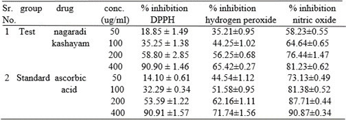
Data are represented as mean ±SD.
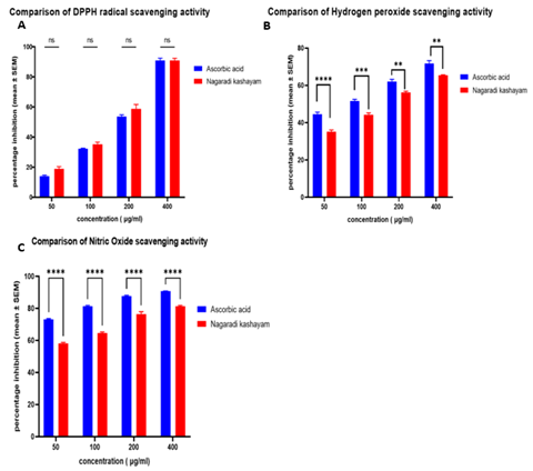
Fig. 1. Effect of Nagaradi Kashayam on DPPH, hydrogen peroxide, and nitric oxide scavenging. n = 3, all values are expressed as mean ± SEM. One way ANOVA followed by Tukey multiple comparison test was done for statistical comparison. The asterisk mark is considered statistically significant. ns - non significant; **** indicates p value < 0>
Effect of Nagaradi Kashayam on body weight, liver weight, and liver morphology
In this study, Fig. 2. illustrates the effect of Nagaradi kashayam on the body weight (A) and liver wet weight (B) of rats. In comparison to the normal control group, disease-group rats showed a significant increase in body weight during the 10th week. On the other hand, nagaradi kashayam treatment significantly reduced the high-fat diet-induced gain in body weight. Also, liver weight was significantly increased in the disease group, whereas the nagaradi kashayam (NK) treatment group showed a significant reduction in liver weight in comparison to the disease group. When the morphological changes of the liver were compared, the fatty liver clearly increased in mass and had a pale appearance in the disease group, as shown in Fig. 3, in comparison to the normal reddish-brown liver of the control group. This colour change was due to the accumulation of lipid in the liver, while the gross appearance of the liver in the kashayam treatment groups was more similar to the normal control group. A mild fatty change was observed in the liver of the low-dose Kashayam-treated group.
Table 2. represents the body weight changes of animals during the period of study in response to the treatments given.
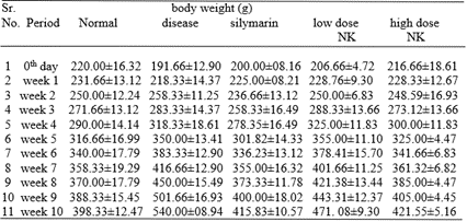
Data are represented as mean ±SD.
Table 3. represents the liver wet weight of various treatment groups at the end of study.
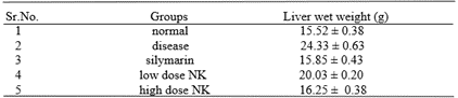
Data are represented as mean ±SD.
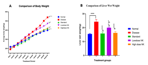
Fig 2. (A) The effect of nagaradi kashayam on body weight. n = 6. The asterisk mark is considered statistically significant. a**** denotes p <0>

Fig. 3: Photographs of gross rat liver specimens from various treatment groups A. normal control; B. disease control; C. silymarin-treated group; D. low-dose kashayam-treated group; E. high-dose kashayam-treated group.
BIOCHEMICAL ANALYSIS
Estimation of SGOT, SGPT, and ALP, total cholesterol, and triglycerides
The hepatic markers such as serum SGOT, SGPT, and ALP were significantly increased in high-fat-fed disease rats in comparison to the normal control group (Fig. 4). While treatment with Nagaradi Kashayam showed a significant reduction in these enzyme levels, The total cholesterol was evaluated for all groups. A significant difference was observed in the kashayam treatment group when compared to the disease group (Fig. 4D). Also, the triglyceride level was higher in the disease group. In all the other treatment groups, the triglyceride level was significantly lower (Fig. 4E). This suggests that the rise in serum triglyceride and total cholesterol levels due to the high fat feeding was suppressed by the treatment with nagaradi kashayam.
Estimation of MDA, GSH
MDA concentration in liver was significantly elevated in high fat diet group (fig 5A). The treatment groups showed significant decrease in MDA level compared to disease control group. When comparing GSH levels (fig 5B), disease group showed a significant increase due to increased oxidative stress in high fat diet-induced fatty liver. Nagaradi kashayam low dose and high dose treated groups showed a dose-dependent increase in GSH activity.
Table 4. represents the serum levels of SGOT, SGPT, ALP, TG and total cholesterol of various treatment groups.
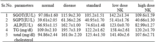
Data are represented as mean ±SD.
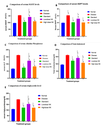
Fig 4. Effect of Nagaradi kashayam on SGOT, SGPT, ALP, total cholesterol, and triglycerides (A-E). n=4, all values are expressed as mean ± SEM. One way ANOVA followed by Tukey multiple comparison test was done for statistical comparison. The asterisk mark is considered statistically significant. a**** indicates p value < 0>
Table 5. represents the MDA and GSH levels obtained from liver homogenate of all treatment groups.
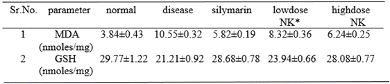
Data are represented as mean ±SD.
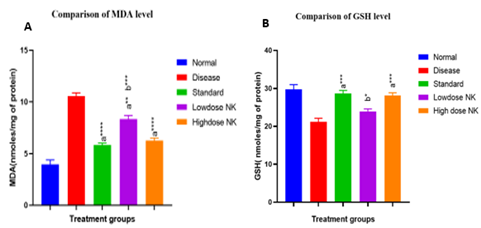
Fig 5. Effect of Nagaradi Kashayam on MDA (A) and GSH (B). n = 4, all values are expressed as mean ± SEM. The asterisk mark is considered statistically significant. a*** indicates p value < 0>
Histopathological studies
In H&E staining (Fig. 6), the normal group showed normal liver architecture, where as in the disease group, many globular clear fat vacuoles were observed. The marked arrow indicates fat vacuoles. The presence of small vacuoles in the low dose kashayam group indicated mild fatty changes. The histology of liver in high dose kashayam group was more similar to normal control group. When compared to the normal, the oil red o-stained section (fig 7), showed accumulation of lipid which is stained as red globules in disease group. The marked arrow represents fat globules. The standard treated group and high dose kashayam treated groups were found to be protected from fatty liver from histological assessment.
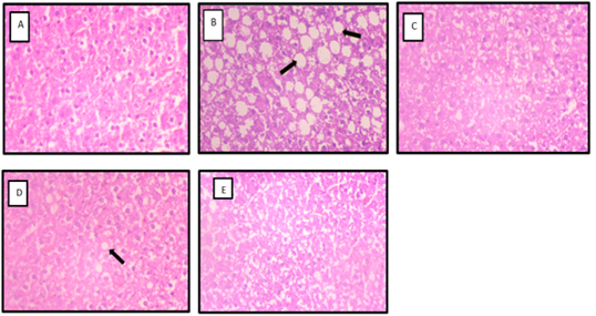
Fig 6. Photomicrographs of H & E-stained sections of rat liver (H & E x 400) A. Normal control, B. Disease control, C. Silymarin treated group, D. Low dose kashayam treated group, E. High dose kashayam treated group.
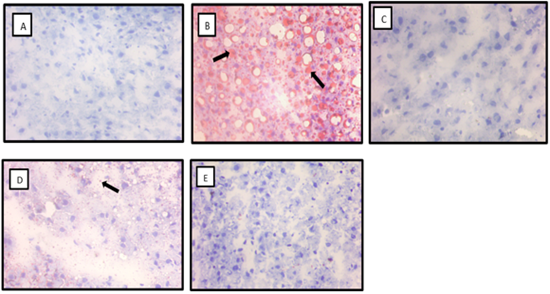
Fig 7. Photomicrographs of Oil Red O-stained sections of rat liver (Oil Red O x 400) A. Normal control, B. Disease control, C. Silymarin treated group, D. Low dose kashayam treated group, E. High dose kashayam treated group.
DISCUSSION
The use of plants as a source of medicine has been an ancient practice, and it is an important component of the health care system in India. Traditional ayurvedic medicines, especially polyherbal formulations, play a vital role in treating a variety of diseases. A large population depends on this system of practice due to its cost-effectiveness and few or no known side effects. The phytochemical screening of the formulation showed the presence of phytoconstituents such as carbohydrates, tannins, flavonoids, and saponins, which supports the earlier studies. In previous studies, the antioxidant activity of constituent herbs of Nagaradi Kashayam was reported. The current study investigated the free radical scavenging effect of Nagaradi Kashayam in DPPH, hydrogen peroxide, and nitric oxide and showed significant activity. It is assumed that the antioxidant activity might be due to the presence of flavonoids. HF-diet-fed rats showed an increase in body weight compared to the control rats. The weight gain may be attributed to the high calories in the diet. This obesogenic diet model represents the most preferred model for obesity and NAFLD as it mimics the obesity pattern in humans. Obesogenic diets are known to induce NAFLD through metabolic syndrome and insulin resistance, which results in an increased flow of FFA into the liver. Lipid oxidation and the TCA cycle are increased in NAFLD, indicating that hepatocytes enhance oxidation when counteracting lipid overload. The excess reducing equivalents generated by beta oxidation cannot be resolved in the mitochondrial chain, resulting in higher ROS generation. ROS accumulation can trigger hepatic stress pathways and lead to progression to steatohepatitis (NASH). The free radicals produced cause lipid peroxidation and mitochondrial GSH depletion. As shown in this study, the lipid peroxidation product, malondialdehyde, levels in the liver were higher in the disease group. The lower levels of GSH in the liver might be associated with the increased oxidative stress in high-fat diet-fed rats. Nagaradi Kashayam significantly increased the level of GSH and showed a significant reduction in MDA levels compared to the disease control group. This indicates that Nagaradi Kashayam can provide a defence system against ROS-induced hepatic stress. The oxidative damage in tissues can lead to hepatocellular damage, as evident by the elevated levels of liver marker enzymes such as SGOT, SGPT, and ALP, which are considered markers of hepatocellular dysfunction. The elevated levels of these enzymes were observed in HF-fed rats. The nagaradi kashayam treatment suppressed oxidative stress-mediated tissue damage and normalised serum levels of SGOT, SGPT, and ALP enzymes. This finding of our study supports the results of previously published articles that reported the hepatoprotective activity of individual constituent herbs of Tinospora cordifolia and ginger. In this study, fatty liver was confirmed by histopathological assessment. Numerous fat vacuoles were found in the liver sections of HF-fed rats in both H&E and oil red o staining. High-fat-fed rats developed hyperlipidemia, which was confirmed by the elevated serum levels of total cholesterol and triglycerides.
CONCLUSION
In conclusion, this study showed that Nagaradi kashayam inhibited the high-fat diet-induced oxidative stress, attenuated levels of total cholesterol and triglycerides, and suppressed hepatic steatosis in NAFLD rats. However, the identification of the active principles responsible for the activities and the mechanisms of action are yet to be studied in detail. Further studies need to be carried out to find out the benefits of in a clinical set up.
ABBREVIATIONS
ALP: Alkaline phosphatase; FFA: Free fatty acid; GSH : reduced glutathione; HF: High fat; MDA: Malondialdehyde; NAFLD: Non-alcoholic fatty liver disease; NASH: Non-alcoholic steato hepatitis; NK: Nagaradi kashayam; ROS: Reactive oxygen species; SGOT: Serum glutamate oxaloacetate transaminase; SGPT: Serum glutamate pyruvate transaminase; TBA: Thiobarbituric acid; TCA: Tricarboxylic acid. TG: Triglycerides.
CONFLICT OF INTEREST STATEMENT
We the authors would like to declare that there is no conflict of interest regarding the publication of this paper.
ACKNOWLEDGEMENT
The research was conducted in the college of pharmaceutical sciences, Govt. Medical College Thiruvananthapuram. We gratefully acknowledge the logistic support provided by the department of pharmaceutical sciences, Govt. Medical College Thiruvananthapuram.
REFERENCE

Stephy George, Shifila A. N., Evaluation Of The Hepatoprotective Effect Of Nagaradi Kashayam (Amruthotharam) In High Fat Diet Induced Model Of Fatty Liver Disease In Male Wistar Rats, Int. J. of Pharm. Sci., 2024, Vol 2, Issue 8, 2542-2554. https://doi.org/10.5281/zenodo.13208953
 10.5281/zenodo.13208953
