
We use cookies to ensure our website works properly and to personalise your experience. Cookies policy
Bengal School of Technology, Sugandha, Delhi Road, Hooghly- 712102, West Bengal, India
Exosome-based drug delivery has emerged as a transformative approach in modern therapeutics due to the natural biological origin, biocompatibility and unique intercellular communication capabilities. These nano-sized extracellular vesicles (30–150 nm), released by almost all cell types, carry proteins, lipids and nucleic acids that enable targeted signaling and molecular transport. Their intrinsic ability to cross biological barriers, including the blood–brain barrier, low immunogenicity and high physiological stability make them superior to many synthetic nano-carriers such as liposomes or polymeric nanoparticles. Exosomes protect encapsulated therapeutic cargo from enzymatic degradation and enhance pharmacokinetics, making them promising carriers for small molecules, proteins, peptides, and nucleic acids. Exosome technical advancements have enabled surface modification for targeted distribution and optimized loading tactics such as passive pre-loading and active post-loading procedures. Ultracentrifugation, size-exclusion chromatography, immune-affinity capture, and microfluidics are all important approaches for isolating high-purity vesicles for therapeutic application. Exosomes have great therapeutic promise in a variety of areas including cancer, neurological disorders, inflammatory illnesses, and renal problems. Their innate tropism and ability to carry genes and proteins emphasize their importance in regenerative medicine and precision treatment. Despite this potential, there are still issues with large-scale production, standardization, low yield, heterogeneity and regulatory uncertainty. Continued breakthroughs in bioengineering, separation procedures and storage stability are critical for clinical translation. Overall, exosome-based drug delivery is a promising and adaptable technology that has the potential to revolutionize targeted therapy and personalised medicine.
Drug distribution plays a vital role in ensuring both the Efficacy and Safety of therapeutic agents. Over the years, various Nanocarrier systems such as Liposomes, Micelles, Dendrimers, Polymeric Nanoparticles, and Inorganic Nanoparticles have been developed to enhance drug delivery. These systems aim to improve the Pharmacokinetic and Pharmacodynamic (PK–PD) profiles of drugs, thereby increasing therapeutic effectiveness while minimizing off-target effects and toxicity.[1] Despite these advances, several limitations remain challenges include achieving precise organ targeting, overcoming toxicity associated with certain chemical and physical properties, and avoiding unfavorable immune responses. To address these issues, researchers are now exploring innovative alternatives.
Among these, Exosomes have recently emerged as a highly promising approach in nanomedicine. Exosomes are nanosized Extracellular Vesicles (30–100 nm) naturally secreted by various cells and found in biological fluids such as blood, cerebrospinal fluid, urine, and saliva.[2] Their unique structural and functional properties make them highly attractive for drug delivery. Composed of a lipid bilayer encapsulating proteins, nucleic acids, and lipids, exosomes mediate intercellular communication by transferring bioactive molecules like proteins, mRNAs, and miRNAs to recipient cells, thereby modulating cellular signaling and function.[3] Notably, their ability to cross the blood–brain barrier (BBB) enhances neurological drug delivery while allowing repeated administration with minimal toxicity. Exosomes offer several advantages over traditional delivery systems, including high stability, excellent biocompatibility, low immunogenicity, and the ability to traverse biological barriers for precise, targeted delivery.[4] They protect encapsulated therapeutics from degradation and improve stability, making them ideal carriers for diverse bioactive molecules. Recent research has rapidly expanded the use of exosomes for delivering drugs, nucleic acids, and proteins through both passive and active loading methods. Surface modification with ligands or antibodies further enhances their targeting capability [5]. Beyond drug delivery, exosomes are being explored in vaccine development, gene and protein therapy, regenerative medicine, and cancer treatment.[6] This review aims to summarize current progress in exosome-based delivery systems, including their composition, engineering strategies, and therapeutic applications. It also highlights their commercial potential and future opportunities for advancing targeted and effective therapeutic interventions.[7]
Exosomes:
Exosomes, a specialized subtype of Extracellular vesicles (EVs), were initially identified as cellular mechanisms for disposing of unwanted materials. Today, however, they are recognized as vital mediators of intercellular communication, transferring nucleic acids, proteins, and metabolites to target cells. These nanosized vesicles originate from endosomes membrane-bound compartments within the cell and are enriched with a diverse range of bioactive molecules, including lipids, proteins, growth factors, nucleic acids, and metabolites.[8]
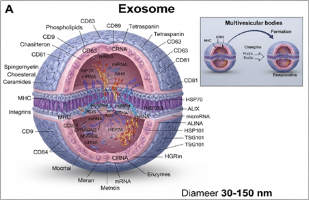
Figure 1: Structure of Exosomes
Exosome formation occurs through three key steps:
Nearly every cell type in the human body secretes exosomes, which are nano spherical, bilayer lipid enclosed structures. Once released, they circulate through systemic pathways, enabling widespread physiological communication.
Types of Exosomes:
Exosomes can broadly be classified into Three categories:
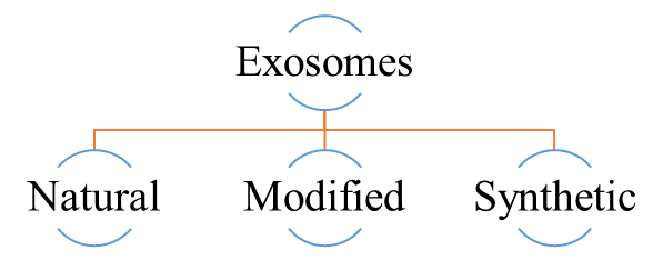
Natural Exosomes:
Natural exosomes originate from either animal or plant sources. Animal-derived exosomes are categorized into those from normal and tumor cells [9]. Various cell types—such as epithelial, endothelial, MSCs, macrophages, dendritic, and immune cells—naturally secrete exosomes, with their biological origin dictating specific functions. For instance, macrophage-derived exosomes can influence the tumor microenvironment and exhibit anticancer effects, whereas MSC-derived ones act as therapeutic and drug delivery vehicles [6,10]. Exosomes are found in biofluids like milk, serum, blood, and urine, and have been explored for drug delivery, such as paclitaxel-loaded milk exosomes. Additionally, exosomal miRNAs show diagnostic value, aiding in conditions like spinal cord injury and endometrial cancer.
Modified Exosomes:
Modified exosomes are natural exosomes that have been engineered to enhance therapeutic potential. Modifications may involve changes to the internal structure (to improve drug loading or cargo protection) or alterations of the external surface (to achieve targeted delivery and better biodistribution) [11].
Artificial Exosomes (Synthetic Exosomes):
Synthetic exosomes are created to mimic natural exosomes. They are typically produced using either cell-based methodologies or lipid-bio membrane fabrication techniques, enabling the design of customizable delivery vehicles.
Figure 2: Types of Exosomes
Composition of Exosomes:
EVs can be broadly divided into two categories: small (exosomes) and large (microparticles/micro vesicles, oncosomes, ectosomes, or apoptotic bodies). Although micro vesicles, which make up the most diverse population, have a diameter of between 50 to 1000 nm, large EVs are plasma membrane-derived and vary from approximately 100 to 1000 nm. Apoptotic bodies, the biggest subtype of EVs (>1000 nm), are formed during the cell fragmentation process during apoptosis. Although the biogenesis mechanisms and sizes of these big EVs are similar, their functional cargos and roles are not.[12] Although there is some overlap between the two, tiny EVs are mostly distinguished by their endosome origin and smaller size. Exosomes have a dimension of are created by consecutive invagination of the plasma membrane, which eventually leads to the formation of intracellular multivesicular bodies (MVBs) made up of Intraluminal vesicles (ILVs), with a range of approximately 30 to 150 nm (average of approximately 100 nm). ILVs are eventually discharged as exosomes once the plasma membrane and MVB fuse.[13] The expression of particular marker proteins, such as the tetraspanins CD63, CD81, and CD9, is typically used to identify exosomes. Additionally, exosomes inherently carry in addition to RNA species including messenger RNA (mRNA), microRNA (miRNA), ribosomal RNA (rRNA), and transfer RNA (tRNA), DNA cargo includes mitochondrial DNA (mt DNA), double stranded DNA (dsDNA), and single stranded DNA (ssDNA). Additionally, they contain a number of cell-derived components, such as cytosolic and cell-surface proteins and metabolites. Lipids such as sphingomyelin, glycosphingolipids, cholesterol, phosphatidylserines (PS), and ceramide is also abundant in exosomes. Surprisingly, the kind and condition of parental cells have a significant impact on the composition profile of exosomes.[14]
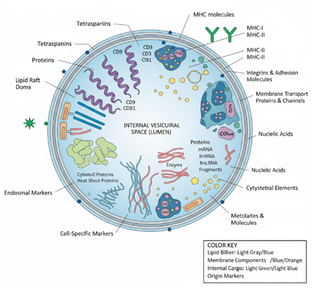
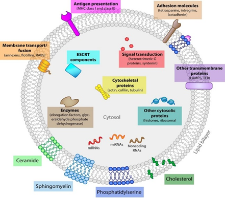
Figure 3: Composition of Exosome.
Advantages and Disadvantages of Exosomes Delivery System:
Advantages:
Disadvantages:
Exosome-based drug delivery offers natural, biocompatible, and highly targeted therapeutic potential but faces major challenges in large-scale production, standardization, and safety validation. [17]
Biogenesis of Exosomes:
It is crucial to identify the processes behind exosome biogenesis since doing so may open up new avenues for therapeutic intervention. There are already many known mechanisms for MVB biogenesis. Neutral sphingomyelinase and ceramide have been shown to be essential for the formation of the Intravehicular membrane of MVBs. It is the endosomal sorting complex for transport (ESCRT) machinery that has been the most thoroughly documented pathway for MVB biogenesis. Syndecan has also been linked to exosome secretion through its interactions with many ESCRT proteins, syntenin, and Alix. Phospholipids, tetraspanins, and the Ras-related protein GTPase Rab are a few more complex proteins that are involved in the biogenesis process; nevertheless, a better understanding of their exact mechanisms is necessary, especially In-Vivo. Different approaches to exosome synthesis, enrichment, and/or concentration may result in differences in the regulatory factors linked to exosome biogenesis being identified. [18]
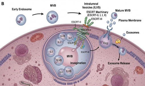
Figure 4: Exosome Biogenesis
Cellular Origins of Exosomes:
Exosomes derived from different cell sources possess unique characteristics that influence their therapeutic potential. Selecting the right donor cell is therefore essential for designing effective exosome-based delivery systems. Human embryonic kidney (HEK) cell exosomes are preferred for their high yield, low immunogenicity, and suitability for systemic delivery due to broad tissue compatibility and ease of genetic modification. Immune cell-derived exosomes, such as those from dendritic cells, carry immune-stimulatory molecules (MHC I/II, co-stimulatory proteins) and serve as potent carriers for cancer immunotherapy [19]. Mesenchymal stem cell (MSC)-derived exosomes exhibit intrinsic therapeutic effects—anti-inflammatory, antifibrotic, and pro-angiogenic—though scalability remains challenging; modified hESC-MSCs expressing c-myc can enhance yield without compromising quality. Tumor-derived exosomes mainly serve diagnostic roles but may also act as immune adjuvants or targeted carriers in anticancer therapy due to their natural tumor tropism [20].
Figure 5: Cellular Origins of Exosomes:
Isolation of Exosome:
As previously stated, vesicles are released by all cells; however, their quantity, composition, and physicochemical characteristics might vary. The gold standard differential centrifugation, filtering, size exclusion chromatography, immunoaffinity capture-based, polymer precipitation, and microfluidics-based methods are among the various methods now employed for exosome isolation. Covering the characteristics and principles of isolation, their benefits and drawbacks, and instances of the application of these isolation approaches. [21] It is still challenging for a particular method to overcome all the related issues, such as batch-to-batch variance, limited yield, or low purity, despite the development of exosome purification methodologies. A thorough characterization of exosomes is essential, specifically with regard to their size, shape, concentration, presence of exosome-enriched indicators, and absence of contaminants.[22]
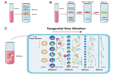
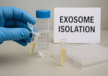
Figure 6: Exosome Isolation
Table 1: Comparison of Techniques for Isolating Exosomes [23]
|
Method |
Core Concept |
Advantages |
Drawbacks |
Recovery Level |
Purity Level |
Sample Size Needed |
|
Ultracentrifugation / Density Gradient |
Uses very high centrifugal force to sediment exosomes. |
Cost-effective (minimal reagent use). |
Labor-intensive, lengthy, may pull down non-exosomal material. |
Low |
High |
Large |
|
Differential Centrifugation |
Sequential spins at increasing speeds separate vesicles by size. |
Maintains particle integrity; avoids introducing external markers. |
Requires advanced instruments, technically demanding. |
High |
Low |
Large |
|
Polymer-based Precipitation |
Chemical agents induce exosomes to aggregate and fall out of solution. |
Simple workflow, preserves vesicle shape, no structural damage. |
Risk of polymer contamination in later analyses. |
High |
Medium |
Small |
|
Ultrafiltration |
Filtration through membranes with specific pore sizes. |
Rapid, economical, and scalable. |
Possible vesicle loss due to membrane adhesion or clogging. |
Medium |
Medium |
Medium |
|
Immunoaffinity Capture |
Antibodies bind to exosome surface markers for selective isolation. |
Highly specific, straightforward method. |
Expensive; may compromise vesicle integrity. |
Low |
High |
Small |
Exosome Manufacturing Processes:
In terms of cell culture and purification, the manufacturing (production and purification) of exosomes, a unique type of biotherapeutic, is comparable to that of biologics. In order to produce exosomes, the parent cell line must be cultured, harvested from the conditioned media, and separated or purified from process-related impurities as extracellular vesicles. Upstream and downstream are the two phases of the exosome production process.[23]
Upstream Processing:
Efficient large-scale exosome production requires selecting an optimal cell source and culture medium. Common donors include MSCs, dendritic cells, HEK293, and 293T, with MSC-derived exosomes prized for their therapeutic properties. MSCs can be isolated from bone marrow, adipose tissue, or umbilical cord, and early screening ensures desired biological effects.[24] Serum-free media are preferred over conventional supplements like FBS or hPL to avoid contamination, though media composition must be optimized to maintain cell behavior. Production systems include static culture flasks and dynamic bioreactors; hollow-fiber bioreactors enable continuous harvesting and improve yield, scalability, and consistency.[25]
Downstream Processing:
Downstream processing of exosomes involves removing debris, concentrating media, and isolating vesicles using ultracentrifugation, size-exclusion chromatography (SEC), microfiltration, tangential flow filtration (TFF), or immunoaffinity capture.[26] Methods separate exosomes by size, density, or surface markers, with selection depending on product goals and upstream complexity. Ultracentrifugation is conventional but may cause aggregation or damage. TFF and hollow-fiber ultrafiltration offer gentler, scalable alternatives with better biomolecule retention and purity. Immunoaffinity capture provides high specificity but is less scalable, while SEC efficiently removes protein and lipoprotein contaminants, reducing particle and ferritin content compared with ultracentrifugation. [27,28]
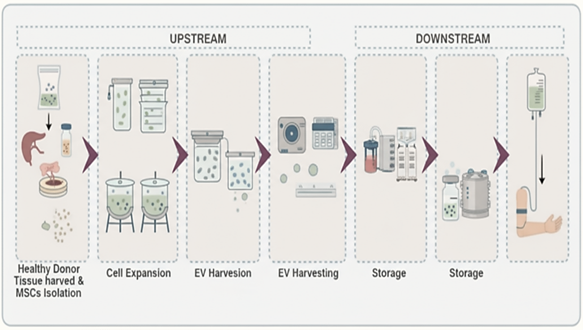
Figure 7: Exosome Manufacturing Processes
[Upstream & Downstream Processing]
Fill Finish:
Following exosome purification, the final product needs to be cryopreserved in a storage buffer that preserves the integrity of the exosomes' vesicles and kept in an appropriate container closure system. Commonly, cryopreservation at (- 800C) with cryoprotectants is employed to improve the stability of proteins and cells after freezing and lessen osmotic damage.[29]
Exosome-Based Drug Loading Strategies:
The lipid bilayer of exosomal vesicles functions as a protective shield, preventing the degradation of their cargo during circulation in the bloodstream. Despite this advantage, the same bilayer structure and the natural molecular composition of exosomes pose challenges for efficient drug loading. Two primary strategies are commonly employed: passive loading and active loading.[30] Passive loading, also called preloading, relies on donor cells that have been pretreated with the drug, which then release drug-containing exosomes without the need for further manipulation of the vesicles. In contrast, active loading (or post loading/remote loading) involves incubating purified exosomes with a drug, enabling direct incorporation. Studies indicate that active loading typically achieves a higher drug-to-vesicle ratio due to energy-driven transport processes and is generally more effective for hydrophobic compounds than for hydrophilic ones.[31]
Table 2: Comparative Analysis of Exosome Drug Loading Techniques: Benefits and Limitations [28,32-36]
|
Passive Loading |
Drug Loading Method |
Mechanism |
Primary Benefits |
Major Limitations |
|
Exosome/Drug Incubation |
Cargo molecules diffuse across exosomal or cellular membranes. |
Straightforward technique without compromising membrane integrity. |
Limited loading capacity. Possible cytotoxicity to donor cells. |
|
|
Donor Cell/Drug Incubation |
Free drugs are internalized by donor cells, and incorporated into exosomes. |
Simple, non-invasive process maintaining membrane stability. |
Inefficient drug loading. Risk of cytotoxic effects to donor cells. |
|
|
Sonication |
Mechanical shear force generates micropores in exosome membranes. |
Enables higher cargo encapsulation than incubation alone. |
Can disrupt membrane structure, impede scalability and cause aggregation of exosome contents. |
|
|
Extrusion |
Exosomes undergo repeated recombination, mixing cargo and surface structures. |
Provides efficient and uniform encapsulation of cargo. |
May compromise immune invisibility of exosomes, increasing recognition by phagocytic cells. |
|
|
Active Loading |
Freeze–Thaw Cycles |
Cycles of freezing and thawing facilitate membrane fusion. |
Efficient for loading drugs, proteins, and peptides directly into exosomes. |
Repeated cycles can degrade proteins and aggregate exosomes, with lower efficiency than alternatives. |
|
Electroporation |
Electric fields create transient pores in exosome membranes. |
Achieves high drug loading efficiency. |
Cargo aggregation and reduction in loading efficiency present persistent challenges. |
|
|
Membrane Permeabilizer Incubation |
Membrane disruptors (e.g., cholesterol, saponin) form pores on exosome surfaces. |
Improved cargo encapsulation over passive methods. |
Saponin poses toxicity risk in vivo and requires further purification to eliminate residuals. |
Delivery Pathways of Exosomes:
Various administration routes have been explored to deliver therapeutic agents or cargo-loaded exosomes to specific tissues or organs. These include Intravenous and Intratumoral methods, both of which can result in systemic distribution of the exosomes. The choice of administration route plays a crucial role in determining how exosome-associated drugs are distributed within the body.[37]
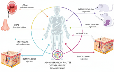
Figure 8: Exosomes Administration Routes
Table 3: Optimizing Exosome Administration: Therapeutic Benefits and Disease Applications [37, 38]
|
Routes of Administration |
Targeted Disease |
Advantages |
|
Intravenous |
Stroke, Parkinson’s disease, traumatic brain injury, acute kidney injury, antitumor therapies (prostate and breast cancer). |
Most common route for systemic administration of exosomes. |
|
Intraperitoneal |
Bronchopulmonary dysplasia, Autoimmune type 1 diabetes |
Allows the loading of larger EV doses. |
|
Oral |
Facilitates resolution of colitis, arthritis |
Convenient administration route for patients. |
|
Intranasal |
Brain parenchyma, Brain cancer, Encephalitis (inflammation of the brain), Parkinson’s disease therapy |
Suitable for EV delivery into the brain, surpassing the blood brain barrier. |
|
Intratumoral |
Glioblastoma multiforme, antitumor therapies |
More effective strategy for antitumor therapies due to higher EV retention in tumors. |
Evaluation of Exosomes:
Exosomes are classified according to their physical, chemical, functional, structural, and biological characteristics [39]. Exosomes may require further characterization for specialized applications, such as in enzyme replacement therapy (ERT), where they carry drugs or biological chemicals to the site of action. To synthesize consistent exosomes with appropriate composition, structural, and functional features, exact processes must be followed.
The methodologies for characterization of exosomes are given below.
Nanoparticle Tracking Analysis (NTA):
Nanoparticle tracking analysis (NTA) quantifies particle diameter by using particle light scattering and Brownian motion. The hydrodynamic diameters are calculated by observing particles' simultaneous, individual Brownian motion. Particles are imaged in discrete locations, making it feasible to distinguish between sample sizes. Fluorescent labelling may be used by NTA to quantify fluorescence and estimate antigen presence in exosomes [40].
Dynamic Light Scattering Technique (DLS):
Dynamic Light Scattering (DLS), also known as Photon Coherence Spectroscopy, measures particle size and dispersion. The system detects particles by analyzing optical signals generated by particle light scattering. This technique is good for forecasting exosome size; however, it lacks source data. The technique's main limitation is that bigger particles might interfere with the detection of smaller particles in formulations. As a result, the size distribution obtained using this approach is not ideal [41].
Atomic Force Microscopy (AFM):
Cryogenic transmission electron microscopy (cryo-TEM) is a widely used method for determining vesicle sizes; however, it is not without limitations. Obstacles include the high cost of the equipment, the need for knowledge for sample preparation, imaging, and data interpretation, and the limited number of visible particles in pictures. To address these issues, atomic force microscopy (AFM) provides a viable alternative. AFM can reveal extracellular vesicles' shape, size, and other biophysical properties [44]. It also maps the mechanical characteristics of exosomes and determines their size distribution. The cantilever beam tip is used to scan the surface with sub-nanometer resolution. AFM can measure structure, biomechanics, and abundance.
Microscopy Study:
Transmission Electron Microscopy (TEM):
Transmission electron microscopy (TEM) analyses particle shape and structure using an accelerated electron beam. TEM pictures of exosomes provide an approximation of their size. Furthermore, preparing a TEM sample might affect the shape of exosomes. Scientists developed the "Cryo-TEM" approach to overcome the limitations of traditional techniques. Cryo-TEM is a sophisticated method that eliminates sample preparation effects [45]. Exomes are spherical in form, whereas exosomes are diverse. In some cases, the electron beam may kill biological material. Isolated exosomes have a cup-shaped structure when analyzed by TEM, but frozen exosomes have circular shapes when investigated using cryo-TEM [46].
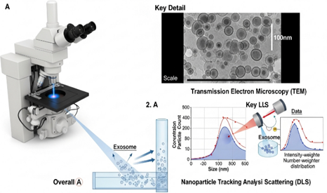
Figure 9: Exosomes Physical Characterization
Scanning Electron Microscopy (SEM):
Scanning Electron Microscopy (SEM) detects low-energy electrons emitted from near the sample surface. It can provide valuable sample data such as morphology, composition, surface texture, and roughness [47]. SEM may reveal surface features of exosomes, including size, shape, and morphology. Backscattered electrons (BSE) detectors may be useful in some circumstances, particularly for exosomes with heavy metal labelled surfaces [48]. This method collects electrons from deeper depths beneath a sample's surface, providing information on its composition and topography.
Enzyme-Linked Immunosorbent Assay (ELISA):
A plate-based enzyme-linked immunosorbent assay may detect and quantify proteins, peptides, hormones, and antibodies. Although it can identify exosomes, it requires more samples and is less sensitive. This test, which uses ELISA, is intended to yield precise cancer exosome counts. It also detects exosomes in plasma, serum, and urine using various antibodies [49]. It also detects exosomes in plasma, serum, and urine using various antibodies.
Fluorescence Correlation Microscopy (FCM):
Microfluidic-dependent Fluorescence Correlation Microscopy (FCM) uses a specific antibody to collect exosomes, which are then tagged with a fluorescent dye and analyzed using a plate reader. FCM may be utilized to construct a microfluidic chip for immunocapture and quantitative exosome analysis [50].
Colorimetric Detection:
The colour intensity of a chromogenic material determines the particles detected by calorimetry. Exosomes were collected utilizing microfluidic technology, and exomes were recognized and quantified using ELISA. This method has been effectively used by scientists and researchers to identify exosomes released by cancer cells [51].
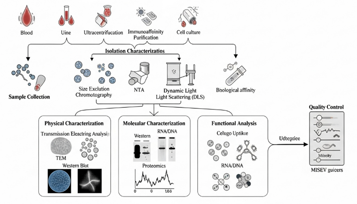
Figure 10: Evaluation of Exosomes
Exosomes as Novel Drug Delivery Systems:
Advanced drug delivery technologies, such as liposomes and polymeric nanoparticles, encapsulate pharmaceuticals for anticancer, antiviral, and antifungal therapy, but they struggle with biocompatibility, long-term stability, and immune targeting [52]. Exosomes are nanoscale delivery vehicles that provide benefits such as biocompatibility, biodegradability, low toxicity, target selectivity, tiny size, membrane fusion ability, extended half-life, and minimum immune reaction. They can be programmed to carry proteins, peptides, nucleic acids, and other substances between cells [53].
Small Molecules:
Exosomes are effective small-molecule therapeutic carriers with nanoscale dimensions, minimal toxicity, and great biocompatibility. They increase targeted administration by demonstrating enhanced pharmacokinetics and anticancer efficacy; for example, doxorubicin-loaded exosomes have higher dispersion and cellular absorption [54]. Exosomes loaded with doxorubicin outperformed free and liposomal forms in terms of cell uptake and biodistribution, but exosomes loaded with curcumin increased solubility, stability, and bioavailability [55].
Exosomal curcumin reduces IL-6 and TNF-α levels and increases survival in septic shock models, relative to free curcumin. It also lowers the number of CD11bGr-1 cells in the lungs, indicating that hydrophobic medicines like curcumin are more effectively delivered [56]. Exosomes can spontaneously traverse the blood-brain barrier, increasing medication distribution to the brain while decreasing MPS clearance. Unlike manufactured nanocarriers, which are restricted by nanotoxicity and poor brain dispersion even after PEGylation, exosomes improve the permeability and therapeutic effectiveness of centrally acting medicines.
Large Molecule (Protein and Peptide Delivery):
Exosomal drug delivery allows for the transfer of big molecules like proteins and peptides for therapeutic and diagnostic reasons while also eliminating undesirable biological components including proteins, lipids, and nucleic acids [57]. Exosomes are suitable carriers for proteins and peptides because of their inherent capacity to transfer endogenous proteins outside the cell [58]. Recent research indicates that in Parkinson's disease, exosomes containing the antioxidant catalase can penetrate the BBB, improving disease outcomes [59].
Nucleic Acids:
Exosomes may transfer DNA and RNA to particular cells, causing genetic alterations in both normal and pathological situations. They effectively alter cell activity by targeting the cytoplasm, making them potentially useful tools for gene therapy and genetic medication delivery [60].
Small Interfering RNAs (siRNAs):
SiRNA modulates target genes in genetic treatment, although there are stability and circulation concerns. Using targeted carriers improves distribution to particular cells, overcoming these restrictions [21]. Exosomes, with their minimal immunogenicity and great RNA transport capabilities, are good siRNA carriers. Lamp2b-modified dendritic and HEK-293 exosomes deliver targeted siRNA to cancer cells, indicating therapeutic potential.
Exosomes as Emerging Therapeutic Tools:
Exosomes are nanosized extracellular vesicles (30-150 nm) released by most cells and containing proteins, lipids, and nucleic acids that govern intercellular communication. Their biocompatibility, minimal immunogenicity, and capacity to pass across barriers such as the blood-brain barrier make them ideal natural drug delivery vehicles. Exosome engineering advancements have shown considerable potential for targeted treatment and regenerative medicine in illnesses like as cancer, neurological, cardiovascular, and inflammatory diseases.
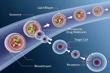
Figure 11: Exosomes Drug Delivery Vehicle
Cancer Disease:
Exosomes are potential cancer therapies due to their innate tumour-targeting and communication capacities. Using biomarkers such as ZIP4, HSP60, and EphrinA2, they penetrate tumours for efficient medication administration and diagnostics. They are more effective when transported via CD47. Exosomes containing siRNA, miR-122, or CRISPR/Cas9 can decrease tumours and improve chemosensitivity, while integrins, TGF-β, VEGF, and IL-8 may increase metastasis. Exosome production or uptake inhibition can restrict tumour spread, highlighting their dual significance in cancer treatment [62].
In Neurological Diseases:
Parkinson’s Disease:
Parkinson's disease (PD), the second most prevalent neurodegenerative condition after Alzheimer's, is characterized by α-synuclein aggregation, leading to motor and cognitive symptoms. Exosomes show potential as biomarkers and therapeutic carriers due to their capacity to cross the BBB, assisting with diagnosis and treatment, yet clinical obstacles remain for their full utilization [63].
Inflammatory Disease:
Inflammation protects against infection, but it can also cause illness when it becomes severe. It is controlled by immune cells, cytokines, and chemokines, with exosomes playing important immunoregulatory functions. Exosomal alterations, such as IFN-regulated SLC22A5, are used as biomarkers for inflammatory illnesses such cancer, IBD, diabetes, obesity, rheumatism, and neurological disorders. Exosomes are easily separated from saliva or urine and allow for non-invasive testing; for example, higher exosomal MIF is linked to PDAC metastasis, whereas intestinal and macrophage exosomes impact IBD development [64].
Renal Diseases:
The kidney is vital for maintaining bodily homeostasis, and exosomes help with renal physiology by modulating cell-cell interactions inside nephrons. According to studies, urinary exosomes perform a dual role in renal function and immunity, protecting the urinary system against infections. They include innate immune proteins including as lysozyme-C, dermcidin, mucin-1, calprotectin, and myeloperoxidase, which prevent the development of both infective and non-infective E. coli strains [64].
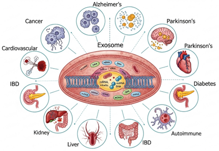
Figure 12: Therapeutic applications of Exosomes in various Diseases
Challenges and Constraints of Exosome-Derived Drug Delivery Approaches:
Exosomes offer great potential as natural drug delivery vehicles due to their biocompatibility and ability to cross biological barriers; however, several challenges limit their clinical application. Their extraction and purification processes are complex, yielding low quantities and lacking standardized high-purity isolation methods. Moreover, exosomes exhibit limited loading capacity for therapeutic molecules, and their endogenous contents can provoke immunogenic responses. Following in vivo administration, they are rapidly cleared from systemic circulation, primarily by macrophages in the spleen and liver. While exosomes can bypass endosomal and lysosomal degradation pathways—offering an advantage over synthetic carriers like liposomes—their use in non-conventional routes (e.g., ophthalmic or transmucosal) remains underexplored due to issues with tissue penetration and stability. Additionally, exosome heterogeneity in size, composition, and origin complicates characterization, standardization, and large-scale production. Overcoming these biological, technological, and regulatory hurdles is crucial for the successful clinical translation of exosome-based drug delivery systems.
Future Directions and Opportunities in Exosome-Based Drug Delivery:
The potential of exosome vesicles as drug delivery systems largely depends on the source and type of the parent cells. Due to their natural ability to deliver therapeutic payloads to recipient cells through endogenous uptake mechanisms, exosomes are considered highly promising carriers for drug delivery. However, several challenges must be addressed before exosomes can be scaled up for clinical applications. These include the development of reliable and scalable isolation methods, efficient drug-loading strategies, and standardized storage protocols. Long-term storage stability remains a critical concern. Studies suggest that techniques such as lyophilization with cryoprotectants like trehalose can preserve exosomal integrity and maintain the stability of their endogenous contents, including proteins and RNA. Another key challenge is ensuring that exosome-based therapeutics comply with regulatory requirements for clinical approval. Unlike other nanomedicines, regulatory frameworks for exosome-based therapeutics are still underdeveloped, which may slow their translation to clinical use.
Although research on exosome-based drug delivery is still in its early stages, ongoing advancements in characterization techniques and targeted delivery strategies are expected to overcome these obstacles, paving the way for the safe and effective clinical application of exosome therapeutics
CONCLUSION:
Exosome-based drug delivery has emerged as a promising approach for targeted and efficient therapeutic delivery. Owing to their natural origin, biocompatibility, and ability to cross biological barriers, exosomes offer significant advantages over conventional nanocarriers. They can effectively transport drugs, nucleic acids, and proteins to specific cells, reducing off-target effects and improving treatment outcomes. Looking forward, the integration of nanotechnology, synthetic biology, and bioengineering could lead to next-generation exosome-based therapeutics. With continued research and innovation, exosomes hold immense promise as natural, customizable, and efficient carriers for precision medicine, potentially revolutionizing the future of drug delivery and disease management.
REFERENCES


Paramita Dey, Syed Asraful Kadir, Sayan Samanta, Exosome Based Drug Delivery: Emerging Trends and Therapeutic Potential, Int. J. of Pharm. Sci., 2025, Vol 3, Issue 12, 608-628. https://doi.org/10.5281/zenodo.17804904
 10.5281/zenodo.17804904