
We use cookies to ensure our website works properly and to personalise your experience. Cookies policy
College of Pharmaceutical Sciences, Government Medical College, Kozhikode, Kerala, 673008, India.
Transdermal drug delivery systems (TDDS) have emerged as a favorable alternative to conventional routes due to their non-invasive nature, improved patient compliance, and controlled drug release. However, the effectiveness of TDDS is often limited by the skin’s outermost layer—the stratum corneum—which serves as a major barrier, especially for drugs with high molecular weight or poor lipophilicity. To overcome this, microneedle (MN) technology has emerged as a promising third-generation transdermal approach. MNs are microscopic, needle-like structures that create transient microchannels in the skin, enhancing drug penetration without reaching nerve-rich deeper layers, thus making the process virtually painless. They enable effective delivery of various therapeutic agents, including small molecules, peptides, proteins, and vaccines. Microneedles are classified into five main types: solid, coated, dissolving, hollow, and hydrogel-forming—each with distinct mechanisms for drug loading and release. The type and fabrication method significantly influence delivery efficiency, release kinetics, and patient comfort. Various fabrication techniques such as micromolding, lithography, and 3D printing are employed to produce microneedles with precise dimensions and suitable materials. These methods enable customization for specific applications. Microneedles combine the benefits of injections and transdermal patches, offering improved compliance, painless administration, and targeted delivery. Despite their advantages, challenges such as scale-up, biocompatibility, and regulatory issues persist. This review offers a comprehensive overview of microneedle technology, focusing on classification, mechanisms, fabrication, advantages, challenges, and applications. Overall, microneedles represent an innovative and versatile platform in pharmaceutical and biomedical fields.
Transdermal drug delivery has been a growing focus in pharmaceutical research due to its potential advantages over conventional oral and intravenous routes. The skin, being the largest organ in the human body, serves as a protective barrier against water loss, injury, and infection1. However, the outermost layer of the skin, the stratum corneum, limits the penetration of many drugs, restricting the number of therapeutics that can be effectively delivered via this route. Despite these challenges, transdermal delivery remains an attractive approach due to its pain-free administration, controlled drug release, and reduced systemic side effects2. To overcome the low permeability limitation of the stratum corneum, advanced transdermal drug delivery strategies have emerged3. The evolution of transdermal systems has been categorized into three generations: the first generation relied on drugs that could naturally penetrate the skin, the second generation utilized chemical and physical enhancers to improve permeability, and the third generation focused on disrupting the skin barrier using microneedles, thermal ablation, and electroporation. Among these, microneedle drug delivery systems (MNDS) have gained significant attention as a minimally invasive and highly effective approach4. Microneedles are micron-scale projections designed to create temporary micro channels in the skin, facilitating the delivery of both small and large molecules, including vaccines, biologics, and therapeutic agents.1 Unlike traditional hypodermic needles, microneedles offer the advantage of painless drug administration, reduced risk of infections, and improved patient compliance. Various microneedle designs—such as solid, coated, dissolving, hollow, and hydrogel-based microneedles—have been developed to optimize drug delivery efficiency. Additionally, advancements in microneedle fabrication techniques, including three-dimensional (3D) printing, electroporation, and cavitational ultrasound, have further expanded their potential applications3. This review aims to explore the principles, types, fabrication methods, and applications of microneedle drug delivery systems while discussing emerging trends and challenges in the field.
The skin, the largest organ in the human body, plays a vital role in protecting against external threats, including water loss, physical injury, and microbial invasion. It covers an area of approximately 1.5 to 2.0 square meters in adults and makes up about 16% of total body weight. This multifaceted organ not only forms a protective barrier but also helps maintain homeostasis and performs sensory, thermoregulatory, and immune functions1. Structurally, the skin is composed of three primary layers: the epidermis, dermis, and hypodermis, which work together to regulate permeability and contribute to its barrier properties. The epidermis, which lacks vasculature, is the outermost layer and measures between 60 and 800 µm in thickness. Its uppermost sublayer, the stratum corneum, comprises about 15–20 layers of specialized, anucleated keratinocytes. With a thickness of 10–20 µm, the stratum corneum functions as the primary barrier, preventing water loss and blocking the entry of external substances. Beneath the epidermis lies the dermis, which is 3 to 5 mm thick and contains nerve endings, sweat glands, lymphatic vessels, and hair follicles. The hypodermis, or subcutaneous tissue, lies below the dermis and is composed primarily of adipose and connective tissue, providing insulation, cushioning, and a network of larger blood vessels that facilitate systemic drug absorption2. Despite the skin’s barrier function, it serves as an appealing site for drug administration due to its accessibility and potential to deliver various therapeutics, including vaccines, biomolecules, and small molecules. However, the lipid-rich and hydrophobic nature of the stratum corneum limits drug penetration and bioavailability5. Drugs applied to the skin can penetrate via three potential pathways: through the sweat ducts, via the hair follicles and sebaceous glands, or directly across the stratum corneum. The effectiveness of drug delivery depends on factors such as the lipid solubility and molecular weight of the therapeutic agents. Understanding these pathways and overcoming the natural barriers of the skin are key aspects of transdermal drug delivery research3. The structure of skin with its different layers are depicted in Figure 1.
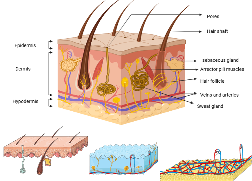
Figure 1: structure of skin: Cross-sectional view of human skin showing the epidermis, dermis, and hypodermis along with key structures like hair follicles, sebaceous glands, sweat glands, blood vessels, and arrector pili muscles.
In the past two decades, there has been growing interest in developing delivery systems that enhance the penetration of drugs through the deeper layers of the skin. This progress has expanded the use of hydrophilic and macromolecule drugs in the treatment of various deep tissue and dermatological conditions. Several physical and pharmaceutical techniques have been explored for transdermal drug delivery (TDD), including sonophoresis, electroporation, iontophoresis, microneedles (MNs), chemical enhancers, and transdermal formulations2. These strategies have demonstrated the ability to improve the absorption of hydrophilic and macromolecule drugs by bypassing the gastrointestinal tract, avoiding first-pass metabolism, and increasing patient adherence due to their non-invasive nature. However, the major challenge to the widespread application of transdermal delivery lies in overcoming the stratum corneum, the outermost layer of the epidermis, which acts as a significant barrier to drug penetration. To address this challenge, substantial research has been directed toward developing methods that either enhance penetration passively or actively or bypass the stratum corneum altogether2. Transdermal drug delivery involves the direct application of drugs to the skin, where they penetrate through the stratum corneum and pass through the underlying epidermis and dermis. When the drug reaches the dermal layer, it becomes available for systemic absorption. The primary goal of this method is to facilitate drug diffusion across the skin and into the bloodstream in a controlled manner4. The history of transdermal drug delivery has been categorized into four distinct generations, as described by Prausnitz and Langer. The first generation primarily relied on patch-based systems that enabled low drug delivery through natural diffusion. The second generation focused on chemical enhancers to improve drug penetration. The third generation introduced advanced technologies such as microneedles, electroporation, and thermal ablation, which could bypass or disrupt the stratum corneum, thereby allowing more targeted drug delivery. The fourth and most recent generation integrates drug delivery with sensing technologies, enabling precise control over the release of pharmaceutical agents4. Different types of transdermal drug delivery systems are illustrated in Figure 2.
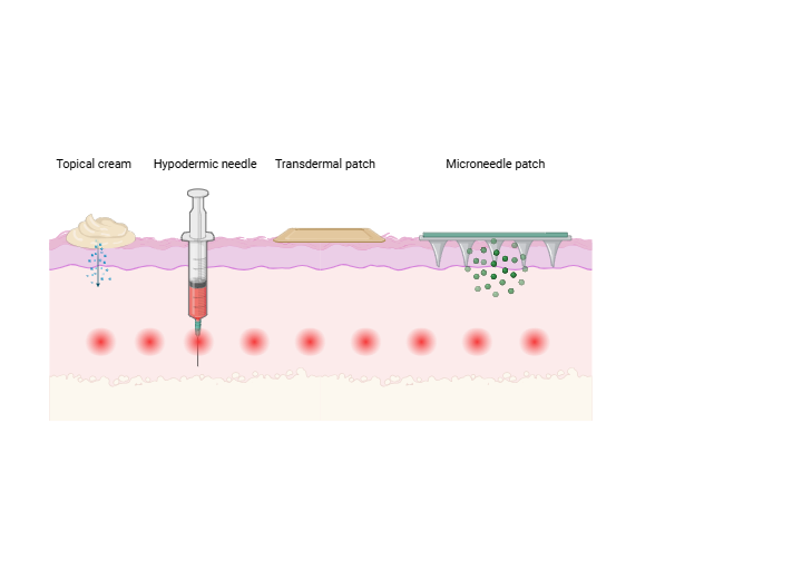
Figure 2: Comparison of drug delivery methods across the skin: topical cream, hypodermic needle, transdermal patch, and microneedle patch showing varying penetration depths and delivery mechanisms.
Microneedles are micrometer-scale needle-shaped structures that can be used to deliver drugs or vaccines through the skin. Microneedles are painless because they have minimal or no interaction with nerve endings in the papillary dermis. Some microneedles are made of a drug to be delivered to the body but are shaped into a needle so they will penetrate the skin and ranges in size, shape, and function but are all used as an alternative to other delivery methods like the conventional hypodermic needle or other injection apparatus1,6. Microneedles are microstructures that are sharp and robust enough for skin penetration, made using microelectromechanical systems (MEMS) technology. MEMS provided a platform for microfabrication of compact miniaturized medical devices for human health screening, monitoring, and diagnostic purposes. The application of microneedle patches to the skin produces micro sized pathways for transporting molecules, including biomedical antigens and cells7. Transdermal drug delivery through microneedle is illustrated in Figure 3.
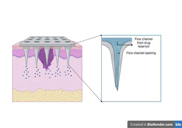
Figure 3: microneedle as transdermal drug delivery system: Schematic of a microneedle system showing drug delivery through flow channels from the reservoir into the skin layers via microneedle openings.
Microneedles (MNs) are an innovative transdermal drug delivery technology designed to overcome the skin's barrier properties while ensuring patient compliance. These microscopic, three-dimensional structures, typically less than 1000 µm in length, create transient microchannels in the skin, enabling efficient drug permeation without reaching pain receptors. Unlike conventional hypodermic needles, which are often associated with pain and reduced patient acceptance, microneedles offer a minimally invasive, self-administrable alternative with enhanced bioavailability. Various types, including solid, coated, dissolving, hydrogel, and hollow microneedles, have been developed using fabrication techniques such as photolithography, micro-molding, and laser ablation6. Structure and application of a microneedle patch on skin surface is illustrated in Figure 4.
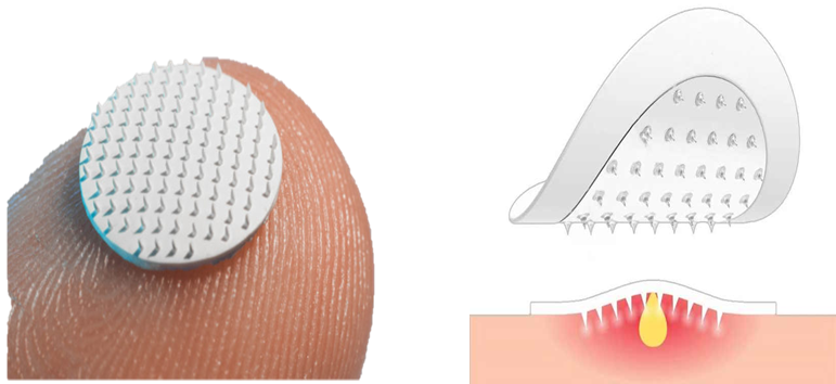
Figure 4: microneedle patch: Application and structure of a microneedle patch showing its placement on the skin surface and penetration into the upper skin layers for drug delivery.
The concept of microneedles has evolved over the years. In 1905, Dr. Ernst Kromayer used motor-powered dental burs to treat skin conditions like scarring and hyperpigmentation. The first mention of microneedles appeared in 1921 when Chambers used needles to inject into an egg’s nucleus. By the 1960s, interest grew in delivering drugs via the stratum corneum, leading to the introduction of the microneedle concept in the 1970s. In 1979, the first transdermal patch for delivering scopolamine was approved. In 1998, silicon microneedles were fabricated using ion etching and photolithography, marking a breakthrough in transdermal drug delivery research. By 2004, microneedle arrays were developed to pierce the skin, enabling further exploration of solid, coated, hollow, dissolvable, and hydrogel-forming microneedles. In 2005, dissolvable microneedles were first used for transdermal drug delivery, and the first microneedle clinical trial was completed in 2007. Recent advances include the use of 3D printing and additive manufacturing to produce low-cost microneedle molds for large-scale production4.
The diffusion mechanism governs the topical route of medication distribution. The skin is momentarily disturbed in the microneedle drug delivery device. In order to administer enough medication to provide the necessary therapeutic response, hundreds of microneedles are arranged in arrays on a tiny patch, similar to a typical transdermal patch that is sold in stores. It avoids the barrier layer by penetrating the stratum corneum. When the medication reaches the site of action, it immediately penetrates the epidermis or higher dermis layer, enters the systemic circulation, and produces a therapeutic effect8.
Size and shape: Microneedles are small, with lengths ranging between 20µm and 2000 µm, short enough to penetrate the epidermis without reaching deeper nerve endings, minimizing pain and discomfort.
Material composition: Microneedles can be fabricated from various materials like silicon, metal, ceramics, biodegradable polymers, or hydrogels depending on the intended application.
Mechanical strength: Microneedles must exhibit enough mechanical strength to pierce the stratum corneum without bending, breaking, or causing skin damage.
Drug loading and release mechanism: The design influences drug release, whether immediate (e.g., coated microneedles) or sustained (e.g., hydrogel-based microneedles).
Biocompatibility: Materials used in microneedles, especially dissolving MNs, must be biocompatible and safe for long-term applications9.
Advantages of microneedles
Minimal invasiveness: Microneedles penetrate the stratum corneum without reaching nerve-rich deeper layers, minimizing pain and discomfort.?
Biocompatibility: Materials used (e.g., polymers, metals, ceramics) are typically biocompatible, reducing the risk of adverse reactions.?
Versatility: Microneedles are suitable for delivering a wide range of therapeutics, including small molecules, proteins, vaccines, and nucleic acids.?
Controlled drug release: Microneedles allow for precise control over drug delivery, enabling sustained or immediate release based on design.?
High patient compliance: The non-invasive nature and ease of application of microneedles improve patient adherence compared to traditional injections.?
Scalability and cost-effectiveness: Microneedles can be manufactured using cost-effective techniques like photolithography, 3D printing, and molding, making them scalable for mass production.?
Minimal risk of infection: Due to their small size and minimal skin penetration, microneedles pose a lower risk of infection compared to hypodermic needles.?
Self-administration potential: Microneedle patches can be easily applied by patients themselves without the need for trained medical personnel.?
Customizable geometry and design: Microneedles can be tailored in terms of length, shape, and density to suit specific applications, such as targeting different skin layers.?
Safety and minimal residual waste: Dissolvable and biodegradable microneedles leave no sharp waste, enhancing safety and environmental friendliness 10.
Disadvantages of microneedles
Skin reactions: Common adverse effects include transient erythema (redness), eema (swelling), and pain at the application site, typically resolving within a week.?
Post-Inflammatory Hyperpigmentation (PIH): Individuals with darker skin tones may experience PIH, leading to uneven skin coloration following treatment.?
Risk of infections: Although minimal, there remains a potential for infections, especially if MNs are used on compromised skin or without proper hygiene.?
Material sensitivities: Allergic reactions can occur due to sensitivity to materials used in MN fabrication, such as certain metals or polymers.?
Limited depth of penetration: MNs may not be suitable for delivering drugs intended for deeper tissues, as their penetration depth is restricted to the superficial layers of the skin11.
Types Of Microneedles
Table 1: summary of types of microneedles12
|
Category |
Material |
Fabrication |
Advantages |
Limitation |
|
Solid MNs |
Metals, silicon, ceramics |
Laser cutting, etching, photolithography |
High mechanical strength, |
Infection, inflammation |
|
Coated MNs |
Metals, silicon, ceramics |
Spray coating, dip coating, piezoelectric inkjet printing |
High mechanical strength, single-step application |
Lower drug capacity |
|
Hollow MNs |
Metals, silicon, polymers |
Microelectromechanical systems, 3D printing |
High scalability, high stability |
Complex and expensive fabrication method |
|
Hydrogel MNs |
Crosslinking polymers |
Micro molding, 3D printing |
Unbreakable, multifunctional |
Insufficient mechanical strength and toxicity |
|
Dissolvable MNs |
Biodegradable and biocompatible polymers |
Micro molding, drawing lithography, 3D printing |
Scale-up fabrication, High biocompatibility |
Limit dosing and inconsistent pharmacokinetics |
Solid MNs: They follow a "poke and patch" approach. MNs create micro-channels in the skin, followed by drug application using a transdermal patch. They can also collect interstitial fluid (ISF) for analysis.
Hollow Microneedles (MNs): They feature a hollow tip filled with a drug, which is deposited into the epidermis upon insertion and the drug release will follow “poke and flow” method.
Coated MNs: Use the "coat and patch" method, where the drug formulation is coated on the MNs and dissolves upon skin penetration to release the drug.
Dissolving MNs: Made from biodegradable polymers, they dissolve after insertion, delivering the drug in a simple one-step process.
Hydrogel-Forming MNs: These MNs swell upon skin contact, forming channels for sustained drug delivery or fluid collection13. (Figure 5)
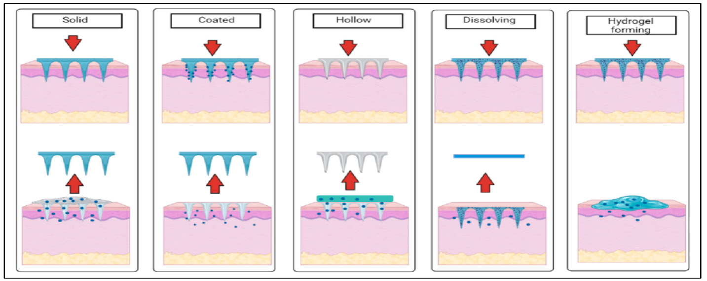
Figure 5: Schematic illustration of different types of microneedles used in transdermal drug delivery systems. Solid microneedles (poke and patch) – create micro channels in the skin followed by topical drug application. Coated microneedles (coat and poke) – drug coated onto solid microneedles is released upon skin insertion. Hollow microneedles – allow liquid drug to flow through a bore directly into the skin. Dissolving microneedles – made of biodegradable polymers that dissolve and release the drug payload. Hydrogel-forming microneedles – swell upon skin insertion and enable drug release from an attached reservoir through the hydrated matrix.12
The first microneedles investigated for drug administration through the skin were solid types, utilizing the “poke and patch” mechanism. This approach involves using the needles to create temporary microholes in the skin, followed by the application of a drug-containing patch14. Solid microneedles are mostly used to create microscopic pores in the skin that increase the permeability of drugs. By creating channels in the stratum corneum, the outermost layer of the skin that is often impervious to most drugs, these microneedles serve as a pretreatment step rather than delivering the medication. Once these channels are established, medications can be applied as lotions, gels, creams, or ointments to the treated area. Deeper skin penetration and better drug distribution to the underlying layers are made possible by this. This passive diffusion improves the drug's bioavailability and systemic absorption15. Several researchers are also investigating stainless steel microneedles. Following the installation of stainless steel MN arrays, improved distribution of captopril and metoprolol tartrate was investigated8. Stainless steel, silicon, titanium, polymethyl methacrylate (PMMA), and polylactic acid (PLA) are common materials used to make solid microneedles because they improve their mechanical strength, sharpness, and accuracy. Compared to hollow microneedles, solid microneedles are easier to make, have better mechanical qualities, and have sharper points 4. Solid microneedles have also been used by dermatologists in collagen induction treatment, a cosmetic procedure that increases the creation of collagen in the skin to rejuvenate it. These microneedles have certain drawbacks despite their advantages, which include excellent mechanical qualities, sharp tips, and ease of production. When coated with pharmaceuticals, they might show poor drug loading capability and problems with dose accuracy. However, compared to conventional intramuscular injections, solid microneedles have demonstrated efficacy in vaccine delivery because of their capacity to elicit a strong and sustained immune response16. Additionally, solid microneedles coated with drugs act as both penetration enhancers and drug storage. When these microneedles are inserted into the skin, they transfer the medication coating to the dermis, ensuring a rapid release of active ingredients. This feature is particularly useful for drugs that must start working immediately. However, problems including limited biological compatibility, potential skin irritation, and precise dosing still need to be fixed. Solid microneedles' simple design and manufacturing method, along with their ability to improve drug delivery efficiency, make them a popular choice for dermatological applications and transdermal drug delivery (TDD) systems2. (Figure 6)
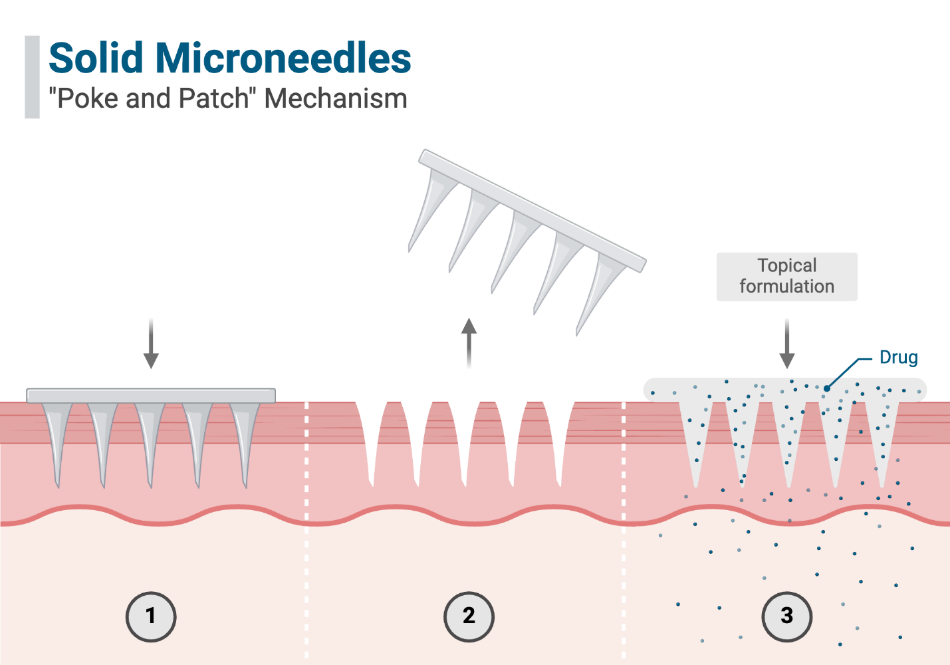
Figure 6: Schematic representation of solid microneedles used in the coat-and-patch method; where microneedles first create microchannels in the skin (1), are then removed (2), and a topical drug formulation is applied over the site for enhanced transdermal delivery through the created channels (3).
Hollow microneedles differ significantly from solid microneedles due to their design, which includes an empty lumen or internal cavity for the storage and delivery of liquid drug formulations. These microneedles have tiny openings at their tips, allowing the direct deposition of drugs into the viable epidermis or dermis layer upon insertion6. Because of this characteristic, they are especially well-suited for delivering high molecular weight substances that could otherwise find it difficult to pass through the skin's natural barrier, like proteins, vaccines, and oligonucleotides. Hollow microneedles actively distribute medications by pressure- or diffusion-driven flow, in contrast to solid microneedles that depend on passive diffusion. According to clinical needs, this permits fast medication infusion or continuous, extended therapeutic activity through controlled, time-dependent drug release17. Hollow microneedles are useful for administering liquid vaccinations against illnesses including influenza, anthrax, and Japanese encephalitis because they may contain a larger amount of drug than other kinds of microneedles4. By enhancing antigen consumption and lymphatic absorption, they also strengthen immunological responses. Because they resemble conventional hypodermic syringes, these microneedles offer a less unpleasant and painful substitute. Its manufacture is more difficult, nevertheless, because an ideal interior cavity with precise dimensions needs to be made. Usually between 50 and 70 micrometers in diameter, the interior aperture must be made to prevent leakage or blockage during medicine delivery6. Nevertheless their potential, hollow microneedles have several drawbacks, such as problems with insertion precision, mechanical stability, and possible obstruction or leakage during injection. Compared to solid microneedles, their widespread use has been considerably constrained by these technical issues. However, throughout time, improvements in materials science and microneedle fabrication methods including photolithography, laser cutting, and three-dimensional laser ablation have enhanced their functionality, durability, and design. Hollow microneedles are potential instruments for vaccine delivery and bio sensing applications, where they can harvest interstitial fluid for diagnostic purposes, by precisely delivering doses by optimizing factors like aspect ratio and flow rate2. (Figure 7).
Figure 7: Illustration of hollow microneedle-mediated drug delivery used in the poke and flow method; , involving microneedle insertion (1), drug infusion from a reservoir through hollow channels into the skin (2), and subsequent microneedle removal (3).
Coated microneedles
Coated MNs employ the "coat and patch" method, where the drug formulation is coated onto the MNs before skin application. Upon penetration, the coating dissolves, depositing the drug into the skin13. Active pharmaceutical ingredients (APIs) are incorporated onto the surface of coated microneedles in order to get over the restrictions that come with drug loading in solid microneedles. The drug, usually in a dry form, is applied to the microneedle shaft using specific coating procedures such layer-by-layer deposition, dip-coating, or spray-coating. Excipients such as polymers, stabilizers, and surfactants may be used in these coatings to enhance the drug layer's adherence, homogeneity, and release profile. The drug coating quickly dissolves after being put into the skin, allowing the active ingredient to enter the skin's living layers and begin working as a medication. Coated microneedles' quick drug release makes them appropriate for uses needing instant drug action, like administering vaccines, managing pain, or administering emergency therapies18. Delivering accurate, premeasured doses of powerful medications in little amounts while reducing the risk of overdose is one of the main benefits of coated microneedles. Coated microneedles are also helpful for delivering high-potency substances like peptides, DNA vaccines, and antigens in microliter amounts since the drug is restricted to the surface. There are still issues, though, mainly with the uniformity of drug distribution on the microneedle surface and the possibility of drug loss during handling or storage. To improve these systems' stability and distribution effectiveness, researchers are also looking into biocompatible and bioadhesive coatings. Despite these difficulties, coated microneedles have enormous promise for usage in therapeutic fields like vaccination, dermatology, and cosmetics because of their low invasiveness, user-friendliness, and increased patient compliance4. (Figure 8).
Figure 8: Schematic representation of coated microneedle-mediated drug delivery using the "coat and poke" approach. The drug is coated onto the surface of solid microneedles (left), which are inserted into the skin. Upon insertion (middle), the drug rapidly dissolves off the microneedle surface into the skin layers. The microneedles are then withdrawn, leaving no residue (right).
Dissolving microneedles-
Dissolving microneedles are a new type of microneedle that combines the drug and needle matrix into a biodegradable and biocompatible polymer to overcome the drawbacks of conventional microneedles. Typically, these microneedles are made of substances that dissolve entirely in the skin after being injected, such as hyaluronic acid, polyvinyl alcohol (PVA), carboxymethyl cellulose, or polyvinylpyrrolidone (PVP). During the manufacturing process, the medicine is integrated into the matrix, guaranteeing that the microneedle will dissolve and deliver the whole dose. The microneedles disintegrate in a matter of minutes to hours after being inserted into the skin, allowing the medication to be released gradually. By removing the need to remove the needle, this device lowers the possibility of needle reuse, biohazardous waste, or unintentional damage19. Their versatility and safety have drawn a lot of attention, particularly in applications like vaccine distribution, where they have demonstrated the ability to boost immunogenicity by specifically targeting immune cells in the epidermal layer of the skin. Their application in gene therapy and cancer treatment has also increased due to their capacity to transport macromolecules, such as proteins, peptides, and nucleic acids19. They also give thermolabile medications better stability, sometimes doing away with the necessity for cold-chain storage. Despite their benefits, problems with drug loading capacity, mechanical strength, and dissolution time still need to be addressed. The performance and scalability of dissolving microneedles are continuously being enhanced by advancements in polymer chemistry, micro molding methods, and sophisticated drug formulations, making them a viable instrument for next transdermal drug delivery (TDD) systems.20, 21, 2219, 23, 24 (Figure 9).
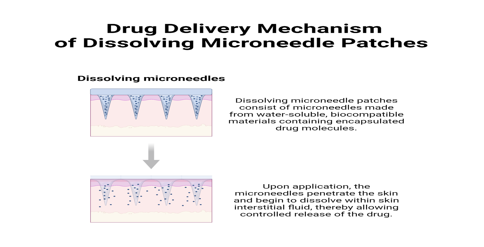
Figure 9: illustration of dissolving microneedle drug delivery; the microneedles, made from biodegradable and water-soluble polymers, are inserted into the skin where they rapidly dissolve upon contact with interstitial fluid, releasing the encapsulated drug directly into the dermal layer. No sharp waste remains after application.
Hydrogel-forming microneedles
One cutting-edge kind of microneedle technology intended for regulated and prolonged medication administration is hydrogel-forming microneedles. These microneedles do not dissolve or break down when inserted into the skin, in contrast to coated or dissolving microneedles. Rather, they expand when they come into contact with interstitial fluid (ISF), creating a network of porous hydrogel that permits the passage of medications from a reservoir that is attached. Hydrogel-forming microneedles, which are usually composed of hydrophilic, biocompatible polymers like polyethylene glycol (PEG), polyvinyl alcohol (PVA), or alginate, establish long-lasting micro channels in the skin that allow for continuous drug delivery over hours or even days25. Delivering hydrophilic and hydrophobic medications, such as proteins, peptides, nanoparticles, and small molecules, is one of the main benefits of hydrogel-forming microneedles. In applications like glucose sensing for diabetic patients, they can also be utilized to harvest interstitial fluid for diagnostic purposes, allowing for real-time health monitoring. The microneedles reduce the possibility of skin irritation, infection, or negative reactions since they stay in place both before and after medication administration. Furthermore, hydrogel-forming microneedles can be designed to behave in a stimuli-responsive manner, in which the concentration of glucose, pH, or temperature causes the release of a medication25. However, to guarantee effective skin penetration without causing early swelling, the mechanical strength of hydrogel-forming microneedles needs to be carefully adjusted. The goal of ongoing research is to improve the hydrogel matrix's tunability so that drug release kinetics may be precisely controlled and production is made easier. Because hydrogel-forming microneedles may combine drug delivery and diagnostic capabilities, they hold great potential as a platform technology in the areas of wearable biosensing devices, customized medicine, and chronic illness management25. Swellable MNs are composed of biocompatible hydrogels that constantly absorb bodily fluids until they are saturated. Although post-processing is frequently necessary, these hydrogels are safe for ISF extraction and biomarker detection. Recently designed porous MNs gather ISF through interconnected pores, allowing fluids to be transported directly and analyzed by biosensors13 (Figure 10).
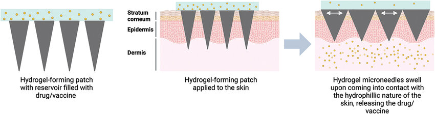
Figure10: Illustration of hydrogel-forming microneedle drug delivery. Upon insertion into the skin, the dry microneedles (left) absorb interstitial fluid and swell (middle), forming a continuous hydrogel network that enables controlled drug release from an attached drug reservoir. The microneedles are removed after the drug is delivered (right), leaving no solid residue in the skin.
Methods Of Fabrication
Manufacturing methods for MNs differ, and different MN kinds call for distinct methods. Microneedle (MN) arrays can be fabricated using various techniques, including laser ablation, micro-molding, additive manufacturing, injection molding, chemical etching, micromachining, and lithography-electroforming-replication26.
Laser ablation
This method makes it possible to create MN arrays by removing material from a substrate using a concentrated optical light beam. Lasers of many varieties, including CO2, UV excimer, and femtosecond lasers, have been used for a variety of purposes at the micro and nanoscales. It takes 10 to 100 nanoseconds for laser ablation to reach the material's burn point, making it a quick and efficient procedure. It provides a low-heat, non-contact method for shaping metals. Thermal impacts, however, have the ability to change the mechanical characteristics and structure of MN, which could result in cracking or decreased fatigue resistance. Furthermore, lasers are expensive and not appropriate for production on a wide scale4 (Figure 11).
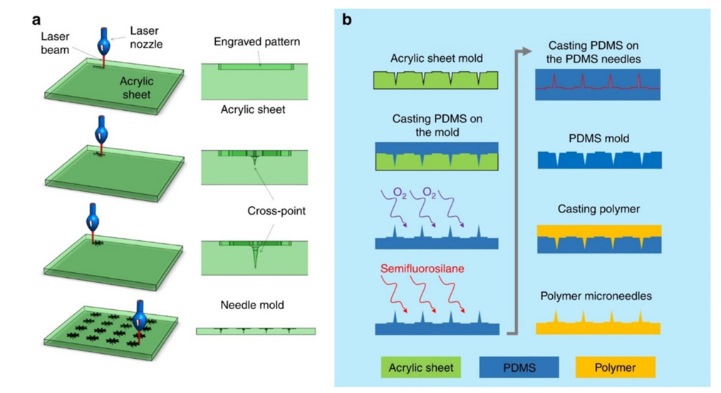
Figure 11: laser ablation4
Lithography
The most popular technique for transferring geometric patterns onto a substrate is photolithography, which is particularly useful in microelectronics. It is essential to MN manufacturing and micromachining. Glass, metal, ceramics, and polymers can all be used in this procedure to create accurate designs with smooth vertical sidewalls. However, it requires a sophisticated cleanroom environment, is expensive (30–35% of integrated circuit fabrication expenses), and takes a lot of time4 (Figure 12).
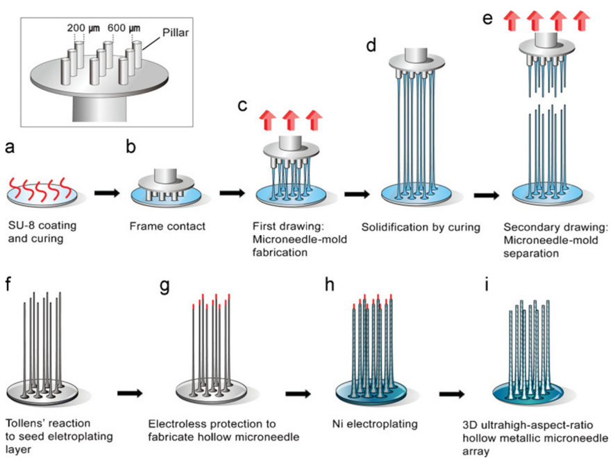
Figure 12: lithograpghy4
Micro-Molding
Making copies of a master mold with a polymer solution—often combined with active medicinal ingredients—is known as micro-molding27. Because of its low cost, excellent repeatability, scalability, and straightforward technique, it is a popular and economical way to produce MNs in large quantities. The procedure consists of five steps: creating the female mold, filling it with bubble-free polymer, solidifying the polymer, removing the MN patch, and preparing the master MN template. Despite its effectiveness, it might have trouble regulating drug dose, penetration depth, and polymer mechanics26. Acupuncture microneedles were utilized to create the master template in a micro molding investigation. To modify the aspect ratio, the acupuncture microneedles were affixed to a plastic substrate with holes that were carefully drilled. A polydimethylsiloxane (PDMS) mold made via replica molding was then used to manufacture a biodegradable polylactic acid (PLA) microneedle array. This procedure illustrated the promise of this economical micro molding method for a range of microneedle applications and showed successful transdermal drug delivery28 (Figure 13).
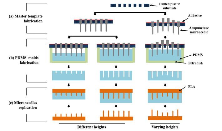
Figure 13: micro molding28
3D Printing (3DP) and Microneedles (MNs):
The idea of personalized medicine is supported by 3D printing (3DP), which makes it possible to create medications with precise dosages, forms, and release schedules. It makes it possible to create MNs with intricate geometries, ideal dimensions, and enhanced mechanical qualities for more effective drug administration and skin penetration. By enabling trans-nasal brain-targeted delivery of medications, this technology broadens the range of MN uses beyond conventional transdermal drug delivery systems (TDDSs). 3DP is a promising method for enhanced TDDSs because of its capacity to optimize drug loading, solubility, stability, and controlled release through the refinement of MN structures. Its drawbacks include a slow rate of production, difficulties with high drug loading, inefficient large-scale manufacture, and a restricted supply of excipients. Notwithstanding these limitations, 3DP is a pharmaceutical technology that is developing quickly and has a lot of promise29. The creation of microneedles (MNs), an essential part of minimally invasive drug delivery systems (TDDS), has been revolutionized by 3D printing technology. In contrast to conventional techniques (molding, etching, and grinding), 3D printing provides speed, accuracy, flexibility, and cost-effectiveness, which makes it perfect for tailoring MN designs for particular medical uses.
Types of 3D printing methods utilized in fabrication in microneedles 29.
Various 3D printing techniques are used based on the intended qualities, material suitability, and MN application:
Stereo lithography (SLA):
This process creates high-resolution MNs with sharp tips for accurate drug delivery using photo polymer resins.
Digital Light Processing (DLP):
Suitable for precise MN designs, DLP provides quick prototyping and biocompatible materials.
Fused Deposition Modeling (FDM):
This thermoplastic-based technique is less expensive than SLA and DLP, although it has a lower resolution.
Additional methods for additive manufacturing:
These include material extrusion, binder jetting, and powder bed fusion, which provide MN manufacturing even more versatility. 3D printing technology continues to advance MN manufacturing, enhancing precision, customization, and integration with biosensors. By overcoming challenges like material compatibility and improving scalability, 3D printing is poised to play a key role in the development of next-generation drug delivery systems and personalized healthcare solutions29.
Types of microneedles and their common fabrication techniques
Different types of microneedles and their common methods of fabrication are summarized in the Table 2.
Table 2: fabrication methods of microneedles8,30
|
Type of MNS |
Fabrication Techniques |
|
Solid microneedles (silicon, metal ,polymer, ceramic)
|
Silicon dry-etching Isotropic etching Wet etching Acid etching Three-dimensional laser ablation Laser cutting Metal electroplating methods Photolithography Ceramic micro molding and sintering Lithography |
|
Coated microneedles |
Dipping (layer by layer) Spraying |
|
Dissolving microneedles |
Micro molding |
|
Hollow mionedles |
Micro- electromechanical systems (MEMS) techniques: laser micromachining Deep reactive ion etching of silicon, an integrated lithographic molding technique Deep X-ray photolithography Wet chemical etching and micro-fabrication |
Characterization techniques used for microneedles
Optical Microscopy: To observe the geometry, shape, and surface morphology of the microneedles4.
Scanning Electron Microscopy (SEM): For detailed surface morphology and structural integrity at the microscale and also to measure dimensions like needle height, base diameter, tip sharpness, inter-needle spacing etc4.
Mechanical Testing (Compression Test): To evaluate the mechanical strength, breakage force, and insertion capability of microneedles4.
Differential Scanning Calorimetry (DSC): For thermal behavior, especially the glass transition temperature (Tg) of polymers244.
Fourier-Transform Infrared Spectroscopy (FTIR): To assess the chemical compatibility and functional group integrity of polymers4.
Skin insertion testing (ex vivo or synthetic skin models): To confirm whether microneedles can successfully penetrate the skin barrier424.
Drug Release and Swelling Studies: To study dissolution/swelling behavior of dissolvable and hydrogel-forming microneedles424.
Challenges Of Microneedle Technology
Features of the API
The Active Pharmaceutical Ingredient's (API) specific characteristics determine whether MNs are created for drug delivery. In order to provide equal distribution and consistent and effective drug administration through the skin, the API must first exhibit high solubility inside the microneedle formulation. Stability is essential for the API to maintain its effectiveness over time; it must be able to withstand environmental effects and co-formulation elements. Furthermore, a reduced molecular weight facilitates improved skin penetration, making smaller molecules better suited for microneedle delivery. For a microneedle delivery system to be safe and well-tolerated, the API must be biocompatible, meaning it must not irritate skin or trigger allergic reactions5.
Danger of infection and allergy
MNs are less painful than injections, but because so many various materials are involved in their production, there is a greater chance of infection and allergies. Based on the same idea, Eva et al. created a microneedle patch using a water-based combination and investigated hairless mice. The findings of studies on inflammation/irritation biomarkers and transdermal water loss showed that MNs were safe and had no negative side effects, thus care should be taken to screen the best materials for MNs. However, a greater guarantee for reducing the risk of microbial infiltration is offered by the creation of MNs containing antibacterial chemicals.16
Low loading of drugs
The tiny size of the microneedle array patch prevents heavy drug loading. Strong cosmetic components are loaded as a result, or the requirement for many MNs increases. Pneumatically propelled medication, such as subcutaneous drug injection, has been made possible by the development of hollow MNs, which has removed this barrier to more efficient drug administration. The hollow MNs' design, however, may potentially drastically lower mechanical strength in usage, resulting in maximal fracture rates. It also prevents the regular discharge of medications from the hollow structure following insertion because to subcutaneous tissue blockage16.
Limited Application Area:
Microneedles can target specific skin sites, but drug metabolism may cause diffusion to nearby tissues. For treatments that cover a vast region, they are less effective. MNs were created to effectively deliver both small and large molecules because the skin's outer barrier restricts penetration; the technology used depends on the medicine and condition16.
Applications Of Microneedle Technology
Microneedle (MN) technology has gained significant attention in transdermal drug delivery due to its ability to painlessly and effectively penetrate the skin’s stratum corneum. This minimally invasive approach facilitates the delivery of a wide range of therapeutic agents, including vaccines, biologics, and small-molecule drugs. The following sections highlight key applications of MN technology:
Transdermal drug delivery
By increasing skin permeability, MN technology makes transdermal medication administration more effective. MNs enable the delivery of poorly skin-permeable medications, including hydrophilic compounds, proteins, and peptides, in contrast to traditional transdermal patches. Numerous studies have shown that MNs increase the bioavailability of medications like insulin, alendronate, and lidocaine31, 32, 3330.
Vaccination
For the delivery of vaccines, microneedle patches present a viable substitute for traditional injections. By focusing on immuno-rich areas of the skin, they improve antigen uptake and provide more potent immune responses at lower dosages. Research on COVID-19, polio, and influenza vaccinations has demonstrated that MN-based delivery improves stability, lowers refrigeration needs, and facilitates self-administratio3435, 32.
Pain-free diabetes management
MNs are being researched for painless insulin administration and minimally invasive glucose monitoring. Controlled insulin release is made possible by hollow and dissolving MNs, which helps with hypoglycemia and patient adherence to diabetes treatment. Compared to subcutaneous injections, clinical trials employing MN-based insulin delivery devices have shown quick absorption and sustained activity1333.
Cancer therapy
MNs are a viable strategy for targeted and localized medication administration in the treatment of cancer. They have been used to administer chemotherapy drugs such cisplatin, 5-fluorouracil, and doxorubicin. Research has demonstrated that MNs can improve tumor-specific medication accumulation and lessen overall toxicity63312 .
Gene and nucleic acid delivery
For use in gene therapy, microneedles make it easier to distribute DNA, siRNA, and mRNA. They are useful for gene-editing treatments and genetic vaccinations because of their capacity to move big macromolecules through the skin effectively and without deterioration. For instance, mRNA vaccines based in Minnesota have been investigated for HPV and COVID-19 vaccination36.
Wound healing and regenerative medicine
Regenerative medicine and tissue engineering have also investigated MN technology. Skin regeneration and wound healing can be improved by dissolving MNs that are packed with growth factors and stem cells. Recent research has demonstrated that MN-based fibroblast growth factor administration speeds up collagen formation and skin healing37,38
Treatment of alopecia
Microneedles improve drug administration and scalp penetration, making them a promising less invasive therapy option for alopecia. Micro needling’s mechanical stimulation boosts blood flow and growth factor release, which encourages the regeneration of hair follicles. Micro needling dramatically increases drug absorption when used in conjunction with topical medications such as minoxidil or platelet-rich plasma (PRP). Novel formulations have shown effective skin insertion, prolonged drug release, and high biocompatibility, such as hydrogel microneedles loaded with sage extract or curcumin-zinc frameworks (ZnMOF). In comparison to conventional creams, minoxidil-loaded tri-layer dissolving microneedles (TDMN) demonstrated better penetration and skin retention while exhibiting no symptoms of toxicity or irritation. These results demonstrate how microneedles may be used to treat androgenetic alopecia and alopecia areata in a targeted and patient-friendly manner13.
Cosmetic and dermatological applications
In dermatology, MN technology is frequently utilized to deliver skin-lightening, acne, and anti-aging chemicals. Deeper skin penetration by active compounds like retinol, hyaluronic acid, and peptides increases their efficacy. Additionally, MN-based devices are employed in microneedling procedures to heal scars and promote collagen formation16,3940.
Neurological drug delivery
MNs are being investigated for non-invasive brain medication delivery. Drugs like dopamine and neuropeptides can be transported across the blood-brain barrier (BBB) by MN-based systems, potentially providing therapies for Alzheimer's and Parkinson's diseases22.
Microneedle-based cardiac delivery systems
Therapeutics can now be delivered to internal organs minimally invasively without open surgery thanks to a unique microneedle (MN) device that was inspired by the jaw mechanism of a snake. Exosome-loaded MNs were successfully placed to infarcted cardiac tissue using hyaluronic acid (HA) for sustained release and methacrylated hyaluronic acid (MeHA) for structural strength. This strategy enhanced heart function and encouraged angiomyogenesis in animal models, indicating the potential of MNs for targeted, organ-specific drug delivery41
Microneedle in allergy treatment
Because microneedles avoid the gastrointestinal system and liver metabolism, they present a promising method for the transdermal delivery of anti-allergy drugs like montelukast. This technique avoids the first-pass effect and helps prevent stomach discomfort. Recent research has shown that, in comparison to nanoparticles alone, montelukast nanoparticles administered by microneedles had improved skin penetration on rat abdomen models. Microneedles successfully penetrate the epidermal barrier, allowing for direct drug administration to the systemic circulation, even if they do not intrinsically enhance drug targeting. This enhances the total medication targeting potential by enabling nanoparticles to effectively reach inflammatory cells and receptors42
Commercially available microneedle systems
Microneedle systems such as acne microneedle patch, birth control microneedle patch, under eye patch, derma roller, flu vaccine delivery transdermal patch, micro needling treatment for alopecia, microneedle tattooing etc. are available in use recently. Figure depicts some of these systems available in market. (Figure 14).
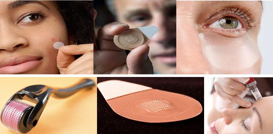
Figure 14: Commercially available microneedle systems: acne microneedle patch, birth control microneedle patch, under eye patch, derma roller, flu vaccine delivery transdermal patch, micro needling treatment
CONCLUSION
Microneedle (MN) technology has revolutionized the landscape of transdermal drug delivery by offering a minimally invasive, pain-free, and effective alternative to traditional routes. The development of various MN types—solid, hollow, coated, dissolving, and hydrogel-forming—has expanded the applicability of this technology across diverse therapeutic areas, from vaccine administration and diabetes management to cancer therapy and dermatological treatments. Despite certain limitations such as limited drug loading capacity and potential for skin irritation, advancements in fabrication methods including 3D printing and smart hydrogels are steadily addressing these challenges. Continued research into biocompatible materials, stimuli-responsive systems, and integration with biosensors will further enhance the safety, efficiency, and versatility of microneedles, making them a cornerstone of future personalized healthcare strategies.
REFERENCES


Anagha P.*, Arya M. M., Dr. Manoj K., Exploring Microneedles: A Minimally Invasive Frontier in Therapeutic Delivery, Int. J. of Pharm. Sci., 2025, Vol 3, Issue 7, 2956-2979. https://doi.org/10.5281/zenodo.16281125
 10.5281/zenodo.16281125