View Article

Abstract

Nanoemulsions have emerged as promising drug delivery systems due to their ability to enhance the solubility and bioavailability of poorly water-soluble drugs. This study aims to develop and evaluate nanoemulsions of Saquinavir, a protease inhibitor with low aqueous solubility, to improve its dissolution rate and therapeutic efficacy. Nanoemulsions were formulated using Capryol 90 as the oil phase, Tween 80 as the surfactant, and Transcutol P as the co-surfactant. A pseudo-ternary phase diagram was constructed to determine the optimal composition for a stable nanoemulsion. Characterization of the prepared nanoemulsions included particle size analysis, zeta potential, polydispersity index (PDI), pH measurement, thermodynamic stability, and in vitro drug release studies. The optimized formulation exhibited a droplet size of less than 200 nm, a negative zeta potential ensuring stability, and enhanced solubility compared to pure Saquinavir. In vitro dissolution studies revealed a significantly higher drug release from the nanoemulsion compared to the unprocessed drug. Stability studies confirmed the physical and chemical stability of the formulation under accelerated conditions. The findings of this study suggest that Saquinavir nanoemulsions could be a viable approach to improve its bioavailability and therapeutic effectiveness in HIV treatment.

Keywords

Nanoemulsion, Saquinavir, Bioavailability, Drug Delivery, Solubility Enhancement

Introduction

Saquinavir is a protease inhibitor used in combination antiretroviral therapy for the treatment of HIV/AIDS. However, its clinical efficacy is hindered by low aqueous solubility (~4 mg/L) and poor bioavailability (~4%), necessitating the use of high oral doses, which can lead to side effects and resistance development. Nanoemulsions, submicron-sized dispersions (20-200 nm), have demonstrated significant potential in enhancing drug solubility, permeability, and bioavailability1-3. Their high surface area-to-volume ratio ensures improved absorption and reduced drug metabolism, making them ideal for lipophilic drugs like Saquinavir. This study focuses on developing and characterizing nanoemulsions for the effective delivery of Saquinavir using lipid-based excipients and surfactants4-7.

2. MATERIALS AND METHODS

2.1. Materials

Saquinavir (API) from Aurobindho Pharma Limited, Capryol 90, Labrafac PG, Transcutol P , Olive oil Gattefosse products, Tween 80, Tween 60, Span 80, PEG 400, Polyvinyl alcohol, potassium chloride, Methanol are laboratory grade.

2.2. Determination of λmax and Calibration Curve

The maximum absorbance wavelength (λmax) of Saquinavir was determined using a UV-Visible spectrophotometer by scanning the drug solution in methanol within a wavelength range of 200-400 nm. The λmax was found to be 240 nm.

A calibration curve was plotted by preparing standard Saquinavir solutions in methanol at different concentrations (5-50 µg/mL) and measuring absorbance at λmax 240 nm. A linear regression equation was obtained, ensuring the method’s suitability for drug estimation in nanoemulsions8-10.

2.3. Solubility Studies

To select an appropriate oil, surfactant, and co-surfactant, Saquinavir was added in excess to various excipients, vortexed, and equilibrated for 72 hours. Samples were centrifuged and analyzed using a UV-Visible spectrophotometer at λmax = 240 nm11-14.

2.3. Construction of Pseudo-Ternary Phase Diagram

Nanoemulsion regions were identified using aqueous titration method, where oil, surfactant, and co-surfactant (Smix) were mixed in different ratios (1:9 to 9:1) and titrated with water. The mixtures were visually assessed for clarity and stability15-17.

2.4. Formulation of Nanoemulsions

Nanoemulsions were prepared using the aqueous titration method, with Capryol 90 (oil), Tween 80 (surfactant), and Transcutol P (co-surfactant). The oil phase was added dropwise into the aqueous phase under continuous stirring and homogenization17-20.

2.5. Characterization Studies

2.5.1. Particle Size and Zeta Potential Analysis

The size and surface charge were measured using Dynamic Light Scattering (DLS) and Zetasizer (Malvern ZS 90)24-26.

2.5.2. Thermodynamic Stability

Nanoemulsions were subjected to centrifugation (3500 rpm for 30 min) and freeze-thaw cycles (-20°C to 25°C) to assess their physical stability24-26.

2.5.3. In Vitro Drug Release Studies

Drug release was evaluated using a USP dissolution apparatus (Type I, basket method) in simulated gastric fluid (pH 1.2) at 37°C ± 0.5°C. Aliquots were analyzed at λmax = 240 nm24-26.

2.5.4. Accelerated Stability Studies

Nanoemulsions were stored at 40°C ± 2°C, 75% RH for three months and analyzed for droplet size, zeta potential, and drug content21-24.

3. RESULTS AND DISCUSSION

The UV-Visible spectroscopic analysis of Saquinavir confirmed a maximum absorption wavelength (λmax) of 240 nm. This wavelength was selected for all subsequent quantitative estimations of the drug in nanoemulsion formulations. The calibration curve was established by preparing a series of standard solutions of Saquinavir in methanol with concentrations ranging from 5 to 50 µg/mL. The absorbance of these solutions was measured at λmax = 240 nm (fig-1), and a linear regression equation was derived. The plot of absorbance versus concentration exhibited a strong linear correlation with an R² value of 0.998 (fig-2), confirming the accuracy, precision, and reliability of the analytical method for drug quantification.

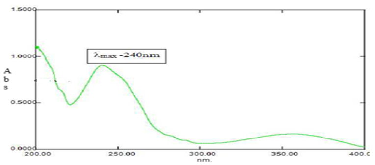
Fig-1: Scan Spectrum of Saquinavir

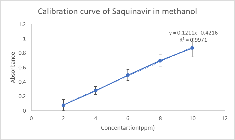
Fig-2: Calibration curve of Saquinavir (n=3)

To determine the most suitable oil, surfactant, and co-surfactant for nanoemulsion formulation, solubility studies were conducted using various excipients. Among the tested excipients, Capryol 90 demonstrated the highest drug solubility (46.39 mg/mL), followed by Tween 80 (56.98 mg/mL) as the surfactant and Transcutol P (41.71 mg/mL) as the co-surfactant. These excipients were selected for nanoemulsion formulation due to their superior drug solubilization capacity and emulsification efficiency.

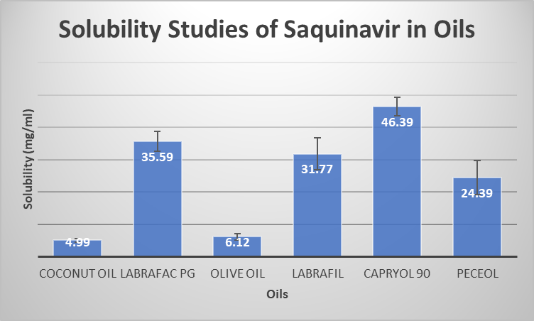
Fig-6: Solubility of Saquinavir in Oils

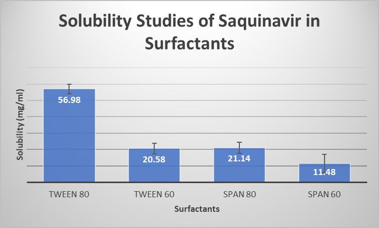
Fig-7: Solubility of Saquinavir in Surfactants

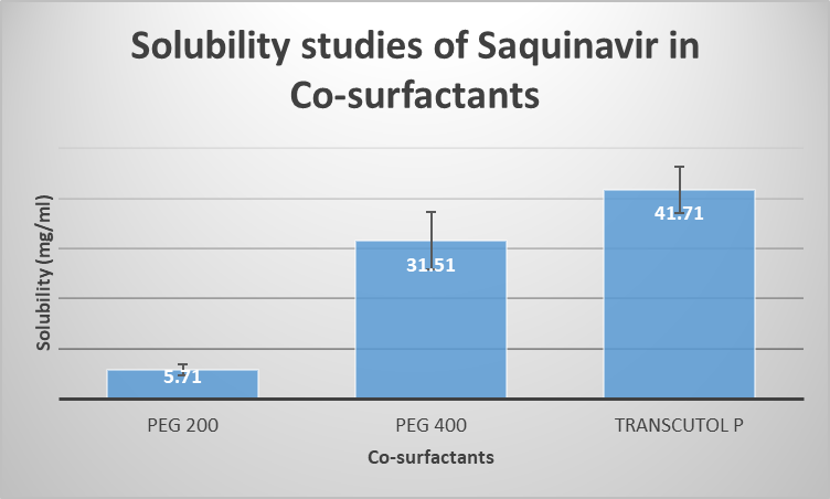
Fig-8: Solubility of Saquinavir in Co-surfactant

To optimize the formulation, a pseudo-ternary phase diagram was constructed to determine the ideal surfactant-to-co-surfactant (Smix) ratio for nanoemulsion formation. The phase diagram revealed that Smix ratios of 3:1 and 4:1 resulted in the widest nanoemulsion region, suggesting that these ratios provided the greatest stability and emulsification efficiency. Based on these findings, nanoemulsions containing Capryol 90 as the oil phase, Tween 80 as the surfactant, and Transcutol P as the co-surfactant were developed using the aqueous titration method.

he optimized Saquinavir-loaded nanoemulsion was evaluated for particle size, polydispersity index (PDI), zeta potential, and pH stability. The mean droplet size of the nanoemulsion was measured at 180 ± 10 nm, confirming its suitability for enhanced drug dissolution and absorption. The polydispersity index (PDI) was found to be 0.243, indicating a narrow size distribution and homogeneity of the nanoemulsion droplets. The zeta potential was recorded at -27.5 mV, suggesting a highly stable formulation with good electrostatic repulsion between droplets, preventing aggregation and coalescence. The pH of the nanoemulsion was measured at 6.8, making it compatible with oral administration without causing irritation to the gastrointestinal tract.

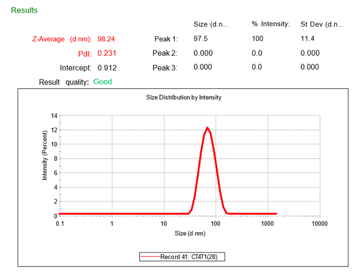
Fig-15: Droplet size of CT4T1(2:8)

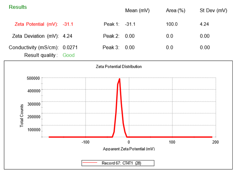
Fig-16-: Zeta potential of CT4T1(2:8)

The in vitro drug release profile of the formulated nanoemulsion was evaluated using a USP dissolution apparatus (Type I, basket method) in simulated gastric fluid (pH 1.2) at 37°C ± 0.5°C. The drug release study demonstrated that 90% of Saquinavir was released from the nanoemulsion within 120 minutes, while the pure drug exhibited only 30% drug release over the same period. The significant enhancement in drug release rate can be attributed to the nano-sized droplets, which increase the surface area available for dissolution and improve Saquinavir’s solubility and permeability in the gastrointestinal tract.

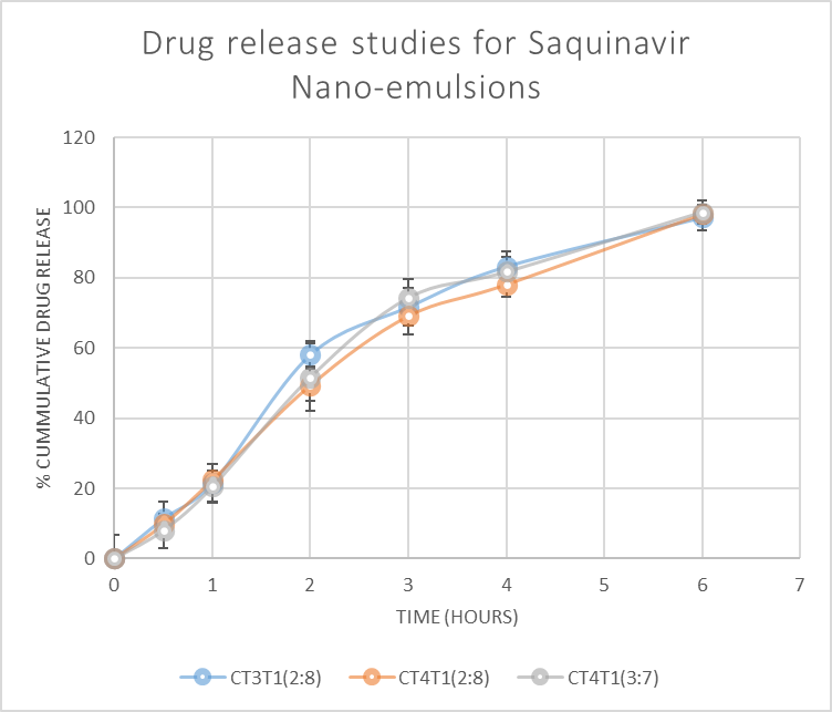
Fig-19: Drug release studies for Saquinavir Nanoemulsions

The drug release continued to increase at later time points, with differences in release rates and extent observed between the formulations. By the end of the 6-hour study period, CT3T1(2:8) had released 97.9% of the drug with a standard deviation of ±3.38%, CT4T1(2:8) had released 99.1% with a standard deviation of ±3.18% and CT4T1(3:7) had released 99.3% with a standard deviation of ±3.23%. Based on the results, CT4T1(2:8) is chosen for the accelerated stability studies.

To assess the stability of the nanoemulsion, accelerated stability studies were conducted under ICH guidelines (40°C ± 2°C, 75% relative humidity) for three months. The formulation was periodically analyzed for changes in droplet size, zeta potential, and drug content. The results indicated that the nanoemulsion maintained its physicochemical properties with no significant changes in particle size, charge, or drug content, confirming its high stability under stress conditions. Additionally, thermodynamic stability tests, including centrifugation and freeze-thaw cycling, showed no signs of phase separation, flocculation, or precipitation, further demonstrating the robust stability of the nanoemulsion formulation.

In summary, the optimized Saquinavir nanoemulsion exhibited superior solubility, rapid drug release, and excellent stability compared to the pure drug. The small droplet size, low PDI, and stable zeta potential suggest that nanoemulsion technology can significantly improve the bioavailability of Saquinavir, potentially enhancing its therapeutic efficacy in HIV treatment. These findings indicate that nanoemulsions could serve as a viable approach to overcome the solubility and bioavailability limitations associated with Saquinavir, thereby improving patient outcomes in antiretroviral therapy.

CONCLUSION

This study successfully developed Saquinavir-loaded nanoemulsions, demonstrating improved solubility, enhanced drug release, and stability. The CT4T1(2:8)-optimized formulation (Capryol 90, Tween 80, and Transcutol P) exhibited a particle size of 98.24 nm with 0.231 PDI, a stable zeta potential of -31.1 mV, and a significant enhancement in dissolution rate. These findings suggest that nanoemulsions are a viable strategy for improving the bioavailability of Saquinavir in HIV therapy.

REFERENCES

  1. Azeem, A., Rizwan, M., Ahmad, F. J., Iqbal, Z., Khar, R. K., & Aqil, M. (2009). Nanoemulsion components screening and selection: A technical note. AAPS PharmSciTech, 10(1), 69–76. https://doi.org/10.1208/s12249-008-9178-9
  2. Forgiarini, A., Esquena, J., Gonzalez, C., & Solans, C. (2001). Formation of nano-emulsions by low-energy emulsification methods at constant temperature. Langmuir, 17(7), 2076–2083. https://doi.org/10.1021/la001362n
  3. Gupta, A., Eral, H. B., Hatton, T. A., & Doyle, P. S. (2016). Nanoemulsions: Formation, properties, and applications. Soft Matter, 12(11), 2826–2841. https://doi.org/10.1039/C5SM02958A
  4. Komaiko, J., & McClements, D. J. (2016). Formation of food-grade nanoemulsions using low-energy preparation methods: A review of available methods. Comprehensive Reviews in Food Science and Food Safety, 15(2), 331–352. https://doi.org/10.1111/1541-4337.12189
  5. Mason, T. G., Wilking, J. N., Meleson, K., Chang, C. B., & Graves, S. M. (2006). Nanoemulsions: Formation, structure, and physical properties. Journal of Physics: Condensed Matter, 18(41), R635–R666. https://doi.org/10.1088/0953-8984/18/41/R01
  6. McClements, D. J. (2013). Nanoemulsions. Soft Matter, 8(6),17–19. https://doi.org/10.1039/C2SM06903B
  7. Solans, C., & Solé, I. (2012). Nano-emulsions: Formation by low-energy methods. Current Opinion in Colloid & Interface Science, 17(5), 246–254. https://doi.org/10.1016/j.cocis.2012.07.003
  8. Shakeel, F., Baboota, S., Ahuja, A., Ali, J., Shafiq, S., & Khan, S. (2008). Nanoemulsions as vehicles for transdermal delivery of aceclofenac. AAPS PharmSciTech, 9(4), 1137–1145. https://doi.org/10.1208/s12249-008-9150-8.
  9. Ratna, R., Yadav, V., & Kumar, A. (2015). Method development and its validation for estimation of Saquinavir hydrochloride by using UV spectroscopy. International Journal of Pharmaceutical Sciences and Research, 6(5), 2154-2160.
  10. Patel, M. R., Patel, R. B., Parikh, J. R., & Solanki, A. B. (2011). Nanoemulsions: An advanced technology for drug delivery. International Journal of Pharmaceutical Sciences and Nanotechnology, 3(1), 633–648.
  11. Mason, T. G., Wilking, J. N., Meleson, K., Chang, C. B., & Graves, S. M. (2006). Nanoemulsions: Formation, structure, and physical properties. Journal of Physics: Condensed Matter, 18(41), R635–R666. https://doi.org/10.1088/0953-8984/18/41/R01.
  12. Chang, C., & McClements, D. J. (2014). Influence of emulsifier type on the stability of vitamin E-enriched emulsions. Food Research International, 62, 662–671. https://doi.org/10.1016/j.foodres.2014.03.030
  13. Solans, C., & Solé, I. (2012). Nano-emulsions: Formation by low-energy methods. Current Opinion in Colloid & Interface Science, 17(5), 246–254. https://doi.org/10.1016/j.cocis.2012.07.003
  14. Banerjee, S., & Chen, H. (2019). Emulsion-based drug delivery systems: Concepts, design, and applications. Journal of Controlled Release, 293, 90–107. https://doi.org/10.1016/j.jconrel.2018.12.020
  15. Ghosh, V., Mukherjee, A., & Chandrasekaran, N. (2013). Formulation and characterization of plant essential oil-based nanoemulsions: Evaluation of antimicrobial, antioxidant, and cytotoxic activities. International Journal of Nanomedicine, 8, 1–13. https://doi.org/10.2147/IJN.S38352
  16. Jaiswal, M., Dudhe, R., & Sharma, P. K. (2015). Nanoemulsion: An advanced mode of drug delivery system. Biotechnology Advances, 33(6), 1046–1060. https://doi.org/10.1016/j.biotechadv.2015.01.008
  17. Kakran, M., Sahoo, N. G., & Li, L. (2011). Fabrication of nanoparticles and their applications in drug delivery: A review. Current Pharmaceutical Biotechnology, 12(9), 1320–1331. https://doi.org/10.2174/138920111798281106
  18. Mittal, H. K., & Kumar, M. (2016). Nanoemulsions as versatile drug delivery carriers for cancer treatment: A review. International Journal of Pharmaceutical Sciences and Nanotechnology, 9(2), 3257–3268. https://doi.org/10.37285/ijpsn.2016.9.2.
  19. Jafari, S. M., McClements, D. J., & Decker, E. A. (2017). Nanoemulsions: Formulation, applications, and characterization. Critical Reviews in Food Science and Nutrition, 57(16), 3390–3406. https://doi.org/10.1080/10408398.2015.1067591
  20. Tadros, T., Izquierdo, P., Esquena, J., & Solans, C. (2004). Formation and stability of nano-emulsions. Advances in Colloid and Interface Science, 108–109, 303–318. https://doi.org/10.1016/j.cis.2003.10.023
  21. Izquierdo, P., Esquena, J., Tadros, T. F., Dederen, C., Garcia, M. J., Azemar, N., & Solans, C. (2004). Formation and stability of nano-emulsions prepared using the phase inversion temperature method. Langmuir, 20(23), 10372–10376. https://doi.org/10.1021/la048146q
  22. Danaei, M., Dehghankhold, M., Ataei, S., Hasanzadeh, M., Javanmard, R., Dokhani, A., Khorasani, S., & Mozafari, M. R. (2018). Impact of particle size and polydispersity index on the clinical applications of lipidic nanocarrier systems. Pharmaceutics, 10(2), 57. https://doi.org/10.3390/pharmaceutics10020057
  23. Jaiswal, M., Dudhe, R., & Sharma, P. K. (2015). Nanoemulsion: An advanced mode of drug delivery system. Biotechnology Advances, 33(6), 1046–1060. https://doi.org/10.1016/j.biotechadv.2015.01.008

Reference

  1. Azeem, A., Rizwan, M., Ahmad, F. J., Iqbal, Z., Khar, R. K., & Aqil, M. (2009). Nanoemulsion components screening and selection: A technical note. AAPS PharmSciTech, 10(1), 69–76. https://doi.org/10.1208/s12249-008-9178-9
  2. Forgiarini, A., Esquena, J., Gonzalez, C., & Solans, C. (2001). Formation of nano-emulsions by low-energy emulsification methods at constant temperature. Langmuir, 17(7), 2076–2083. https://doi.org/10.1021/la001362n
  3. Gupta, A., Eral, H. B., Hatton, T. A., & Doyle, P. S. (2016). Nanoemulsions: Formation, properties, and applications. Soft Matter, 12(11), 2826–2841. https://doi.org/10.1039/C5SM02958A
  4. Komaiko, J., & McClements, D. J. (2016). Formation of food-grade nanoemulsions using low-energy preparation methods: A review of available methods. Comprehensive Reviews in Food Science and Food Safety, 15(2), 331–352. https://doi.org/10.1111/1541-4337.12189
  5. Mason, T. G., Wilking, J. N., Meleson, K., Chang, C. B., & Graves, S. M. (2006). Nanoemulsions: Formation, structure, and physical properties. Journal of Physics: Condensed Matter, 18(41), R635–R666. https://doi.org/10.1088/0953-8984/18/41/R01
  6. McClements, D. J. (2013). Nanoemulsions. Soft Matter, 8(6),17–19. https://doi.org/10.1039/C2SM06903B
  7. Solans, C., & Solé, I. (2012). Nano-emulsions: Formation by low-energy methods. Current Opinion in Colloid & Interface Science, 17(5), 246–254. https://doi.org/10.1016/j.cocis.2012.07.003
  8. Shakeel, F., Baboota, S., Ahuja, A., Ali, J., Shafiq, S., & Khan, S. (2008). Nanoemulsions as vehicles for transdermal delivery of aceclofenac. AAPS PharmSciTech, 9(4), 1137–1145. https://doi.org/10.1208/s12249-008-9150-8.
  9. Ratna, R., Yadav, V., & Kumar, A. (2015). Method development and its validation for estimation of Saquinavir hydrochloride by using UV spectroscopy. International Journal of Pharmaceutical Sciences and Research, 6(5), 2154-2160.
  10. Patel, M. R., Patel, R. B., Parikh, J. R., & Solanki, A. B. (2011). Nanoemulsions: An advanced technology for drug delivery. International Journal of Pharmaceutical Sciences and Nanotechnology, 3(1), 633–648.
  11. Mason, T. G., Wilking, J. N., Meleson, K., Chang, C. B., & Graves, S. M. (2006). Nanoemulsions: Formation, structure, and physical properties. Journal of Physics: Condensed Matter, 18(41), R635–R666. https://doi.org/10.1088/0953-8984/18/41/R01.
  12. Chang, C., & McClements, D. J. (2014). Influence of emulsifier type on the stability of vitamin E-enriched emulsions. Food Research International, 62, 662–671. https://doi.org/10.1016/j.foodres.2014.03.030
  13. Solans, C., & Solé, I. (2012). Nano-emulsions: Formation by low-energy methods. Current Opinion in Colloid & Interface Science, 17(5), 246–254. https://doi.org/10.1016/j.cocis.2012.07.003
  14. Banerjee, S., & Chen, H. (2019). Emulsion-based drug delivery systems: Concepts, design, and applications. Journal of Controlled Release, 293, 90–107. https://doi.org/10.1016/j.jconrel.2018.12.020
  15. Ghosh, V., Mukherjee, A., & Chandrasekaran, N. (2013). Formulation and characterization of plant essential oil-based nanoemulsions: Evaluation of antimicrobial, antioxidant, and cytotoxic activities. International Journal of Nanomedicine, 8, 1–13. https://doi.org/10.2147/IJN.S38352
  16. Jaiswal, M., Dudhe, R., & Sharma, P. K. (2015). Nanoemulsion: An advanced mode of drug delivery system. Biotechnology Advances, 33(6), 1046–1060. https://doi.org/10.1016/j.biotechadv.2015.01.008
  17. Kakran, M., Sahoo, N. G., & Li, L. (2011). Fabrication of nanoparticles and their applications in drug delivery: A review. Current Pharmaceutical Biotechnology, 12(9), 1320–1331. https://doi.org/10.2174/138920111798281106
  18. Mittal, H. K., & Kumar, M. (2016). Nanoemulsions as versatile drug delivery carriers for cancer treatment: A review. International Journal of Pharmaceutical Sciences and Nanotechnology, 9(2), 3257–3268. https://doi.org/10.37285/ijpsn.2016.9.2.
  19. Jafari, S. M., McClements, D. J., & Decker, E. A. (2017). Nanoemulsions: Formulation, applications, and characterization. Critical Reviews in Food Science and Nutrition, 57(16), 3390–3406. https://doi.org/10.1080/10408398.2015.1067591
  20. Tadros, T., Izquierdo, P., Esquena, J., & Solans, C. (2004). Formation and stability of nano-emulsions. Advances in Colloid and Interface Science, 108–109, 303–318. https://doi.org/10.1016/j.cis.2003.10.023
  21. Izquierdo, P., Esquena, J., Tadros, T. F., Dederen, C., Garcia, M. J., Azemar, N., & Solans, C. (2004). Formation and stability of nano-emulsions prepared using the phase inversion temperature method. Langmuir, 20(23), 10372–10376. https://doi.org/10.1021/la048146q
  22. Danaei, M., Dehghankhold, M., Ataei, S., Hasanzadeh, M., Javanmard, R., Dokhani, A., Khorasani, S., & Mozafari, M. R. (2018). Impact of particle size and polydispersity index on the clinical applications of lipidic nanocarrier systems. Pharmaceutics, 10(2), 57. https://doi.org/10.3390/pharmaceutics10020057
  23. Jaiswal, M., Dudhe, R., & Sharma, P. K. (2015). Nanoemulsion: An advanced mode of drug delivery system. Biotechnology Advances, 33(6), 1046–1060. https://doi.org/10.1016/j.biotechadv.2015.01.008

Photo
Dasa Mani Deepika
Corresponding author

Centre for Pharmaceutical Sciences, UCESTH, Jawaharlal Nehru Technological University, Hyderabad, Kukatpally, Telangana, India. 500085

Photo
M. Sunitha Reddy
Co-author

Centre for Pharmaceutical Sciences, UCESTH, Jawaharlal Nehru Technological University, Hyderabad, Kukatpally, Telangana, India. 500085

Photo
K. Anie Vijetha
Co-author

Centre for Pharmaceutical Sciences, UCESTH, Jawaharlal Nehru Technological University, Hyderabad, Kukatpally, Telangana, India. 500085

Dasa Mani Deepika, M. Sunitha Reddy, K. Anie Vijetha, Formulation and In-Vitro Evaluation of Saquinavir Nanoemulsion, Int. J. of Pharm. Sci., 2025, Vol 3, Issue 9, 1173-1180. https://doi.org/10.5281/zenodo.17092376

More related articles
Formulation and Evaluation of Effervescent Tablets...
Dhanashree Nale, Sunita Sanap, Yuvraj Lokhande, Kedar Shinde, Roh...
Chiral HPLC Method Development And Validation For ...
SHAIK MASTANAMMA, Chunduri Akhila, M S DHANDAPANI, ...
Improvement Of Blood Glucose Control With Artifici...
Swati Dilipsingh Thakur , Kishor V. Otari , Ajay Y . Kale , ...
Related Articles
Treatment Of Mild Chronic Hypertension During Pregnancy...
Ramkishor Sahu, Ranjeet Baghel, Somprabha Madhukar, Shruti Rathore, ...
Exploring Novel Pyrimidine Derivatives: Design, Synthesis, Characterization, and...
Avinash Patil, Prashant Chavan, Amitkumar Rawal, Sandeep Patil, ...
Severe Health Effect of Malaria and its Treatment...
Sontakke Pradip, Waghmare Vyankatesh, Farande Rushikesh, Swami Bhujayya, Shinde Devanand , Shaikh Sh...
Formulation and Evaluation of Effervescent Tablets Using Natural Excipients: A N...
Dhanashree Nale, Sunita Sanap, Yuvraj Lokhande, Kedar Shinde, Rohan Shinde, Nitiksha Jadhav, ...
More related articles
Formulation and Evaluation of Effervescent Tablets Using Natural Excipients: A N...
Dhanashree Nale, Sunita Sanap, Yuvraj Lokhande, Kedar Shinde, Rohan Shinde, Nitiksha Jadhav, ...
Improvement Of Blood Glucose Control With Artificial Pancreas In Coronavirus Dis...
Swati Dilipsingh Thakur , Kishor V. Otari , Ajay Y . Kale , ...