View Article

Abstract

Wound healing is a dynamic and multifaceted biological process involving cellular, molecular, and tissue-level mechanisms. Despite advances in modern medicine, effective management of chronic wounds particularly those associated with diabetes, immune dysfunction, and aging remains a significant clinical challenge. Chronic wounds, including pressure sores and diabetic foot ulcers, contribute substantially to global morbidity, mortality, and healthcare costs. Current treatment options are often inadequate, resulting in prolonged healing, increased infection risk, and higher rates of amputation. This underscores the urgent need for alternative, cost-effective, and efficient wound-healing agents. Cinnamomum tamala (Indian bay leaf), a plant widely used in traditional medicine for its antimicrobial properties, has shown promise in preliminary studies related to wound care. However, there is limited scientific evidence to validate its therapeutic potential in wound healing. This study aims to investigate the wound healing properties of Cinnamomum tamala using in vitro models to assess its efficacy in promoting wound closure. Additionally, standard toxicity assays will be employed to evaluate the safety profile of the plant extract. By providing empirical data on the effectiveness and safety of Cinnamomum tamala, this research seeks to bridge the gap between traditional medicine and modern clinical applications. The findings may contribute to the development of novel, natural, and affordable wound healing treatments that can significantly benefit patients suffering from chronic wounds worldwide.

Keywords

Chronic wounds, Morbidity, Cinnamomum tamala, contaminants, targeted drug delivery, bioavailability.

Introduction

Wound healing is a dynamic and complex process that involves several cells, molecular, and tissue-level processes. Even with advancements in medical technology, wound healing is still a major clinical challenge, especially when it comes to diabetes, chronic wounds, and compromised immune function. For many years, Indian bay leaf, or But nothing is known about its potential for wound healing. Millions of people around the world suffer from chronic wounds, which contributes to a high morbidity, mortality, and financial burden of wound treatment. Globally, chronic wounds like pressure sores and diabetic foot ulcers place a heavy cost on healthcare systems. A wound is characterized as harm to living tissue that interferes with the body's natural ability to form and function. This disruption is caused by a breakdown in cellular and anatomical continuity, which leads to a loss of physiological or protective tissue integrity. Physical, chemical, thermal, microbiological, or immunological damage are the most common causes of wounds. Wounds can have significant social and economic repercussions for patients and their families in addition to the medical harm they cause. They frequently result in physical limitations like decreased mobility and function, along with excruciating pain, low self-esteem, anxiety and despondency, and, in certain situations, early mortality.

Wounds, particularly those affecting the dermal layer, can seriously impair the skin's natural physiology. The extent of damage is mostly determined by how well the body recovers from these tissue injuries, which may alter the anatomical structure of the skin. The wounded area is restored and repaired by a coordinated set of cellular and molecular processes called wound healing. This healing cascade has a distinct trajectory, and numerous classification schemes have been developed to complex, dynamic process in tissue repair. Procedure of wound healing is categorized differently by different writers. According to some definitions, the initial stage is inflammation, which is followed by proliferation, and the remodeling phase ends with repair. The justification for looking into Cinnamomum tamala's capacity to heal wounds and its potential as a new kind of wound treatment. The purpose of the study is to use in vitro models to assess how well Cinnamomum tamala extract promotes wound healing. Additionally, using common toxicity tests, the study will evaluate the toxicity and safety of Cinnamomum tamala extract.

Since Cinnamomum tamala has been used for generations in traditional medicine, the analysis is important since it attempts to provide scientific proof of its ability to heal wounds.  The study may help create new, natural, and reasonably priced wound healing treatments that will help millions of individuals with chronic wounds around the world.  In conclusion, this study intends to explore Cinnamomum tamala's wound healing activity since it may help create new, natural, and reasonably priced wound healing treatments that could help millions of individuals with chronic wounds around the world.

METHODOLOGY

Materials

The kind donation of Cinammomum tamala Plant came from Chengannur  botanical garden, Chengannur, Kerala While Ethanol was purchased from Rajarambapu Patil Sahakari Sakhar karkhana, Islampur, H2SO4, Million’s Reagent, Chloroform , Mayer’s Reagent, Acetic Anhydride, Glacial Acetic Acid and Ferric Cyanide solution was purchased from Research Lab., Fine Chem Industries, Mumbai. Methanol and Acetone high purity and analytical grade were guaranteed for all other compounds utilized in the investigation.

Plant Material Collection

Cinnamomum tamala was likely chosen for the study due to its historical significance and wound healing properties. Despite its traditional use, there is limited scientific evidence on the wound healing properties of Cinnamomum tamala plant was procured from botanical garden, Chengannur, Kerala and authenticated in Botany Department at Balwant College Vita, Maharashtra, India.

Drying and Grinding

After being shade-dried, the leaves were dried for 20 minutes at sixty degrees in a hot air oven (Shri Krishna Surgicals). Using a mixer grinder, the dried leaves were ground into a coarse powder. (Mahalaxmi electronics).

Preparation of Extract

Extraction in pharmaceutics is the process of isolating active pharmaceutical ingredients (APIs) from plant, animal, or microbial sources. Extraction is essential for extracting the active components from the bulk material, which ensures the safety, effectiveness, and purity of medications. Using Soxhlet apparatus (Shri Krishna Surgicals), dried leaves of Cinnamomum tamala were extracted with ethanol using a progressive solvent extraction process over a 48-hour period. The ethanolic extract was placed in a number-colored container and subjected to direct sunshine.

Soxhlet Extraction Procedure

  • The first stage of analysing plant components is extraction. Chemical substances are isolated or separated from plant tissues using this method. There exist multiple extraction processes, and the selection of one is contingent upon:
  • The behaviour of the chemical ingredients.
  • The various form of plant being used.
  • Specific purpose of the extraction.

A heated organic solvent is used in the continuous extraction process known as Soxhlet extraction. This technique works especially well when contaminants are insoluble and the target compound is only weakly soluble in the selected solvent. An effective, unsupervised process is made possible by Soxhlet extraction.

Invented in 1879 by Franz von Soxhlet, the Soxhlet extractor consists of three key components:

  1. Percolator (Boiler and Reflux): Helps the solvent circulate.
  2. Thimble: Usually composed of cotton or thick filter paper, it contains the solid material that needs to be removed.
  3. Syphon Mechanism: Finishes the extraction cycle by periodically draining the solvent from the thimble.
  4. Thimble: After inserting the substance containing the target component into the thimble, it is put in the Soxhlet extractor's main chamber.
  5. Round Bottom Flask (RBF) or Distillation Flask: This flask contains the extraction solvent that will be utilized in the procedure.
  6. Heating Mantle: To warm the solvent, the distillation flask is set on a heating element.
  7. Reflux Condenser: Positioned above the extractor, the condenser facilitates the cooling and condensation of solvent vapours.
  8. Water Inlet & Outlet: Connected to the condenser, these allow for the continuous flow of water to regulate the condenser’s temperature.

Formulation method

The thimble is filled with the solid material, usually in a cotton bag. Next, a flask containing the extraction solvent is placed on top of the Soxhlet extractor, with the condenser attached above. The solvent vaporizes when heated to its reflux point. The vapours rise via the distillation arm, condense, and drip into the chamber that holds the solid material thimble. The chosen chemical fills the chamber after dissolving into the heated solvent. When the chamber reaches a particular level, the solvent is drained back into the distillation flask by the siphon mechanism. More of the target molecule dissolves with each cycle of this process, which is repeated numerous times over hours or even days. In the distillation flask, the target component is concentrated after 72 cycles, or 48 hours. Following extraction, the extracted component is obtained by removing the solvent, usually by evaporating it in direct sunlight. The part of the extracted substance that is insoluble is left in the thimble and is often thrown away.

Evaporation of Extract

After using the Soxhlet procedure, the ethanolic extract was transferred into a porcelain dish and exposed to direct sunshine for two days.

Solubility of Extract

In scientific investigations, solubility is a crucial physical characteristic, especially when creating different formulations. For a particular temperature, it is the maximum amount of solute that may dissolve in a given amount of solvent. Because solubility has a major impact on a drug's bioavailability, it is an important consideration in drug formulation.

A compound's molecular structure and the solution's conditions affect how soluble it is. Lipidophilicity, hydrogen bonding, molecular volume, crystal energy, and ignitability are all influenced by the structure and dictate how effectively a substance dissolves. In contrast, the conditions of the solution are affected by factors such as temperature, time, ionic strength, co-solvents, additives, and pH. In the process of finding and developing new drugs, poor solubility can seriously reduce productivity. An effective medicine must reach its target at sufficient concentrations. The pharmacologic potency and permeability of a chemical are therefore directly related to its minimum allowable solubility. Because it gives the most accurate evaluation of solubility without the impact of other factors, solubility experiments are usually carried out at room temperature (25 °C). Generally speaking, solubility increases with system temperature, which could skew the results.

Characterization

FTIR Study

FTIR spectrophotometer (Mahalaxmi Scientifics) was used to record the Fourier Transform Infrared (FTIR) spectra of Cinnamomum tamala extract. It is a crucial instrument for evaluating the drug's purity. The fundamental peaks in the FTIR spectrum indicate the drug's chemical makeup. To determine the chemical stability and purity of Cinnamomum tamala, an FTIR analysis was conducted.

Scratch Assay

The sample ability to heal wounds was assessed using L929 cells in vitro cell migration experiments. Basically, 2 x 10⁵ cells/mL were planted onto 6-well plates, and they were grown all night long. Following a Dulbucco's Phosphate Buffered Saline (DPBS) wash of the cells, a scratch was made using a sterile 200 µL tip. To remove the detached cells and other cellular debris, the cells were rinsed with DPBS. 100 µL of the sample and 5 µg/mL of the positive control, cipladine, were added to the cells, and they were then grown for 24 hours. One popular drug for wound care is cipladine. The negative control cells received no treatment. Photographs taken with a digital camera and an inverted microscope revealed the cell movement and morphological changes. Each experiment was run in three duplicates (n ¼ 3). The width of the scratch and the closure of the wound were examined using SAGLO software at different intervals (48 hours).

In-Vitro Assay

To determine the cytotoxicity and biocompatibility of Cinnamomum tamala extract, an in vitro cell viability assay was conducted using the L929 mouse fibroblast cell line. This assay helps assess whether the extract is safe for use in wound healing by evaluating its effect on the metabolic activity of fibroblasts.

Cell Line and Culture Conditions

  • Cell Line: L929 (Mouse Connective Tissue Cell Line) was obtained from a certified cell bank and used for cytotoxicity assessment due to its relevance in connective tissue and dermal models.
  • Culture Medium:
    1. Dulbecco's Modified Eagle Medium (DMEM) – High Glucose
      • Cat. No.: 11965-092 (Thermo Fisher Scientific)
      • Enriched with essential amino acids, vitamins, and glucose to support fibroblast growth.
    2. Fetal Bovine Serum (FBS)
      • Brand: Gibco, Invitrogen
      • Cat. No.: 15240-062
      • Used at a concentration of 10% (v/v) to provide growth factors and nutrients.
    3. Antibiotic-Antimycotic Solution (100X)
      • Brand: Thermo Fisher Scientific
      • Cat. No.: 15240-062
      • Used at 1% (v/v) to prevent bacterial and fungal contamination.

Procedure

        1. The mouse connective tissue cell line L929, which originated at the National Center for Cell Sciences (NCCS), Pune, was cultivated in DMEM Medium with 10% fetal bovine serum added.
        2. Cells at a concentration of 1 × 10⁴ cells/ml were incubated in culture media for 24 hours at 37 °C and 5% CO2.
        3. Cells were seeded into 96 wells and tissue culture grade microplates at a density of 70 μl, 104 cells/well in 100 μl of culture medium, and 100 μl of a. Samples (10–100 µg/ml), respectively.
        4. The cell line was incubated in control wells with DMSO (0.2% in PBS). Each sample was incubated in triplicate. In order to determine the percentage of live cells after culture and the survival rate of control cells, controls were maintained.
          Thermo Scientific BB150 CO2 incubators were used to cultivate cell cultures for 24 hours at 37 °C and 5% CO2.
        5. The media was completely removed after incubation, and 20 μl of MTT reagent (5 mg/min PBS) was added.
        6. Following MTT injection, cells were grown in a CO2 incubator for four hours at 37°C.
        7. Checked for formazan crystal formation in the wells using a microscope. Only living cells could change the yellowish MTT into a dark formazan. Following the medium's complete elimination. 200 μl of DMSO was added, and the mixture was wrapped in aluminum foil and incubated at 37.0°C for 10 minutes.
        8. Three duplicate measurements of each sample's absorbance at 570 nm were made using an Elisa microplate reader (Benesphera E21).

In Vitro Wound Healing Activity: Scratch Assay

To evaluate the wound healing potential of Cinnamomum tamala extract, an in vitro scratch assay was performed using the L929 fibroblast cell line, a widely used model for dermal wound healing studies due to its high proliferative and migratory capacity.

Materials and Cell Culture Conditions

  • Cell Line: L929 mouse fibroblast cells (ATCC® CCL-1™) were cultured under standard conditions (37°C, 5% CO?, 95% humidity).
  • Culture Media: Cells were maintained in Dulbecco’s Modified Eagle Medium (DMEM) supplemented with:
    • 10% Fetal Bovine Serum (FBS)
    • 1% Antibiotic-Antimycotic Solution (containing penicillin, streptomycin, and amphotericin B)
  • Washing Buffer: Dulbecco’s Phosphate Buffered Saline (DPBS) was used for washing and cleaning prior to the assay.

Scratch Assay Procedure

  1. Cell Seeding: L929 fibroblasts were seeded in a 6-well plate at a density of approximately 5 × 10? cells/well and incubated until a confluent monolayer was formed (usually 24–48 hours).
  2. Scratch Creation: A straight-line scratch (wound) was introduced manually in the center of the monolayer using a sterile 200 μL pipette tip. Detached cells and debris were removed by gently washing the wells with DPBS.
  3. Treatment:
    • Cells were then incubated with serum-free DMEM containing different concentrations of Cinnamomum tamala extract.
    • A negative control (media without extract) and a positive control (media with known wound healing agent, if applicable) were also maintained.
  4. Incubation: The cells were incubated for up to 24 hours, and images of the scratch area were taken at 0 hours, 12 hours, and 24 hours using an inverted phase-contrast microscope.
  5. Wound Closure Measurement: The width of the scratch at each time point was measured using ImageJ software.

Organoleptic Properties

The Cinnamomum tamala extract was subjected to a detailed macroscopic evaluation to document its organoleptic properties. These parameters are essential for quality control, standardization, and reproducibility of the extract in future pharmacological or clinical applications. The physical condition, color, taste, texture, and odor were assessed visually and through sensory evaluation as follows:

  • Physical Condition: The extract was observed to be in a semi-solid (or liquid/paste, depending on your sample) form at room temperature, indicating ease of handling and potential for formulation into topical preparations.
  • Colour: The extract exhibited a deep brown (or specify actual shade) coloration, which may be attributed to the presence of polyphenolic compounds and tannins typically found in Cinnamomum tamala.
  • Taste: Upon organoleptic evaluation, the extract presented a pungent, slightly bitter taste, consistent with the known flavor profile of bay leaf constituents, particularly eugenol and cinnamaldehyde.
  • Texture: The extract had a smooth and homogenous consistency, free from particulate matter, indicating effective filtration and solvent removal during preparation.
  • Odour: The extract emitted a strong aromatic scent, characteristic of Cinnamomum tamala, with notes of cinnamon and clove, reflecting the volatile oil content.

Phytochemical Screening

  • Test for tannins: - To check for tannins, boil 1 milliliter of Cinnamomum tamala leaf extract in 20 milliliters of water, then filter the mixture. The test was examined for brownish green or a blue-black hue when a few drops of 0.1% FeCl3 were added.
  • Saponin test (foam test):  Give the dry powder or medication extract a good shake with water.  Stable, long-lasting foam was seen.
  • Flavonoid test: Five ml of diluted ammonia solution and a part of the Cinnamomum tamala leaf were put into a test tube, followed by concentrated H2SO4. The extract's yellow hue indicated the presence of flavonoids. Later, the yellow colouring goes away.
  • Steroid test: In a test tube, combine 1 milliliter of Cinnamomum tamala leaf extract with 2 millilitres of H2SO4 and 2 millilitres of acetic anhydride.  When steroids were present, the colour shifted from violet to blue or green.
  • Salkowski's test (terpenoids test): Carefully mixing 3 ml of conc. H2SO4 with 5 ml of Cinnamomum tamala leaf extract in 2 ml of chloroform creates a layer. A reddish-brown colouring of the interface was observed to signify the presence of terpenoids.
  • Triterpene test: After adding 2 millilitres of concentrated H2SO4, 1 milliliter of acetic anhydride and 1 milliliter of Cinnamomum tamala leaf extract were added to 1 milliliter of chloroform.  When triterpenoids are present, a reddish violet colour forms.
  • Alkaloids are tested using Mayer's test: Mayers regent applied to 1 milliliter of Cinnamomum tamala leaf extract at the test tube's side. Alkaloids are indicated by the formation of a creamy or white precipitate.
  • Modified Borntragetr's test for anthraquinones glycoside:  Add five ml of 5% FeCl3 and five ml of dil. HCl to five ml of extract.  In a boiling water bath, heat for five minutes.  After cooling, add chloroform and mix thoroughly.  After removing the organic layer, apply the same amount of diluted ammonia.  The ammoniacal layer is reddish-pink.
  • Keller-Killani test for cardiac glycosides: Five milliliters of Cinnamomum tamala leaf extract were mixed with two milliliters of glacial acetic acid that contained one drop of FeCl3 solution. An underlayment of 1 milliliter of concentrated H2SO4 was utilized. The cardenolide deoxy sugar feature is indicated by an interface brown ring.
  • Coumarin test: When 2 ml of Cinnamomum tamala leaf extract is mixed with three ml of 10% NaOH in a test tube, a yellow hue is produced, signifying the presence of coumarins.
  • Emodin test: The various extracts of Cinnamomum tamala leaves were mixed with two milliliters of NH4OH and three milliliters of benzene. 

RESULT AND DISCUSSION

FTIR Study

FTIR spectra of Cinnamomum tamala extract were recorded using FTIR (Bruker). The spectrum was recorded over range of wave number 3500 to 1000 cm-1. The characteristic absorption peaks of Cinnamomum tamala were shown in figure 8.5. The values of major peaks in FTIR spectrum of Cinnamomum tamala extract are mentioned in Table 8.5. The observed characteristic peaks confirm the presence of key fictional groups, ensuring the purity of drug.

The FTIR spectral analysis of Cinnamomum tamala confirmed the presence of characteristic functional groups through their corresponding absorption peaks. The observed peaks closely matched the standard values, with minor variations attributed to instrumental factors or environmental conditions. At 2922.94 cm-1, the C-H stretching vibration was detected. At 1443.10 cm-1, the C-H bending peak was detected. The observed C-H bending was 1366.57 cm-1.
Overall, the detected peaks validate the chemical integrity and purity of Cinnamomum tamala by confirming the existence of important functional groups linked to the plant. Since the minor peak shifts fall within allowable bounds, the medication will continue to be pure and unaltered structurally.

Figure 1: FTIR Spectrum of Cinnamomum tamala Extract.

Table 1: FTIR Spectrum Major Peak Values and Respective Functional Group

Sr. no.

Functional group

Indication

Wavelength cm-1

1

O-H

-

3328.08

2

C-H

Stretching

2922.94

3

C=O

-

1715.75

4

C=O/C=C

-

1610.28

5

C=C

-

1514.53

6

C-H

Bending

1443.10

7

C-H

Bending

1366.57

8

C-O/C-C

-

1267.16

9

C-O/C-C/C-N

-

1034.233

In-vitro scratch assay

Percentage (%) of cells that moved in the direction of the wound and helped it close microscopically images representing the In vitro wound healing nature of Sample: L929 cells were incubated in presence or absence of Samples and standard drug Cipladine and images were captured at 48 hours. According to images and results Sample showed Moderate activity Percentage (%) of cells that moved in the direction of the wound and helped it close.

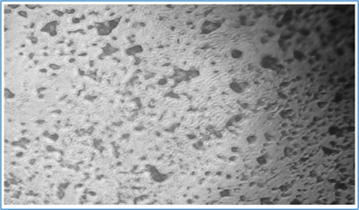
Figure 2: Normal Morphology of L929


       Control - Wound Scratch                                       After 48 hours - Control      


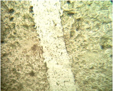
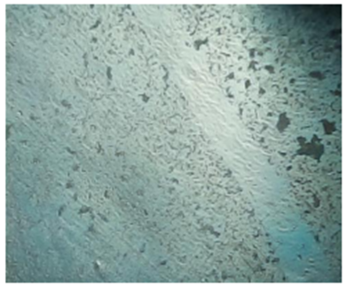
             Standard - Wound Scratch (Cipladine)                 After 48 hours - Standard (Cipladine)

           
  Sample - Wound Scratch                                 After 48 hours - Sample

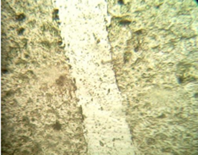
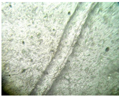
Table 2 Percentage (%) of Cells Reduction in Wound Closure.

Sample

Reading 0 hours (mm)

Mean

Reading 48 hours (mm)

Mean

Percentage of cell Reduction

Control

10.00

09.98

10.05

10.01

9.79

9.80

9.70

9.76

-

Standard Cipladine-5 (µg/mL)

10.00

09.98

10.05

10.01

1.22

1.20

1.24

1.22

87.81

Sample

10.00

09.98

10.05

10.01

4.50

4.60

4.70

4.60

54.04

In-vitro Cell Viability Assay: MTT

Principle of Assay: Since mitochondrial succinate dehydrogenase enzymes in living cells can change the yellow water soluble substrate 3-(4, 5-dimethylthiazol-2-yl)-2, 5-diphenyltetrazoliumbromide (MTT) into an insoluble, purple formazan product that can be measured using spectrophotometry, this colorimetric assay is based on this ability. Since MTT can only be reduced by metabolically active cells, the level of activity serves as a barometer for the viability of the cells.

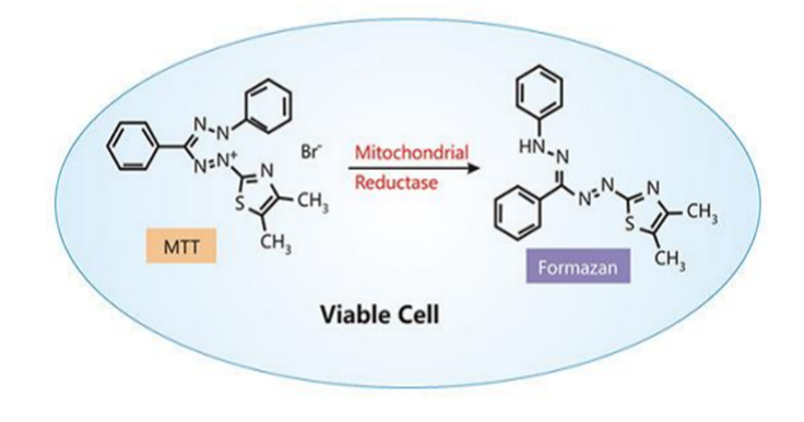
Figure 2: Principle Reaction of Cell Viability Assay.

Table 3: Effects of Sample against L929 (Mouse Connective Tissue Cell Line) by MTT Assay

Sr.no

Concentration (µg/ml)

Absorbance (O D)

Cell viability (%)

1

2

3

Average

1

Control

2.107

2.114

2.11

2.110

-

2

Sample

10


1.256


1.254


1.251


1.253


59.39

40

1.45

1453

1.459

1.454

68.91

100

1.526

1.529

1.53

1.528

72.42

The % cell viability of standard and Sample solution is given in table. Ethanolic extract of Cinnamomum tamala showed concentration dependent cell viability. The Maximum cell viability of Cinnamomum tamala extract was found to be 68.91% at 40 µg/ ml and 72.42% at 100 µg/ml respectively, where minimum % cell viability of sample was found to be 59.39% at 10 µg/ml. This study showed that Cinnamomum tamala extract has good cell viability. Overall, the MTT assay conducted on normal cells demonstrated a dose-dependent response to the tested compound. At higher concentrations, the cells maintained high viability, as indicated by strong purple formazan formation, suggesting minimal cytotoxic effects. As the concentration increased, a gradual increase in cell viability was observed, reflected by raise in absorbance values. However, even at higher doses, normal cells retained a considerable level of metabolic activity, indicating that the compound exhibited low toxicity toward normal cells. These findings suggest that the tested compound may be selectively toxic and relatively safe for normal cells at therapeutic concentrations.

Organoleptic property

Solubility of Extract

Solubility studies revealed that Cinnamomum tamala is easily soluble in ethanol and chloroform and slightly soluble in water. Solubility studies confirmed Cinnamomum tamala extract’s high Solubility in ethanol, chloroform and limited in water.

Table 4: Solubility of Extract

Solvent

Solubility

Water

Slightly  Soluble

Ethanol

Soluble

Chloroform

Soluble

Phytochemical Screening

The physical characterization of Cinnamomum tamala was carried out to confirm its basic physico-chemical properties. Visual observation confirmed that the extract exists as a semisolid, appearing as blackish green with an aromatic Odour and Bitter-sweet taste.

Table 5: Organoleptic Properties

Sr. no

Physical Properties

Methods

Descriptions

1

Physical state

Visual observation

Semi solid

2

Colour

Visual observation

Blackish green

3

Odour

Smelling

Aromatic

4

Taste

Tasting

Bitter-sweet

5

Texture

-

Smooth

Qualitative Analysis of Phytochemicals

A preliminary phytochemical analysis of Cinnamomum tamala leaves revealed the presence of tannin content, saponin levels, flavonoids, also terpenoids and triterpenoids, alkaline compounds, amines, the antioxidant polyphenol, sugars, coumarins are and these pigments.

Table 6: Phytochemicals Present in the Extract (+) Present (++) Present in higher concentration (-) absent.

Sr.no.

Phytochemicals

Ethanolic extract

1

Tannin

++

2

Saponin

++

3

Flavonoids

++

4

Steroids

++

5

Terpenoids

++

6

Triterpenoids

++

7

Alkaloids

+

8

Anthraquinones

++

9

Polyphenol

++

10

Glycoside

++

11

Coumarins

++

12

Emodin

-

13

Anthocyanins

+

CONCLUSION

The extracted Cinnamomum tamala exhibited promising wound healing activity, as evidenced by its ability to enhance collagen synthesis, improve tissue strength, and accelerate wound closure. The study provides valuable insights into the extraction and wound healing activity of Cinnamomum tamala, highlighting its potential as a natural remedy for wound healing. The organoleptic properties of Cinnamomum tamala extract are consistent with its traditional use. The extract's pleasant aroma and flavour profile may contribute to its wound healing activity.

The ethanol extract's higher solubility may facilitate better penetration of the extract's bioactive compounds into the wound site, promoting tissue repair and regeneration. The phytochemical constituents of Cinnamomum tamala extract may work synergistically to promote tissue repair, reduce inflammation, and enhance collagen synthesis. The study provides a foundation for further research on the pharmacological and therapeutic properties. The FTIR spectra revealed the presence of various functional groups, including hydroxyl, carbonyl groups, which are characteristic of phenolic compounds, flavonoids, and Terpenoids, etc. The extract may promote wound healing by enhancing cell growth, differentiation, and migration. From the scratch assay it was concluded that the sample showed significant wound healing activity. At the different doses of extract, the carried out MTT assay for wound healing activity against L929 cell line. The extract showed good cell Viability activity at significant concentration against L929 (Mouse Connective Tissue Cell line) when compared to Control. Over all study highlight the potential of Cinnamomum tamala as a natural remedy for wound healing, particularly in the management of chronic wounds, diabetic foot ulcers and burn wounds.

Conflict of interest

There is no conflict of interest

REFERENCES

  1. Tyavambiza C, Dube P, Goboza M, Meyer S, Madiehe AM, Meyer M. Wound healing activities and potential of selected African medicinal plants and their synthesized biogenic nanoparticles. Plants (Basel) 2021; 10:2635. https://doi.org/10.3390/plants10122635.
  2. H. K. Nagar, A. K. Srivastava, R. Srivastava, M. L. Kurmi, H. S. Chandel, and M. S. Ranawat, “Pharmacological investigation of the wound healing activity of Cestrum nocturnum (L.) ointment in wistar albino rats,” Journal of Pharmaceutics, vol. 2016, Article ID 9249040, 8 pages, 2016
  3.  Järbrink K, Ni G, Sönnergren H, Schmidtchen A, Pang C, Bajpai R, et al. Prevalence and incidence of chronic wounds and related complications: A protocol for a systematic review. Syst Rev 2017; 6:1?7.
  4.  Guo S, Dipietro LA. Factors affecting wound healing. J Dent Res 2010; 89:219?29. doi: 10.1177/0022034509359125.
  5. Anne Stamm, kestin reimers, Sarah Strau, Peter Vogt and Thomas Scheper: Lliyana pepelanova. In-vitro wound healing assays-state of the art. BN 2016; 17(1-2); 79-87.
  6. Singer, A.J.; Clarck, R.A.F. Cutaneous wound healing. N. Engl. J. Med. 1999, 341, 738-746.
  7. Broughton, G.; Janis, J.E.; Attinger, C.E. The basic science of wound healing. Plast. Reconstr. Surg. 2006, 117 (Suppl. 7), S12-S34.
  8. Andrade, M.G.L. Alterações do processo de cicatrização de queimaduras em indivíduos diabéticos. Rev. Bras. Queimaduras 2013, 12, 42-48.
  9. Brem, H.; Tomic-Canic, M. Cellular and molecular basis of wound healing in diabetes. J. Clin. Investig. 2007, 117, 1219-1222.
  10. Isaac, C. Processo de cura das feridas: Cicatrização fisiológica. Rev. Med. 2010, 89, 125-131.
  11. Mandelbaum, S.H.; Di Santis, E.P.; Mandelbaum, M.H.S.A. Cicatrization: Current concepts and auxiliary resources-Parte I. An. Bras. Dermatol. 2003, 72, 393-410.
  12. Mallefet, P.; Dweck, A.C. Mechanism of Wound Healing Examined. Personal. Care 2008, 9, 75-83.
  13. Pober, J.S.; Sessa, W.C. Evolving functions of endothelial cells in inflammation. Nat. Rev. Immunol. 2007, 7, 803-815.
  14. Kumar, V.; Abbas, A.K.; Aster, J.C. Robbins Patologia Básica, 9th ed.; Elsevier: Rio de Janeiro, Brazil, 2013; p. 928.
  15. Lamano, T.L.C. Patologia Geral. Inflamação; Universidade de São Paulo: Ribeirão Preto, Brazil, 2008.
  16. Rodero, M.P.; Khosrotehrani, K. Skin wound healing modulation by macrophages. Int. J. Clin. Exp. Pathol. 2010, 3, 643–653.
  17. Ross, R.; Odland, G. Human wound repair. II. Inflammatory cells, epithelial-mesenchymal interrelations, and fibrogenesis. J. Cell Biol. 1968, 39, 152–168.
  18. Kim, M.H.; Liu, W.; Borjesson, D.L.; Curry, F.-R.E.; Miller, L.S.; Cheung, A.L.; Liu, F.-T.; Isseroff, R.R.; Simon, S.I. Dynamics of neutrophil infiltration during cutaneous wound healing and infection using fluorescence imaging. J. Investig. Dermatol. 2008, 128, 1812–1820.
  19. Deppermann, C.; Cherpokova, D.; Nurden, P.; Schulz, J.-N.; Thielmann, I.; Kraft, P.; Vögtle, T.; Kleinschnitz, C.; Dütting, S.; Krohne, G.; et al. gray platelet syndrome and defective thrombo-inflammation in Nbeal2-deficient mice. J. Clin. Investig. 2013, 123, 3331–3342.
  20. Strodtbeck, F. Physiology of wound healing. Newborn Infant. Nurs. Rev. 2001, 1, 43–52.
  21. Mendonça, R.J.; Coutinho-Neto, J. Aspectos celulares da cicatrização. An. Bras. Dermatol. 2009, 84, 257–262.
  22. Waldrop, J.; Doughty, D. Wound-healing Physiology. In Acute and Chronic Wounds: Nursing Management; Mosby Inc.: London, UK, 2000; pp. 17–39.
  23. Enoch, S.; Leaper, D. Basic science of wound healing. Surgery 2007, 26, 31–37.
  24. Eckes, B.; Nischt, R.; Krieg, T. Cell-matrix interactions in dermal repair and scarring. Fibrogenesis Tissue Repair. 2010, 3, 4.
  25. Reinke, J.M.; Sorg, H. Wound Repair and Regeneration. Eur. Surg. Res. 2012, 49, 35–43.
  26. Waldrop, M.A.; Kolb, S.J. Current treatment options in neurology-SMA therapeutics. Curr. Treat Options Neurol. 2019, 21, 25.
  27. Eming, S.A.; Brachvogel, B.; Odorisio, B.; Koch, M. Regulation of angiogenesis: Wound healing as a model. Prog. Histochem. Cytochem. 2007, 42, 115–170.
  28. Beldon, P. Basic science of wound healing. Surgery 2010, 28, 409–41.
  29. Delavary, B.M.; van der Veer, W.M.; van Egmond, M.; Niessen, F.B.; Beelen, R.H. Macrophages in skin injury and repair. Immunobiology 2011, 216, 753–762.
  30. Young, A.; McNaught, C.E. The physiology of wound healing. Surgery 2011, 29, 475–479.
  31. Galiano, R.D.; Michaelis, J.; Dobryansky, M.; Levine, J.P.; Gurtner, G.C. Quantitative and reproducible murine model of excisional wound healing. Wound Rep. Regen. 2004, 12, 485–492.
  32. Galkowska, H.; Wojewodzka, U.; Olszewski, W.L. Chemokines, cytokines, and growth factors in keratinocytes and dermal endothelial cells in the margin of chronic diabetic foot ulcers. Wound Rep. Regen. 2006, 14, 558–55.
  33. Eo, H.; Lee, H.J.; Lim, Y. Ameliorative Effect of Dietary Genistein on Diabetes Induced Hyper-Inflammation and Oxidative Stress During Early Stage of Wound Healing in Alloxan Induced Diabetic Mice. Biochem. Biophys. Rev. Commun. 2016, 478, 1021–1027.
  34. Park, J.H.; Kim, S.; Hong, H.S.; Son, Y. Substance P promotes diabetic wound healing by modulating inflammation and restoring cellular activity of mesenchymal stem cells. Wound Rep. Reg. 2016, 24, 337–348.
  35. Romana-Souza, A.P.; Nascimento, A.P.; Monte-Alto-Costa, A. Propranolol improves cutaneous wound healing in streptozotocin-induced diabetic rats. Eur. J. Pharmacol. 2009, 611, 77–84.
  36. Kant, V.; Gopal, A.; Pathak, N.N.; Kumar, P.; Tandan, S.K.; Kumar, D. Antioxidant and anti-inflammatory potential of curcumin accelerated the cutaneous wound healing in streptozotocin-induced diabetic rats. Int. Immunopharmacol. 2014, 20, 322–330.
  37. Sharma A, Khanna S, Kaur G, Singh I. Medicinal plants and their components for wound healing applications. Futur J Pharm Sci 2021; 7:53. https://doi.org/10.1186/s43094?021?00202?w
  38.  Alam G, Singh MP, Singh A. Wound healing potential of some medicinal plants. Int J Phar Sci Rev Res 2011; 9:136?45.
  39. Majumder Pulak et al, an ethano-phytochemical and pharmacological review on novel Indian medicinal plants used in herbal formulations, International Journal of Pharmacy and Pharmaceutical Sciences, 2013, Vol. 5(4): 74-83.
  40. Anonymous. (1950). Wealth of India, PID. Council of Scientific and Industrial Research.
  41. Tiwari, S., & Talreja, S. (2020). Importance of Cinnamomum tamala in the treatment of various diseases. Pharmacognosy Journal, 12(6s), 1792-1796.
  42. Giri M, Chahal P, Shipra, Gupta G, Tabassum. A Comprehensive Review of Cinnamomum tamala. Adv Tradit Med. 2022;23.
  43. Ananthakrishnan R, Ettickal S, Kumar S, Koranappallil B, Kumar R. Comparative Chemical Profiles of Essential Oil Constituents of Eight Wild Cinnamomum Species from India's Western Ghats. Chem Biodivers., 2018, 15(11).
  44. Upadhyay RK. Therapeutic and Pharmaceutical Potential of Cinnamomum tamala. RRJPPS.2017;6(3):18-28.
  45. Bendre AV, Tare MS, Kamble KV, et al. Diuretic activity of Cinnamomum tamala. Research Journal of Pharmacy and Technology. 2010;3(1):289-290.
  46. 8. Shah M, Panchal M. Ethnopharmacological properties of Cinnamomum tamala- A review. 2010;5(3):141-144.
  47. Hassan Waseem et al, Antimicrobial Activity of Cinnamomum tamala Leaves, Journal of Nutritional Disorders & Therapy, 2016, 6(2): 2-5, DOI: 10.4172/2161-0509.1000190.
  48. Upadhyay K Ravi, Therapeutic and Pharmaceutical Potential of Cinnamomum tamala, Research Reviews in Pharmacy and Pharmaceutical Sciences, 2017, 6(3): 18-26.
  49. Tiwari, S., Talreja, S. Do you think disease and disorder are same?–here is the comparative review to brash up your knowledge. J. Pharm. Sci. & Res. Vol.
  50. 12(4), 2020, 462-468.
  51.  Hassan Waseem et al, Antimicrobial Activity of Cinnamomum tamala Leaves, Journal of Nutritional Disorders & Therapy, 2016, 6(2): 2-5, DOI: 10.4172/2161-0509.1000190.
  52.  Rao V Pasupuleti et al, Cinnamon: A Multifaceted Medicinal Plant, Hindawi Publishing Corporation, 2014, DOI- http://dx.doi.org/10.1155/2014/642942
  53.  Mal D et al, Chemical Constituent of Cinnamom umtamala: An Important Tree Spices, Int.J.Curr.Microbiol.App.Sci, 2018, 7(4): 648-651.
  54. Singh Jyoti et al, Phytochemical screening and antimicrobial studies of Cinnamomum tamala, J. Global Trends Pharm Sci, 2019, 10(3): 6606 – 6612
  55. Enyedi, B., & Niethammer, P. Mechanisms of epithelial wound detection. Trends Cell Biol. 25, 398-407 (2015).
  56.  Tyavambiza C, Dube P, Goboza M, Meyer S, Madiehe AM, Meyer M. Wound healing activities and potential of selected african medicinal plants and their synthesized biogenic nanoparticles. Plants (Basel) 2021; 10:2635. https://doi.org/10.3390/plants10122635
  57.  Järbrink K, Ni G, Sönnergren H, Schmidtchen A, Pang C, Bajpai R, et al. Prevalence and incidence of chronic wounds and related complications: A protocol for a systematic review. Syst Rev 2017; 6:1?7.
  58.  Lampugnani MG: cell migration into a wounded area invitro methods in Mol. Biol 1999; 96: 177-182.
  59. Harwood Laurence M. Mody, Christopher (13 Jun 1989), experimental organic chemistry principles and practice (illustrated ed), wiley.blackwell.pp.122-125.15BN 978-0-632/02017/1.
  60. Harborne JB. Phytochemical Methods; A guide to modern techniques of plant Analysis. 2nd ed, London New York; 1973.

Reference

  1. Tyavambiza C, Dube P, Goboza M, Meyer S, Madiehe AM, Meyer M. Wound healing activities and potential of selected African medicinal plants and their synthesized biogenic nanoparticles. Plants (Basel) 2021; 10:2635. https://doi.org/10.3390/plants10122635.
  2. H. K. Nagar, A. K. Srivastava, R. Srivastava, M. L. Kurmi, H. S. Chandel, and M. S. Ranawat, “Pharmacological investigation of the wound healing activity of Cestrum nocturnum (L.) ointment in wistar albino rats,” Journal of Pharmaceutics, vol. 2016, Article ID 9249040, 8 pages, 2016
  3.  Järbrink K, Ni G, Sönnergren H, Schmidtchen A, Pang C, Bajpai R, et al. Prevalence and incidence of chronic wounds and related complications: A protocol for a systematic review. Syst Rev 2017; 6:1?7.
  4.  Guo S, Dipietro LA. Factors affecting wound healing. J Dent Res 2010; 89:219?29. doi: 10.1177/0022034509359125.
  5. Anne Stamm, kestin reimers, Sarah Strau, Peter Vogt and Thomas Scheper: Lliyana pepelanova. In-vitro wound healing assays-state of the art. BN 2016; 17(1-2); 79-87.
  6. Singer, A.J.; Clarck, R.A.F. Cutaneous wound healing. N. Engl. J. Med. 1999, 341, 738-746.
  7. Broughton, G.; Janis, J.E.; Attinger, C.E. The basic science of wound healing. Plast. Reconstr. Surg. 2006, 117 (Suppl. 7), S12-S34.
  8. Andrade, M.G.L. Alterações do processo de cicatrização de queimaduras em indivíduos diabéticos. Rev. Bras. Queimaduras 2013, 12, 42-48.
  9. Brem, H.; Tomic-Canic, M. Cellular and molecular basis of wound healing in diabetes. J. Clin. Investig. 2007, 117, 1219-1222.
  10. Isaac, C. Processo de cura das feridas: Cicatrização fisiológica. Rev. Med. 2010, 89, 125-131.
  11. Mandelbaum, S.H.; Di Santis, E.P.; Mandelbaum, M.H.S.A. Cicatrization: Current concepts and auxiliary resources-Parte I. An. Bras. Dermatol. 2003, 72, 393-410.
  12. Mallefet, P.; Dweck, A.C. Mechanism of Wound Healing Examined. Personal. Care 2008, 9, 75-83.
  13. Pober, J.S.; Sessa, W.C. Evolving functions of endothelial cells in inflammation. Nat. Rev. Immunol. 2007, 7, 803-815.
  14. Kumar, V.; Abbas, A.K.; Aster, J.C. Robbins Patologia Básica, 9th ed.; Elsevier: Rio de Janeiro, Brazil, 2013; p. 928.
  15. Lamano, T.L.C. Patologia Geral. Inflamação; Universidade de São Paulo: Ribeirão Preto, Brazil, 2008.
  16. Rodero, M.P.; Khosrotehrani, K. Skin wound healing modulation by macrophages. Int. J. Clin. Exp. Pathol. 2010, 3, 643–653.
  17. Ross, R.; Odland, G. Human wound repair. II. Inflammatory cells, epithelial-mesenchymal interrelations, and fibrogenesis. J. Cell Biol. 1968, 39, 152–168.
  18. Kim, M.H.; Liu, W.; Borjesson, D.L.; Curry, F.-R.E.; Miller, L.S.; Cheung, A.L.; Liu, F.-T.; Isseroff, R.R.; Simon, S.I. Dynamics of neutrophil infiltration during cutaneous wound healing and infection using fluorescence imaging. J. Investig. Dermatol. 2008, 128, 1812–1820.
  19. Deppermann, C.; Cherpokova, D.; Nurden, P.; Schulz, J.-N.; Thielmann, I.; Kraft, P.; Vögtle, T.; Kleinschnitz, C.; Dütting, S.; Krohne, G.; et al. gray platelet syndrome and defective thrombo-inflammation in Nbeal2-deficient mice. J. Clin. Investig. 2013, 123, 3331–3342.
  20. Strodtbeck, F. Physiology of wound healing. Newborn Infant. Nurs. Rev. 2001, 1, 43–52.
  21. Mendonça, R.J.; Coutinho-Neto, J. Aspectos celulares da cicatrização. An. Bras. Dermatol. 2009, 84, 257–262.
  22. Waldrop, J.; Doughty, D. Wound-healing Physiology. In Acute and Chronic Wounds: Nursing Management; Mosby Inc.: London, UK, 2000; pp. 17–39.
  23. Enoch, S.; Leaper, D. Basic science of wound healing. Surgery 2007, 26, 31–37.
  24. Eckes, B.; Nischt, R.; Krieg, T. Cell-matrix interactions in dermal repair and scarring. Fibrogenesis Tissue Repair. 2010, 3, 4.
  25. Reinke, J.M.; Sorg, H. Wound Repair and Regeneration. Eur. Surg. Res. 2012, 49, 35–43.
  26. Waldrop, M.A.; Kolb, S.J. Current treatment options in neurology-SMA therapeutics. Curr. Treat Options Neurol. 2019, 21, 25.
  27. Eming, S.A.; Brachvogel, B.; Odorisio, B.; Koch, M. Regulation of angiogenesis: Wound healing as a model. Prog. Histochem. Cytochem. 2007, 42, 115–170.
  28. Beldon, P. Basic science of wound healing. Surgery 2010, 28, 409–41.
  29. Delavary, B.M.; van der Veer, W.M.; van Egmond, M.; Niessen, F.B.; Beelen, R.H. Macrophages in skin injury and repair. Immunobiology 2011, 216, 753–762.
  30. Young, A.; McNaught, C.E. The physiology of wound healing. Surgery 2011, 29, 475–479.
  31. Galiano, R.D.; Michaelis, J.; Dobryansky, M.; Levine, J.P.; Gurtner, G.C. Quantitative and reproducible murine model of excisional wound healing. Wound Rep. Regen. 2004, 12, 485–492.
  32. Galkowska, H.; Wojewodzka, U.; Olszewski, W.L. Chemokines, cytokines, and growth factors in keratinocytes and dermal endothelial cells in the margin of chronic diabetic foot ulcers. Wound Rep. Regen. 2006, 14, 558–55.
  33. Eo, H.; Lee, H.J.; Lim, Y. Ameliorative Effect of Dietary Genistein on Diabetes Induced Hyper-Inflammation and Oxidative Stress During Early Stage of Wound Healing in Alloxan Induced Diabetic Mice. Biochem. Biophys. Rev. Commun. 2016, 478, 1021–1027.
  34. Park, J.H.; Kim, S.; Hong, H.S.; Son, Y. Substance P promotes diabetic wound healing by modulating inflammation and restoring cellular activity of mesenchymal stem cells. Wound Rep. Reg. 2016, 24, 337–348.
  35. Romana-Souza, A.P.; Nascimento, A.P.; Monte-Alto-Costa, A. Propranolol improves cutaneous wound healing in streptozotocin-induced diabetic rats. Eur. J. Pharmacol. 2009, 611, 77–84.
  36. Kant, V.; Gopal, A.; Pathak, N.N.; Kumar, P.; Tandan, S.K.; Kumar, D. Antioxidant and anti-inflammatory potential of curcumin accelerated the cutaneous wound healing in streptozotocin-induced diabetic rats. Int. Immunopharmacol. 2014, 20, 322–330.
  37. Sharma A, Khanna S, Kaur G, Singh I. Medicinal plants and their components for wound healing applications. Futur J Pharm Sci 2021; 7:53. https://doi.org/10.1186/s43094?021?00202?w
  38.  Alam G, Singh MP, Singh A. Wound healing potential of some medicinal plants. Int J Phar Sci Rev Res 2011; 9:136?45.
  39. Majumder Pulak et al, an ethano-phytochemical and pharmacological review on novel Indian medicinal plants used in herbal formulations, International Journal of Pharmacy and Pharmaceutical Sciences, 2013, Vol. 5(4): 74-83.
  40. Anonymous. (1950). Wealth of India, PID. Council of Scientific and Industrial Research.
  41. Tiwari, S., & Talreja, S. (2020). Importance of Cinnamomum tamala in the treatment of various diseases. Pharmacognosy Journal, 12(6s), 1792-1796.
  42. Giri M, Chahal P, Shipra, Gupta G, Tabassum. A Comprehensive Review of Cinnamomum tamala. Adv Tradit Med. 2022;23.
  43. Ananthakrishnan R, Ettickal S, Kumar S, Koranappallil B, Kumar R. Comparative Chemical Profiles of Essential Oil Constituents of Eight Wild Cinnamomum Species from India's Western Ghats. Chem Biodivers., 2018, 15(11).
  44. Upadhyay RK. Therapeutic and Pharmaceutical Potential of Cinnamomum tamala. RRJPPS.2017;6(3):18-28.
  45. Bendre AV, Tare MS, Kamble KV, et al. Diuretic activity of Cinnamomum tamala. Research Journal of Pharmacy and Technology. 2010;3(1):289-290.
  46. 8. Shah M, Panchal M. Ethnopharmacological properties of Cinnamomum tamala- A review. 2010;5(3):141-144.
  47. Hassan Waseem et al, Antimicrobial Activity of Cinnamomum tamala Leaves, Journal of Nutritional Disorders & Therapy, 2016, 6(2): 2-5, DOI: 10.4172/2161-0509.1000190.
  48. Upadhyay K Ravi, Therapeutic and Pharmaceutical Potential of Cinnamomum tamala, Research Reviews in Pharmacy and Pharmaceutical Sciences, 2017, 6(3): 18-26.
  49. Tiwari, S., Talreja, S. Do you think disease and disorder are same?–here is the comparative review to brash up your knowledge. J. Pharm. Sci. & Res. Vol.
  50. 12(4), 2020, 462-468.
  51.  Hassan Waseem et al, Antimicrobial Activity of Cinnamomum tamala Leaves, Journal of Nutritional Disorders & Therapy, 2016, 6(2): 2-5, DOI: 10.4172/2161-0509.1000190.
  52.  Rao V Pasupuleti et al, Cinnamon: A Multifaceted Medicinal Plant, Hindawi Publishing Corporation, 2014, DOI- http://dx.doi.org/10.1155/2014/642942
  53.  Mal D et al, Chemical Constituent of Cinnamom umtamala: An Important Tree Spices, Int.J.Curr.Microbiol.App.Sci, 2018, 7(4): 648-651.
  54. Singh Jyoti et al, Phytochemical screening and antimicrobial studies of Cinnamomum tamala, J. Global Trends Pharm Sci, 2019, 10(3): 6606 – 6612
  55. Enyedi, B., & Niethammer, P. Mechanisms of epithelial wound detection. Trends Cell Biol. 25, 398-407 (2015).
  56.  Tyavambiza C, Dube P, Goboza M, Meyer S, Madiehe AM, Meyer M. Wound healing activities and potential of selected african medicinal plants and their synthesized biogenic nanoparticles. Plants (Basel) 2021; 10:2635. https://doi.org/10.3390/plants10122635
  57.  Järbrink K, Ni G, Sönnergren H, Schmidtchen A, Pang C, Bajpai R, et al. Prevalence and incidence of chronic wounds and related complications: A protocol for a systematic review. Syst Rev 2017; 6:1?7.
  58.  Lampugnani MG: cell migration into a wounded area invitro methods in Mol. Biol 1999; 96: 177-182.
  59. Harwood Laurence M. Mody, Christopher (13 Jun 1989), experimental organic chemistry principles and practice (illustrated ed), wiley.blackwell.pp.122-125.15BN 978-0-632/02017/1.
  60. Harborne JB. Phytochemical Methods; A guide to modern techniques of plant Analysis. 2nd ed, London New York; 1973.

Photo
Mrs Sonali Gurav
Corresponding author

Assistant professor Adarsh College of pharmacy vita

Photo
Preeti Kale
Co-author

Research Scholar, Adarsh College of pharmacy, vita

Photo
Snehal Jamdade
Co-author

Research Scholar, Adarsh College of pharmacy, vita

Photo
Shreya kamble
Co-author

Research Scholar, Adarsh College of pharmacy, vita

Photo
Shubham Patil
Co-author

Research Scholar, Adarsh College of pharmacy, vita

Photo
Arati Mali
Co-author

Research Scholar, Adarsh College of pharmacy, vita

Photo
Aishwarya Kachare
Co-author

Research Scholar, Adarsh College of pharmacy, vita

Photo
Pratik Kale
Co-author

Research Scholar, Adarsh College of pharmacy, vita

Sonali Gurav, Preeti Kale, Snehal Jamdade, Shreya kamble, Shubham Patil, Arati Mali, Aishwarya Kachare, Pratik Kale, In-Vitro wound healing activity using cell line by extraction of Cinnamomum tamala, Int. J. of Pharm. Sci., 2025, Vol 3, Issue 5, 4993-5007. https://doi.org/10.5281/zenodo.15556450

More related articles
A Review on Gastroretentive Floating Tablet: A Too...
Mansi Khedekar, Amit Sontakke, Dr. K. R. Biyani , ...
Formulation And Evaluation of Carvedilol Melt?In?M...
Pranav Mahamuni , Dr. V. M. satpute, S.R.Ghodake, Someshwar More,...
Nanorobots-The Future of Medicine ...
Dr. Paramita Dey, Susmita Ghosh, Shreya Das, ...
Formulation and Evaluation of Captopril Loaded Mouth Dissolving Films for Rapid ...
Gaikwad Mayuri, Fufane Aditi, Gadekar Kalpesh, Dhumse Pradip, Taral M. N., ...
Novel Approaches in Herbal Medicine Administration: The Role of Targeted Drug De...
Priti pagar , Khushi borwal , pachpute D. S. , tufail dana , manohar nikam , Dipali Kothawade, ...
Niosomes: A Review Of Drug Delivery System...
Pradnya H. Kapse, Vrushali S. Gangurde, Khanderao Jadhav, Rishikesh Bachhav, ...
Related Articles
Evaluation of Analytical Techniques for Curcumin Quantification: A Comprehensive...
Jansi Patel, Chalsi Patel, Mikita Purohit, Dip Patel, Romit Limbachiya, Dr. Khushbu Patel, ...
Review Paper On: Nanostructured Lipid Carriers [NLCs] In Cosmeceutcals ...
Gayatri Bahatkar , Krutika Patre , Mansi Takare, Manisha ghatare, Mansi Raut, ...
Formulation and In-Vitro Evaluation of Venlafaxine Loaded Polymers Based Nanopar...
Parbati Kumari Shah , Pankaj Chasta, Dr. Peeyush Jain, ...
More related articles
Formulation And Evaluation of Carvedilol Melt?In?Mouth Tablet Using Mucoadhesive...
Pranav Mahamuni , Dr. V. M. satpute, S.R.Ghodake, Someshwar More, ...
Nanorobots-The Future of Medicine ...
Dr. Paramita Dey, Susmita Ghosh, Shreya Das, ...
Formulation And Evaluation of Carvedilol Melt?In?Mouth Tablet Using Mucoadhesive...
Pranav Mahamuni , Dr. V. M. satpute, S.R.Ghodake, Someshwar More, ...
Nanorobots-The Future of Medicine ...
Dr. Paramita Dey, Susmita Ghosh, Shreya Das, ...