Department of Biotechnology, Nims Institute of Allied Medical Science and Technology, Nims University Rajasthan, Jaipur-303121.
Hyperthermia has emerged as a promising adjunctive therapy for cancer due to its ability to selectively target malignant cells while preserving nearby healthy tissues. The integration of nanoparticles (NPs) has significantly enhanced the precision and effectiveness of hyperthermia treatments. Various nanoparticles, including carbon-based structures, magnetic nanoparticles (MNPs), and gold nanoparticles (AuNPs), act as localized heat generators when exposed to external stimuli such as radiofrequency waves, alternating magnetic fields, or near-infrared (NIR) light. These agents deliver controlled heating directly to tumor sites while minimizing systemic side effects. Advances in nanoparticle engineering have further improved their heat conversion efficiency, tumor-targeting capabilities, and biocompatibility. Functionalization of nanoparticle surfaces with tumor-specific ligands or antibodies enables active targeting of cancer cells, thereby reducing off-target effects. Moreover, studies have demonstrated that combining hyperthermia with immunotherapy, chemotherapy, or radiation therapy can produce synergistic therapeutic outcomes. Recent research highlights the application of nanoparticle-mediated hyperthermia in treating cancers such as glioblastoma, breast cancer, and prostate cancer. Nonetheless, challenges remain in minimizing toxicity and designing nanoparticles that can efficiently detect and target tumor cells. Broader clinical adoption is also hindered by regulatory challenges and the high costs associated with nanoparticle synthesis. Early findings from clinical trials investigating the safety and efficacy of this approach are promising. Moving forward, the development of advanced imaging techniques for real-time monitoring, optimized treatment protocols, and multifunctional nanoparticles with theranostic properties will be crucial. Nanoparticle-induced hyperthermia represents a transformative strategy in oncology with the potential to significantly improve cancer treatment outcomes.
Cancer remains one of the leading causes of death worldwide, primarily driven by genetic mutations that promote uncontrolled cell proliferation and metastasis—a process known as carcinogenesis. Key features of cancer cells, such as sustained proliferation, metastatic potential, resistance to apoptosis, and the induction of angiogenesis at tumor sites, make the disease exceptionally challenging to treat. Conventional therapies, including chemotherapy, radiation therapy, and surgery, often suffer from drawbacks such as limited efficacy and collateral damage to healthy tissues, leading to growing interest in the development of novel therapeutic strategies [1]. Heat-based therapies have been explored since ancient times, as cancer cells are generally more sensitive to elevated temperatures compared to normal cells. However, traditional hyperthermia methods have often failed to achieve sufficient tumor selectivity and anti-tumor efficacy when used alone. Recent advances in the engineering of functional nanomaterials capable of generating localized hyperthermia have revitalized the concept of hyperthermia (HT) as a viable cancer treatment [2]. Moreover, research has demonstrated that HT can activate heat shock responses in tumor cells, contributing to its therapeutic effects [9]. HT treatments are generally categorized based on temperature: thermal ablation (temperatures of 45?°C or higher), mild hyperthermia (between 40 and 42?°C), and moderate hyperthermia (ranging from 42 to 45?°C) [10]. When used at mild to moderate levels, HT leads to temporary and reversible changes in tissue function, depending on both temperature and exposure time—collectively known as the thermal dose. This is often quantified using the metric of cumulative equivalent minutes at 43?°C (CEM43), with a typical clinical target being 43?°C sustained for up to one hour [3]. Mild to moderate hyperthermia induces a range of biological effects, including increased blood circulation and oxygen supply, modifications in protein structures, shifts in metabolic activity, stimulation of anti-tumor immune responses, and inhibition of DNA repair mechanisms. Improved oxygenation at the tumor site, resulting from elevated blood flow, enhances the production of reactive oxygen species, thereby boosting the effectiveness of radiation therapy [4]. Additionally, hyperthermia acts as a chemosensitizer by promoting drug accumulation and potency through better vascular perfusion and reduced DNA repair capacity [5]. It also stimulates immune responses against tumors by initiating temperature-sensitive immune pathways [6]. As a result, when used alongside primary treatments, hyperthermia can heighten cellular susceptibility to damage and contribute to more favorable therapeutic outcomes [7]. At temperatures exceeding 45?°C, hyperthermia (HT) induces irreversible cellular injury and leads to tumor destruction. When applied for short durations, this process—known as thermal ablation—results in coagulation and protein denaturation, ultimately causing cell death primarily through necrosis. Due to its direct cytotoxic effects, thermal ablation holds potential as a standalone cancer treatment. HT techniques are generally categorized into local, regional, and whole-body therapies depending on the targeted treatment area. Local HT is often used for small, surface-level or intra-cavitary tumors and typically employs energy sources like radiofrequency (RF), microwave (MW), or ultrasound delivered through specialized applicators [8]. In some localized treatments, a segment of the patient’s blood is externally heated and then reintroduced into the body to elevate the temperature of specific regions such as limbs or internal cavities. Whole-body HT, on the other hand, involves methods like thermal baths to uniformly raise the entire body temperature to around 43?°C. Despite significant insights into temperature-based effects across various cancer types, challenges such as uneven tumor heating and unintended damage to surrounding tissues continue to limit the broader application of HT [9].
Hyperthermia as an Adjuvant to Radiation and Chemotherapy
Hyperthermia (HT) is gaining recognition as an effective supportive treatment when used alongside radiation and chemotherapy in cancer therapy. When tissue temperatures are increased to a range of 40?°C to 45?°C, cancer cells become more vulnerable to conventional treatments [10]. In the case of radiation therapy, HT improves oxygen delivery to tumor tissues by increasing local blood flow. This enhanced oxygenation boosts the formation of reactive oxygen species, making radiation more effective at damaging cancer cell DNA. Additionally, HT disrupts cellular mechanisms responsible for DNA repair, making it harder for cancer cells to recover from radiation-induced injury. When paired with chemotherapy, HT improves drug delivery and retention in the tumor environment by increasing vascular permeability and promoting blood circulation [11]. This leads to higher concentrations of chemotherapy drugs in the targeted area and amplifies their toxic effects on tumor cells. Together, these mechanisms help to overcome treatment resistance and improve the overall success of cancer therapies, highlighting HT’s value as a complementary approach [12]. It is well established that even a modest rise in temperature can increase the sensitivity of tumor cells to both radiation and chemotherapy, and in some cases, reduce their viability altogether. Despite the promising potential of combining hyperthermia with conventional cancer treatments, its overall clinical adoption has remained limited [13]. One of the most notable and effective applications of heat-based therapy is in the treatment of intraperitoneal metastases, particularly those arising from ovarian cancer. In this context, hyperthermia is used to enhance the performance of certain chemotherapeutic agents [14]. The advantage of non-targeted or diffuse heating in this technique lies in its ability to broadly affect multiple tumor sites. However, a major limitation has been the difficulty in consistently delivering a uniform and sufficient thermal dose to all metastatic lesions, which restricts the full therapeutic potential of the approach [10]. Traditional chemotherapy has faced limitations due to its inability to selectively target cancer cells, often leading to damage in healthy tissues and reducing its overall therapeutic index (TI). For this reason, chemotherapy has mostly been applied as a complementary treatment alongside surgery or radiation in managing solid tumors. The advent of localized hyperthermia—particularly magnetic nanoparticle hyperthermia (mNP)—has introduced a promising strategy to enhance the effectiveness of chemotherapy by concentrating both heat and drug activity directly within tumor tissues. This targeted thermal approach has the potential to reduce the required chemotherapy dosage, thereby limiting damage to surrounding healthy cells. Significant advances have been made in directing chemotherapeutic agents specifically to tumors and controlling their release at the intended site [11]. Nanoparticles, especially those based on iron oxide and related materials, are being actively developed as drug delivery platforms. These systems can facilitate either sustained or externally triggered drug release, with activation mechanisms that respond to factors such as temperature, pH changes, electric fields, ultrasound, ultraviolet light, or radiation. However, extensive in vivo studies are still necessary to fully establish the safety and therapeutic efficacy of these nanoparticle-based technologies [12]. The ideal integration of chemotherapy and hyperthermia depends on several factors, including tumor type, drug dosage, clinical setting, and the applied temperature. For example, certain chemotherapeutic agents, such as cisplatin, have demonstrated effectiveness at relatively lower hyperthermic temperatures. In contrast, studies indicate that alkylating agents like cyclophosphamide, ifosfamide, and melphalan exhibit optimal therapeutic activity at around 41.5°C [13]. However, the ideal timing for the administration of hyperthermia alongside chemotherapy remains under investigation. Studies have shown that after the heat source is removed, blood flow to the treated area can remain elevated for a period, even at temperatures just 2–3°C below the baseline. This temporary increase in circulation could enhance the delivery of chemotherapy drugs to the heated tissues, potentially improving therapeutic outcomes [14]. Two primary mechanisms have been proposed to explain how hyperthermia enhances the effects of therapeutic radiation: (1) Ionizing radiation causes DNA damage, while heat impairs the proteins responsible for DNA repair, thereby exacerbating the damage [15]. Additionally, cells located in the hypoxic regions of tumors are generally more resistant to radiation compared to those in oxygen-rich areas, and hyperthermia can help eliminate these resistant cells. Several factors, such as temperature, the duration of hyperthermic treatment, and the timing of radiation relative to hyperthermia, influence the interaction between these two treatments [12]. Due to the complexity of these interactions, achieving an optimal combination of ionizing radiation and hyperthermia remains a challenge. However, early studies suggest that combining radiation therapy with magnetic nanoparticle (MNP)-based hyperthermia holds promise. Moreover, integrating the delivery of radionucleotides through magnetic nanoparticles with nanoparticle-induced hyperthermia could offer a promising therapeutic strategy [15].
Magnetic Nanoparticles in Hyperthermia Cancer Treatment
Magnetic nanoparticles (MNPs) have attracted considerable interest in cancer therapy, especially in the area of magnetic hyperthermia a treatment strategy where MNPs generate localized heating within tumor tissues upon exposure to an alternating magnetic field (AMF) [16].These nanoparticles, typically on the scale of 1-100 nm, possess magnetic properties that allow them to be directed to tumor sites, where they induce controlled heating, damaging cancer cells while minimizing the impact on healthy tissue. The development of various types of magnetic nanoparticles has opened up promising avenues for improving the effectiveness of cancer treatment, especially in combination with chemotherapy, radiation therapy, or as standalone treatments [17].
1. Iron Oxide Nanoparticles (Fe?O? and Fe?O?):
Iron oxide nanoparticles are the most widely utilized magnetic nanoparticles for hyperthermia applications. They consist of iron in two oxidation states: Fe(III) in maghemite (Fe?O?) and a mixed Fe(II)/Fe(III) composition in magnetite (Fe?O?). These nanoparticles possess outstanding magnetic characteristics and are highly effective at producing localized heat when exposed to an alternating magnetic field [18].
Advantages Biocompatibility: Iron oxide nanoparticles are biocompatible, non-toxic, and easily biodegradable, making them suitable for use in biomedical applications [19].Magnetic properties: The superparamagnetic nature of iron oxide nanoparticles allows them to respond to external magnetic fields without retaining residual magnetization once the field is removed, thereby preventing undesirable aggregation within the body [20].Versatility: They can be functionalized with various ligands, drugs, or targeting molecules to enhance tumor targeting, drug delivery, or imaging [21]. Examples: Magnetite (Fe?O?) and Maghemite (Fe?O?) are widely used due to their favorable magnetic properties and biocompatibility [22].
2. Cobalt Ferrite Nanoparticles (CoFe?O?): Cobalt ferrite nanoparticles consist of cobalt (Co) and iron (Fe) in the form of a spinel structure. These nanoparticles offer higher coercivity and saturation magnetization compared to iron oxide nanoparticles, making them more effective in inducing heat during hyperthermic treatment [23]. Higher magnetic properties: Cobalt ferrite nanoparticles have higher saturation magnetization, meaning they can generate greater heat in response to AMF, which can enhance the effectiveness of hyperthermia treatment [24]. Stability: They exhibit good thermal stability, allowing them to function effectively at higher temperatures [25].
Challenges: Toxicity concerns: Cobalt-based nanoparticles may exhibit some toxicity in certain biological systems, which necessitates careful evaluation of their biocompatibility before clinical use [24].
3. Nickel Ferrite Nanoparticles (NiFe?O?): Nickel ferrite nanoparticles are composed of nickel (Ni) and iron (Fe) in a ferrite structure. These nanoparticles possess excellent magnetic properties and are another promising candidate for hyperthermic cancer treatment [26].Advantages: High Curie temperature: Nickel ferrite nanoparticles have a higher Curie temperature compared to other magnetic nanoparticles, which allows them to maintain their magnetic properties at elevated temperatures [27].Enhanced heating efficiency: They exhibit strong magnetic response to AMF, which enables effective local heating in tumor tissues [28].Challenges:Toxicity concerns: While nickel ferrite nanoparticles show good magnetic properties, nickel is a potentially toxic material, and further studies are required to assess their safety for in vivo applications [29].
4. Magnesium Ferrite Nanoparticles (MgFe?O?): Magnesium ferrite nanoparticles are composed of magnesium (Mg) and iron (Fe) and are characterized by moderate magnetic properties compared to other ferrites. These nanoparticles have been studied for their potential in hyperthermic therapy due to their biocompatibility and lower toxicity [30].Advantages: Biocompatibility: Magnesium ferrite nanoparticles are less toxic compared to cobalt and nickel ferrites, making them a safer option for biomedical applications [31].Effective heat generation: Though their magnetic properties are typically weaker than iron oxide or cobalt ferrite nanoparticles, they can still generate sufficient heat for hyperthermia treatment [32]. Challenges: Weaker magnetic properties: Compared to other types of ferrite nanoparticles, magnesium ferrite exhibits lower magnetic susceptibility, which can reduce its heating efficiency under an alternating magnetic field [33].
5. Core-Shell Magnetic Nanoparticles
Descr: Core-shell magnetic nanoparticles consist of a magnetic core (usually iron oxide or other magnetic materials) coated with a non-magnetic material, such as silica, gold, or polymer coatings. This structure allows for better stability, biocompatibility, and functionalization [34].Advantages: Enhanced stability and reduced toxicity: The coating helps protect the magnetic core from degradation and reduces the potential toxicity of the nanoparticles, improving their safety profile [35]. Multi-functional capabilities: Core-shell nanoparticles can be loaded with therapeutic agents (e.g., chemotherapy drugs) or imaging agents, allowing for multi-modal cancer treatment, including drug delivery, thermal therapy, and diagnostic imaging [36]. Targeting capabilities: The coating can be functionalized with targeting ligands, which improves the specificity of the nanoparticles toward cancer cells, thus enhancing the therapeutic outcome [37].Examples: Iron oxide core with a silica or gold shell: This is one of the most studied configurations due to the dual benefit of effective magnetic hyperthermia and the possibility of further functionalization [38].
6. Lanthanide-Based Magnetic Nanoparticles: Lanthanide-based magnetic nanoparticles, such as those composed of gadolinium (Gd), are a class of magnetic nanoparticles that combine the magnetic properties of rare earth elements with high stability. These nanoparticles are often used in both hyperthermia and imaging applications [39].Advantages: High magnetic susceptibility: Lanthanide-based nanoparticles have strong magnetic properties, enabling efficient heat generation during hyperthermia treatment [40]. Multi-modal functionality: These nanoparticles are often used in combined therapies, providing both imaging (e.g., MRI) and treatment (hyperthermia) [39].Challenges:Cost and availability: Lanthanides are expensive and may be limited in their widespread use due to cost and supply constraints [38].Biocompatibility: While gadolinium-based nanoparticles are used in imaging, their use in therapeutic applications requires extensive testing to ensure minimal toxicity [40].
7. Spinel Ferrites (MFe?O?, where M = Mn, Zn, Cu):
Spinal ferrites are a class of magnetic nanoparticles where the metal (M) in the spinel structure is replaced with elements such as manganese (Mn), zinc (Zn), or copper (Cu). These materials exhibit tunable magnetic properties, allowing for optimization based on the specific therapeutic needs [41].Advantages: Tailorable magnetic properties: The metal composition of magnetic nanoparticles can be modified to fine-tune their magnetic properties, enabling more precise control over the amount of heat generated during exposure to an alternating magnetic field (AMF) [42]. Good heating efficiency: Spinel ferrites are capable of efficient heat generation, making them effective for hyperthermic cancer therapy [43].Challenges: Variable stability: The stability of spinel ferrites can vary depending on the metal used, which may influence their long-term effectiveness and safety [44]. Magnetic nanoparticles offer significant promise in the field of hyperthermic cancer treatment due to their ability to generate localized heat, enabling targeted therapy with minimal side effects. The choice of nanoparticle type depends on various factors such as magnetic properties, biocompatibility, and the ability to functionalize and target cancer cells [45]. Among the most widely used are iron oxide nanoparticles, due to their excellent safety profile and magnetic properties, while core-shell nanoparticles provide additional flexibility for multi-modal therapies [46]. Continued research and development in this area are essential for optimizing the properties of these nanoparticles and overcoming challenges such as toxicity, stability, and uniformity of heat delivery.
Table: …Illustration of types of nanoparticles.
|
Type of Magnetic Nanoparticle |
Magnetic Properties |
Biocompatibility |
Results |
Applications |
|
Iron Oxide Nanoparticles (Fe?O?, Fe?O?) |
High magnetic susceptibility, superparamagnetic behavior. |
Excellent biocompatibility, widely considered safe for in vivo use. |
Most commonly used in hyperthermia therapy. |
Targeted drug delivery, imaging, and hyperthermia treatment. |
|
Cobalt Ferrite Nanoparticles (CoFe?O?) |
Stronger magnetic properties than iron oxide, higher heating efficiency. |
Moderate biocompatibility, more toxicity concerns than iron oxide. |
High coercivity and saturation magnetization. |
Deep tumor heating, potential for hyperthermia treatments requiring high temperatures. |
|
Nickel Ferrite Nanoparticles (NiFe?O?) |
Strong magnetic properties, effective heat generation. |
Lower biocompatibility than iron oxide, requires more study. |
High Curie temperature, allowing for stability at elevated temperatures. |
Hyperthermic therapy, potential for high-temperature treatments. |
|
Magnesium Ferrite Nanoparticles (MgFe?O?) |
Moderate magnetic properties. |
Good biocompatibility and lower toxicity than cobalt or nickel. |
Biocompatible and less toxic. |
Mild hyperthermia treatments, less common in high-temperature treatments. |
|
Core-Shell Magnetic Nanoparticles |
Magnetic core provides heating, with functionalized shells enhancing drug delivery. |
Biocompatible when designed correctly, shell materials like silica and gold improve safety. |
Enhanced stability and reduced toxicity due to protective shells. |
Targeted therapy, dual-function (hyperthermia + drug delivery), imaging. |
|
Lanthanide-Based Magnetic Nanoparticles (e.g., Gd-based) |
Strong magnetic properties, suitable for both hyperthermia and MRI. |
Gadolinium-based nanoparticles are used in imaging, but their use in therapy requires further testing for biocompatibility. |
High magnetic susceptibility and stability. |
Multi-modal therapies (e.g., imaging + hyperthermia), advanced cancer treatments |
|
Spinal Ferrites (MnFe?O?, ZnFe?O?, CuFe?O?) |
Tunable magnetic properties allow for heat optimization. |
Biocompatibility varies depending on the metal used in the spinel structure. |
Tunable magnetic properties. |
Hyperthermia therapy, potential for customized applications depending on the metal composition. |
Magnetic Nanoparticle Heating Mechanism
The primary goal of designing magnetic nanoparticles (MNPs) for cancer therapy is to optimize their ability to absorb energy when exposed to an alternating magnetic field (AMF), thereby generating localized hyperthermia within tumor tissues [47]. This focused heating can be used to directly induce thermal cytotoxicity in cancer cells or to enhance the effectiveness of adjunctive therapies like chemotherapy or radiotherapy. Beyond their therapeutic potential, magnetic nanoparticles are also widely employed as contrast agents in magnetic resonance imaging (MRI) [48]. However, it is essential to recognize that nanoparticles optimized for imaging purposes may not exhibit the same degree of thermal response under AMF, and the reverse is also true. In fact, some nanoparticles with the highest specific absorption rate (SAR) which is a key indicator of heating efficiency that may face challenges related to safety and biocompatibility, limiting their clinical applications [49]. Achieving effective hyperthermia requires maximizing the specific absorption rate (SAR) of nanoparticles. The SAR is influenced by various physicochemical factors, such as the particle composition, size, shape, magnetic anisotropy, and saturation magnetization, in addition to external variables like the frequency and amplitude of the applied alternating magnetic field (AMF) [50]. There are four primary mechanisms by which energy is converted into heat when magnetic materials are exposed to an AMF:
Dielectric Losses: In non-conductive materials, the oscillating electric field can cause dielectric polarization, generating heat due to the lag in polarization response. However, this mechanism is more relevant in bulk dielectric materials and contributes minimally to nanoparticle heating in biological applications [51].
Eddy Current Losses: In highly conductive materials, the changing magnetic field induces circulating currents (eddy currents), which generate heat due to electrical resistance [50]. While effective in macroscopic systems, this mechanism is inefficient at the nanoscale due to the small volume of individual particles. Moreover, eddy currents can cause non-selective heating of surrounding tissues, potentially leading to off-target damage [52].
Brownian Relaxation (Frictional Heating): This mechanism involves the physical rotation of anisotropic magnetic nanoparticles suspended in a fluid, with their magnetic dipoles aligning to the alternating magnetic field. The frictional interactions between the rotating particles and the surrounding medium lead to the dissipation of energy in the form of heat [53]. Brownian motion, a key contributor to this heating, is strongly influenced by the viscosity of the surrounding medium and the hydrodynamic size of the nanoparticles. In cases where the particles are immobilized or within cells, particularly in solid tumors, their Brownian motion is significantly reduced, limiting the contribution to the overall heating effect [54].
Néel Relaxation and Hysteresis Losses: This is the most significant mechanism for nanoparticle heating. It involves the internal realignment of magnetic domains within the nanoparticle without physical rotation [55]. For superparamagnetic nanoparticles, energy is dissipated through rapid flipping of magnetic moments (Néel relaxation). For larger or ferromagnetic particles, hysteresis losses occur due to the lag between magnetization and demagnetization cycles under AMF [56]. The area within the hysteresis loop in a magnetization vs. field strength graph corresponds to the energy lost as heat during each cycle. This is highly influenced by particle coercivity and magnetic anisotropy [57].
Factors Affecting the Heating Efficiency of Magnetic Nanoparticles
The ability of magnetic nanoparticles to generate heat when exposed to an alternating magnetic field (AMF) is influenced not only by the material properties but also by a range of intrinsic and extrinsic factors [58]. These factors collectively determine the Specific Absorption Rate (SAR), which measures the thermal energy emitted by the nanoparticles and is usually expressed in watts per gram (W/g) [59]. Optimizing the SAR is crucial for ensuring both the effectiveness and safety of hyperthermia-based cancer therapies.
1. Particle Size
The size of the nanoparticle core greatly influences its magnetic behavior: Superparamagnetic particles (usually < 20 nm): These have no permanent magnetization outside the AMF but exhibit strong magnetic responses when the field is applied. They heat via Néel relaxation, where the magnetic moment flips inside the particle without physical rotation. Single-domain ferromagnetic particles (20–100 nm): These particles may generate heat through hysteresis loss, which is generally more efficient but can lead to residual magnetization if not controlled. Multi-domain particles (>100 nm): They often exhibit lower heating efficiency and may aggregate, reducing performance and biocompatibility.
There is an optimal size range (typically 10–30 nm) where heating efficiency is maximized without compromising biocompatibility or causing aggregation.
2. Particle Shape and Anisotropy
The shape of magnetic nanoparticles influences their magnetic anisotropy, which refers to the directional dependence of a material's magnetic properties. Spherical particles are easier to produce but may have lower heating efficiency. Rod-shaped or disc-like particles show higher magnetic anisotropy, leading to improved SAR due to greater resistance to moment flipping and enhanced hysteresis behavior. Anisotropic shapes also impact how the particle interacts with its environment (e.g., flow, rotation, cellular uptake). More anisotropic particles generally offer better heating but may be harder to synthesize consistently.
3. Magnetic Properties (Coercivity, Saturation Magnetization) :
Coercivity refers to the resistance of a magnetic material to demagnetization. Particles with moderate coercivityare ideal for hyperthermia as they can generate heat through hysteresis without becoming permanently magnetized. Saturation Magnetization (Ms) is the maximum magnetic response of the particle. A high Ms indicates that the particle can absorb and convert more magnetic energy into heat under AMF. High magnetization enhances heating potential but must be balanced with safety and particle stability.
4. Surface Coating and Functionalization
Magnetic nanoparticles are often coated to: Prevent aggregation, improve biocompatibility and circulation time and Enable targeted delivery to tumor cells using ligands or antibodies. However, the coating can also affect heating by: Altering the hydrodynamic size, which affects Brownian relaxation. Acting as a thermal insulator, reducing heat transfer to surrounding tissues .A well-chosen surface coating balances biocompatibility with efficient heat conduction.
5. Applied Magnetic Field Parameters (AMF)
Frequency (f): Usually in the range of 100–500 kHz for clinical applications. Higher frequencies can lead to better heating but may also cause discomfort or damage to normal tissues.
Amplitude (H): Field strength must be high enough to stimulate the magnetic particles but not so strong that it causes heating of healthy tissues due to eddy currents or dielectric losses. To ensure patient safety, the product of frequency and field strength (H × f) is typically kept below 5 × 10? A·m?¹·s?¹, known as the Brezovich limit. The effectiveness and safety of hyperthermia strongly depend on tuning AMF conditions to match the properties of the nanoparticles.
6. Medium Viscosity and Tumor Environment
Brownian relaxation, which involves physical rotation of the nanoparticles, depends heavily on the viscosity of the medium. In dense or solid tumor environments, physical movement may be restricted, reducing heating via this mechanism.
Néel relaxation, however, is independent of the medium's viscosity, making it more reliable for in vivo applications where movement is limited. Understanding the tumor’s physical microenvironment helps determine the most effective heating mechanism.
7. Aggregation and Dispersion Stability
When nanoparticles aggregate, their effective surface area and individual magnetic behavior change, often resulting in a drop in SAR. Aggregation also leads to: Reduced tumor penetration, Unpredictable heating patterns and Potential toxicity due to accumulation in non-target tissues.Surface modification, PEGylation, or using surfactants can improve dispersion and prevent aggregation.
Synthesis of Magnetic Nanoparticles (MNPs)
A commonly used method for synthesizing magnetite (Fe?O?) nanoparticles is the co-precipitation technique, which involves mixing ferric (Fe³?) and ferrous (Fe²?) ions in a 2:1 molar ratio under alkaline conditions (pH 8–14) in the absence of oxygen. This reaction typically takes place in a non-oxidizing environment to avoid unwanted side reactions and to ensure the correct stoichiometry of the resulting magnetite.
General Reaction:
Fe2+ + 2Fe3+ + 8OH− → Fe3O4 + 4H2O.
To prevent the oxidation of Fe?O? into Fe(OH)?, an oxygen-free (anaerobic) environment is essential. In the presence of oxygen, magnetite may further react as shown:
Fe3O4+0.25O2+4.5H2O→3Fe (OH)2
This oxidation reduces the stability of the magnetite phase and alters its magnetic properties, which can be undesirable for biomedical applications.
Controlling Particle Characteristics
The reaction kinetics during co-precipitation plays a critical role in determining the size, distribution, and magnetic behaviour of the nanoparticles. These properties can be controlled by separating the process into two distinct phases:
Nucleation Phase: This is initiated by creating a supersaturated salt solution, which allows rapid and simultaneous formation of nanoparticle nuclei.
Growth Phase: After nucleation, the focus shifts to the controlled growth of these nuclei. Limiting further nucleation ensures a narrow size distribution of particles.
Tunable Properties
By adjusting various reaction parameters such as pH, temperature, ionic strength, reaction time, and stirring speed scientists can tailor:
These modifications are essential for optimizing MNPs for specific biomedical uses, such as magnetic resonance imaging (MRI), targeted drug delivery, or magnetic hyperthermia.
Specifications for Magnetic Nanoparticles in Cancer Hyperthermia Therapy
Magnetic nanoparticles (MNPs), when subjected to an alternating magnetic field (AMF), can induce localized hyperthermia in tumor tissues. This thermal approach either directly induces cytotoxicity or enhances tumor sensitivity to conventional treatments like chemotherapy and radiotherapy. However, several technical and biological specifications must be met for this strategy to be clinically effective and safe.
Key Requirements for Clinical Viability
Magnetic nanoparticles must exhibit low toxicity and high biocompatibility. To prevent rapid clearance via the reticuloendothelial system (RES) and renal filtration, surface modification—often with polymers or biocompatible coatings—is essential. These coatings also help reduce immune recognition and prolong circulation time [60].
2. Targeted Heating Using AMF
To avoid collateral damage to healthy tissues, the applied AMF must be spatially focused to selectively heat only those nanoparticles accumulated within tumor tissue. It is crucial that nanoparticles deposited in off-target organs such as the liver and kidneys remain unheated during the procedure [61].
3. High Thermal Conversion Efficiency
The nanoparticles must possess a high specific absorption rate (SAR) to efficiently convert magnetic energy into heat. This is necessary to achieve cytotoxic thermal doses—typically measured as cumulative equivalent minutes at 43°C (CEM43) exceeding 60—without excessively heating surrounding healthy tissues [62].
4. In Vivo Visibility and Monitoring
For real-time localization and tracking, nanoparticles should be engineered for non-invasive imaging, such as magnetic resonance imaging (MRI) or optical fluorescence. This allows for precise placement verification and better control during therapy [63].
5. Thermal Feedback During Treatment
Tumor temperature must be continuously monitored throughout the hyperthermia session to ensure consistent therapeutic heating. This accounts for physiological changes like variable blood perfusion and thermal adaptation, which can alter local heat distribution [64].
6. Optimization of Treatment Parameters
The treatment’s effectiveness depends on a number of factors, including nanoparticle dosage, treatment frequency, duration, and delivery methods. Accurate monitoring and feedback are essential for optimizing these parameters and improving therapeutic outcomes [65].
7. Targeting Mechanisms for Metastatic Disease
For treating dispersed or metastatic tumors, nanoparticles should be functionalized with targeting ligands such as antibodies, peptides, or small molecules. This enhances their accumulation in tumor tissue while sparing normal cells and increases treatment specificity [66].
8. Role as Targeted or Adjuvant Therapy
While magnetic nanoparticle hyperthermia (mNPH) shows potential as a stand-alone treatment, its greatest promise likely lies in combination therapies—as an adjunct to chemotherapy or radiation, or as part of targeted therapies. These applications warrant further in-depth clinical investigation [67].
Targeting of Nanoparticles in Cancer Therapy
Magnetic and other therapeutic nanoparticles can be directed toward cancerous tissues through two primary strategies: passive targeting and active targeting. These approaches enhance the selectivity, efficacy, and safety of treatments like hyperthermia, chemotherapy, and radiotherapy by concentrating therapeutic agents at tumor sites while minimizing systemic exposure [68]. The efficacy of magnetic nanoparticle-based hyperthermia in cancer treatment heavily depends on the ability to selectively accumulate nanoparticles within tumor tissues. This selectivity ensures that therapeutic effects, such as localized heating, are maximized at the tumor site while minimizing damage to surrounding healthy tissues. To achieve this, two primary targeting approaches are employed: passive targeting and active targeting. These strategies differ in their mechanisms but are often used synergistically in modern therapeutic systems [69].
Passive Targeting
Passive targeting leverages the pathophysiological characteristics of solid tumors. A key phenomenon supporting this approach is the Enhanced Permeability and Retention (EPR) effect, first described by Maeda and Matsumura [70]. This effect occurs due to the abnormal and leaky blood vessels found in tumor tissues, which feature fenestrations ranging from 100 nm to 2 μm in size.These gaps in the blood vessel walls allow nanoparticles, particularly those in the size range of 10–200 nm, to extravasate from the bloodstream into the tumor interstitium. Furthermore, poor lymphatic drainage in tumors leads to prolonged retention of these particles within the tumor microenvironment [71]. The effectiveness of passive targeting is largely dependent on the physicochemical properties of the nanoparticles, including their size, surface charge, and shape. Nanoparticles that are too small (<10 nm) are rapidly cleared through renal filtration, while those that are too large (>200 nm) may be sequestered by the liver and spleen via the mononuclear phagocyte system [72]. Neutral or slightly negative surface charges and deformable shapes can enhance circulation time and tissue penetration. However, one limitation of passive targeting is its non-specific nature. While it capitalizes on tumor physiology, it does not ensure direct interaction with cancer cells, which can reduce therapeutic efficacy [73].
Active Targeting
To address the limitations of passive targeting, active targeting strategies have been developed. These strategies involve functionalizing the surface of nanoparticles with molecules that specifically bind to receptors or biomarkers overexpressed on cancer cells [73]. This ligand-receptor interaction promotes the cellular uptake of nanoparticles via receptor-mediated endocytosis, thereby improving the intracellular delivery of therapeutic agents or enhancing localized heating through magnetic fields [74]. Various ligands are used in active targeting, including small molecules (such as folic acid), peptides, monoclonal antibodies, and nucleic acid-based aptamers. For instance, folate-functionalized nanoparticles are effective in targeting folate receptor-positive cancers such as ovarian and breast cancer [75]. Monoclonal antibodies, like trastuzumab, can be conjugated to nanoparticles to target HER2-positive breast cancer cells. These antibody-functionalized nanoparticles exhibit high specificity and can achieve precise delivery of therapeutic agents with minimal off-target effects. Aptamers, which are small single-stranded oligonucleotides, offer an alternative targeting strategy due to their high affinity and specificity for target molecules, low immunogenicity, and ease of synthesis [76].
Synergistic Targeting Approaches
Modern nanoparticle systems often combine both passive and active targeting strategies to maximize therapeutic impact. Passive targeting ensures that nanoparticles accumulate at the tumor site due to the EPR effect, while active targeting enables specific cellular internalization by recognizing cancer-specific markers [77]. This dual approach not only increases the selectivity of treatment but also enhances nanoparticle uptake, retention, and efficacy in cancer cells. Moreover, advanced developments in targeting involve stimuli-responsive nanoparticles that release their therapeutic payloads in response to internal (e.g., pH, enzyme activity) or external (e.g., magnetic fields, light, heat) cues [78]. These smart systems enable controlled and site-specific therapy, further minimizing systemic toxicity and improving treatment precision.
Future Directions in Targeting
The future of nanoparticle targeting lies in the integration of multimodal functionalities. Efforts are underway to design nanoparticles that not only deliver drugs or induce hyperthermia but also allow for real-time imaging and diagnostics (theranostics) [79]. Such platforms use agents visible in MRI, PET, or fluorescence imaging, providing clinicians with tools to track delivery, assess accumulation, and evaluate therapeutic response in real-time. Furthermore, advancements in bioinformatics and molecular profiling of tumors could facilitate the development of personalized targeting systems, where nanoparticle surface ligands are tailored to the specific receptor expression profile of an individual’s tumor [80]. The effective targeting of magnetic nanoparticles is central to the success of hyperthermia-based cancer therapies. While passive targeting benefits from the intrinsic nature of tumor vasculature, active targeting adds specificity and depth to nanoparticle-cell interactions [81]. The combination of both strategies, along with novel smart and multifunctional designs, holds significant promise for improving the precision, safety, and efficacy of nanoparticle-mediated cancer treatments [82].
Types of Hyperthermia Treatment
Although hyperthermia (HT) therapy holds great promise in clinical cancer treatment, achieving precise control over local temperature remains a significant challenge. Nonetheless, three primary types of HT are commonly used in clinical settings: whole-body HT, regional HT, and local HT. Each of these methods has distinct applications, advantages, limitations, and potential side effects, which impact their clinical implementation [83].

Figure: ……Types of Hyperthermia.………
Whole-Body Hyperthermia (WBH)
Whole-body HT is a well-established treatment for metastatic cancer, particularly in patients with disseminated disease. This approach typically involves placing the patient in a thermal chamber or using heating blankets that gradually elevate the body’s temperature. Whole-body hyperthermia (WBH) involves exposing the entire body to elevated temperatures, typically ranging from 39–42°C. This approach aims to boost the immune system’s capacity to target and destroy cancer cells, as well as to increase tumor sensitivity to radiation or chemotherapy [84]. Despite its potential, WBH is associated with significant side effects, including gastrointestinal issues such as diarrhea, nausea, and vomiting, as well as cardiac complications like thrombosis, myocardial ischemia, and even myocardial failure. These adverse effects limit its broader clinical application, particularly in patients with preexisting health conditions [85].
Regional Hyperthermia (RHT)
Regional HT is commonly used to treat cancers located in specific body regions, such as intra-abdominal tumors or peritoneal metastases. This form of hyperthermia often involves the perfusion of the peritoneal cavity with heated fluids, which may also contain chemotherapeutic drugs. This combination aims to target the tumor site more effectively by enhancing the cytotoxicity of the drugs and increasing the sensitivity of cancer cells to radiation. However, the challenge with regional HT lies in ensuring adequate temperature distribution to the tumor area, as uneven heating can cause damage to surrounding healthy tissues. Despite its utility, regional HT also has side effects, such as gastrointestinal distress and potential cardiovascular risks [86].
Local Hyperthermia (LHT)
Local HT refers to the treatment of a specific tumor or localized area of the body, where heat is applied directly to the target tumor tissue, minimizing the impact on healthy surrounding tissue. Local HT can be applied through three main methods: external, luminal, and interstitial hyperthermia.
1.External Hyperthermia:
In external HT, heat is applied to the tumor through the skin using external sources like microwaves, radiofrequency radiation, high-intensity ultrasound, or lasers. This method is primarily used for superficial tumors, as it can heat tissues at or near the skin surface [87]. However, a significant challenge with this approach is the uneven distribution of heat, which may result in both underheating and overheating of tumor and surrounding tissues, causing potential harm to healthy cells.
Temperatures applied in external HT typically range from 39 to 42°C, which are effective for sensitizing tumor cells, but localized heating above 43 to 45°C can lead to cellular necrosis, presenting a risk to surrounding healthy tissue [88].
2.Luminal Hyperthermia:
This approach is primarily used for colorectal cancer and involves the use of specialized probes that are inserted into the luminal spaces, such as the rectum, to directly heat the tumor site. Luminal HT can target tumors in internal body cavities and is less invasive than interstitial methods. However, the technique is constrained by its ability to heat only areas that are directly accessible through the lumen, limiting its applicability to certain types of tumors [89].
3.Interstitial Hyperthermia:
Interstitial HT involves the direct insertion of a metal antenna, often made of ferromagnetic materials, into the tumor tissue. The antenna generates heat when exposed to an alternating magnetic field (AMF), providing targeted thermal energy to the tumor. While this method is effective in heating deeper tumors, it is highly invasive and can cause substantial discomfort or necrosis in surrounding healthy tissues, potentially damaging tissues up to 1-2 cm away from the heat source [90]. This technique’s invasive nature and the potential for side effects underscore the need for improved strategies to reduce collateral damage to healthy tissues [91].
Challenges and Future Directions
All three main types of hyperthermia face notable challenges, especially concerning temperature regulation and associated side effects. Achieving precise control over the temperature at the tumor site while minimizing harm to adjacent healthy tissues remains a significant obstacle. Consequently, there is increasing interest in innovative approaches that employ magnetic nanoparticles (MNPs) for targeted and localized heating. MNP-based hyperthermia offers several advantages, including the ability to control the location and intensity of heat using external magnetic fields. By targeting tumor-specific receptors with functionalized MNPs, it is possible to focus heat directly on cancer cells, enhancing the therapeutic efficacy and reducing damage to healthy tissues. This emerging approach has the potential to address the key limitations of conventional HT treatments and improve patient outcomes significantly.
Cellular and Molecular Aspects of Hyperthermia (HT)
Hyperthermia (HT) involves the application of elevated temperatures to selectively damage cancer cells, and its cellular and molecular impacts are essential to understanding how it works as a cancer treatment strategy. Several cellular changes are triggered by heat exposure, which ultimately disrupt the balance of cellular homeostasis and can lead to cell death. The extent of damage varies with the temperature and duration of the exposure, with temperatures above 42°C typically causing cell death, whereas moderate temperatures between 39 and 42°C often do not result in immediate fatal damage but can sensitize cells to other treatments like radiation and chemotherapy [92][93].
Protein Denaturation and Aggregation
One of the primary mechanisms of cell damage induced by HT is protein denaturation. At elevated temperatures, proteins undergo unfolding, exposing hydrophobic regions that are normally buried inside the protein structure. This unfolding can lead to protein aggregation, which disrupts cellular function and promotes cellular stress. The aggregates formed can interfere with normal protein functions and even bind to healthy proteins, further exacerbating the damage. This denaturation process disrupts protein functions that are crucial for maintaining cellular integrity, and misfolded proteins can accumulate if the cellular protein quality control mechanisms (like proteasomal and lysosomal degradation) fail to clear them effectively [94][95].
Disruption of the Nuclear Matrix and Chromatin
HT also affects the nuclear matrix and chromatin structure. Elevated temperatures lead to compression of the nuclear matrix, which, in turn, causes irreversible changes to chromatin. These alterations hinder essential processes such as DNA replication, transcription, and repair mechanisms. Heat-induced damage to key molecules involved in DNA repair, such as DNA polymerases α and β, disrupts the cell cycle and oftn leads to cell cycle arrest. This arrest can ultimately result in cell death, particularly through apoptosis or necrosis. Additionally, the degradation of proteins via proteasomal and lysosomal pathways further intensifies these nuclear alterations, impairing the cell’s ability to recover [96][97].
Metabolic and Membrane Disruption
In addition to protein and DNA damage, HT disrupts other essential cellular structures, including the cytoskeleton and cell membranes. Heat can cause the cytoskeleton to destabilize, impairing cell structure and movement, which is particularly important for cellular functions like division and migration. Furthermore, membrane permeabilitybecomes dysregulated under heat stress. Elevated temperatures increase the intracellular concentrations of sodium (Na+), hydrogen (H+), and calcium (Ca2+), which disrupts the balance of ions across the cell membrane, leading to oxidative stress and metabolic dysfunction [98]. The disruption of the oxidative phosphorylation pathway further reduces the cell's energy production, making it difficult for the cell to maintain vital functions, contributing to cell death [99].
Differential Sensitivity Between Tumor and Normal Cells
One of the key reasons hyperthermia (HT) is an effective cancer treatment is the differential sensitivity to heat between tumor and normal cells. Tumor cells tend to be more susceptible to thermal cytotoxicity compared to normal cells, largely due to the irregular vascularization, hypoxic conditions, and acidic microenvironments present within tumors. These factors make tumor cells more vulnerable to heat, even though normal cells generally exhibit greater heat tolerance [100]. As a result, HT is considered a noninvasive cancer treatment option, as it selectively targets tumor cells while minimizing damage to surrounding healthy tissue.
Immunological Responses and HT as an Adjuvant Therapy
HT can also trigger an immune response, contributing to its therapeutic efficacy. By inducing fever-like conditions, HT stimulates the immune system to attack cancer cells. Studies show that temperatures as low as 38.5°C can initiate immune responses, while temperatures between 39–43°C are optimal for activating antitumor immunity. HT can be employed not only as a direct cancer treatment but also as an adjuvant immunotherapy [101]. Heat-induced stress activates the production of pro-inflammatory cytokines and heat shock proteins (HSPs), which are essential for immune system activation. These proteins stimulate the function of antigen-presenting cells (APCs), including dendritic cells (DCs) and macrophages, thereby boosting both innate and adaptive immune responses. This enhanced immune activity targets the tumor microenvironment and improves the effectiveness of cancer treatments [101][102].
Role of Heat Shock Proteins (HSPs)
HSPs are essential molecular chaperones that help cells cope with stress, and their production is upregulated by heat. HSP72, for instance, is an HSP that is particularly important for developing thermotolerance. It helps normal cells withstand heat stress by promoting protein folding and preventing aggregation. Interestingly, the expression of HSP72 varies between normal and cancerous cells. In a study involving breast cancer cells (MCF-7) and normal human dermal fibroblasts, it was found that normal cells expressed more HSP72 than cancer cells when exposed to 43°C for 30 minutes [103]. Additionally, while HSP72 is located in the cytoplasm of cancer cells, in normal cells, it is primarily found in the nucleus, which is linked to the development of thermotolerance in those cells. This suggests that cancer cells are more vulnerable to HT-induced apoptosis, while normal cells may have a better ability to survive heat exposure due to their higher HSP72 expression [104].
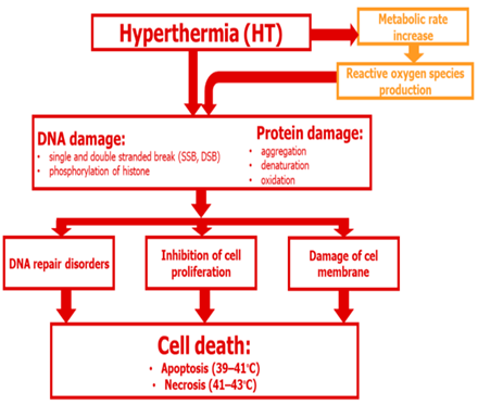
Figure: Metabolic pathway of Hyperthermia………
HT-Induced Apoptosis
Apoptosis is a regulated form of cell death that plays a vital role in maintaining cellular homeostasis, controlling tissue growth, and eliminating damaged or unnecessary cells. Hyperthermia (HT) has been shown to trigger apoptosis in various cancer cell lines, including leukemic and cervical carcinoma cells, typically when exposed to temperatures ranging from 42–44°C [105]. However, some cell types are more resistant to thermal stress than others. HT-induced apoptosis involves three primary apoptotic pathways: the intrinsic (mitochondrial-mediated) pathway, the extrinsic (death receptor-mediated) pathway, and the endoplasmic reticulum stress-mediated pathway. Each of these pathways activates caspases, which are essential enzymes responsible for executing apoptosis [104].
Intrinsic Pathway: Mitochondrial Mediated Apoptosis
The intrinsic pathway plays a central role in hyperthermia (HT)-induced apoptosis. This pathway is regulated by the Bcl-2 family of proteins, which includes both pro-apoptotic proteins (e.g., Bax, Bak, Bid) and anti-apoptotic proteins (e.g., Bcl-2, Bcl-xL). When cells are exposed to heat stress, Bid, a pro-apoptotic protein, facilitates the translocation of Bax to the mitochondrial membrane [106]. This results in the formation of pores in the membrane and the release of cytochrome c into the cytosol, a critical event in the initiation of apoptosis [55]. In cancer cells, such as HeLa cells, HT at 42–43°C induces an upregulation of pro-apoptotic proteins like Bax, Bak, Puma, and Noxa, while anti-apoptotic proteins like Bcl-2 and Bcl-xL are downregulated. The shift in the balance of these proteins drives the cell toward apoptosis, with caspase 3 activation leading to chromatin condensation and subsequent cell death [107][108].
Extrinsic Pathway: Death Receptor-Mediated Apoptosis
The extrinsic pathway involves death receptors on the cell surface, such as Fas and TNF receptors, which bind to their respective ligands and initiate a signaling cascade that activates caspase 8. This, in turn, activates downstream caspases, leading to apoptosis. While this pathway can be modulated by hyperthermia (HT), the intrinsic mitochondrial pathway is generally regarded as the primary mechanism driving apoptosis in cancer cells subjected to thermal stress [109].
Endoplasmic Reticulum Stress Pathway
Hyperthermia (HT) also induces endoplasmic reticulum (ER) stress, resulting in the accumulation of misfolded proteins within the ER. This triggers the unfolded protein response (UPR), which can activate caspases and lead to cell death. The ER stress pathway is closely interconnected with both the intrinsic mitochondrial pathway and the induction of apoptosis by HT.
Necrosis vs. Apoptosis
While temperatures ranging from 40–45°C can trigger apoptosis, higher temperatures generally drive cells toward necrosis. Necrosis is a form of cell death marked by the loss of membrane integrity and the subsequent inflammatory response. Necrosis occurs when the damage is too extensive for the cell to repair itself and is typically accompanied by the release of cellular contents that can cause inflammation in the surrounding tissue [110].
Pro-Apoptotic Proteins and Heat Shock Proteins (HSPs)
Heat stress also activates several pro-apoptotic proteins, including Bim and Noxa, which further promote mitochondrial membrane permeabilization and cell death. Heat shock proteins (HSPs), especially HSP72, play a crucial role in regulating apoptosis. While HSP72 helps protect normal cells from thermal stress by assisting in protein folding and preserving cellular integrity, it can also promote apoptosis in tumor cells when their ability to withstand heat is exceeded [61].
Advantages of Hyperthermia-Based Treatment with Iron Oxide Nanoparticles
Iron oxide nanoparticle-based hyperthermia is an advanced approach that combines the benefits of targeted therapy, minimal invasiveness, and local temperature control. This technique leverages the magnetic properties of iron oxide nanoparticles to produce localized heat when exposed to an alternating magnetic field (AMF). This heat raises the temperature of the tumor while minimizing damage to surrounding healthy tissues [62][63].
Selective Targeting
One of the primary advantages of iron oxide nanoparticle-based hyperthermia is its ability to selectively target cancer cells. By functionalizing the nanoparticles with specific ligands or antibodies, they can be engineered to recognize and bind exclusively to cancer cells, thereby minimizing damage to surrounding healthy tissues. This selectivity significantly reduces the off-target effects commonly associated with conventional cancer treatments like chemotherapy and radiation [65][66]. Furthermore, magnetic guidance can be employed to concentrate nanoparticles at the tumor site, ensuring that they reach the target with high precision.
Improved Tumor Microenvironment
Hyperthermia has been shown to improve the tumor microenvironment by enhancing oxygenation and blood flow, which makes cancer cells more sensitive to treatment. This is especially beneficial when hyperthermia is combined with other therapies like chemotherapy and radiation, as it can enhance their effectiveness. By improving the tumor’s response to treatment, hyperthermia provides a synergistic effect that increases the overall therapeutic outcomes [67].
Multimodal Therapy and Drug Delivery
Another notable advantage of this approach is its ability to integrate with drug delivery systems. Iron oxide nanoparticles can be designed to carry therapeutic agents directly to the tumor site, enabling controlled and localized drug release upon heating. This combination of drug delivery and hyperthermia reduces the necessary dosage of chemotherapy agents, thus minimizing side effects while improving treatment efficacy [68][69].
Biocompatibility and Safety
Iron oxide nanoparticles are biocompatible and can be safely metabolized by the body. Once administered, the nanoparticles break down into iron ions, which are naturally processed by the body. To further enhance safety and efficacy, surface modification techniques have been developed to improve their circulation time, stability, and decrease immunogenicity, making them suitable for clinical use [70][71].
Imaging Capabilities
In addition to their therapeutic potential, iron oxide nanoparticles are valuable in imaging techniques, particularly magnetic resonance imaging (MRI), due to their magnetic properties. This dual functionality enables real-time monitoring of nanoparticle distribution and treatment progress, ensuring precise and effective therapy [72].
Challenges and Future Directions
Despite these advantages, several challenges remain. Accurate temperature regulation is essential to avoid thermal damage to surrounding healthy tissues. Moreover, concerns about the long-term accumulation and clearance of nanoparticles from the body need to be addressed through further research. Additionally, the high cost and limited availability of this technology could hinder its widespread adoption in clinical settings [73]. However, ongoing advancements in nanoparticle engineering, functionalization, and delivery systems are gradually overcoming these limitations. Preclinical and clinical studies are confirming the safety and efficacy of iron oxide nanoparticle-based hyperthermia, and it holds great potential for becoming a standard part of cancer treatment protocols [74].
CONCLUSION
Iron oxide nanoparticle-based hyperthermia presents a ground breaking approach to cancer treatment with its ability to deliver highly focused, minimally invasive therapy. Unlike conventional cancer treatments, this method leverages the thermal and magnetic properties of nanoparticles, allowing for precise tumor ablation while significantly reducing side effects. The ability of hyperthermia to specifically target tumors, combined with other treatment modalities such as imaging and drug delivery, significantly enhances its therapeutic potential. This multimodal approach shows promise in improving treatment outcomes by synergistically increasing the efficacy of traditional therapies like chemotherapy and radiation. The ongoing advancements in nanoparticle design, functionalization, and delivery systems are gradually overcoming key challenges such as temperature regulation, clearance of nanoparticles, and cost-effectiveness. These innovations pave the way for iron oxide nanoparticle-induced hyperthermia to become a more viable option in clinical cancer care. As these technologies continue to evolve, they offer the potential to revolutionize cancer treatment by providing localized, targeted therapies that minimize systemic toxicity and harm to healthy tissues. Moreover, the adaptability of this treatment, including its integration with real-time monitoring and multimodal applications, underscores its potential to redefine cancer care. The promise of personalized, targeted treatment with reduced side effects could ultimately enhance the quality of life for cancer patients and offer hope for improved long-term survival rates. As research and clinical validation progress, iron oxide nanoparticle-based hyperthermia may significantly contribute to the future of cancer therapies, making it a transformative and essential tool in the fight against cancer. With further investigation and innovation, this technique stands poised to provide safer, more efficient, and highly effective treatment options for a wide range of cancers, revolutionizing the therapeutic landscape and offering new hope for cancer patients globally.
REFERENCES


Bablu Malhotra, J. Anuradha, Dr. R. Sanjeevi*, Nanotheranostics: Nanoparticle Fortified Hyperthermia for Efficient Cancer Treatment, Int. J. of Pharm. Sci., 2025, Vol 3, Issue 7, 4244-4268. https://doi.org/10.5281/zenodo.16629638
 10.5281/zenodo.16629638
