College of Pharmaceutical Sciences, Govt Medical College, Kozhikode.
The eye is a highly delicate and intricate organ protected by multiple structural and physiological barriers that play a crucial role in preserving ocular homeostasis. But these same barriers—the blood-ocular barriers, corneal epithelium, tear film, and conjunctival clearance—present serious obstacles to efficient drug administration, especially in achieving and maintaining therapeutic drug concentrations at the site of action. Conventional ocular formulations, such as eye drops and ointments, suffer from a low bioavailability because of their restricted permeability and quick precorneal clearance. Ocular inserts have become a viable substitute in this regard, providing better patient compliance, longer residence duration, and controlled and sustained medication release. Ocuserts [ocular inserts] are defined as sterile preparations, multilayered, solid or semisolid devices placed in the cul-de-sac or conjunctival sac and whose size and shape are designed especially for ophthalmic application. These deliver the drug at a constant rate by a diffusion mechanism. This review provides a comprehensive overview of ocular inserts including history, advantages, disadvantages, classification of ocular inserts, their design strategies, mechanisms of drug release, evaluation methods, recent advancements aimed at overcoming ocular barriers for enhanced therapeutic efficacy and additionally, it highlights different polymers that are commonly employed in the ocular inserts and offers an update of related research work on the ocular inserts.
Eyes and vision are beautiful windows to the world. Together, they provide a better perception of their surroundings. However, the eyes are susceptible to a wide variety of fungal, bacterial, and viral infections. 1Ocular drug delivery systems (ODDS) have drawn interest recently due to their potential for treating a range of eye conditions.2 The fundamental objective of developing these systems is to optimize drug retention duration, improve ocular permeability, and accomplish targeted delivery for enhanced therapeutic outcomes.3 Effective drug absorption is hampered by the complex anatomy, physiology, and barrier function of the cornea.4 As a result, only a small fraction of the drug, effectively 5% or even less of the instilled dose, is ocularly absorbed.5 Achieving an ideal drug concentration at the active site for an adequate period is the major obstacle to designing ocular drug delivery systems with excellent therapeutic efficacy. 6Maintaining the therapeutic drug level in the tear film or site of action requires frequent instillations of eye drops. However, using very concentrated solutions frequently can cause harmful side effects, including corneal surface damage.7 Various drug-delivery devices have been designed for ocular applications to tackle the concerns regarding topical medications.8 Ocular inserts are one example of such a device. “These are defined as sterile, thin, multilayered, drug-impregnated preparations with solid or semisolid consistency intended to lie beneath the upper or lower eyelid, where a diffusional technique would release the medication” 9The primary goal of ophthalmic inserts is to extend the ocular residence time and thereby guarantee a sustained release ideal for ocular drug delivery.10 The thickness, drug selection, and drug release time are the main areas where this device has to be improved.11
History of Ocular Inserts
Alza Corporation created Ocusert, the first ocular implant, in the 1970s. It was designed to provide controlled drug release for the treatment of glaucoma, specifically delivering pilocarpine for a maximum of 5-7 days to reduce intraocular pressure.12 In contrast to conventional eye drops, the Ocusert was a tiny, flexible device that was inserted into the conjunctival sac. The drug diffused from an internal reservoir that contained alginate as an excipient through the two surrounding ethylene vinyl acetate (EVA) membranes, allowing for prolonged drug release over a week and increasing patient compliance. Due to the negative side effects of pilocarpine and discomfort difficulties with the 500-μm-thick device, this device has subsequently been withdrawn.13 Three components are commonly found in all varieties of ocuserts: "an outer annular ring" for effortless handling and precise insertion; "a central drug reservoir" where the drug is integrated in a polymer; and "a rate controlling membrane" that ensures the regulated release of drugs from the reservoir.14
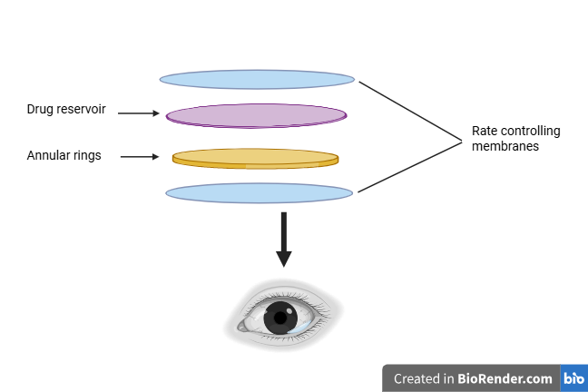
Fig.1 Ocusert
Advantages Of Ocusert15,16
Disadvantages17,18
Mechanism Of Drug Release from Ocular Inserts
Drugs are continuously released via the membrane into the tear fluid as a part of the diffusion mechanism. If the insert has pores and is a solid, non-erodible body, the drug could be released through diffusion through the pores. In the soluble devices, the primary mechanism involved in the dissolution is polymer swelling. When the insert is placed in the eye, water from the tear fluid penetrates the matrix, leading to swelling, polymer chain relaxation, and finaly, drug diffusion. The dissolution of the matrix depends on the polymer structure, with linear amorphous polymers dissolving faster than cross-linked or partially crystalline polymers. 19
2. Osmosis
An insert with a transverse impermeable elastic membrane that separates the interior into a first and second compartment is used to explain the osmosis mechanism. Semi-permeable and elastic membranes enclose the first compartment, whereas an impermeable membrane encloses the second compartment. There is a drug release hole in the insert's impermeable wall. While the second compartment serves as a drug reservoir, the first compartment holds a solute that is impervious to the membrane. Water diffuses and expands the first compartment while contracting the second, driving the drug through the opening when it is placed in the aqueous environment of the eye. 20
A bio-erodible matrix that distributes the drug makes up the functional unit of an insert's body. The matrix bio-erodes when the insert comes into contact with the tear fluid, delivering the medication in a regulated and sustained manner. Although the drug may be distributed uniformly throughout the matrix, it is thought that a more controlled release happens when the drug is superficially concentrated in the matrix. 21
Classification Of Ocular Inserts
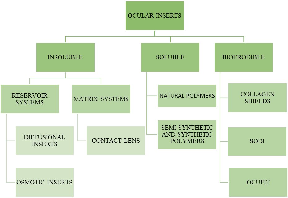
These are made up of insoluble polymers and can deliver the drug at a controlled and predetermined rate, but they need to be removed from the site. 22 They are classified into two categories
These reservoir systems can release the drug by diffusion or an osmotic process. It contains a drug-containing liquid, gel, colloid, semisolid, solid matrix, or carrier, in that order. Carrier materials might be hydrophilic, hydrophobic, organic, natural, or synthetic polymers. They have been sub-classified into 23
1. Diffusional inserts, e.g., 'Ocuserts'
2. Osmotic inserts.
a. Diffusional Inserts
The Diffusional inserts/Ocuserts system is a ground-breaking medication delivery technique based on a porous membrane, where drugs are released via a diffusional release mechanism. It is made up of a central drug reservoir that is protected by a specifically made semipermeable/microporous membrane that allows the drug to diffuse out of the reservoir at a precise rate. The tear fluid, which seeps through the membrane until adequate internal pressure is achieved, regulates the release of drugs. Its activity can be explained by Fick's diffusion equation. 24
J=DA dc/dx.
Where J: solute flux.
D: Diffusion coefficient for the medication within the polymer membrane.
A: Membrane area.
dc/dx: Drug concentration gradient within the membrane along the direction of drug flow.
B. Osmotic Inserts
Osmotic inserts are divided into two types and typically consist of the core encircled by a peripheral segment. 25
Type 1:
The central element consists of a singular reservoir containing a drug, optionally accompanied by an additional osmotic solution, which is dispersed within a polymer matrix and encircled by distinct minute deposits. The second peripheral component of these inserts is a cover film composed of an insoluble semi-permeable polymer. Holes are created when the polymer matrix ruptures due to osmotic pressure. The medication can be liberated from deposits close to the device's surface through these perforations.
Type 2:
The central part consists of two separate compartments, which are used to hold drug and the osmotic solute. The osmotic solute reservoir is enclosed by a semi-permeable membrane, while the drug reservoir is enclosed by an elastic impermeable membrane. The active ingredient is forced through the single drug release orifice when the crack diffuses into the osmotic compartment, where an osmotic pressure causes the elastic membrane to stretch and compress the drug-containing compartment.
Contact lenses are the primary representative of the second category, the matrix system, which is a specific set of insoluble ophthalmic devices. It is made up of hydrophilic or hydrophobic polymers that are covalently cross-linked to produce a three-dimensional network or matrix that can hold onto solid materials, water, or aqueous medication solutions. By absorbing water, the hydrophilic or hydrophobic polymer swells. Until a final swelling (equilibrium) is achieved, the elastic retroactive forces that arise along the chains or crosslinks fight the swelling brought on by the osmotic pressure of the polymer segments. 26
Contact Lenses
The original purpose of contact lenses, which are formed structures, was vision correction. By soaking them in medication solutions beforehand, their utility has been expanded to include possible drug delivery systems. This system's primary benefit is its ability to concurrently release medication and correct vision. According to Refojo, contact lenses should be divided into five categories. Rigid, Semi-rigid, Elastomeric, Soft hydrophilic, Bio-polymeric. Rigid contact lenses are made of rigid polymers that are difficult to permeate by moisture and oxygen. Gas-permeable polymers like cellulose acetate butyrate are used to alleviate, but are uncomfortable to wear and not suitable for long-term drug administration. In such a scenario, soft hydrophilic lenses are developed for continuous drug release. Soft hydrophilic contact lenses are popular due to their ease of fitting and better tolerance. The hydrophilic or lipophilic nature of the drug determines its inclusion in contact lenses. The dosage, soaking duration, and drug concentration in the solution all affect drug release.27
2. Soluble Ocular Inserts
The advantage of these soluble inserts is that they are completely soluble, meaning that the intervention is limited to insertion and does not require removal from the site of application. They can be classified into two types. 28
Natural polymers are the basis for the first kind of soluble inserts. Collagen, a natural polymer widely used in the soluble ophthalmic implants. The medication is progressively released from the interstitial spaces between the collagen molecules as the collagen degrades and thereby providing a better drug delivery.
The second kind of soluble insert is typically based on synthetic polymers like polyvinyl alcohol, Polycaprolactone, Polymethacrylate, or semi-synthetic polymers like cellulose derivatives. Eudragit, a polymer often used for enteric coating, can be employed as the coating agent of the insert to reduce the release rate.29
3. Bio-Erodible Inserts
These inserts are composed of bio-erodible polymers, such as cross-linked gelatine derivatives and polyester derivatives, which dissolve when chemical bonds are hydrolyzed. The ability to adjust the rate of erosion of these bio-erodible polymers by altering their final structure during synthesis and by adding cationic or anionic surfactants is a significant benefit. However, erodible systems might have vastly different rates of erosion, and inflammatory responses might occur depending on each patient's physiology and lacrimation patterns. It might be brought on by degradation products and residual solvents from the polymer production process.
Some significant ocular inserts that are either commercially available (SODI) or in advanced stages of development (collagen shields, Ocufit, and Minidisc) are covered in the paragraphs that follow. 30
A. Soluble Ophthalmic Inserts (Sodi)
SODI, a tiny oval wafer created by Soviet scientists, was intended for cosmonauts who were unable to administer eyedrops while in weightless settings. Collagen shields and SODI represent the first contemporary resurgence of gelatin "lamellae," which vanished from pharmacopoeias in the late 1940s. Russian chemists and ophthalmologists worked together to develop the SODIs, which resulted in the creation of a novel soluble copolymer known as ABE in 1976. Following rigorous preclinical and clinical testing, ABE was used to produce SODI industrially as oval-shaped, sterile thin films weighing 15–16 mg and color-coded for over 20 common ophthalmic drugs. ABE also produced the maximum concentration of medicines in rabbit ocular tissues. The film softens in 10–15 seconds after being inserted into the upper conjunctival sac, forms a polymer clot, and releases the medication within an hour. 31
B. Collagen Shields
Collagen implants were suggested as gentamicin delivery systems and tear substitutes by Bloomfield et al. in 1977 and 1978. They discovered that the collagen insert had the highest concentration of gentamicin in the rabbit eyes' tears, cornea, and sclera, after three hours. Collagen shields impregnated with gentamicin and dexamethasone have been used in other therapies; these have a 10-hour delivery period that is similar to subconjunctival injections. However, collagen shields have limitations such as irritation and visual problems, and it worsen corneal ulcers caused by alkali burns. Small particles of collagen suspended in a 1% methylcellulose medium make up a novel preparation known as collasomes, which offers the same therapeutic benefits as collagen shields but without the drawbacks. To sum up, collagen is a useful substance for a number of medical uses. 32,33
The Ocufit is a silicone elastomer rod-shaped device that is 25–30 mm long and 1.9 mm in diameter, made to fit the human conjunctival fornix. Compared to oval, flat inserts, its cylindrical shape is more effective at lowering expulsion rates. Ocufit's combination of long retention and continuous medicine delivery is demonstrated by the fact that 70% of placebo devices remain in the upper fornix of participants. Patients' capacity to keep the inserts was unaffected by active conditions such as corneal ulcers, trachoma, episcleritis, anterior uveitis, bacterial, allergic, and adenoviral conjunctivitis, or scarring. In an in vitro study, tetracycline-loaded inserts released 45% of the medication over 14 days, with a first-day burst and a steady rate throughout the rest of the period. The ocufit is presently being developed for infants and young children. 34
d. Minidisc Ocular Therapeutic System
Like a tiny (4-5 mm) contact lens, the Minidisc Ocular Therapeutic System (OTS) includes a convex and concave face that greatly adapts to the sclera of the eye. According to reports, the device's specific size and form make it simple to fit under the upper or lower lid without sacrificing oxygen permeability, comfort, or eyesight. The Minidisc was said to take less time and require less manual skill to place than the Lacrisert, another common insert. The device has been tested in a variety of forms, including erodible, non-erodible hydrophobic, and non-erodible hydrophilic. 35
Preparation Of Ocular Inserts
1. Solvent Casting Methods
This method entails making numerous batches in different amounts. A suitable solvent is used to dissolve the polymer. After adding plasticizer to the mixture while stirring constantly, a precisely measured quantity of medicine is added to the mixture, creating a homogenous dispersion. Using an inverted funnel, the resulting mix was transferred onto a petri dish and coated. This allows for uniform and slow evaporation at room temperature until the film has dried. A cork borer is used to cut the dry films into the appropriate shapes and sizes. Airtight containers are used to store the prepared ocuserts. 36–38
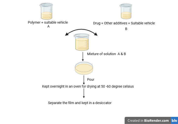
Fig. 2 Solvent casting method
To create a clear solution, the polymer is immersed in a 1% (v/v) carboxylic acid solution for roughly twenty-four hours. After adding the required amount of drug to the previously mentioned solution, the conjugate is dissolved in the polymer solution for fifteen minutes using a vortex mixer. The plasticizer is absorbed by the solution. To enable the film to regulate the pace of growth and eliminate air bubbles from the solution, a viscous solution was made and allowed to sit for half an hour. After being poured onto flat glass molds, the membranes were allowed to cool for two hours. A water-soluble, nontoxic, non-irritating adhesive is used to push the dry film into the desired size and shape, and the matrix is positioned between the flow control membranes. They are wrapped in aluminium foil and stored in a desiccator. 39,40
3. Hot Melt Extrusion
This method passes the medication and polymer through a 60# mesh before weighing and blending them. The mixture is then treated with a plasticizer. After that, the mixture is loaded into and drained from the melter tank. The materials were carefully measured, cut, wrapped in aluminium foil, heat-sealed, and gamma-sterilized.41 The quality of the cutting surfaces is significantly impacted by the precise fusion of the polymer formulation and the chosen cutting technique, which makes cutting the insert a crucial phase. With the help of the CaliCutTM post-extrusion technology, polymer strands may be accurately calibrated and cut into precisely sized inserts. Ocular inserts are produced using this next-generation modular device. It is easier to fabricate ocular dosage forms with the appropriate size when HME and CaliCut are combined. When compared to traditional eye drops, the formulation of ocular inserts is incredibly cost-effective.42

Fig. 3 Hot melt extrusion method 43
Customized pharmaceutical dosage forms for a range of uses have been made using 3DP. When HME and FDM-3DP are combined, it may be possible to produce patient-centered medications with complex forms, distinctive designs, and specialized release properties for use in the eyes. Examine the possibility of combining HME with FDM-3DP technologies to create specialized, long-lasting CIP-HCL inserts for use in the eyes. Additionally, they used a design of experiment (DOE) technique to investigate how various printing process factors affected specific important quality features of prepared inserts, such as drug release. The limitations of traditional ophthalmic dosage forms and the problems of previous crude technologies, including solvent casting methods for ocular insert fabrication, may be solved by this additive manufacturing technology.44
Evaluation of Ocuserts
Shadambikar et al conducted the determination of drug content by dissolving the optimized Ocusert in a solvent system by probe sonication. The obtained sample is centrifuged, the supernatant is adequately diluted, and the drug content is analyzed spectrophotometrically. 45
2. Swelling Index
To determine the swelling index, inserts are randomly selected and weighed initially and then placed into freshly prepared STF media at 37 ± 0.2 °C. For a maximum of 30 minutes, the insert was taken out every 5 minutes, the excess media was wiped off with filter paper, and the insert was weighed again. The swelling index was calculated as46
SI ( % ) = Weight of swollen insert − Initial weight of the ocular insert × 100
Initial weight of the ocular insert
3. Sterility Test
Following in vitro drug release experiments, the chosen formulation was sterilized using gamma or UV radiation. Prepared sterile and unsterile Ocusert solutions are taken out and aseptically transported to the soybean-casein digest medium and fluid thioglycollate media, respectively. This is followed by an incubation period of at least 14 days at 20°C to 25°C for the soybean-casein digest medium and 30°C to 35°C for the fluid thioglycolate. 47
4. Tensile Strength
Tensile strength of ocuserts refers to the maximum stress that the ocusert can withstand while being stretched before it breaks. 48
Tensile strength = Force at break (N)
Cross-sectional area (mm2)
5. Percent Moisture Absorption
This is done to assess the ocuserts' physical integrity or stability in humid environments. Each batch's ocuserts were weighed before being put in aluminum chloride-filled desiccators. Three days later, the ocuserts were taken out and weighed once more. 49
Percent of moisture absorption = Final weight - Initial weight x 100
Initial weight
6. Percent Moisture Loss
This is performed to verify the ocuserts' integrity when they are dry. The ocuserts were weighed and then placed in desiccators with anhydrous calcium chloride inside. Three days later, these ocuserts were taken out and weighed once more. 50
Percent moisture loss = Initial weight – Final weight x 100
Initial weight
7. In Vitro Drug Release Study
1. Bi-chamber donor-receiver compartment model
A commercial semi-permeable membrane is used in a bi-chamber donor-receiver compartment model for in vitro release experiments. The donor compartment is an open cylinder to which the cellophane membrane is attached after being pre-soaked in a dissolution medium (STF Ph 7.2) for the entire night. To simulate in vivo circumstances, such as a corneal epithelial barrier, a semi-permeable membrane is employed. To replicate tear volume, 0.7 ml of water is added to the donor compartment along with the ocular insert. The surface of the membrane is in contact with a reservoir compartment that contains 25 millilitres of pH 7.4 phosphate buffer. Continuous stirring of the dissolving liquid is done with a magnetic stirrer. After the samples are removed from the receptor compartment, an equivalent volume of pH 7.4 phosphate buffer is added. Samples are analysed spectrophotometrically 51
2. Open Flow Through Apparatus
The apparatus mimics the constant flow of tears in an in vitro diffusion cell, but it is unable to duplicate the eye's constant blinking. It includes two fluted glass adaptors and a 2 ml glass tube. It is attached to a reservoir that contains PVC flexible tubes and isotonic phosphate buffer saline (IPBS) with a pH of 7.4. The head of the reservoir is maintained constant while a valve regulates the buffer flow rate. Air bubbles are eliminated by allowing a tiny amount of fluid to flow away. A thin, circular Teflon disk with an ocular insert adhered to it exposes only one surface to the dissolution media. After inserting the disk into the cell, the fluid temperature is kept at 37±1°C.The drug content is analysed using a spectrophotometer at regular intervals.52
3. The inserts are placed in a beaker containing defined quantity of phosphate buffer pH 7.4 kept at 37±1°C, under control stirring. At various intervals sample are withdrawn and analyzed spectrophotometrically.53
8. Ex Vivo Permeation Study
Using a diffusion cell, the ex vivo permeation study is conducted. The goat cornea is taken out of the eye and placed on a diffusion cell so that the corneal side comes in touch with the ocusert in the donor compartment. The receptor fluid is constantly agitated with a magnetic stirrer to keep its temperature at 37 ± 0.5ºC. Sample is taken out of the receiver compartment at predetermined intervals and subjected to spectrophotometric analysis. An equivalent volume of tear fluid is added to each sample that is removed. 54
9. In Vivo Drug Release Study
Before the in vivo study, the ocuserts were sterilized using UV light. After sterilization, ocuserts are stored in a polyethylene bag using forceps within the sterilization chamber. The experiment included albino rabbits of either sex. Ocuserts for in vivo research are placed in the cul-de-sac of the lower conjunctiva. In order to assess the drug content, the ocuserts were carefully removed after 30, 60, 90, 120, and 150 minutes. 55
10. Stability Studies
According to International Council for Harmonisation (ICH) guidelines, stability studies are designed to estimate a pharmaceutical product’s shelf life by accelerating degradation, typically through elevated temperature and humidity conditions. These studies facilitate the evaluation of critical parameters such as physical appearance, mechanical durability, and drug content over time. Sankar et al performed a stability study of diclofenac ocuserts by placing inserts in a glass container and wrapping them in aluminum foil, then storing them in a stability chamber maintained at 40?°C and 75% relative humidity (RH). Assessments were conducted at predetermined intervals 0, 1, 2, 3, and 6 months to monitor changes in physical appearance, percentage weight variation, folding endurance, and drug release profiles.56
11. Ocular Irritation Test
Nair et al performed an ocular irritation test of timolol ocusert by placing the test substance into the conjunctival sac of one eye of the test animal. To prevent the material from being lost, the lids were then gently held together for about a second. At intervals of 1, 24, 48, and 72 hours on the first day and 7, 14, and 21 days, the test eye was examined for alterations in the cornea, iris, conjunctiva, and chemosis relative to the control.57
Novel Strategies That Can Be Incorporated into Ocuserts
The rapidly developing field of nanotechnology is led by solid lipid nanoparticles, which offer a number of potential applications in drug delivery. Solid lipid nanoparticles are colloidal drug delivery systems ranging in size from 10 – 500 nm. They are made up of solid lipids, so that they are well tolerated by the body. The advantages of SLNPs include their high stability, improved mucoadhesive property, low toxicity to the body, high drug-loading capacity with little drug leakage, drug targeting, and controlled and/or prolonged drug release. They are lipophilic and tiny; therefore, they can easily cross biological barriers.58–61
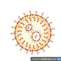
Fig.4 Solid lipid nanoparticles
Ramadan et al developed vildagliptin solid lipid nanoparticle incorporated ocuserts for treating diabetic retinopathy by the double emulsion/melt dispersion method. Sustained drug release by VLD-SLNPs-OCU may be a useful substitute for traditional oral dosing forms, resulting in less systemic adverse effects and less variation in plasma drug levels. In addition to reducing blood sugar, VLD-OCUs may enhance retinal blood flow, which makes them a potentially helpful treatment option for DRP.62
Nanofibers are incredibly thin fibers made of polymers that range in diameter from 1 to 1000 nanometers. Drugs can be administered once or twice a day by using polymeric fibers and controlled-release administration routes. This improves patient adherence and prevents harmful plasma peak concentrations that may occur from often administering immediate-release formulations. Excellent stability, targeted delivery, high drug-loading capacity, large surface area, reduced toxicity, enhanced mechanical capabilities, and suitability for the delivery of medications that are sensitive to temperature are just a few benefits that come with small nanofibers. Nanofibers can be prepared using a variety of methods, including electrospinning, melt spinning, force spinning, and emulsion spinning.63–65

Fig.5 Nanofibers
For prolonged ocular administration, Taghe et al. created macrolide-loaded nanofibrous inserts using cellulose acetate and polycaprolactone as the base. To achieve a prolonged and increased release of azithromycin for the ocular drug delivery system, polycaprolactone and cellulose acetate nanofibers carrying the antibacterial macrolide were effectively created by electrospinning.66
3. Glycerosomes
Significant quantities of glycerol, phospholipid, water, and other active components make up glycerosomes, which are vesicular drug delivery vehicles. Increased efficacy, improved stability, enhanced absorption, drug targeting at specific areas, and delivery at a predetermined rate are just a few advantages that glycerosomes provide. There are numerous known methods for producing glycerosomes, including solvent spherule, detergent elimination, reverse-phase evaporation, thin film hydration, and others. Researchers are currently investigating the potential of glycerosomes as nanocarriers for both natural and manufactured bioactive medications.67–70
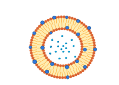
Fig.6 Glycerosomes
Naguib et al, developed 3D printed ocusert laden with ultra-fluidic glycerosomes of ganciclovir for the management of ocular cytomegalovirus retinitis. In this study, UFGs containing phospholipid, cholesterol, sodium taurocholate, and glycerol were successfully synthesized using the ethanol injection method. Using PLA filament and a CAD design, a 3D printed ocusert was produced. The optimal UFGs showed increased permeability through the ocular tissue in contrast to edge-activated vesicles and conventional glycerosomes. The drug's ocular bioavailability was increased and its release was sustained for five days by the Ocusert that included the optimal GCV-loaded UFGs. To put it briefly, the results indicate that using an ocusert loaded with GCV-loaded UFGs to treat CMV-induced retinitis has promising potential.71
4. Liposomes
The spherical vesicles known as liposomes have an aqueous core around by one or more concentric phospholipid bilayers.. Liposomes are a potent medication delivery vehicle since they are harmless and biodegradable. Because of their structural adaptability, biocompatibility, biodegradability, non-toxicity, and non-immunogenicity, liposomes are regarded as effective drug delivery vehicles. Because phospholipids in solution have an amphiphilic nature that resembles that of natural cell membranes, liposomes and mammalian cell membranes can interact well, facilitating effective cellular uptake. Liposomes also can self-assemble, contain enormous pharmacological payloads, and have a variety of physicochemical and biophysical properties that can be altered to influence their biological characteristics.72–75
Fig. 7 Liposome
A 3-D printed ocular insert containing liposomal moxifloxacin was created and assessed by Duman et al. The purpose of this study was to examine the enhanced bioavailability and controlled release effect of liposomal moxifloxacin-containing 3D-printed ocular inserts, as well as their appropriateness for ocular administration. The advantages of using a 3D printer in conjunction with liposome technology have been assessed. Compared to free moxifloxacin, liposomal moxifloxacin has demonstrated superior particle size and homogeneity. Liposomes successfully reduced the concentration of free drug, as anticipated.76
Polymers Employed in Ocular Inserts
1. Gelatin
Because of its inherent qualities, such as biocompatibility, biodegradability, drug targeting to the desired site, controlled release, improving the bioavailability of topical administration, delivering a variety of agents, and improving patient compliance, A versatile delivery route for therapeutically active substances to the ocular surface is gelatin. Micro and nano gelatin-based fabrications, namely hydrogels, nanoparticles, nanocomposites, microspheres, scaffolds, and ocular adhesives, applications are being thoroughly researched for their ocular applications. Gelatin is a viable biomaterial for ocular research because of all these innovative techniques.77,78 Lee et al. developed and characterized an ocular insert made of gelatin that contained tropicamide and phenylephrine. This study suggests that Gelfoam is an adaptable carrier for both topical and systemic drug delivery through the ocular route.79 Gelatin-based ocular inserts are recommended primarily because to their great biocompatibility. Furthermore, gelatin's breakdown products are safe. As a result, it has been used extensively in ocular applications.But in a physiological setting, gelatin swells as a hydrocolloid and quickly releases the payload. Gelatin must be crosslinked to prevent the rapid release of medications from devices based on it. Formaldehyde, acrylamides, epoxides, carbodiimides, and glutaraldehydes are examples of bifunctional crosslinkers. Since leftover crosslinkers provide major toxicity concerns, the crosslinking technique employed for gelatin is crucial for drug release, degradation kinetics, and—above all—biocompatibility. As an alternative, Gelatin methacryloyl (GelMa) can be created by joining methacryloyl groups to the main amino groups of Gelatin. Thereafter, when Irgacure® 2959 is present, this can be crosslinked using UV light.During the crosslinking procedure, the methacryloyl groups are ultimately integrated into the polymeric matrix. GelMa crosslinking has been widely employed in tissue engineering and is comparatively benign 80
2. Chitosan
Chitosan is a naturally occurring biodegradable polymer that has been thoroughly studied because of its potent mucoadhesive properties. Due to the ionic interactions between the chitosan and the anionic ocular mucosa drug’s mucoadhesion and ocular retention are greatly enhanced. Therefore, a chitosan-based nanoparticulate technology can decrease the number of ocular injections required and improve long-term patient compliance. Chitosan relaxes the strong bonds between cells, increasing permeability. Additionally, it is made by deacetylating fungal cell walls and crustacean exoskeletons, which results in a low production cost and little ecological impact. Research on stimuli-responsive biological delivery systems may find chitosan, in particular, to be a potentially valuable platform due to its extraordinary swelling behaviors across a range of physiological conditions.81,82 Jadhav et al formulated and evaluated a polymer-coated bimatoprost chitosan matrix ocular insert for lowering intraocular pressure in rabbits by the solvent casting method. The major goal of this study was to attain a sustained drug release.83 Said et al. proposed ocular mucoadhesive and biodegradable sponge-like inserts to accurately and continuously administer voriconazole. A pH within the safe ocular range and good in vitro mucoadhesive properties are characteristics of the optimum formulation. Rapid hydration and gelling, good mucoadhesive behavior when injected into the eye, high ocular safety and biocompatibility, sustained antifungal activity compared to the drug suspension, and biodegradation were all encouraging in vivo results. Therefore, it might be regarded as an exceptional carrier for VCZ ocular administration.84
3. Sodium Alginate
A naturally occurring polymer, alginate is derived from bacteria and marine brown seaweeds. In the field of drug delivery, alginate has been extensively researched as an excipient. Sodium alginate has several benefits for ocular therapeutic targeting, such as ion-sensitive in situ gelation, non-toxic and biodegradable behavior, and the polymer's mucoadhesive properties. Alginate's quick gelation increases ocular medicine absorption and prolongs ocular residence, which lowers the frequency of drug administration. Alginate's widespread availability and alluring physicochemical characteristics have prompted pharmaceutical researchers to investigate more innovative approaches to ocular drug targeting.85,86 Desiato et al developed an ocular antibiotic-loaded soluble film insert by the solvent casting method. The study's main goal was to utilize biocompatible excipients to make an entirely soluble ocular film insert that would enhance the distribution of levofloxacin.87
4. Tamarind Seed Polysaccharide
Tamarind seed polysaccharide (TSP), a naturally occurring compound that is derived from Tamarindus indica seeds, has demonstrated significant promise because of its strong biocompatibility, safety profile, improved mucoadhesion, and physico-chemical characteristics. Due to these qualities, it is now utilized in dilating eye drops for eye examinations as well as formulations for the treatment of bacterial keratitis, glaucoma, and dry eye disease.88,89 A metformin insert made of tamarind seed polysaccharide was created by Reghu et al. The major goal of the study was to create an ocular insert of MET using a carbohydrate-based polymer that would guarantee compatibility, ocular retention, and gradual release for improved ocular burn management.90
5. Guar Gum
Guar gum (GG) is a widely employed polymer in a variety of ocular drug delivery systems. Additionally, guar gum has a considerable bioadhesive power and overcomes the rapid departure from the ocular region by extending the corneal residence period. Because of the extended corneal surface retention, it has demonstrated encouraging outcomes in ocular administration. 91,92 For efficient ocular administration, Kumar S. et al. designed a guar gum-based ofloxacin continuous release ocular implant. Using guar gum as the polymer, the current work aimed to develop a sustained release ocular delivery method for ofloxacin. Ofloxacin's guar gum ocular inserts show notable film-forming capabilities. According to the study, the guar gum-based ocular insert may help ensure that ofloxacin is delivered in a controlled, efficient, and reliable manner. In vitro release experiments revealed zero-order release kinetics for the ocular implants.93
6. Hydroxy Propyl Methyl Cellulose
The cellulose derivative hydroxypropyl methylcellulose (HMPC) has many uses in medication formulations because of its rheological characteristics, transparency, biocompatibility, and water solubility.94 The U.S. Food and Drug Administration has authorized the use of hydroxypropyl methylcellulose (HPMC), a hydrophilic, biocompatible, and mucoadhesive polymer, in eye drops, gel formulations, films, or inserts for the administration of ocular medications. HPMC can be used as an ocular lubricant because of its ability to maintain moisture on the surface of the eyes. So these have superior efficacy in the management of dry eye disease, and they help to preserve physiological corneal density.95 As per recent research, methacrylation modification of polymers can strengthen mucoadhesive power by forming a covalent link between the thiol group (conjunctival mucosa) and the C=C (methacrylate moieties).96 Noori et al.devised an insert for the simultaneous ocular delivery of tinidazole and levofloxacin. This study showed that HPMC incorporated Ocusert had proper physical properties, strength, and mucoadhesion with a high swelling index and sustained release of both drugs for around 12 h.97
7. Hyaluronic Acid
HA's exceptional capacity to absorb and retain water has earned it the moniker "nature's sponge." HA can increase its initial solid volume by a thousand times when it is moistened. Additionally, because of its protective function at the cornea/conjunctiva epithelium and mucus-like rheological behavior, this polymer can be used as an adjuvant in the treatment of dry eye disease. In addition, HA's superior biocompatibility, biodegradability, bioadhesion, viscoelasticity, and receptor interaction qualities make it a viable drug carrier for the treatment of ocular diseases.98,99 To treat dry eye condition, Lokhande et al. created and assessed a biodegradable nanofiber insert that contains hyaluronic acid. Because of its delayed release, which enables a regulated or prolonged release of the medication from the formulation, hyaluronic acid (HA) is used as an API in this study. HA is used as a tissue lubricant to protect the eye's surface and reduce mechanical stress when Dry Eye Syndrome strikes. Additionally, because of its water-retentive qualities, HA enhances water surface wettability, decreases friction during eye blinking, and promotes tear evaporation. It has been hypothesized that dry eye causes an overexpression of CD44 receptors, which HA binds to to stabilize the ocular surface barrier and tear film. Another way that HA works against the elevated inflammatory markers in dry eye is by functioning as an anti-inflammatory agent.100
8. Poly Vinyl Alcohol
PVA is a biodegradable, water-soluble polymer that is frequently used to dissolve hydrophobic medications while offering processing simplicity and chemical resistance. For applications requiring regulated release, PVA's polymer structure offers adjustable permeability. It now appears on the FDA GRAS list and is used in a number of nondegradable eye implants, including Vitrasert®, Retisert®, and Iluvien®. With passive diffusive release from the implant, Vitrasert®, the first FDA-approved PVA and ethylene-vinyl acetate (EVA) copolymer implant for intravitreal treatment of CMV retinitis, offers a therapeutic dose for 6–8 months. Retisert® is a multi-layered implant that treats non-infectious posterior uveitis by delivering controlled release of fluocinolone acetonide for up to 2.5 years through a permeable PVA outer layer. The FDA has approved Iluvien® for the treatment of DME, and clinical trials are currently being conducted to test the delivery technique for other eye disorders, such as wet AMD. PVA is also being investigated for topical medication administration via wafers to the eyes 101,102 Caravaca et al. have devised and validated an insert for the ocular delivery of progesterone. The created insert possesses the mechanical qualities necessary for its ocular use, as well as adaptability, optical clarityand superperior biocompatibility. Experiments on PG release in vitro demonstrate that PG is released in a controlled fashion.103
9. Polycaprolactone
PCL, a biodegradable, semi- crystalline polyester widely utilized in biomedical and pharmaceutical fields due to its low toxicity and ease of processing. A significant study has been done on the benefits of PCL, which include biocompatibility, suitable mechanical strength, affordability, and ease of use with regulated pore size and shape.104,105 For the improved ocular administration of ketorolac tromethamine, Taghe et al. developed polycaprolactone and polymethacrylate nanofibers. The Formulations were used to provide the right amount of mechanical strength and flexibility. In contrast to the 10-hour release of the medication from the eye drop formulation, the in vitro release investigation showed a sustained 6-day release of ketorolac. As a result, developed nanofibrous inserts can be regarded as appropriate solutions for KET sustained delivery, hence lowering the frequency of administration.106
10. Polyacrylic Acid
Many studies have been conducted on the application of polyacrylic acid, a biocompatible and biodegradable polymer, in medicine delivery. The capacity of polyacrylic acid to create hydrogels, its mucoadhesive qualities, and its stimuli-responsive behavior are some of its many qualities that make it a potential material for a variety of applications. Polyacrylic acid's mucoadhesive qualities make it an excellent option for administering medications to the eyes, which could extend the drug's release and improve its bioavailability. Drug delivery systems that release pharmaceuticals in response to particular stimuli, such as variations in pH, temperature, or ionic strength, can be developed using polyacrylic acid's stimulus-responsive nature.107,108 A mucoadhesive ocular implant based on thiolated polyacrylic acid was created by Hornof et al. The ocular insert made of thiolated poly (acrylic acid) is a possible novel solid device for the ocular administration, according the current investigation.The inserts can guarantee a continuous medication release on the ocular surface for an extended amount of time, according to in vivo investigations.109
Table no:1 List of Ocular Inserts Commercially Available 110–114
|
Sl. No |
Drugs |
Class Of Drug |
Polymers |
|
1 |
Aceclofenac |
NSAIDS |
Hydroxy methyl cellulose, Ethyl cellulose |
|
2 |
Acyclovir |
Antiviral agent |
Poly vinyl alcohol, Methyl cellulose, Ethyl cellulose, Carbopol 934 |
|
3 |
Acyclovir |
Antiviral agent |
Methyl cellulose, Hydroxy propyl methyl cellulose, Hydroxy propyl cellulose, Starch |
|
4 |
Brimonidine Tartarate |
IOP Lowering agent |
Poly vinyl alcohol, Ethyl cellulose, Poly vinyl pyrrolidone K-30 |
|
5 |
Ciprofloxacin |
Antiinfective agent |
Hydroxy propyl methyl cellulose, Ethyl cellulose, Methyl cellulose, Poly vinyl pyrrolidone |
|
6 |
Ciprofloxacin |
Antiinfective agent |
Eudragit, Polyvinyl acetate |
|
7 |
Diclofenac sodium |
NSAIDS |
Hydroxy propyl methyl cellulose, Eudragit L 100 |
|
8 |
Fluconazole |
Antifungal agent |
Hydroxy propyl methyl cellulose, Poly vinyl alcohol, Poly vinyl pyrollidone |
|
9 |
Indomethacin |
NSAIDS |
Eudragit L 100, Eudragit RL 100, Hydroxy propyl methyl cellulose , Ethyl cellulose |
|
10 |
Ketorolac tromethamine |
NSAIDS |
Gelatin, Hydroxy propyl methyl cellulose , Ethyl cellulose |
|
11 |
Levobunolol |
β – blocker |
Hydroxy propyl methyl cellulose, Methyl cellulose, Poly vinyl pyrrolidone |
|
12 |
Levobunolol |
β – blocker |
Ethyl cellulose, Eudragit RL 100 |
|
13 |
Levofloxacin |
Antibacterial agent |
Ethyl cellulose, Sodium alginate |
|
14 |
Moxifloxacin |
Antibacterial agent |
Eudragit RL 100, Eudragit RS 100, Sodium methyl carboxy cellulose |
|
15 |
Moxifloxacin |
Antibacterial agent |
Chitosan, Methyl cellulose, Eudragit L 100 |
|
16 |
Natamycin |
Polyene antibiotic agent |
MC, Sodium alginate, Gelatin |
|
17 |
Natamycin |
Polyene antibiotic agent |
Eudragit L 100, Eudragit RL 100, Hydroxy propyl methyl cellulose, Ethyl cellulose |
|
18 |
Norfloxacin |
Antibacterial agent |
Hydroxy propyl methyl cellulose, Ethyl cellulose, Poly vinyl pyrrolidone K-30 |
|
19 |
Ofloxacin |
Antibacterial agent |
Polyethylene oxide, Eudragit L 100 |
|
20 |
Ofloxacin |
Antibacterial agent |
Hydroxy propyl methyl cellulose, Poly vinyl pyrrolidone, Poly vinyl alcohol |
|
21 |
Pefloxacin |
Antibiotic agent |
Eudragit RS 100, Eudragit RL 100, Poly vinyl pyrrolidone K-30 |
|
22 |
Timolol maleate |
Antiglaucoma agent |
Methyl cellulose , Hydroxy propyl cellulose, Eudragit RL 100, Eudragit RS 100, Ethyl cellulose, Poly vinyl pyrrolidone |
CONCLUSION
With several benefits over traditional formulations, such as extended drug release, decreased dosage frequency, increased therapeutic efficacy, and better patient compliance, ocular inserts represent a potential development in the field of ocular drug delivery. The regulated administration of a variety of medications to the anterior and posterior parts of the eye has been investigated using a variety of insert types, including reservoir, matrix, biodegradable, and non-biodegradable inserts. The potential of ocular inserts is still being expanded by continuous research and development, especially in the areas of biodegradable polymers and nanotechnology-based systems, despite certain drawbacks like patient pain or insertion challenges. Ocular inserts have the potential to become a more popular and successful therapeutic option for both acute and chronic ocular conditions with additional clinical data and advancements in patient-centered design.
Conflict Of Interest
Ethics approval and consent to participate - Not applicable
Consent to publication - Not applicable
Availability of data and materials - all data generated or analysed during this study are included in this published article
Competing interests - The authors declare that they have no competing interests
Funding
The authors declare that no funds, grants, or other support were received during the preparation of this manuscript.
ACKNOWLEDGEMENT
Not applicable
Authors Contributions
RS conceptualized the review topic, conducted the literature review, and led to the writing of the manuscript. FN contributed to the literature collection, critically analyzed the data, and assisted in organizing the manuscript structure. AP supported the literature review process, refined the content, and handled formatting, references, and final proofreading.
REFERENCES

Reshma Suresh*, Fathima Nourin K. K., Anju Parambil, Sustained Vision: A Comprehensive Review on Ocular Inserts for Controlled Drug Delivery, Int. J. of Pharm. Sci., 2025, Vol 3, Issue 7, 2825-2849. https://doi.org/10.5281/zenodo.16267541
 10.5281/zenodo.16267541
