
We use cookies to ensure our website works properly and to personalise your experience. Cookies policy
Department of Pharmaceutical Chemistry, Shivajirao S. Jondhle College of Pharmacy, Asangaon, Thane- 421306.
Microbial infections, particularly those caused by Staphylococcus aureus, pose significant challenges due to increasing resistance and the adverse effects of existing treatments. This study investigates the synthesis and characterization of novel naphthalene hydrazone derivatives with enhanced antimicrobial activity. Seven derivatives (NH-1 - NH-7) were synthesized, and their progress was monitored using thin-layer chromatography (TLC). The compounds were characterized by determining their melting points and using various spectroscopic techniques, including infrared (IR) spectroscopy, nuclear magnetic resonance (NMR) spectroscopy, and mass spectrometry. The antimicrobial efficacy of these compounds was evaluated specifically against Staphylococcus aureus with derivative (NH-6) showing most consistent activity, while compounds (NH-3), (NH-4), and (NH-7) showed limited activity. In silico studies, including ADME analysis, Lipinski's Rule of Five and Veber's Rule suggested favorable pharmacokinetic profiles and drug-like properties. This research lays a promising foundation for the development of novel antimicrobial therapies, addressing the urgent need for more effective treatments with fewer side effects.
Microbial agents, also known as microorganisms, include bacteria, viruses, fungi, protozoa, and algae, which are microscopic life forms capable of surviving in diverse environments. While some microbial agents are pathogenic and cause diseases, many are beneficial, aiding in processes such as fermentation, biodegradation, and the synthesis of antibiotics and other bioactive compounds. Their ability to rapidly adapt and evolve makes them essential both as tools and targets in scientific research.1
1.1.1 Types of Microbial Infections
Microbial infections can be classified based on the causative microorganism—bacteria, viruses, protozoa, or parasites—and the site of infection (localized or systemic) as follows :
Table 1. Types of Microbial Infection
|
Types Of Microbial Infection |
||
|
Bacterial Infections |
Viral Infections |
Protozoal & Parasitic Infections |
|
-Respiratory Infections -Skin and Soft Tissue Infections (SSTIs) -Urinary Tract Infections (UTIs) -Gastrointestinal Infections |
-Influenza and Respiratory Viruses -Hepatitis Viruses -Human Immunodeficiency Virus (HIV) -Herpes Viruses |
-Malaria -Amebiasis and Giardiasis -Toxoplasmosis -Helminthic Infections |
These are caused by pathogenic bacteria and can affect various body systems.
Respiratory Infections:
Streptococcus pneumoniae, Haemophilus influenzae and Mycobacterium tuberculosis are common bacterial pathogens causing pneumonia, bronchitis, and tuberculosis respectively.2
Includes cellulitis, impetigo, and abscesses caused by Staphylococcus aureus and Streptococcus pyogenes.3
Commonly caused by Escherichia coli, these infections can affect the bladder (cystitis) or kidneys (pyelonephritis).4
Caused by Salmonella, Shigella, Vibrio cholerae and Clostridioides difficile leading to diarrhea, dysentery and enterocolitis.5
II. Viral Infections
Viruses are obligate intracellular pathogens and can lead to a wide variety of diseases, some self-limiting and others chronic or fatal.
Influenza virus, respiratory syncytial virus (RSV), and coronaviruses affect the respiratory tract.6
Hepatitis A, B, and C viruses infect the liver, with Hepatitis B and C leading to chronic liver disease and hepatocellular carcinoma.7
A retrovirus that targets CD4+ T cells, leading to immunodeficiency and increased susceptibility to opportunistic infections.8
III. Protozoal and Parasitic Infections
These infections are more common in tropical and subtropical regions but can have global impact due to travel and migration.
Caused by Plasmodium species (notably P. falciparum), transmitted by Anopheles mosquitoes, affecting the liver and red blood cells.10
Intestinal protozoal infections caused by Entamoeba histolytica and Giardia lamblia, resulting in dysentery and malabsorption.11
Caused by Toxoplasma gondii, particularly dangerous in pregnant women and immunocompromised individuals.12
Include infections by parasitic worms like Ascaris lumbricoides, Schistosoma spp. and Taenia spp., causing gastrointestinal and systemic symptoms.13
1.2. Antimicrobial Agents
Antimicrobial agents are essential in combating infectious diseases caused by bacteria, viruses, protozoa, and other microorganisms. These agents work by targeting specific microbial components to inhibit growth or cause microbial death. However, their effectiveness is often challenged by resistance development, toxicity, and limited drug availability for certain pathogens.14
1.2.1 Classification of Antimicrobial Agents
Antimicrobial agents are categorized based on their mechanism of action, chemical structure, or spectrum of activity.
Examples: Gentamicin, Amikacin.
Examples: Ciprofloxacin, Levofloxacin.
Examples: Azithromycin, Erythromycin.
Examples: Doxycycline, Tetracycline.
Example: Common combination: Cotrimoxazole (TMP-SMX).
Naphthalene is a polycyclic aromatic hydrocarbon consisting of two fused benzene rings. It is a white, crystalline, volatile solid with a characteristic odor, commonly used in mothballs and as a chemical intermediate in the production of various compounds. Naphthalene is derived from coal tar and petroleum, and its derivatives are widely used in the pharmaceutical industry due to their diverse biological activities.
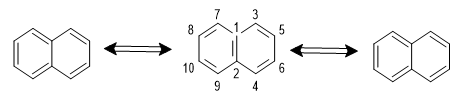
Canonical structures of Naphthalene
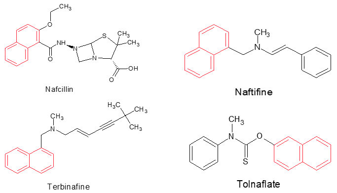
Naphthalene derivatives have shown a wide range of pharmacological activities, making them valuable in the development of therapeutic agents. These derivatives are used in the treatment of several diseases, including infections, inflammation, and cancer. Naphthalene-containing drugs often exhibit enhanced stability and bioavailability, contributing to their effectiveness as therapeutic agents.

Naphthalene Pharmacological Activities
Nafcillin is a beta-lactam antibiotic belonging to the penicillinase-resistant penicillin class. While it does not contain a classical naphthalene ring, its structure includes a naphthalene-related aromatic moiety that enhances resistance to beta-lactamase degradation. Nafcillin is primarily used to treat infections caused by Staphylococcus aureus (non-MRSA strains) by inhibiting bacterial cell wall synthesis through binding to penicillin-binding proteins (PBPs), leading to cell lysis and death.
Naftifine is a synthetic allylamine antifungal agent with a naphthalene ring system. It is applied topically to treat dermatophyte infections such as tinea corporis and tinea pedis. Naftifine acts by inhibiting squalene epoxidase, a critical enzyme in the ergosterol biosynthesis pathway, thereby leading to membrane dysfunction and fungal cell death.
Terbinafine, another allylamine antifungal, contains a substituted naphthalene nucleus. It is effective against dermatophytes, molds, and some yeasts. Its mechanism of action involves inhibition of squalene epoxidase, causing toxic accumulation of squalene and depletion of ergosterol, leading to fungal cell membrane disruption and death. Terbinafine is used both topically and systemically, especially in the treatment of onychomycosis.
Tolnaftate is a thiocarbamate antifungal drug used topically for superficial fungal infections such as athlete’s foot and ringworm. While not a pure naphthalene derivative, tolnaftate includes an aromatic system with similar physicochemical properties. It is believed to inhibit squalene epoxidase, like naftifine and terbinafine, interfering with ergosterol biosynthesis and compromising fungal membrane integrity.
Hydrazones are a class of organic compounds characterized by the functional group R1 C=NNHR. They are typically formed by the condensation reaction of hydrazines with aldehydes or ketones. Hydrazones possess a wide range of biological activities, including antimicrobial, antitumor, anti-inflammatory, and antitubercular properties, making them valuable scaffolds in medicinal chemistry.
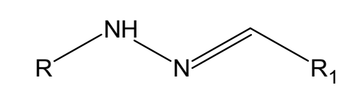
Hydrazone derivatives have been developed into various therapeutic agents due to their ability to interact with biological targets through hydrogen bonding, metal coordination, and other interactions. These compounds are versatile in their pharmacological profiles, leading to the development of drugs for different therapeutic applications.
Molecular hybridization is a concept in medicinal chemistry that involves the combination of two or more pharmacophores from different bioactive molecules into a single hybrid compound. This approach is used to develop new drugs with improved efficacy, reduced side effects, and enhanced pharmacokinetic properties. The resulting hybrid molecules are designed to interact with multiple biological targets, potentially offering synergistic therapeutic effects.

Molecular Hybridization
In Silico Studies
2.1. Calculation of Lipinski's Rule of Five, Veber’s rule.
2.1.1. Lipinski's Rule of Five: It was established by Christopher A. Lipinski in 1997, offers a framework for determining the drug-likeness and oral bioavailability of compounds. According to Lipinski's Rule of Five, a compound is more apt to be orally active if it meets these criteria.15
2.1.2. Veber's Rule: It is a guideline used in drug design and pharmacokinetics to predict the oral bioavailability of a drug. According to Veber's Rule:
Table 2. Lipinski rule and Veber’s rule of Compounds NH -1 – NH-2
|
Compound Codes |
Lipinski rule of five |
Veber’s rule |
|||||
|
M Log P |
Mol. Wt. (g/mol) |
HBA |
HBD |
Violations |
Total polar surface area (Ų) |
No. of rotatable bonds |
|
|
NH-1 |
3.97 |
246.12 |
1 |
1 |
0 |
24.39 |
3 |
|
NH-2 |
3.60 |
276.33 |
2 |
1 |
0 |
33.62 |
4 |
|
NH-3 |
4.48 |
280.05 |
1 |
1 |
0 |
24.39 |
3 |
|
NH-4 |
4.21 |
260.33 |
1 |
1 |
0 |
24.39 |
3 |
|
NH-5 |
4.59 |
325.20 |
1 |
1 |
0 |
24.39 |
3 |
|
NH-6 |
3.70 |
291.30 |
3 |
1 |
0 |
70.21 |
4 |
|
NH-7 |
4.21 |
260.33 |
1 |
1 |
0 |
24.39 |
3 |
2.2. In silico ADME (Absorption, Distribution, Metabolism and Excretion)
2.2.1. In silico ADME Evaluation: It play an indispensable role in determining the pharmacokinetic attributes and drug-like qualities of newly designed Naphthalene hydrazone derivatives. To facilitate this, the SwissADME tool (https://www.swissadme.ch/) was utilized to compute an array of physicochemical parameters, alongside assessing pharmacokinetic properties of these molecules.16 Ultimately, this strategic approach accelerates the drug development cycle, increasing the likelihood of identifying effective new Antimicrobial agents.
2.2.2. GI Absorption (Gastrointestinal Absorption)
If compounds show good predicted GI absorption, they could potentially be developed into oral antimicrobial drugs for treating systemic Staphylococcus infections. Poor absorption might indicate the need for alternative routes of administration, like topical or intravenous.
2.2.3. BBB Permeation (Blood-Brain Barrier Permeation)
If compounds are predicted to cross the BBB, they might be useful in treating CNS infections caused by Staphylococcus. If they don't cross the BBB, they would be less likely to cause CNS-related side effects.
2.2.4. P-Glycoprotein Substrate
The compounds are predicted to be P-gp substrates, they might be less effective at treating Staphylococcus infections because they could be expelled from microbial cells or human cells, leading to lower intracellular concentrations. Knowing this could guide you in modifying the chemical structure to avoid P-gp recognition or in considering co-administration with P-gp inhibitors.
Predictive models can help determine which CYP enzymes metabolize your compounds. This is crucial for understanding how quickly your compounds might be metabolized in the body and whether they could interact with other drugs.
CYP1A2: Involved in the metabolism of drugs like caffeine, theophylline, and certain antidepressants. It can be induced by smoking and certain dietary compounds.
CYP2C19: Metabolizes drugs such as proton pump inhibitors (e.g., omeprazole), antiepileptics (e.g., diazepam), and certain antiplatelet drugs (e.g., clopidogrel). Genetic polymorphisms in CYP2C19 can lead to variable drug responses.
CYP2C9: Metabolizes nonsteroidal anti-inflammatory drugs (NSAIDs) like ibuprofen, antidiabetic drugs (e.g., tolbutamide), and anticoagulants (e.g., warfarin). Genetic variations in CYP2C9 can affect drug metabolism, leading to altered drug efficacy or toxicity.
CYP2D6: Involved in the metabolism of a wide range of drugs, including beta-blockers (e.g., metoprolol), antidepressants (e.g., fluoxetine), and opioids (e.g., codeine). CYP2D6 is highly polymorphic, leading to significant interindividual variability in drug metabolism.
CYP3A4: The most abundant and important enzyme in drug metabolism, CYP3A4 metabolizes about 50% of all marketed drugs, including statins, certain antivirals, and calcium channel blockers. It is susceptible to induction or inhibition by various drugs, leading to potential drug-drug interactions. The compounds are predicted to be metabolized by these enzymes, particularly CYP3A4, they might have a shorter half-life and could interact with other drugs metabolized by the same enzymes. Understanding this can help in predicting potential drug-drug interactions and adjusting dosing regimens.
SwissADME tool predict the skin permeability of your compounds, important for topical or transdermal antimicrobial treatments. The compounds are predicted to have good skin permeability, they could be formulated into topical creams or ointments for treating Staphylococcus infections on the skin. If the permeability is low, systemic delivery might be more effective.
Table 3: In silico ADME properties of compounds NH -1 – NH-2
|
Compound codes |
Pharmacokinetic properties |
||||||||
|
GI Absorp-tion |
BBB Permea -tion |
P-glyco protein sub-strate |
CYP 1A2 |
CYP 2C19 |
CYP 2C9 |
CYP 2D6 |
CYP 3A4 |
Log Kp (skin perm-eation, cm/s) |
|
|
NH-1 |
High |
Yes |
No |
Yes |
Yes |
Yes |
Yes |
No |
-4.64 |
|
NH-2 |
High |
Yes |
No |
Yes |
Yes |
Yes |
Yes |
No |
-4.61 |
|
NH-3 |
High |
Yes |
No |
Yes |
Yes |
Yes |
No |
No |
-4.16 |
|
NH-4 |
High |
Yes |
No |
Yes |
Yes |
No |
Yes |
No |
-4.57 |
|
NH-5 |
High |
Yes |
No |
Yes |
Yes |
Yes |
No |
No |
-4.39 |
|
NH-6 |
High |
Yes |
No |
Yes |
Yes |
Yes |
No |
No |
-4.80 |
|
NH-7 |
High |
Yes |
No |
Yes |
Yes |
No |
Yes |
No |
-4.23 |
Experimental Work
3.1. General Remarks:
3.2. Experimental Procedure
Step 1: Synthesis Of Naphthalene Hydrazone
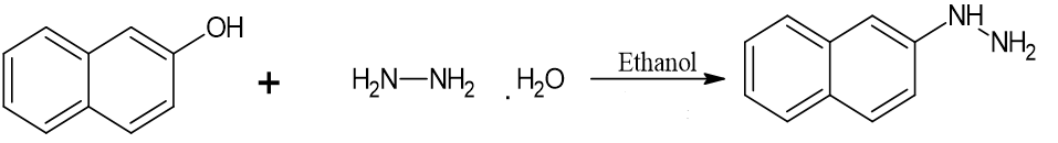
Beta – Naphthol Hydrazine hydrate Naphthalene Hydrazone
Procedure for step 1: Take (1.4g; 0.01mole) beta naphthol in a 250ml RBF. Add 10ml of hydrazine hydrate in it. Then reflux the mixture for 16-18 hrs. Pour the reaction mixture in crushed ice. Filter the precipitate and recrystallise with ethanol.
Results: Molecular formula: C10H10N2.
TLC: Solvent system (n-hexane-0.8 ml : ethyl acetate-0.2ml) (Rf = 0.66)
Melting point: 118-120oC
Percentage yield: 84.33%
STEP 2: Synthesis Of Naphthalene Hydrazone Derivatives (NH-1 – NH-2)
Procedure for step 2:
Take (1.76g; 0.01mole) Naphthalene hydrazine in a 250ml RBF. Add equimolar quantity of substituted benzaldehyde/acetophenone in it. Add few drops of glacial acetic acid. Add 10ml of ethanol as a solvent. Then reflux the mixture for 4 - 20hrs. Filter the precipitate and recrystallize with ethanol.
TLC: Solvent system (n-hexane-0.8 ml: ethyl acetate-0.2ml)
3.3. Structure Of Synthesized Derivatives
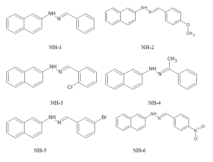
3.4. Melting Point, Chemical Formulae and RF Values of Synthesized Derivatives
Table 4. Melting Point, Chemical Formulae and RF Values of compounds NH-1 NH-2
|
Sample code |
Substituted R group |
Mol. formula |
Melting Point 0C |
Mol. Wt. g/mol |
% Yield |
Rf value |
||
|
NH-1 |
R=H |
R1=H |
R2=H |
C17H14N2 |
90-92 |
246.30 |
62.33 |
0.65 |
|
NH-2 |
R=H |
R1=H |
R2=OCH3 |
C18H16N2O |
113-117 |
276.33 |
77.02 |
0.76 |
|
NH-3 |
R=H |
R1=H |
R2=Cl |
C17H13N2Cl |
202-204 |
280.75 |
67.50 |
0.56 |
|
NH-4 |
R=CH3 |
R1=H |
R2=H |
C18H16N2 |
105-108 |
260.33 |
70.58 |
0.61 |
|
NH-5 |
R=H |
R1=H |
R2=Br |
C17H13N2Br |
197-200 |
325.20 |
74.55 |
0.70 |
|
NH-6 |
R=H |
R1=H |
R2=NO2 |
C17H13N3O2 |
118-121 |
291.30 |
65.23 |
0.75 |
|
NH-7 |
R=H |
R1=H |
R2=CH3 |
C18H16N2 |
167-169 |
260.33 |
70.60 |
0.63 |
Spectral Analysis
4.1. IR Spectrum
4.1.1. IR spectrum of NH-1
3275 cm?¹ (N-H stretch-secondary amine), 3057 cm?¹ (Aromatic C-H stretch-sp²), 1687 cm?¹ (C=N stretch - imine). IR confirms the presence of hydrazone (C=N) and aromatic system.
4.1.2. IR Spectrum of NH-2
3445 cm?¹ (N-H stretch - secondary amine), 3073 cm?¹ (Aromatic C-H stretch), 1691 cm?¹ (C=N stretch), 2949 cm?¹ (Aliphatic C-H stretch), 1152 cm?¹ (C-O stretch). IR confirms both aromatic and aliphatic features along with a C-O group.
4.1.3. IR Spectrum of NH-3
3407 cm?¹ (N-H stretch), 3034 cm?¹ (Aromatic C-H stretch), 1699 cm?¹ (C=N stretch), 733 cm?¹ (C-Cl stretch). IR confirms presence of hydrazone and chloro-substitution.
4.1.4. IR Spectrum of NH-4
3410 cm?¹ (N-H stretch), 3243 cm?¹ (Aromatic C-H stretch), 1649 cm?¹ (C=N stretch), 2903 cm?¹ (Aliphatic C-H stretch). IR confirms aromatic and aliphatic groups along with imine linkage.
4.1.5. IR Spectrum of NH-5
3243 cm?¹ (N–H stretching - secondary amine), 3050 cm?¹ (sp² C–H stretch -aromatic rings), 1662 cm?¹ (C=N stretching - imine group). The IR spectrum shows key functional groups: the N–H stretch confirms presence of –NH–, the aromatic C–H stretch supports the aromatic nature, and the C=N stretch indicates the formation of a hydrazone linkage.
4.1.6. IR Spectrum of NH-6
3259 cm?¹ (N–H stretch of secondary amine), 3087 cm?¹(sp² C–H stretch of aromatic group), 1692 cm?¹ (C=N stretch of imine). The IR data of NH-6 confirms the presence of a secondary amine (N–H), aromatic system (sp² C–H), and a C=N bond, indicating successful formation of the hydrazone structure.
4.1.7. IR Spectrum of NH-7
3277 cm?¹ (N–H stretch of secondary amine), 3056 cm?¹ (sp² C–H stretch of aromatic group), 1695 cm?¹ (C=N stretch of imine). NH-7 shows characteristic stretches for secondary amine, aromatic rings, and imine (C=N), confirming its expected hydrazone framework.
4.2. Mass Spectrum
4.2.1. Mass Spectrum of NH-1
The mass spectrum of NH-1 shows a molecular ion peak at m/z 247.12, which corresponds to the (M+1) peak of the compound. This confirms the presence of a molecular weight close to 246.11 g/mol, matching the calculated molecular mass of NH-1. This peak validates the molecular structure and confirms the compound's identity.
4.2.2. Mass Spectrum of NH-2
The observed molecular ion peak at m/z: 277.00 corresponds to the (M+1) peak, confirming the presence of the expected molecule. The shift is due to the natural abundance of heavier isotopes (like 13C). The spectrum validates the structure and confirms the presence of oxygen (likely from a carbonyl or ether group).
4.2.3. Mass Spectrum of NH-4
The mass spectrum of NH-4 shows a prominent molecular ion peak at m/z 261.33, which corresponds to the (M+1) peak. This confirms the molecular weight close to the calculated value of 260.11 g/mol, supporting the proposed structure of the compound and validating its molecular formula.
4.3. 1HNMR
4.3.1. 1HNMR Spectrum of NH-4
δ ppm 8.7 (s, 1H) (NH proton of –NH–NH– group - deshielded due to hydrogen bonding), δ ppm 7.6–7.8 (m, 3H) (Aromatic protons of naphthalene ring), δ ppm 7.1–7.5 (m, 4H) (Remaining aromatic protons of naphthalene), δ ppm 6.9–7.1 (m, 5H) (Aromatic protons of phenyl ring), δ ppm 3.8 (s, 3H) (Methyl group attached to aromatic ring).
Antimicrobial Activity
5.1. Kirby-Bauer Well Diffusion Method:
Kirby-Bauer method is a standardized disc diffusion test used to determine how effective antibiotics are against bacteria by measuring the zone of inhibition around antibiotic discs on an agar plate.
5.1.1. Principle of Kirby-Bauer Method:
Antibiotic-impregnated paper disks are placed on the surface of a Mueller-Hinton agar plate that has been uniformly inoculated with the test bacterium. The antibiotic diffuses radially outward through the agar. As it diffuses, it establishes a concentration gradient. If the bacterium is sensitive to the antibiotic, a zone of inhibition (area with no bacterial growth) forms around the disk. The diameter of this zone is measured and compared to standardized charts to determine if the bacterium is susceptible, intermediate, or resistant to the antibiotic.
5.1.2. Test Procedure:
The Kirby-Bauer method is based on the principle of antibiotic diffusion and bacterial growth inhibition.
1. Inoculation:
A pure culture of bacteria is uniformly spread across the surface of a Mueller-Hinton agar plate to create a lawn of bacterial growth.
2. Application of Antibiotic Discs:
Paper discs impregnated with known concentrations of different antibiotics are placed on the surface of the inoculated agar.
3. Diffusion of Antibiotic:
The antibiotic begins to diffuse radially from the disc into the surrounding agar. The concentration of the antibiotic is highest near the disc and decreases as it moves further away.
4. Interaction with Bacteria:
If the bacteria are sensitive to the antibiotic, their growth will be inhibited in the area where the antibiotic concentration is high enough. If the bacteria are resistant, they will grow right up to the edge of the disc.
5. Zone of Inhibition:
After incubation (usually 18–24 hours at 35–37°C), a clear circular area (zone of inhibition) appears around the disc if the antibiotic is effective. The diameter of this zone is measured in millimeters.
6. Interpretation:
The measured zone is compared to standard CLSI/EUCAST guidelines to categorize the bacteria as:Sensitive (S), Intermediate (I), Resistant (R)
5.2. Antimicrobial Activity against Staphylococcus aureus

5.3. Results and Interpretation: Measure the zone of inhibition.
Table 5. Antimicrobial Activity of Synthesized Derivatives (NH-1 – NH-7)
|
Antimicrobial activity |
|||
|
Zone Of Inhibition in mm |
|||
|
Derivatives |
25µl |
50µl |
100µl |
|
NH-1 (Benzaldehyde) |
NI |
NI |
NI |
|
NH-2 (Anisaldehyde) |
NI |
NI |
NI |
|
NH-3 (2-Chloro Benzaldehyde) |
NI |
NI |
16 |
|
NH-4 (Acetophenone) |
NI |
13 |
16 |
|
NH-5 (3-Bromo Benzaldehyde) |
NI |
NI |
NI |
|
NH-6 (4-Nitro Benzaldehyde) |
12 |
13 |
16 |
|
NH-7 (4-Methyl Benzaldehyde) |
NI |
NI |
17 |
|
Note: NI-No Inhibition, Strain no. - SA04 Staphylococcus aureus |
|||
Table 6. Antimicrobial Activity of Standard Antibiotic – Gentamicin
|
Control |
AB (Gentamicin 10mcg) |
DMSO |
|
Staphylococcus aureus |
23 |
NI |
RESULTS AND DISCUSSION
6.1.1. Lipinski’s Rule of 5 And Veber’s Rule
6.1.2. ADME Analysis
All compounds were predicted to inhibit CYP1A2, CYP2C19, CYP2C9 and CYP2D6. None of the compounds inhibited CYP3A4.
6.1.3. Synthesis And Characterization of Substituted Naphthalene Hydrazone Derivatives
6.1.4. Antimicrobial Activity Against Staphylococcus aureus
CONCLUSION
Additionally, exploring how these compounds work could help make them more effective and reduce any side effects.
REFERENCES

Alnaj Thange*, Akshata Wasnik, Shivam Yadav, Sonal Yadav, Chetana Mayekar, Sunayana Ghodgaonkar, Bhagyashree Chaudhari, Synthesis, Characterization and Antimicrobial Evaluation of Some Novel Naphthalene Hydrazone Derivatives, Int. J. of Pharm. Sci., 2025, Vol 3, Issue 7, 2927-2943. https://doi.org/10.5281/zenodo.16278633
 10.5281/zenodo.16278633