View Article

Abstract

The human skin microbiome plays a pivotal role in sustaining cutaneous health through barrier fortification, immunological modulation, and microbial equilibrium. This review consolidates existing insights into the diversity, spatial heterogeneity, and functional dynamics of skin-associated microbes, with particular emphasis on the manner in which intrinsic and extrinsic factors—including age, hygiene practices, ultraviolet radiation exposure, and dietary habits—modulate the microbial landscape. Illustrative case studies from Indian populations and site-specific microbial assessments further elucidate the impact of genetic background, lifestyle variables, and environmental context. Microbial dysbiosis has been implicated in a spectrum of dermatological disorders, including atopic dermatitis, acne vulgaris, psoriasis, and chronic wounds, wherein disturbances in microbial communities contribute to heightened inflammation and compromised epidermal integrity. Emerging therapeutic modalities that incorporate prebiotics, probiotics, postbiotics, and engineered bacterial strains offer compelling alternatives to conventional treatments. Notably, “triple biotic” formulations—which integrate all three biotic components—are garnering increased attention for their synergistic efficacy in restoring cutaneous microbial homeostasis. Despite these advancements, several critical limitations persist. The field currently lacks a sufficient volume of clinical trials to substantiate the long-term safety, therapeutic efficacy, and regulatory viability of engineered or synbiotic skin-based therapies. Furthermore, the absence of standardized methodologies, harmonized clinical endpoints, and comprehensive data-sharing protocols continues to obstruct the successful clinical translation of microbiome-based innovations into standard dermatological practice. By synthesizing foundational microbiome science with emerging clinical and bioengineering developments—including triple biotic strategies and microbial engineering—this review delineates both the therapeutic potential and current constraints of microbiome-directed interventions, and provides a prospective framework for their future application in dermatology.

Keywords

Microbial Dysbiosis, Case Studies, Triple Biotic Formulations, Symbiotic Skin Therapies, Engineered Bacteria

Introduction

Recent progress in microbiome research has underscored the significant influence of skin-resident microbial communities on cutaneous health, immune function, and barrier physiology. As understanding of these complex microbial ecosystems has expanded, there has been a growing interest in therapeutic interventions aimed at supporting or reinstating microbial equilibrium—particularly through the application of prebiotics, probiotics, and postbiotics. These approaches, whether employed independently or in combination, present a more targeted and sustainable alternative to conventional skincare strategies, which often rely on broad-spectrum antimicrobials or symptomatic relief. Among these emerging innovations, the integrative concept of combining prebiotics, probiotics, and postbiotics into a cohesive “triple biotic” formulation has gained notable momentum. This multifactorial strategy is designed not only to replenish beneficial microbial taxa and their bioactive metabolites but also to foster a conducive microenvironment that promotes their persistence and functional activity on the skin. Although this field remains in an early developmental phase, accumulating clinical data has begun to substantiate the dermatological benefits of such combinatory interventions. For example, recent clinical trials involving triple biotic products have reported measurable improvements in parameters such as skin hydration, barrier integrity, and microbial diversity. Nonetheless, these outcomes are distributed across varying formulations, microbial strains, and evaluation endpoints, highlighting the necessity for broader validation and mechanistic elucidation. Concurrently, advances in synthetic biology and microbial engineering have facilitated the creation of precision-targeted live biotherapeutics and bioactive microbial systems, capable of reshaping the skin microbiota with unprecedented specificity. These engineered bacterial platforms introduce novel therapeutic avenues for managing chronic inflammatory dermatoses, augmenting skin immune defenses, and delivering therapeutic agents directly at the epidermal surface.  This review compiles and examines the current body of evidence regarding triple biotic approaches for enhancing skin health, incorporating findings from in vitro experiments, in vivo animal models, and human clinical studies. Particular focus is placed on the impact of specific microbial combinations on community composition, host immune responses, and dermatological outcomes such as inflammation, hydration, and barrier functionality. By critically analyzing and organizing this multidisciplinary literature, the review seeks to clarify the present state of microbiome-focused dermatological interventions, while identifying key areas that warrant further exploration to unlock their full therapeutic potential.

1.1Definition and composition of the skin microbiome

The skin microbiome denotes the intricate and dynamic consortium of microorganisms that inhabit the human skin, functioning as a crucial interface between the host and the external environment. This microbial community comprises a diverse array of taxa—including bacteria, fungi, viruses, archaea, and mites—that coexist with host epidermal cells. Most of these organisms exhibit commensal (nonharmful) or mutualistic (beneficial) relationships with the host, although certain taxa may become pathogenic under specific environmental or physiological conditions.

Major Microbial Groups

1. Bacteria

Bacteria constitute the most abundant members of the skin microbiome and are predominantly represented by four phyla: Actinobacteria (36–51%), Firmicutes (24–34%), Proteobacteria (11–16%), and Bacteroidetes (6–9%).

  • Moist anatomical regions (e.g., axillae, groin) are primarily colonized by Staphylococcus (Firmicutes) and Corynebacterium (Actinobacteria).
  • Sebaceous (lipid-rich) zones, such as the face and back, favor lipophilic taxa like Cutibacterium (formerly Propionibacterium), which metabolize sebum.
  • Dry regions (e.g., forearms, legs) typically demonstrate higher microbial diversity but reduced overall biomass relative to moist or sebaceous sites.

These bacterial communities contribute to immune modulation, pathogen competition, and maintenance of the skin barrier.

2. Fungi

Fungal populations are predominantly composed of lipophilic Malassezia species, which preferentially colonize sebaceous zones such as the scalp and facial skin. While these yeasts form part of the normal flora, dysregulated overgrowth is associated with conditions such as seborrheic dermatitis and dandruff.

3. Viruses

The skin virome primarily comprises bacteriophages, which infect bacterial hosts, and eukaryotic viruses such as papillomaviruses and polyomaviruses. These viral elements can influence bacterial community structure and functionality.

4. Mites

Demodex species—most notably D. folliculorum and D. brevis—reside within hair follicles and sebaceous glands. Generally benign, they have been implicated in certain inflammatory dermatoses, such as rosacea.

5. Archaea

Although present in relatively low abundance, archaea have been identified predominantly in sebaceous skin regions. Their precise functional roles remain under investigation.

Spatial and Temporal Variation 

The skin microbiome exhibits pronounced site specificity, as each anatomical region presents a unique combination of sebum, moisture, and pH, resulting in distinct microbial assemblages. For instance:

The forehead and upper back are dominated by lipid-metabolizing species.

The antecubital fossa (inner elbow) supports organisms adapted to moist environments.

  • It also varies over time, influenced by age, puberty, and environmental exposures.
  • Newborns acquire their first microbiome from maternal skin, vaginal, or cesarean sources, which evolves during early life.

1.2 Functions and Importance in Skin Health

The skin microbiome is not simply a passive colonizer; it performs active functions essential for homeostasis, defense, and repair.

A. Barrier Protection and Competitive Exclusion

Commensal microbes protect the host by occupying ecological niches, thereby preventing pathogen overgrowth through competitive exclusion. Many skin bacteria produce antimicrobial peptides (AMPs)—for example, Staphylococcus epidermidis secretes phenol?soluble modulins that inhibit Staphylococcus aureus colonization . These AMPs complement host?derived peptides, bolstering the skin’s chemical barrier against invading microbes .

B. Immune Education and Modulation

 The microbiome plays a critical role in educating the immune system, particularly innate immunity. Skin commensals continually interact with keratinocytes, Langerhans cells, and other immune cells to calibrate immune responses . Through pattern recognition receptors (e.g., Toll?like receptors), microbes stimulate low?level cytokine production that maintains immune tolerance, preventing unnecessary inflammation while priming defenses against real threats . Stimulate host defenses only when needed.

C. pH Regulation and Acid Mantle Formation

Skin commensals contribute to the formation and maintenance of the acid mantle, a thin, slightly acidic film (pH?4.5–5.5) on the stratum corneum that inhibits pathogen colonization . They generate free fatty acids and other acidic metabolites (e.g., lactic acid) via sebum and sweat metabolism, sustaining an environment hostile to many opportunistic microbes . This acid mantle also optimizes the activity of pH?dependent enzymes crucial for lipid synthesis and barrier repair .

D. Wound Healing and Tissue Repair

Certain commensals enhance wound healing by promoting keratinocyte proliferation and migration. Cutibacterium acnes, for instance, produces short?chain fatty acids that drive epidermal regeneration and modulate local inflammation . Staphylococcus epidermidis can upregulate host AMPs and growth factors that accelerate re?epithelialization, reducing scar formation.

E. Systemic and Neuro?Immune Interactions (Emerging)

Emerging research suggests a skin–brain–microbiome axis, whereby skin microbiota influence, and are influenced by, psychological stress and systemic immune responses . Stress?induced hormonal changes can shift microbial composition, which in turn may affect cutaneous barrier integrity and inflammation, linking psychological states to skin disorders

1.3 Skin Microbiome variation in Indian Families.

Family Structure and Sampling

In India, multi-generational “joint families” coexist under one roof, sharing genetics, diet, lifestyle, and environment, providing a unique natural experiment for skin microbiome studies .A recent investigation sampled axillary sweat from 72 individuals across 15 families (three generations, G1: 65–91 yrs; G2: 41–63 yrs; G3: 13–30 yrs) in two Maharashtrian cities (Pune and Ahmednagar). Rigorous controls (no deodorant, standardized collection) minimized transient contamination.

Taxonomic Composition and Diversity

Across families, the most abundant phyla were Firmicutes (mean ~ 73 %), Proteobacteria (~ 24 %), and Actinobacteria (~ 3 %); dominant genera included Staphylococcus (~ 51 %), Bacillus (~ 16 %), and Pseudomonas (~ 9 %). Overall Shannon α-diversity did not differ significantly by age or sex but showed moderate associations with diet (vegetarian vs. mixed; FDR < 0.1) and location (Pune vs. Ahmednagar; FDR < 0.1).

Influence of Geography, Diet, and Cohabitation

β-Diversity (Bray–Curtis) revealed a highly significant familial signature (PERMANOVA p = 0.001) and a borderline effect of geography (p = 0.07), whereas diet, age, and sex were non-significant. Within-family microbiomes were, on average, more similar than those between families—suggesting that cohabitation (shared environment and behaviors) and/or genetics impart a unique “family microbiome”. A parallel study of Indian patrilineal families also found no clear clustering by age or gender but echoed familial patterns in skin profiles Nature.

1.4 Factor affecting Intrinsic and extrinsic factor.

The skin microbiome is shaped by a complex interplay of intrinsic (host?related) and extrinsic (environmental or behavioral) factors. These determinants modulate microbial diversity, community structure, and functional interactions, thereby influencing skin health and disease propensity.

1.4.1 Intrinsic Factors

  1. Age and Developmental Stage
    • Neonatal colonization begins in utero and at birth, influenced by delivery mode; vaginally delivered infants acquire Lactobacillus and Prevotella, whereas Cesarean?delivered infants are colonized by skin?associated genera such as Staphylococcus and Cutibacterium .
    • Childhood to adulthood: Pre?pubertal skin harbors higher proportions of Firmicutes and Proteobacteria, while sebum?rich adolescent skin favors lipophilic Cutibacterium acnes .
    • Elderly skin shows reduced sebum production and lower microbial diversity, with declines in Cutibacterium and increases in Corynebacterium and Proteobacteria .
  2. Anatomic Site and Skin Physiology
    • Sebaceous (“oily”) sites (face, chest, back) are dominated by Cutibacterium (up to 46%) and certain fungi (Malassezia) due to abundant lipids .
    • Moist sites (axilla, groin) favor Staphylococcus and Corynebacterium (each ~20–28%) given higher humidity and pH neutrality .
    • Dry sites (forearm, leg) are more diverse, with Proteobacteria, Bacteroidetes, and Actinobacteria in roughly equal measures .
  3. Skin Barrier and Immune Factors
    • pH: Slightly acidic (~5.5) surface pH favors commensals like S. epidermidis and inhibits pathogens such as Staphylococcus aureus .
    • Sebum composition: Free fatty acids (e.g., lauric, sapienic) generated by C. acnes acidify the skin and possess antimicrobial activity against Gram?positive pathogens .
    • Antimicrobial peptides: Host?derived AMPs (hBD?1–3, LL?37, RNase?7, S100A7, dermcidin) are modulated by commensals, bolstering the innate barrier .
  4. Genetics and Hormones
    • Sex differences: Female skin—thinner, more acidic, lower sebum—exhibits higher microbial richness. Generally in mens Androgen?driven sebum increases during puberty, favoring lipophilic bacteria.
    • Immune genotype: Variants in genes regulating skin barrier proteins and AMP production affect individual microbial patterns and disease susceptibility . For example, mutations in the filaggrin gene can predispose individuals to atopic dermatitis and alter microbial colonization.

5. Ethnicity

  • Skin Properties: Differences in skin structure, pigmentation, and sweat composition among ethnic groups can affect microbiome composition.

1.4.2 Extrinsic Factors

  1. Hygiene and Personal Care
    • Washing and soaps: Routine cleansing reduces total CFU but can deplete commensals if overused; soap enriched with triclocarban or benzalkonium targets pathogens like group A streptococci .
    • Cosmetics: Moisturizers (lipid?rich) promote lipophilic commensals (Staphylococcus, Cutibacterium); overuse or certain preservatives may decrease overall diversity and trigger irritation or dermatitis .
  2. Antibiotics and Disinfectants
    • Systemic/topical antibiotics (e.g., tetracyclines, macrolides) drastically reduce C. acnes and can cause overgrowth of resistant strains and non?target commensals .
    • Alcohol and chlorhexidine: Alcohol?based hand rubs and chlorhexidine gluconate rapidly kill pathogens without significant long?term diversity loss on healthcare workers’ skin .
    • Occlusive Products: Can create moist environments that favor growth of specific bacteria or fungi.

3.Occupation and Environmental Contact

    • Rural vs. urban: Rural residents exhibit greater skin microbial diversity due to soil and animal contact; urbanization correlates with loss of environmental taxa and gains in indoor?associated microbes .
    • Animal exposure: Pet owners and agricultural workers share microbes (e.g., Roseomonas, Acinetobacter) with their animals, altering nasal and skin communities .

4.Textiles and Occlusion

    • Clothing fabrics serve as reservoirs; occlusive, synthetic garments foster Malassezia overgrowth in moist areas. Washing frequency and detergent choice modulate textile?associated microbiota .

5. Diet

  • Nutritional Status: Diet can influence skin secretions (such as sebum composition) and indirectly affect the skin microbiome.
  • Probiotics/Prebiotics: Consumption of certain foods may influence the skin microbiome through the gut–skin axis.

6.Climate, UV Exposure, and Geography

    • Humidity and temperature: Tropical, humid regions enhance fungal richness (Malassezia, Aspergillus), whereas arid climates reduce diversity .
    • Sunlight: UVB/UVA exposure reduces C. acnes porphyrin production and shifts community composition; some commensals produce UV?protective compounds (e.g., pityriacitrin) .
    • Air Pollution: Pollutants can alter skin pH and barrier function, indirectly affecting microbial communities.
  • Skin microbiome grow in response to long sun exposure and reduce ROS.

MATERIAL AND METHOD

Study Design and Sampling

  • The study focused on assessing the impact of prolonged sun exposure on the skin microbiome of lifeguards, a group with high daily sunlight exposure.
  • Skin swabs were collected from the outer arms of ten lifeguards at two time points: before the summer (May) and after the summer (October).
  • The skin microbiome samples were analyzed to determine changes in microbial composition due to cumulative sun exposure and sea water spray.

Microbiome Analysis

  • Microbial DNA was extracted from the skin swabs and sequenced to identify the bacterial taxa present.
  • Beta diversity (differences in microbial composition) was assessed using Principal Coordinates Analysis (PCOA) of the Jaccard distance matrix and Bray-Curtis similarity, with statistical significance tested via ANOSIM.
  • Alpha diversity (species richness and evenness) was measured using the Shannon diversity index.
  • The Linear Discriminant Analysis Effect Size (LEfSe) method was used to identify taxa that changed significantly in abundance after the summer.

In Vitro Experiments

  • To test UV resistance, Sphingomonas mucosissima (as a model strain) and Escherichia coli K-12 (control) were inoculated onto LB agar plates and exposed to UV lamps at 254 nm and 312 nm, with varying doses.
  • Plates were incubated at 30°C to observe bacterial growth post-irradiation.
  • For antioxidant activity, HaCaT human keratinocyte cells were exposed to UVB radiation to induce reactive oxygen species (ROS). The effect of Sphingomonas culture supernatant on ROS levels was measured and compared to controls.

Results

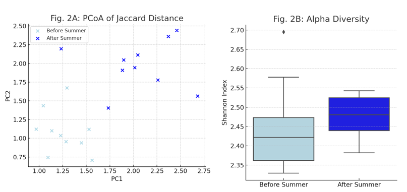
Changes in Skin Microbiome Composition (Fig. 2)

  • Fig. 2A: PCOA analysis of Jaccard distances showed a significant difference in the composition of low-abundance microbial species before and after the summer (ANOSIM, P = 0.004, R = 0.015).
  • Fig. 2B: Shannon diversity index revealed no significant change in overall diversity or evenness (p = 0.65), indicating that the dominant microbiome remained stable, but rare taxa shifted.

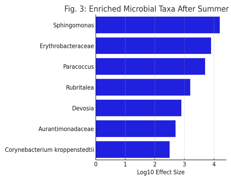
Enrichment of Specific Bacterial Taxa (Fig. 3)

Fig. 3: LEfSe analysis identified specific bacterial taxa that increased in relative abundance after the summer. The figure displays taxa (at family or genus level) present in at least 50% of volunteers, with notable increases in Sphingomonas and Erythrobacteraceae.

Individual Responses in Key Bacterial Families (Fig. 4)

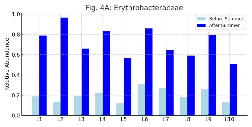
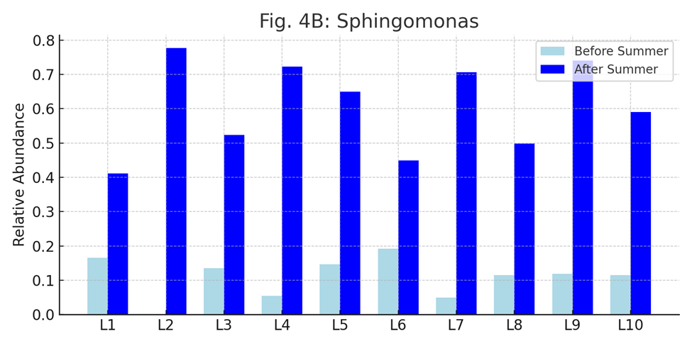
Fig. 4: This figure presents the relative abundances of Erythrobacteraceae (A) and Sphingomonas (B) in each lifeguard before and after the summer. Most participants showed increased levels of these bacteria after prolonged sun exposure.

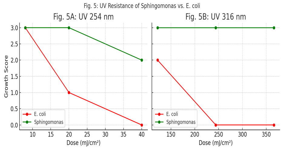
UV Resistance of Sphingomonas (Fig. 5)

  • Fig. 5: Sphingomonas mucosissima demonstrated high resistance to UV irradiation at both 254 nm and 312 nm wavelengths. In contrast, E. coli K-12 could not survive beyond minimal radiation exposure.
  • Sphingomonas tolerated all tested doses, confirming its UV resilience.

DISCUSSION

  • The study demonstrates that while the overall skin microbiome remains stable in terms of dominant species, prolonged sun exposure leads to significant shifts in the composition of less abundant bacteria1.
  • Notably, Sphingomonas and Erythrobacteraceae were enriched after the summer. Both genera are known to produce bioactive compounds (e.g., carotenoids, glycosphingolipids, ubiquinone 10) with antioxidant and photoprotective properties.
  • Sphingomonas, in particular, showed remarkable resistance to UV radiation and its culture supernatant could reduce UVB-induced ROS in human keratinocytes by 25%, suggesting a direct protective effect against oxidative stress.
  • These findings support the hypothesis that the skin microbiome adapts to environmental stressors like UV radiation by enriching for bacteria capable of mitigating damage, potentially providing an additional layer of photoprotection alongside host mechanisms.
  • The results align with previous research indicating that low-abundance species in the skin microbiome are more dynamic and responsive to environmental changes, while the core microbiome remains stable.
  • The study suggests potential applications for harnessing UV-resistant skin bacteria or their metabolites in dermatological and cosmetic products aimed at enhancing skin protection against solar damage.

1.5 Methods of Skin microbiome exploration.

  • All methodologies listed—such as:
  • DNA/RNA extraction from skin swabs or tapes,
  • 16S rRNA/ITS amplicon sequencing,
  • Whole genome (shotgun) metagenomics,
  • Metatranscriptomics,
  • Long-read sequencing (e.g., Nanopore, PacBio),
  • Bioinformatic pipelines for diversity and function—

are culture-independent, as they analyze microbial communities directly using molecular and computational tools.

1.5.1 Ongoing and Proposed Approaches (Table?1)

Step

Current Methods

Ongoing/Proposed Improvements

1.Sampling


Swabs (dry/moistened),

tape-stripping,punch biopsies

Engineered stabilization kits(eg.,

OMNIgene*SKIN),3D-printed

microprojection arrays for spatially resolved sampling

2.Nucleic Acid Extraction


Commercial low-input kits

(Qiagen,MoBio), bead-beating

Or enzymatic lysis

Protocols optimized for ultra-low biomass and resilient cutaneous microbes; enhanced host-DNA depletion strategies

3.Sequencing


Short-read amplicon( 16S rRNA, ITS) on Illumina;  targeted qPCR/RT-qPCR

Long-read/hybrid platform (Nanopore, PacBio) for full-length marker gene; shotgun metagenomics; metatranscriptomics

4.Bioinformatics

Analysis


Alpha/beta diversity; taxonomic classification

Against SILVA, Greengenes.

ASV-based denoising (e.g., DADA2) for single-nucleotide

Resolution; skin specific reference database; multi-omics integration

Note: Table1 highlights how each workflow step is being refined to improve sensitivity, resolution, and functional insight in skin microbiome research.

1.5.2 Assessing Reagents and Cross?Contamination (Table?2)

Control

Applied at

Purpose

Expected Outcome

Extraction    Blank


DNA/RNA extraction and library prep

Reveal DNA from reagents (“kitom”) or plasticware; detect cross-contamination during extraction

Negligible nucleic acid; minimal amplification; low read counts distinct from true samples

Sampling Negative


Collection

Identify environmental or handling contamination at the point of sampling

No or minimal DNA; profile matches ambient background rather than skin

PCR No-Template


Library preparation

Detect contaminants introduced during amplification (reagent, aerosols)

No or minimal amplification product

PCR Positive

Library preparation

Verify amplification reagents and thermocycle performance

Amplicon/library of expected size and yield

Mock Community


Extraction Analysis

Benchmark   pipeline sensitivity,specificity, and quantitative accuracy using defined mixtures of cells, DNA, or in silico genomes

Recovery of expected taxa at correct proportions; metrics of  false positive/negative

Technical Replicates


Extraction, library, sequencing

Quantify variability from low_biomass stochastic effects; improve reproducibility

Consistent profiles across replies; outlier detection

Host Depletion controls

Extraction/library prep

Validate efficacy of host- DNA/RNA removal methods

Enrichment of microbial reads over host reads without introducing bias

Prevention is the best cure and several best practices can be applied to minimize the impact of reagents combinations and cross – contamination on skin microbiome results when adding one or several of the above – mentioned control.

Best Practice: Sequence all controls alongside samples and apply bioinformatic tools e.g., decontam ( amplicon and shotgun metagenomics),SourceTracker, microDecon ( amplicon data), Recentrifuge ( shotgun metagenomics data),Squeegee ( shotgun metagenomics data with the possibility of application to amplicon data )to identify and remove contaminant signatures.

  • Comparative Methods of Skin vs. Gut Microbiome Exploration

The comparative overview of the methodological approaches employed in exploring the skin and gut microbiome. Although many techniques overlap due to shared microbiome principles, there are important distinctions due to the anatomical and biological differences between skin and gut environments. The content is synthesized from quality-controlled pipelines used in large-scale studies and adapted to suit both high- and low-biomass microbial communities.

Table 3: Comparative Methodologies for Skin vs. Gut Microbiome Analysis

Step

Skin Microbiome

Gut Microbiome

Sample Collection

Sterile swabs or tapes from defined areas (e.g., forehead, forearm).

Stool collected in sterile containers.

Post-Collection Handling

Swabs into stabilization buffer or on dry ice.

Stool mailed ambient or cold, then frozen.

Storage Conditions

Stored at –80°C.

Initial –20°C, then –80°C upon lab receipt.

DNA Extraction

Bead beating + low biomass kits (e.g., QIAamp BiOstic).

Bead beating + Arrow Stool DNA or PowerSoil kits.

DNA Quantification

PicoGreen adapted for low DNA yield.

PicoGreen quantification in 96-well format.

16S rRNA Region

V3–V4 or V4 hypervariable regions.

V3–V4 region commonly used.

Library Preparation

PCR amplification, SequalPrep normalization, AMPure XP, LabChip.

Similar process with higher yield.

Sequencing Platform

Illumina MiSeq (2×250 bp).

MiSeq or NovaSeq for deeper coverage.

Reference Databases

SILVA v138.1, Greengenes2; ASV via DADA2.

Same databases and tools (QIIME, Deblur, UPARSE).

Quality Filtering

DADA2 filtering (error ≤2), chimera removal, low abundance filtering.

Same parameters with ≥4K reads/sample threshold.

Normalization

Rarefaction to even depth or VST (CLR, DESeq2).

Similar, often rarefied to 4K reads.

Batch Effect Mitigation

Track metadata (e.g., season, storage time); use ComBat, removeBatchEffect.

Include batch variables in PERMANOVA, multivariate models.

Contamination Controls

Negative/positive controls; beta diversity & host DNA for swaps.

Sex typing qPCR; Aitchison distance for same-sex swaps.

Diversity Analysis

Shannon index (alpha), CLR + Aitchison (beta).

Same methods applied.

Differential Abundance

Compositional models (ANCOM, ALDEx2).

Same statistical tools used.

Multivariate Modeling

PERMANOVA, MaAsLin2.

Identical approaches.

Visualization

Donut plots, heatmaps, PCoA/NMDS.

Same visualization methods.

Table 4: Key Differences at a Glance

Step

Skin Microbiome

Gut Microbiome

Sample Type

Swabs or adhesive tapes from skin sites

Stool samples

Storage

-80°C after collection

Mailed at -20°C, then -80°C in lab

DNA Extraction

Low biomass kits (e.g., QIAamp BiOstic)

High biomass kits (e.g., Arrow Stool DNA kit)

Sequencing Platform

Illumina MiSeq (2×250?bp)

NovaSeq or similar high-throughput platforms

Read Filtering

Higher stringency due to low biomass

Standard filtering parameters

Contamination Checks

Negative/positive controls, swaps

Sex typing, host DNA contamination checks

Taxonomic Resolution

Genus level common

Strain-level possible

Applications

Dermatological studies

GI health, systemic disease

2.The Skin Microbiome in Health and Disease

2.1. Role in maintaining skin homeostasis

The skin functions first and foremost as a physical barrier, preventing pathogen ingress and desiccation while simultaneously providing a habitat for commensal microbes that contribute to cutaneous health . Embedded within this barrier are multiple innate immune defenses—notably antimicrobial peptides such as β-defensins, substance P, and cathelicidins—that exhibit broad-spectrum activity against bacteria, viruses, and fungi by disrupting microbial membranes without harming host cells . These peptides can be constitutively expressed or upregulated in response to injury or microbial challenge; for example, cathelicidin levels are abnormally elevated and processed in rosacea, whereas reduced antimicrobial peptide expression in atopic dermatitis predisposes to infection . Beyond peptides, the skin’s cellular innate defenses—including Langerhans cells, natural killer cells, neutrophils, and resident lymphoid populations—continuously survey for and eliminate invading organisms, secrete cytokines that shape adaptive immunity, and help maintain equilibrium between tolerance of commensals and clearance of pathogens . Together, these structural and immunological mechanisms underpin skin homeostasis, orchestrating a balance between barrier integrity, microbial colonization, and immune responsiveness.

2.2. Dysbiosis and its impact on dermatological conditions

Dysbiosis denotes a state in which the normal balance of the skin microbiome—both in species composition and functional capacity—is disrupted, leading to pathological outcomes .
Such microbial imbalance can impair the epidermal barrier, enhancing transepidermal water loss and permitting allergen or pathogen invasion, which fuels cutaneous inflammation.
Furthermore, dysbiosis skews immune education: commensals that ordinarily induce regulatory T?cells diminish, while pro?inflammatory taxa expand, exacerbating disease susceptibility.
Clinical evidence links dysbiosis to worsened disease severity and chronicity across multiple skin disorders, underscoring its central role in dermatological pathophysiology.

"Dysbiosis is an imbalance in microbial composition, often leading to reduced diversity or pathogenic overgrowth. It has been implicated in various skin disorders, including acne, atopic dermatitis, and psoriasis."

One compelling illustration of microbiome-associated dysbiosis is observed in chronic wounds, particularly diabetic foot ulcers, which serve as a model for studying the disruption and restoration of microbial balance during the healing process. Recent research has shown that:

"The microbial landscape of diabetic ulcers evolves during healing. In a study involving 10 diabetic patients treated with a fluorescein-based galenic therapy (Fluorexin), 16S rRNA sequencing revealed significant shifts in bacterial communities within the ulcer bed over 20 days."

Initially, these chronic wounds are dominated by pathogenic species, such as Staphylococcus aureus, which is often associated with inflammation and delayed wound healing. However, as healing progresses:

"There was a reduction in S. aureus and an increase in beneficial genera such as Corynebacterium, Propionibacterium, Streptococcus, and Pseudomonas."

These microbial shifts occurred without the use of topical or systemic antibiotics, highlighting the potential of non-antibiotic antimicrobial and anti-inflammatory approaches to support the healing process. The therapy also led to:

"A reduced prevalence of opportunistic pathogens like Enterococcus, Finegoldia, and Peptoniphilus."

Furthermore, comparison between microbiota at the ulcer bed (intralesional) and the surrounding skin (perilesional) revealed insights into microbial origins and dynamics:

"Comparison between intralesional (ulcer bed) and perilesional (surrounding skin) microbiota showed no major compositional differences at baseline (t0), supporting the hypothesis that ulcer microbiota may arise from the adjacent skin."

However, notable changes in diversity and genus prevalence occurred post-treatment, with significant variation depending on ulcer location:

"Diversity and genus prevalence did change significantly post-treatment, especially depending on ulcer location (e.g., plantar vs. lower leg), with plantar ulcers showing greater homogeneity."

These findings underscore several critical points:

  • Dysbiosis in chronic wounds involves an enrichment of pathogens and depletion of commensals.
  • Microbiome composition can be modulated without antibiotics to support healing.
  • Localized interventions and wound location influence microbiota dynamics.

As such, the study provides a compelling case for the therapeutic potential of skin microbiome modulation in wound care, and it reinforces the broader relevance of dysbiosis in the development and resolution of dermatological conditions.

3.Skin Microbiome in Dermatological Conditions

3.1. Eczema and atopic dermatitis

Atopic dermatitis (AD) is characterized by marked dysbiosis, notably a sharp decline in overall bacterial diversity and the overrepresentation of Staphylococcus aureus in lesional skin.
Up to 90?% of AD patients are colonized by S. aureus, compared to ~5?% of healthy individuals, and disease flares correlate with increased S. aureus density.
Strain?level profiling reveals that specific S. aureus lineages carry toxins and proteases (e.g., phenol?soluble modulins, bacteriocins) that directly damage the epidermal barrier and trigger inflammation
Conversely, commensals such as coagulase?negative staphylococci and Cutibacterium spp. normally secrete antimicrobial peptides that limit pathogen overgrowth; their depletion in AD permits pathogenic blooms. Emerging metagenomic studies also implicate shifts in the fungal mycobiome—such as increased Malassezia osloensis—and elevated bacteriophage loads during AD flares, underscoring the multi?kingdom nature of dysbiosis in eczema.

  • Therapeutic interventions including topical corticosteroids, antiseptics, bleach baths, and emerging microbiome?modulating agents restore microbial diversity and correlate with clinical improvement.

3.2. Acne vulgaris   

"Acne is a long-lasting inflammatory skin condition that affects sebaceous glands and hair follicles. Skin rashes, including whiteheads, blackheads, pustules, papules, and cysts on the face, chest, back, and shoulders are the primary clinical symptoms.

The main contributory factors to acne vulgaris are:

  • Androgen increase
  • Excessive keratosis at the opening of hair follicles and sebaceous glands
  • Bacterial colonization (notably Cutibacterium acnes)
  • Increased sebum production
  • Possibly endocrine and genetic disorders

Acne is tightly linked to both the abundance and strain diversity of Cutibacterium acnes within the pilosebaceous unit. Acne patients often harbor a higher ratio of acne?associated C. acnes lineages and a lower relative abundance of protective species like C. granulosum. Metagenomic analyses reveal that acneic skin shows enrichment of genes involved in pro?inflammatory metabolite production and biofilm formation, while healthy skin exhibits greater diversity of metabolic pathways that may antagonize C. acnes overgrowth. Cross?talk with Staphylococcus epidermidis is also evident: certain S. epidermidis strains produce antimicrobials that inhibit C. acnes, but in acne, strain?level shifts can turn S. epidermidis into a secondary driver of inflammation via protease secretion.

  • Acne treatments (benzoyl peroxide, retinoids, antibiotics) exert both antimicrobial and microbiome?shaping effects, though concerns about resistance and collateral damage to beneficial taxa have spurred interest in probiotics and phage therapy.

Probiotic Acne Treatment:

  • Lactobacillus plantarum produces antimicrobial peptides that:
    • Reduce inflammation
    • Strengthen the skin's natural defense
    • Inhibit Cutibacterium acnes
  • Improvements in acne lesions have been seen, along with decreases in transepidermal water loss and sebum production, when using  a cream containing Lactobacillus plantarum ferment lysate can effectively and safely cure mild-to-moderate acne vulgaris after four weeks of topical treatment
  • Lactobacillus extract improves:
    • Skin barrier integrity
    • Reduces skin erythema
    • Modulates skin microbiota
  • Ultrasonically treated Streptococcus thermophilus preparations:

Certain lactic acid bacteria, specifically Streptococcus thermophiles, have been found to enhance ceramide production in the stratum corneum when topically applied as a cream. Ceramides are lipids that play a crucial role in maintaining the skin’s barrier function and hydration levels, and this is particularly beneficial for individuals with acne-prone skin because acne treatments can sometimes lead to dryness and irritation. Additionally, it shows antimicrobial activity against Cutibacterium acnes, a bacterium associated with the development of acne.

  • Topical use of probiotics such as Lactobacillus salivarius LS01 and Bifidobacterium breve BR03 is suggested for:
    • Treating rosacea
    • Acting as a microbial barrier against acne-inducing pathogens
  • Probiotics in skincare also reduce skin inflammation and restore microbial balance, providing a natural, less aggressive approach compared to traditional acne treatments.
  • Products : Creams or serums containing live or lysed strains (e.g., Mother Dirt, LaFlore, Gallinee).

3.3 Psoriasis

Psoriasis involves immune dysregulation and microbial imbalance, particularly increased Staphylococcus species and reduced fungal diversity—mainly a dominance of Malassezia restricta and M. globosa. Lesions also show depletion of anti-inflammatory bacteria like Faecalibacterium prausnitzii and enrichment of Streptococcus, promoting Th1/Th17-mediated inflammation. Therapies such as TNF-α and IL-17 inhibitors, and phototherapy, can partially normalize skin (and even gut) microbiota. This suggests that microbial shifts contribute to disease pathology and resolution, reinforcing the relevance of the gut–skin axis in psoriasis management.

Product: Gallinee Soothing Body cream Prebiotic- Alpha-glucan oligosaccharide, inulin promotes beneficial bacteria; suitable for inflammatory skin like psoriasis.

3.4 Other Skin Disorders Linked to Microbiome Imbalance

Rosacea : features increased densities of Demodex folliculorum mites and associated bacteria like Bacillus oleronius, which trigger innate immune activation through proteases and cathelicidin LL?37 upregulation. alongside shifts in S. epidermidis that may produce temperature?dependent virulence factors.
Alterations in the cutaneous and gut microbiome further amplify chronic inflammation;

  • Preliminary trials of probiotics and targeted antimicrobials (topical ivermectin, metronidazole) have shown efficacy PMC.

Hidradenitis Suppurativa (HS): Is marked by decreased alpha diversity and overgrowth of anaerobes such as Porphyromonas and Prevotella in lesional skin, correlating with chronic inflammation in apocrine?rich folds. HS lesions also demonstrate impaired antimicrobial peptide expression and complement dysregulation, compounding microbial invasion and biofilm formation. Broad?spectrum and targeted antibiotics, combined with biologics (adalimumab, secukinumab), offer partial remission, while novel microbiome?targeted approaches remain under investigation.

Vitiligo: In general, the gut microbiota is responsible for the body’s appropriate immunity and defense against harmful microbes. Therefore, alterations that are considered harmful at the intestinal microbiota level may result in infections and autoimmune diseases in a variety of organs outside of the colon, including the skin . A recent study  shows that patients with vitiligo have a different microbial composition from healthy people, with a considerably lower Bacteroidetes to Firmicutes ratio. They also differ significantly from healthy people in 23 blood metabolites, and these metabolites are linked to particular microbial indicators. Commensal bacteria are vital components of the skin microbiome and play a crucial role in skin health. Another study highlights that vitiligo-affected skin exhibits a dysbiosis in microbial community diversity, with lesional areas showing reduced taxonomic richness and evenness. Notably, Actinobacterial species are dominant in normal skin, while Firmicutes species dominate in vitiligo lesions, suggesting that these microbial changes could influence the development and severity of vitiligo.

4.The Interplay Between Skin Microbiome and Skincare

4.1. Effects of traditional skincare products on the skin microbiome

Recent clinical studies have revealed the nuanced impact of various skincare formulations on the skin microbiome. In a split-face, randomized, double-blind study, 26 women applied a preservative-containing regimen (CSP) to one cheek and a preservative-free regimen (PFP) to the other for three weeks. High-throughput sequencing and bioinformatics analyses (QIIME2, DADA2, CLR transformation, ANCOMBC2) identified key microbial shifts and correlations with skin quality metrics.

Key Findings:

  • Core Microbiome Resilience: Dominant species (*Cutibacterium acnes, Corynebacterium kroppenstedtii, Neisseriaceae) remained stable despite product use.
  • Richness Decline with PFP*: Significant species richness decline (p = 0.01) under the preservative-free regimen, affecting rare taxa.
  • Taxonomic Shifts:
  • PFP enriched for Sphingomonas and Neisseria (linked to UV protection and barrier support).
  • CSP showed increased Porphyromonas, Peptoniphilus lacrimalis, and Veillonellaceae (associated with dysbiosis).
  • Microbiome-Skin Correlations:
  • Under PFP, increases in Sphingomonas and Neisseria correlated with reduced fine lines, folds, and redness.
  • CSP showed fewer and less favorable correlations, suggesting a broader antimicrobial effect disrupting beneficial taxa.

4.1.1. Ingredient-Level Effects on Microbiota

  • Salicylic Acid (SA): Often recommended for acne, SA can indiscriminately reduce both pathogenic and beneficial bacteria, potentially impairing the acid mantle and prolonging skin sensitivity.
  • Benzyl Alcohol (BA): Used as a natural preservative; inhibits *S. epidermidis and Propionibacterium spp. in leave-on products, potentially reducing microbial diversity with repeated use.
  • Phenoxyethanol (PE): Common paraben alternative; moderately alters microbiome composition, increasing *Proteobacteria and reducing S. epidermidis in short-term applications.

These insights highlight the need for microbiome-friendly formulations that preserve core communities while selectively targeting pathogens.

4.2. Probiotics, prebiotics, and postbiotics in skincare

4.2.1 Probiotic Lysates as Postbiotics

Topical postbiotics are inactivated microbial preparations—lysates or fermentation broths—rich in peptides, organic acids (e.g. lactic acid), and cell?wall fragments. Unlike live probiotics, they do not require viability, offering enhanced shelf life and safety in formulations .

Lysates of Lactobacillus rhamnosus (LR) have been shown to:

  • Strengthen barrier function by upregulating tight junction proteins (e.g. claudins) and reducing sodium lauryl sulfate–induced permeability.
  • Mitigate oxidative stress through enhanced antioxidant enzyme activity in keratinocytes, protecting against UV?induced lipid peroxidation.

Clinical application of LR lysate in a VHProbi® lotion improved transepidermal water loss (TEWL) and skin hydration over eight weeks in healthy volunteers, demonstrating barrier restoration and soothing effects.

4.2.2 Prebiotics Feeding Resident Microbes

Prebiotics in skincare—typically non?digested oligosaccharides like inulin, arabinogalactan, or galacto?oligosaccharides—serve as fermentable substrates for resident commensals (e.g. Cutibacterium acnes’ benign strains, Staphylococcus epidermidis), promoting their growth over pathogens .

By selectively fueling beneficial microbes, prebiotics:

  • Enhance microbial diversity, which correlates with reduced inflammation in sensitive or atopic skin .
  • Generate endogenous postbiotics, such as short?chain fatty acids (SCFAs) that acidify the stratum corneum (pH?4.5–5.5), strengthening the lipid barrier and inhibiting pathogen colonization .
  • Case Study – Sugarcane Straw Extract as a Prebiotic:
    Carvalho et al. developed a 24?h aerobic in vitro model to screen cosmetic ingredients’ impact on skin microbiota. Their sugarcane straw extract—rich in phenolic acids and flavones—acted as a prebiotic: it preserved overall microbial diversity while shifting species ratios toward S. epidermidis (beneficial) over S. aureus (potentially pathogenic) . This selective modulation suggests its promise as a microbiome?friendly prebiotic in topical formulations.
  • In the same preclinical model, the sugarcane straw extract’s phenolic profile conferred dual action: antioxidant protection and microbial modulation. Its incorporation into topical serums or creams could thus provide both protective (ROS scavenging, anti?aging) and microbiome?balancing benefits, making it an attractive multifunctional    bioactive for dermatological formulations .

4.2.3 Synergistic Formulations

Combining probiotic lysates with prebiotic polymers (synbiotic approach) yields synergistic effects: the lysate delivers signaling molecules that modulate keratinocyte gene expression (e.g. upregulation of ceramide synthesis), while prebiotics sustain repopulation of beneficial taxa. Such formulations are typically delivered as:

  • Serums/essences with high postbiotic concentration (5–10%), low viscosity for deep stratum corneum penetration.
  • Emulsions or creams incorporating encapsulated lysates to protect bioactives from oxidation and enable controlled release.

4.2.4 Skin Brightening and Pigmentation Control

Probiotics and their derivatives have shown promising effects in skin brightening and reducing freckles by modulating pigmentation pathways.

  • Melanin Inhibition :  Probiotic metabolites (e.g., lactic acid, phenylacetic acid) inhibit tyrosinase—the enzyme responsible for melanin production.
  • Gene Regulation: Some strains downregulate melanogenesis-related genes like MITF, TRP-1, and TRP-2.
  • Antioxidant Defense : Enhancement of antioxidant enzymes (SOD, glutathione) reduces oxidative stress, indirectly controlling hyperpigmentation.

Topical probiotics with these effects include Lactobacillus plantarum, Bifidobacterium longum, and their lysates, now used in whitening serums and anti-freckle creams.

4.2.5 Bioconversion Technologies in Probiotic Skincare

A novel approach utilizes bioconversion via probiotic fermentation to enhance the bioactivity of plant extracts. One example is the Bioconverted Chestnut Shell Extract (BCS) fermented with Lactiplantibacillus plantarum SM4.

  • Enhanced Bioactivity:

  * ↑2.6x polyphenols

  * ↑5.4x flavonoids

  * ↑1.3x antioxidant activity

  * ↑1.2x UVA absorption

  * ↓ TRP-1/TRP-2 expression (melanin suppression)

  • Applications : BCS can be integrated into sun care, anti-aging, and skin-brightening products.
  • Mechanism:  Bioactive compounds (e.g., gallic acid, quinic acid) from fermentation inhibit MITF-regulated transcription of pigmentation genes, offering multi-target skin protection.

Table 4: Functional Summary of Probiotic Mechanisms

Function

Mechanism

Whitening


Tyrosinase inhibition, gene downregulation, antioxidant activity

Barrier Support

Ceramide/lipid production via keratinocyte stimulation

Anti-aging

Free radical scavenging, increased hydration

Anti-inflammatory

Suppression of IL-6, TNF-α; restoration of microbiome balance

4.2.6 Commercial Examples Featuring Probiotic Lysates

Well-known cosmetic products utilizing probiotic technologies:

  • Estée Lauder Advanced Night Repair – Bifida ferment lysate (barrier repair, hydration)
  • La Roche-Posay Toleriane – Vitreoscilla ferment (soothing, barrier support)
  • SK-II Facial Treatment Essence – Pitera™ (yeast ferment with anti-aging benefits)
  • L’Oréal Paris Revitalift  – Bifida ferment lysate (revitalization, protection)

These real-world examples demonstrate the growing role of probiotic formulations in dermatological and cosmetic applications.

4.2.7 Triple Biotic Technology: A Case Study in Synbiotic Skincare

Triple Biotic Technology combines probiotics, prebiotics, and postbiotics to synergistically enhance skin health by restoring and protecting the skin microbiome. A recent study demonstrated the clinical efficacy of this approach in improving skin barrier function, hydration, and overall skin quality.

The technology includes:

  • Probiotics: Non-replicative lysates of Lactobacillus, which stimulate keratinocyte renewal and antimicrobial peptide production.
  • Prebiotics: Nutritional substrates like α-glucooligosaccharides that selectively support beneficial skin microbes.
  • Postbiotics: Metabolites such as lactic acid and peptides that reinforce the acid mantle and modulate inflammation.

In a double-blind, placebo-controlled clinical trial, a triple biotic serum was applied to the facial skin of 66 female participants over 28 days. Results showed:

A 40% reduction in visible dryness.A 32% improvement in skin hydration (measured via corneometry).Enhanced microbiome diversity and stability (via 16S rRNA sequencing). Furthermore, metagenomic analyses revealed increased abundance of Staphylococcus epidermidis and Cutibacterium granulosum, both known for their roles in barrier integrity and pathogen resistance. The study emphasized the importance of maintaining microbial balance, as dysbiosis was correlated with reduced hydration and barrier dysfunction. This case exemplifies the potential of synbiotic formulations in dermatology, especially when targeting sensitive, dry, or dysbiotic skin conditions. The combination of all three biotic types ensures immediate barrier support, sustained microbial modulation, and long-term skin health benefits.

5.Engineering Bacteria for Dermatological Applications

5.1. Overview of engineered bacteria and bacterial products

Engineered live biotherapeutic products (eLBPs) are genetically modified microbes designed to perform therapeutic functions at the host interface, such as synthesizing antimicrobial peptides or secreting immunomodulatory factors directly on the skin. These platforms often leverage Generally Recognized As Safe (GRAS) strains—like Lactobacillus or Staphylococcus epidermidis—as chassis for therapeutic gene cassettes. Material science innovations embed these bacteria in hydrogels or microneedle arrays to protect them from environmental stressors and enable controlled release on the skin surface.

Microalgal Photosynthetic Patch.
A living patch composed of the microalga Chlamydomonas reinhardtii immobilized on a fibrin scaffold delivers oxygen directly to hypoxic chronic wounds. In a porcine model, this eukaryotic LBP accelerated re?epithelialization and angiogenesis without adverse immune activation .

Yeast-Based Delivery Systems.
Recombinant Saccharomyces cerevisiae particles loaded with curcumin illustrate eukaryotic cell–mediated drug carriers for topical antioxidant therapy, showing sustained release and enhanced skin penetration in ex vivo human skin assays .

5.2. Genetic engineering approaches for skin microbiome modulation

CRISPR-Cas systems, genetic toggle switches, and quorum-sensing circuits have been adapted to skin eLBPs, allowing bacteria to sense disease biomarkers (e.g., inflammatory cytokines) and respond by producing therapeutic molecules only when needed. Directed evolution and promoter engineering optimize expression levels of payload genes (e.g., anti-Staphylococcal bacteriocins) without imposing fitness costs on the chassis. Safety mechanisms—such as kill-switches triggered by environmental cues—prevent uncontrolled bacterial proliferation outside the target site.

Multivalent GM?Lactococcus lactis.
The L.?lactis MG1363 strain was engineered to co?express human FGF?2, IL?4, and CSF?1, combining pro?angiogenic and immunomodulatory signals. In a Phase?II trial for diabetic ulcers, this cocktail enhanced granulation tissue formation and reduced time to wound closure versus placebo .

Quorum?Sensing “Detect?and?Destroy” in S. epidermidis.
The engineered S. epidermis act like biosensor. These engineered strains can detect signaling molecules    (called autoinducing peptides) that are naturally produced by S. aureus during quorum sensing (a way bacteria communicate).When these signals are detected, the engineered S. epidermidis responds by releasing antimicrobial peptides. This “detect-and-destroy” approach has shown promise in reducing S.aureus in mouse models of atopic dermatitis.

5.3. Applications in targeted therapy for skin disorders   

 5.3.1 Atopic dermatitis (eczema): Topical applications of Roseomonas mucosa genetically modified non?Staphylococcus commensals have entered clinical testing were associated with epithelial repair via TNF?mediated lipid signaling in a small open?label study,and Vitreoscilla filiformis extracts improved disease severity in randomized eczema trials . These programs underscore the potential of both whole?cell and extract?based approaches to rebalance dysbiotic skin ecosystems to secrete anti?inflammatory lipids have reduced inflammation and S. aureus colonization in early?phase trials .

5.3.2 Acne vulgaris: Strains engineered to overproduce antimicrobial peptides against Cutibacterium acnes have demonstrated preclinical efficacy in murine models, paving the way for first?in?human studies .

5.3.3 CXCL12?Producing Limosilactobacillus reuteri (ILP100).
Topical ILP100 accelerated wound healing in diabetic?wound pig models through localized CXCL12 release embedded in a hydrogel, promoting keratinocyte migration and neovascularization. This program has advanced to Phase?I trials with a planned 5?year safety follow?up .

5.3.4 Sphingomyelinase?Overexpressing Coagulase?Negative Staphylococci.
Engineered S. carnosus expressing skin?beneficial sphingomyelinase enzymes serves as a testbed before deploying analogous constructs in S. epidermidis, demonstrating increased ceramide production and barrier restoration in reconstructed human epidermis .

5.3.5 Engineered skin bacteria induce antitumor T cell responses against melanoma

 Recent advancements in synthetic biology and immunology have paved the way forutilizing     engineered skin commensals as therapeutic agents in dermatology. A seminal example of this approach is presented in a study by Chen et al. (2023), wherein Staphylococcus epidermidis—a common human skin bacterium—was genetically engineered to express tumor antigens and used to elicit antigen-specific immune responses against melanoma. This study serves as a    groundbreaking case demonstrating the potential of commensal microbes in targeted therapy for skin-related malignancies. In this study, strains of S. epidermidis were engineered to express either full-length or peptide fragments of the ovalbumin (OVA) antigen and melanoma-associated neoantigens. Upon topical colonization of murine skin, these engineered strains induced robust CD8? and CD4? T cell responses specific to the expressed tumor antigens. Remarkably, these T cells were capable of migrating from the skin to tumor sites—including distal and metastatic lesions—where they infiltrated the tumors and exerted cytotoxic effects. The engineered bacteria demonstrated significant antitumor activity in both subcutaneous and metastatic melanoma models, with further enhancement when combined with immune checkpoint inhibitors such as anti-PD-1 therapy. Crucially, this approach avoided the systemic toxicity and inflammation commonly associated with other immunotherapeutic strategies. The therapeutic effect was found to be dependent on bacterial viability and antigen localization, indicating that active colonization and optimized antigen presentation are essential for effective immune priming.

This case study exemplifies the viability of using engineered skin microbiota not just as passive modulators of immunity, but as active participants in precise, site-directed immunotherapy. It opens a novel therapeutic avenue in dermatology—particularly for cutaneous malignancies like melanoma—by leveraging the skin's existing microbial ecosystem to deliver and sustain localized immune activation.

  • Research Gap 

Despite these promising outcomes, several critical challenges remain unresolved. Notably, the clinical translation of such strategies is in its infancy. There is a lack of extensive clinical trials evaluating the long-term safety, stability, and efficacy of engineered commensal bacteria in human populations. Potential issues such as host-microbiome compatibility, immunogenic tolerance, gene transfer risks, and the regulatory complexity of live biotherapeutic products require rigorous investigation.

  • Furthermore, although S. epidermidis was effective in murine models, inter-species differences in immune responses and microbiome composition may affect translatability to human settings. Ensuring reproducibility, scalability, and consistency in manufacturing and applying these engineered strains for human use poses additional technical and regulatory hurdles.
  • CONCLUSION

This innovative approach marks a critical step toward personalized and microbiome-integrated dermatological treatments. However, addressing the outlined research gaps through well-designed clinical trials, biosafety evaluations, and regulatory frameworks is essential before such therapies can become mainstream in dermatological practice.

5.3.6 Psoriasis Using Engineered Bacteria

Psoriasis, a chronic immune-mediated skin disorder, is characterized by hyperproliferation of keratinocytes and inflammation, often requiring long-term topical treatments with limited patient adherence. Recent advances in synthetic biology have enabled the development of engineered bacteria as targeted delivery vehicles for sustained, localized therapy. A notable example is the work of Montgomery et al. (2024), who investigated the use of Bacillus subtilis as a microbiome-based platform for topical drug delivery. By engineering B. subtilis to express green fluorescent protein (GFP) as a model therapeutic, the researchers evaluated its survival, expression capacity, and safety profile on ex vivo pig skin, human skin tissue cultures, and in vivo mouse models. The engineered strain showed persistent colonization and protein production on the skin for up to 5 days, especially when supported by selective pressures such as malate or kanamycin. Moreover, no significant cytotoxic effects were observed in keratinocyte assays or histological analysis of treated mouse skin. Given B. subtilis’ genetic tractability and status as a GRAS (Generally Recognized as Safe) organism, its application in delivering anti-inflammatory or immunomodulatory agents holds significant promise for managing psoriasis. These findings underscore the feasibility of developing next-generation, microbiome-integrated therapeutics aimed at reducing flare-ups, improving skin barrier function, and enhancing adherence in psoriasis care. Further clinical studies are essential to optimize colonization dynamics, regulatory compliance, and therapeutic payload efficacy.

5.4 Research Gap Highlighted Here:
"Engineered Bacteria: Few clinical trials; limited safety and efficacy data."
There is a pressing need for extensive clinical validation of engineered microbes, with long-term safety, efficacy, and host-microbiome interaction studies still underrepresented in dermatological research.

1. Limited Number of Clinical Trials

Despite promising preclinical data, there is a scarcity of clinical trials evaluating engineered bacteria for dermatological applications. Most studies are in early phases, with few progressing to Phase II or III. This limited clinical evidence hinders our understanding of the efficacy and safety of these therapies in diverse patient populations.

Example: A study involving the application of Staphylococcus hominis A9, a beneficial skin bacterium, showed a significant reduction in Staphylococcus aureus levels in eczema patients. While the results were promising, larger and more comprehensive trials are necessary to confirm these findings and assess long-term outcomes.

2. Insufficient Long-Term Safety Data

Long-term safety profiles of engineered bacterial therapies remain underexplored. Potential risks include unintended colonization, horizontal gene transfer, and immune reactions. For instance, while a case report demonstrated no adverse effects 17 months post-treatment with a photosynthetic scaffold for skin regeneration, such long-term follow-ups are rare. Comprehensive studies are needed to assess the persistence and systemic impacts of these therapies over extended periods.

3. Lack of Standardized Efficacy Endpoints

There is a need for standardized biomarkers and clinical endpoints to evaluate the efficacy of engineered bacterial treatments. Current studies often use varied metrics, making it challenging to compare results across trials. Establishing uniform criteria would facilitate more consistent assessments of therapeutic outcomes.

Example: In trials assessing the use of Roseomonas mucosa for atopic dermatitis, improvements were measured using different scales and patient-reported outcomes. The lack of standardized assessment tools makes it difficult to compare efficacy across studies and hinders the development of clear clinical guidelines.

4. Limited Understanding of Host–Microbiome Interactions

The interactions between engineered bacteria and the host's native microbiome are not fully understood. Factors such as colonization dynamics, competition with resident microbes, and immune responses can influence treatment efficacy and safety. Further research is necessary to elucidate these complex interactions and optimize therapeutic strategies.

Example: A study on the application of Staphylococcus epidermidis demonstrated its potential to inhibit S. aureus colonization through the production of antimicrobial peptides. However, the broader implications of introducing engineered strains into the existing skin microbiome ecosystem remain to be fully explored

About the Research Gap

Description

  • Current State: Most advances in engineering skin bacteria for therapeutic or cosmetic use are at the preclinical or proof-of-concept stage.
  • Gap: There are very few published clinical trials that evaluate the safety, tolerability, and real-world efficacy of these engineered strains in humans.
  • Consequences: Without clinical data, regulatory approval is difficult, and widespread adoption in dermatology is limited. There is also uncertainty about potential side effects, long-term impacts, and interactions with the native skin microbiome.

Why It Matters

  • Patient Safety: Engineered microbes could have unintended effects (e.g., immune reactions, gene transfer).
  • Therapeutic Efficacy: Laboratory success does not always translate to clinical benefit.
  • Regulatory Approval: Authorities require robust clinical data for new biologics or live biotherapeutic products.

Future Work to Address This Gap

1. Design and Conduct Rigorous Clinical Trials

  • Phase I: Assess safety and tolerability in healthy volunteers.
  • Phase II/III: Test efficacy in specific skin conditions (e.g., atopic dermatitis, acne).
  • Endpoints: Include both clinical outcomes (lesion reduction, symptom improvement) and microbiome changes.

2. Develop Standardized Protocols

  • Dosing: Determine optimal application methods and frequencies.
  • Monitoring: Establish guidelines for monitoring adverse events and microbiome shifts.

3. Long-Term Safety Studies

  • Persistence: Track how long engineered strains persist on the skin.
  • Horizontal Gene Transfer: Study potential for gene exchange with native microbes.

4. Personalization Approaches

  • Microbiome Profiling: Match engineered strains to individual microbiome types for maximum benefit and minimal risk.
  1. Regulatory Engagement
  • Dialogue with Agencies: Work with regulators (FDA, EMA) to define requirements for approval of live skin biotherapeutics.

5.4.1 Data?sharing and Metadata Gaps Hindering Clinical Validation

  • Despite encouraging preclinical results for skin?targeted probiotic and engineered strains, nearly half of microbiome studies fail to meet minimum standards for sequence data availability, and the vast majority lack standardized, machine?readable metadata .
  • Poor metadata standardization—with over 3,000 different descriptors for common sample attributes and >80% missing values for key variables (e.g., host age)—makes cross?study comparisons and meta?analyses effectively impossible .
  • Without tiered, FAIR?aligned reporting, even well?designed clinical trials of engineered bacteria cannot be aggregated or re?evaluated, slowing the accumulation of robust safety and efficacy evidence .

Gap?Filling Proposal: Implementing Tier?Based Metadata Standards

  • Adopt the Bronze/Silver/Gold badge framework for all skin?microbiome interventions, ensuring that every clinical study:

1.Bronze: Provides accession IDs and basic MIxS core fields (e.g., sample name, collection date, geographic location) .

2.Silver: Includes recommended MixS checklist fields (e.g., sequencing method, primer sequences) and explicit Data Availability Statements clarifying access conditions .

3.Gold: Publishes raw FASTQ/BCL files alongside full processing scripts in public repositories (e.g., NCBI SRA, ENA) with no login or embargo requirements .

  • Utilize the Mishmash validation tool to automatically verify compliance prior to peer review, providing immediate feedback to authors and journals .
  • Encourage regulators and funding agencies in dermatology to mandate at least Silver?tier compliance for any grant? or IRB?approved trial of engineered bacterial therapeutics .

By embedding these tier?based metadata standards into the clinical?trial pipeline, dermatological research can ensure that every study contributes reusable, comparable, and trustworthy data—thereby accelerating the long?term safety and efficacy validation of engineered skin microbiome therapies.

6.The Role of Diet in Maintaining a Healthy Skin Microbiome

Diet plays a pivotal role in shaping the skin microbiome and, by extension, overall skin health. The connection between diet, gut health, and skin health is increasingly recognized through the concept of the "gut-skin axis," which describes how the gut microbiome communicates with the skin via immune, endocrine, and metabolic pathways. Dietary choices can influence inflammation, microbial diversity, and the skin's barrier function, all of which are crucial for maintaining a balanced and resilient skin microbiome.

A well-balanced diet rich in whole foods, antioxidants, fiber, and phytonutrients supports optimal skin health and helps prevent inflammatory skin conditions. Conversely, diets high in sugar, refined carbohydrates, and processed foods can fuel inflammation and disrupt the balance of both gut and skin microbiota, potentially leading to or exacerbating skin disorders such as acne, eczema, and psoriasis.

  • Mechanisms Linking Diet and Skin Microbiome

Nutritional Deficiencies and Excesses: Deficiencies in vitamins, minerals, and fatty acids, as well as excessive intake of unhealthy fats and sugars, can disrupt the skin’s balance and microbiome, manifesting in various skin problems.

Gut Microbiome Modulation: Dietary fibers and prebiotics promote the growth of beneficial gut bacteria, which in turn produce short-chain fatty acids (SCFAs). SCFAs have anti-inflammatory effects and can strengthen the skin barrier.

Direct Impact on Skin Microbiota: Certain nutrients and bioactive compounds can directly influence the composition and function of the skin microbiome, affecting immune responses and barrier integrity.

6.1. Foods that support a balanced skin microbiome

A variety of foods have been identified as particularly beneficial for supporting a healthy skin microbiome:

  • Probiotic-Rich Foods Examples: Yogurt, kefir, kimchi, sauerkraut, miso, and fermented cheeses23.

Benefits: These foods introduce beneficial bacteria (such as Lactobacillus and Bifidobacterium) into the gut, which can help reduce inflammation, improve skin barrier function, and decrease the incidence of skin conditions like acne and eczema.

  • Prebiotic Foods Examples: Garlic, onions, bananas, asparagus, and whole grains.

Benefits: Prebiotics are non-digestible fibers that serve as food for beneficial gut and skin microbes. They help maintain microbial diversity and support the production of SCFAs, which have anti-inflammatory and barrier-strengthening effects.

  • Polyphenol-Rich Foods Examples: Green tea, berries, dark chocolate, and colorful fruits and vegetables.

Benefits: Polyphenols act as antioxidants, reducing oxidative stress and inflammation in the skin. They also modulate the composition of the microbiome, promoting the growth of beneficial bacteria.

  • Omega-3 Fatty Acids Examples: Fatty fish (like salmon and mackerel), flaxseeds, walnuts, and chia seeds.

Benefits: Omega-3s have strong anti-inflammatory properties and help maintain skin integrity, reduce redness, and support a balanced microbiome.

  • Additional Dietary Considerations High-Protein Diets: Collagen peptides and high-quality proteins may support skin repair and microbial diversity.
  • Fiber-Rich Diets: Whole grains and fiber-rich foods promote the growth of beneficial bacteria and the production of SCFAs, enhancing skin immunity and homeostasis.

Avoidance of Processed Foods: Reducing intake of processed foods, excess sugars, and unhealthy fats can minimize inflammation and prevent the proliferation of pathogenic bacteria. A diet emphasizing probiotic and prebiotic foods, polyphenol-rich produce, and omega-3 fatty acids can significantly benefit the skin microbiome, supporting a healthy skin barrier, reducing inflammation, and preventing or alleviating dermatological conditions. Personalized dietary strategies, guided by healthcare professionals, are recommended to address individual needs and optimize skin health through microbiome modulation

7.Bacterial Products as Skincare Solutions

7.1. Antimicrobial peptides from bacteria

Several skin-resident bacterial species naturally produce antimicrobial peptides (AMPs) that enhance host defense and suppress pathogenic colonization. These bacterially-derived AMPs serve critical functions in maintaining microbial balance and protecting against opportunistic infections. Staphylococcus epidermidis, a dominant skin commensal, synthesizes phenol-soluble modulins (PSMs), which exhibit potent bactericidal activity against Staphylococcus aureus, thereby reducing its colonization and inflammatory potential . S. epidermidis also produces epidermin and other lantibiotics that modulate the skin microbiome and immune response. Cutibacterium acnes secretes short-chain fatty acids like propionic acid and CAMP factors, which not only exhibit antimicrobial effects but also contribute to immune modulation and tissue repair . Staphylococcus hominis produces bacteriocins, including sh-lantibiotics, that selectively inhibit S. aureus, making it a potential candidate for microbiome-based therapeutics in atopic dermatitis. Lactobacillus plantarum, often incorporated in probiotic formulations, generates bacteriocin-like peptides that suppress acne-associated C. acnes, reduce inflammation, and promote skin barrier integrity. These natural peptides from commensal microbes act synergistically with host-derived AMPs (e.g., LL-37, β-defensins), reinforcing the skin’s innate barrier against pathogens.

7.2. Bioactive compounds and their dermatological applications

7.2.1 The white?fleshed pitaya (Hylocereus undatus) extract investigated (IBR?Dragon™) is rich in both prebiotic polysaccharides and classic phytochemical antioxidants, which together confer a suite of skin?beneficial activities.

  • Prebiotic Oligosaccharides

White?fleshed H. undatus contains a high concentration of oligosaccharides—namely raffinose, stachyose, maltopentaose, and maltotriose—that have been shown in vitro to selectively promote the growth of beneficial gut bacteria such as Bifidobacterium and Lactobacillus . When applied topically, these prebiotic sugars similarly foster commensal skin species (Staphylococcus epidermidis, S. hominis) while suppressing pathogens (S. aureus, Cutibacterium acnes) in a competitive coculture model, thereby rebalancing the microbial community toward a healthier state . Clinically, a 1% H. undatus cream increased Faith’s phylogenetic diversity index of facial skin microbiota by 13% versus placebo after 28 days—rising to +22% in volunteers over 45 years—indicating enhanced microbial richness and evenness .

  • Phenolic and Betalain Antioxidants

Beyond oligosaccharides, the extract contains a suite of anti?inflammatory and antioxidant compounds, including betacyanin pigments, p?coumaric acid, vanillic acid, and gallic acid . These molecules scavenge reactive oxygen species in the epidermis, mitigating oxidative stress that otherwise drives inflammation and barrier degradation. Indeed, topical application of 1% H. undatus extract reduced clinical red spot counts by 35% at Day 28 (vs. placebo), lowered dermatologist?graded redness by 4%, and delayed histamine?induced microvascular response onset by 32%, demonstrating robust anti?inflammatory effects in vivo .

  • Collagen?Stimulating and Barrier?Enhancing Effects

In streptozotocin?induced diabetic rats, the aqueous extract stimulated collagen synthesis—evidenced by elevated hydroxyproline and DNA content—and improved tissue tensile strength and epithelialization, hallmarks of accelerated wound healing . In human volunteers, transepidermal water loss (TEWL) decreased by 13% with the active formulation, indicating tighter barrier function and improved hydration retention . Mechanistically, phenolic acids like p?coumaric and gallic acid can upregulate keratinocyte filaggrin and ceramide production, further reinforcing stratum corneum integrity and preventing irritant penetration.

  • Brightening and Anti?Aging Benefits

The antioxidant fraction also yielded measurable improvements in skin luminosity: ITA (Individual Typological Angle) increased by 11%, and L* (lightness) rose by 3% after 28 days of treatment, reflecting reduced pigmentation heterogeneity and a brighter complexion . In parallel, high?resolution PRIMOS imaging revealed an 11% reduction in wrinkle count and 5–7% decreases in wrinkle volume and length around the crow’s?feet, suggesting that antioxidant?driven protection of dermal collagen and elastin fibers can translate into visible anti?wrinkle effects .

Table 5: Summary of Key Bioactive and Their Actions

Compound Class

Examples

Dermatological Action

Prebiotic oligosaccharide

Raffinose, maltotriose,stachyose

Supports commensal growth; rebalances dysbiosis

Betalains

Betacyanin pigments

Potent antioxidant; anti-inflammatory

Phenolic acids

p-Coumaric, vanillic, gallic acids

ROS scavenging; stimulates collagen and ceramide

Collagen stimulators

Hydroxyproline-elevating factors

Promotes dermal matrix synthesis; accelerates healing

Collectively, H. undatus extract’s blend of prebiotic sugars and phytochemical antioxidants offers a multifunctional approach: nurturing a resilient, diverse microbiome while directly protecting and rebuilding skin structure, making it a promising cosmeceutical for inflammation?prone, aging, and dysbiotic skin.

7.2.2Case Study – Sugarcane Straw Extract as a Prebiotic:
Carvalho et al. developed a 24?h aerobic in vitro model to screen cosmetic ingredients’ impact on skin microbiota. Their sugarcane straw extract—rich in phenolic acids and flavones—acted as a prebiotic: it preserved overall microbial diversity while shifting species ratios toward S. epidermidis (beneficial) over S. aureus (potentially pathogenic) . This selective modulation suggests its promise as a microbiome?friendly prebiotic in topical formulations.

  • In the same preclinical model, the sugarcane straw extract’s phenolic profile conferred dual action: antioxidant protection and microbial modulation. Its incorporation into topical serums or creams could thus provide both protective (ROS scavenging, anti?aging) and microbiome?balancing benefits, making it an attractive multifunctional bioactive for dermatological formulations .

7.2.3 Case Study – Vitreoscilla filiformis Extract as a Postbiotic Skincare Agent

Vitreoscilla filiformis, a gram-negative bacterium found in thermal spring water, has gained attention as a postbiotic ingredient in dermatology. Its non-viable lysate has been incorporated into various skincare formulations due to its immunomodulatory, anti-inflammatory, and microbiome-balancing properties. Mechanistically, V. filiformis extract stimulates innate immunity through modulation of Toll-like receptors (TLRs) and promotes the production of antimicrobial peptides such as cathelicidins and defensins. This activity supports the host’s defense system and reduces inflammatory cytokine production in skin conditions like atopic dermatitis (AD).

Clinically, topical formulations containing V. filiformis lysate have demonstrated:Reduction in Staphylococcus aureus colonization on eczematous skin. Improvements in barrier function, including increased ceramide synthesis and decreased transepidermal water loss (TEWL).Enhanced skin hydration and significant improvements in SCORAD and EASI scores. As a non-viable postbiotic, it offers several formulation advantages: heat stability, compatibility with diverse delivery systems (creams, emulsions, sprays), and a strong safety profile. Its dual action—targeting both immune modulation and microbial balance—makes it particularly valuable in treating dysbiosis-driven skin disorders. The success of V. filiformis extract illustrates how bacterial derivatives can bridge cosmetic and therapeutic functions, supporting the broader trend of microbiome-conscious skincare.

7.3 Probiotic and Postbiotic Formulations

7.3.1 Overview and Design Principles

1. Probiotic and postbiotic formulations are designed to support the skin barrier, improve hydration, and reduce inflammation.

2. Lactobacillus rhamnosus lysate has been shown in lab studies to strengthen skin structure by increasing proteins that hold skin cells together.

3. A topical application of fermented Lactobacillus helveticus improved skin moisture and reduced water loss in human skin.

4. Vitreoscilla filiformis lysate helps calm inflammation by encouraging anti-inflammatory signals from skin cells.

5. These approaches are directly relevant to earlier discussions on skin microbiome dysbiosis and loss of barrier integrity, offering targeted strategies to restore microbial and epidermal balance.

7.3.2 Delivery Challenges and Encapsulation Strategies

1. Live probiotics are delicate and can be damaged by heat, oxygen, and preservatives used in skincare products.

2. Freeze-drying is commonly used to preserve probiotics in powder form for stability.

3.Protective capsules made from natural gels (like alginate) help keep probiotics alive and effective in cosmetic creams.

4. Postbiotics do not require live cells, making them easier to formulate and more stable for regular use.

7.3.3 Applications and Product Types

1. Postbiotic creams like those containing LactoSporin (from Bacillus coagulans) have been shown to reduce acne lesions and sebum in clinical studies.

2. Lactobacillus plantarum lysate creams reduced acne symptoms in mild to moderate cases.

3. For sensitive skin, probiotic lotion blends (e.g., VHProbi® Mix R) improved skin hydration and reduced irritation over 28 days.

4. In eczema, topical probiotics have helped reduce redness, itching, and skin bacteria like Staphylococcus aureus.

5. A clinical trial by Sathikulpakdee et al. (2022) found that a lotion containing Lactobacillus paracasei supernatant significantly reduced acne lesion counts and redness, performing similarly to benzoyl peroxide but with fewer side effects.

6. In a study by Myles et al. (2020), topical use of Roseomonas mucosa in children with atopic dermatitis led to a 50% reduction in eczema severity and lowered S. aureus colonization, showing long-term skin improvement.

Table: 6 Treatment Modalities Targeting the Skin Microbiome

Treatment Modality

Delivery Method

Key Components


Applications


Probiotics


Topical application

Live bacteria (Lactobacillus, Bifidobacterium)

Atopic dermatitis, acne, pigmentation, repigmentation

Prebiotics


Oral or topical

Oligosaccharides (e.g., FOS, GOS)

Microbial support, suppression of pathogens

Postbiotics

Topical application

Lysates, bacteriocins, peptides

Antioxidant effects, skin barrier support

7.3.4 Regulatory and Safety Considerations

1. There are no specific regulations for probiotic cosmetics; products must meet general safety standards.

2. Live probiotic ingredients must be proven safe and free of harmful microbes, but postbiotic ingredients are generally easier to approve.

3. Clinical trials report that probiotic and postbiotic skincare is safe and well tolerated, with very few cases of minor irritation.

4. A systematic review by Salem et al. (2018) found that topical probiotics used in cosmetic formulations showed no major adverse effects in human studies, reinforcing their safety.

5. European Union cosmetic regulations require that any ingredient derived from microbial fermentation must meet strict microbial quality criteria to ensure it is non-pathogenic and non-toxic.

7.3.5 Clinical Validation and Future Directions

1. Studies have confirmed that probiotic and postbiotic products can improve hydration, strengthen the barrier, and reduce inflammation.

2. Larger and longer clinical trials are needed to confirm these benefits across broader populations.

3. Future trends include smart delivery systems (like microneedles or responsive hydrogels) and personalized skincare based on individual skin microbiomes.

4. With more scientific support and advanced formulation, probiotic and postbiotic skincare may become key tools in treating common skin concerns.

5. A 2021 study by Knackstedt et al. highlighted the potential of topical probiotics to reduce inflammation and restore microbial balance in chronic skin conditions like psoriasis and rosacea.

6. Innovations such as encapsulated live microbes, pH-responsive vesicles, and postbiotic-enriched creams are being tested in pilot trials, showing early promise for broader dermatologic applications.

7. As discussed in other parts of this review, aligning these microbial strategies with broader trends—such as microbiome-targeted therapies, biomarker-driven personalization, and sustainability in skincare—will help ensure long-term relevance and integration into mainstream dermatology.

8.Clinical Trials and Research in Skin Microbiome Therapies
Over the past three years, the U.S.?Food and Drug Administration has granted its first two approvals for microbiome?based live biotherapeutics—REBYOTA ( RBX2660 ), approved November 30?2022 for prevention of recurrent C.?difficile infection in adults U.S. Food and Drug AdministrationDrugs.com, and VOWST (SER?109), approved April 26?2023 as the first orally administered fecal?microbiota product for the same indication U.S. Food and Drug AdministrationDrugs.com. These landmark approvals underscore how microbiome?based diagnostics and therapeutics are moving rapidly from bench to bedside—and set the stage for analogous efforts in dermatology.

8.1. Advances in microbiome?based dermatology research

The number of registered clinical trials using live biotherapeutic products for skin conditions has grown significantly. Most are in early phases (I/II), targeting conditions like atopic dermatitis, acne, or chronic wounds. These trials use novel endpoints, including microbiome composition, cytokine levels, and lesion imaging, to evaluate both microbial and clinical responses. Compassionate-use studies under FDA Expanded Access protocols have also reported promising outcomes.

  • The skin microbiome’s role in host defense has been elucidated through mechanistic and early clinical studies. Coagulase?negative staphylococci like Staphylococcus epidermidis produce short?chain fatty acids that inhibit histone deacetylases and lipoteichoic acid that activates TLR pathways, both promoting barrier integrity and wound healing . Building on these insights, Staphylococcus hominis A9 (ShA9) was developed as a topical live biotherapeutic: a phase 1 randomized, double?blind trial in adults with S. aureus?positive atopic dermatitis confirmed its safety and demonstrated significant reductions in S. aureus burden and local inflammation, meeting key secondary endpoints.
  • In a pivotal preclinical study, Montgomery et al. (2024) investigated the feasibility of using Bacillus subtilis, a genetically tractable and non-pathogenic skin commensal, as a microbiome-integrated delivery system for topical therapies. By engineering B. subtilis to express green fluorescent protein (GFP) as a model therapeutic, the study assessed key translational parameters such as persistence on skin, protein production, and safety. The bacterium demonstrated sustained colonization and protein expression for up to 4–5 days on ex vivo pig skin and up to 2 days on a full-thickness human skin culture model. In vivo murine experiments further confirmed the persistence and lack of adverse tissue response, while cytotoxicity assays on keratinocytes showed minimal toxicity. To enhance bacterial viability and colonization, the study incorporated environmental modulation strategies, including supplementation with malate—a preferred carbon source—and kanamycin, to which the engineered strain was resistant. These interventions significantly improved persistence and protein output, highlighting the feasibility of engineering microenvironmental advantages for sustained therapeutic action. Although the study used GFP as a proxy for therapeutic agents, the findings lay crucial groundwork for future applications involving the delivery of anti-inflammatory or immunomodulatory molecules for chronic skin diseases such as atopic dermatitis and psoriasis. This research marks a significant advance in microbiome therapeutics by integrating computational modeling, ex vivo, in vitro, and in vivo methodologies to evaluate a living delivery system. While not yet tested in human clinical trials, the approach demonstrates a key step toward translating synthetic biology innovations into microbiome-based dermatological treatments. Importantly, it also addresses regulatory considerations by avoiding excessive disruption to the native microbiota and maintaining Generally Recognized as Safe (GRAS) status with B. subtilis. These findings reinforce the growing potential of engineered skin microbiomes as modular, programmable platforms for future precision dermatology applications.
  • Shotgun Metagenomics & Metatranscriptomics: Enabling species? and strain?level resolution and functional gene profiling, surpassing the limitations of 16S/ITS amplicon approaches.
  • Multi?Omic Integration: Combining metagenomes with metaproteomes and metabolomes to link microbial taxa to bioactive compounds and host responses.
  • Machine Learning: Applying AI to predict disease status from complex microbiome signatures and identify novel biomarkers for targeted therapies
  • Recent advancements in dermatological research have increasingly relied on complex in vitro skin microbiome models to bridge the gap between observational human studies and mechanistic insight into host-microbe interactions. One significant development is the creation of SURFACE (Skin microbiome Reconstruction For Assessment of Cutaneous Effects), a multi-strain human skin microbiome testbed introduced by Maloney et al. This model comprises six representative bacterial strains—Staphylococcus epidermidis, Cutibacterium acnes, Streptococcus thermophilus, Staphylococcus hominis, Rothia dentocariosa, and Corynebacterium striatum—colonizing a reconstructed human epidermal tissue under air-liquid interface conditions for up to seven days. The tissue maintained barrier integrity, as measured by transepithelial electrical resistance (TEER), and histological analysis confirmed stratified epithelial layers with resident microbial colonies. This platform successfully modeled atopic dermatitis-like inflammation by exposing tissues to a pro-inflammatory cytokine cocktail (IL-22, IL-4, IL-13, TNF-α), resulting in tissue barrier breakdown, upregulation of key markers (e.g., S100A9, SERPINB4, DEFB4B), and significant shifts in microbial diversity and abundance. Notably, dysbiosis in this model reflected clinical observations from AD patients, where reduced C. acnes and elevated C. striatum and S. hominis were observed. Such physiologically relevant organ-on-chip models offer a powerful alternative to animal studies by enabling dynamic and controlled evaluation of microbiome-host interactions, microbial therapeutics, and inflammatory disease mechanisms, setting a new standard for preclinical screening of microbiome-based skin treatments.

8.2. Current clinical trials on engineered bacteria for skin health

Several clinical trials have explored the use of engineered bacteria in treating skin conditions:

  • Phase 1/1b:
    • Staphylococcus hominis A9 (ShA9) topical bacteriotherapy (NCT03151148) demonstrated safety in 54 adults over a 1?week application, with plans to expand into longer co?treatment studies .
    • A Phase 1 trial of Ilya Pharma’s emilimogene sigulactibac (ILP100)—a genetically modified Lactococcus strain—aims to assess safety and preliminary efficacy in diabetic foot ulcers (completion December 2027) .
    • Phase?I/II Roseomonas mucosa in Atopic Dermatitis. Small cohorts (n≈15) showed significant EASI and SCORAD score reductions and decreased S. aureus colonization, with favorable tolerability
    • Engineered L. reuteri for Wound Healing: A first-in-human Phase 1 trial indicated that engineered Lactobacillus reuteri expressing CXCL12 accelerated wound healing without adverse effects. long?term safety under surveillance in extension protocol .

Phase 2/3:

    • AOBiome/Maruho’s Nitrosomonas eutropha B244 is planned for atopic dermatitis under topical delivery (no NCT yet) .
    • Siolta Therapeutics’ STM?103H, a multispecies topical formulation, is enrolling mild?to?moderate atopic dermatitis patients (NCT05003804) with anticipated readouts in 2025 .
    • Vedanta Biosciences’ VE303 consortium (eight Clostridia strains) and MRM Health’s MH002 consortium (six strains) are being explored for broader dermatological indications, including graft?versus?host disease with skin involvement .
    • B244 for Pruritus: A Phase 2b trial demonstrated that B244, a topical ammonia-oxidizing bacteria, significantly improved itch severity in atopic dermatitis patients with a favorable safety profile
  • Microbial extracts and postbioticsRDC Clinical’s Maolactin biologic (bioactive proteins) for GI dysfunction (oral delivery) and Aurealis Pharma’s topical Lactococcus cremoris AUP?16 in diabetic foot ulcer both leverage bacterially derived molecules .
  • Research Gap: Despite these promising Phase?I/II data, no pivotal Phase?III trials are registered, and long?term efficacy/safety outcomes remain unpublished .
  • Collectively, these trials represent the field’s shift from correlational studies to mechanisms–metrics–modifiers frameworks, where defined microbial agents are rationally selected, manufactured under GMP, and advanced through regulatory pathways .

9.Ethical and Regulatory Considerations

9.1. Challenges in the regulation of engineered bacteria

The application of engineered bacteria in dermatological therapy presents complex regulatory challenges. These products, often classified as engineered live biotherapeutic products (eLBPs), operate at the intersection of pharmaceuticals, biologics, and devices. As such, current regulatory frameworks are not yet fully equipped to address their unique characteristics—namely, their viability, potential for gene transfer, and interaction with the host microbiome. A significant barrier to clinical translation is the lack of standardized regulatory guidelines. Existing approval pathways for conventional drugs do not readily apply to living microorganisms engineered to deliver therapeutic agents in situ. Issues such as gene stability, microbial persistence, and environmental containment must be considered in regulatory assessments. There is also a need for harmonized potency assays and release criteria, particularly since the therapeutic efficacy of eLBPs can depend on contextual host-microbe interactions.

The manufacturing and quality control of engineered strains pose additional challenges. Ensuring reproducibility, batch consistency, and stability of engineered genetic constructs requires advanced bioprocessing techniques. Approaches such as continuous fermentation, lyophilization, and controlled environmental modulation (e.g., carbon source optimization) are being explored to improve yield and shelf life of eLBPs. Furthermore, defining clinical endpoints and standardized biomarkers for efficacy remains difficult due to the complex and dynamic nature of microbiome-host interactions. Studies such as those involving Staphylococcus hominis A9, Roseomonas mucosa, and Limosilactobacillus reuteri have demonstrated preliminary success, but regulatory bodies require more robust and standardized data to guide approvals. In light of these gaps, there is a pressing need for multidisciplinary collaboration involving microbiologists, dermatologists, regulatory scientists, and bioengineers to develop and validate regulatory frameworks tailored to engineered skin microbiome therapeutics.

9.2. Safety and ethical concerns in microbiome-based treatments

The therapeutic use of microbiome-based products, especially engineered strains, raises a spectrum of safety and ethical concerns. These include risks related to long-term colonization, horizontal gene transfer, and immune modulation. While early-phase trials report minimal adverse effects, comprehensive long-term safety data are scarce. One notable risk is unintended microbiome disruption. The introduction of engineered strains can alter native microbial communities, potentially leading to dysbiosis or resistance emergence. For instance, engineered Staphylococcus epidermidis strains used in melanoma models were effective in eliciting T cell responses but required strict control to avoid uncontrolled colonization or off-target immune effects. Another concern is the potential for immunogenic responses. Although strains like Roseomonas mucosa and Vitreoscilla filiformis have shown therapeutic benefits in atopic dermatitis without major adverse reactions, the variability in host immune responses necessitates personalized approaches and cautious monitoring. Ethically, the transparency of risk communication and informed consent is paramount. Patients should be clearly informed about the live and genetically modified nature of the treatments, including their possible persistence and interaction with host physiology. There is also an ethical imperative to ensure equitable access to microbiome therapies. As these treatments move toward personalization and commercial availability, cost and access disparities could exacerbate existing inequities in dermatological care. Moreover, data sharing and metadata standardization represent crucial ethical considerations. The lack of standardized, machine-readable metadata in microbiome research hampers reproducibility and clinical validation. Implementing structured data standards (e.g., the proposed Bronze/Silver/Gold metadata tiers) and enforcing FAIR principles is essential to build a trustworthy scientific foundation and accelerate safe clinical integration. In conclusion, addressing these safety and ethical challenges through rigorous monitoring, transparent regulation, and inclusive policy development is essential for responsible advancement in microbiome-based dermatology.

10.Future Directions and Challenges

10.1. Personalized microbiome-based skincare

Personalized microbiome-based skincare represents a transformative approach in dermatology that aligns with the principles of precision medicine—customizing treatments based on an individual's unique biological, genetic, and microbial profile. As our understanding of the human skin microbiome deepens, and as sequencing technologies become more accessible, personalized skincare is evolving from a theoretical concept into a tangible, science-driven practice.

The Foundation of Personalization: The Skin Microbiome

Each individual's skin harbors a distinct microbial signature, shaped by genetics, environment, hygiene practices, age, diet, geography, and immune status. This heterogeneity means that even common dermatological conditions, such as eczema, acne, or psoriasis, may have different microbial drivers and immune pathways across individuals. Personalized microbiome-based skincare seeks to:

  • Characterize the patient’s skin microbiome (e.g., relative abundance of Staphylococcus aureus, Cutibacterium acnes, Malassezia species).
  • Identify dysbiosis patterns contributing to disease onset or progression.
  • Match patients with targeted microbial therapies based on their individual microbial and immunological profiles.

Microbiome Profiling and AI Platforms

Next-generation sequencing techniques such as 16S rRNA and shotgun metagenomics allow high-resolution mapping of skin microbial communities. AI-powered platforms then analyze these complex datasets to create actionable insights.

  • HelloBiome : Combines NGS and proprietary AI to provide personalized skincare regimens based on individual microbiome profiles.
  • Eagle Genomics : Collaborates with cosmetic brands to analyze consumer microbiome data for customized product development.

These platforms recommend specific probiotic, prebiotic, or postbiotic ingredients designed to restore microbial balance and optimize skin health.

Therapeutic and Cosmetic Applications                                                                                     An increasing number of cosmetic and dermatological products are incorporating microbiome science:

* Microbiome-gentle cleansers

* Serums enriched with Lactobacillus lysates

* Postbiotic-infused facial masks

Brands like *L’Oréal* and *Codex Labs* are leading in funding research on microbial metabolites such as nitric oxide from ammonia-oxidizers, aiming to improve skin barrier function and reduce inflammation. Clinical-stage products, including *AOBiome’s Nitrosomonas eutropha spray*, are currently being tested in acne and eczema.

Synbiotic Formulations

Advanced products combine prebiotics (e.g., inulin) and engineered probiotics to enhance treatment efficacy. This synbiotic approach leverages prebiotics to support the colonization and activity of applied probiotics.

Multi-Omics Integration  The integration of metagenomics and metabolomics enables predictive modeling that adjusts formulations in response to microbiome dynamics and host skin responses. These data-driven adjustments promise highly precise treatment personalization.

Engineered Bacteria: Precision Tools for Personalized Treatment

Genetically engineered microbes, or engineered live biotherapeutic products (eLBPs), offer powerful tools for personalized interventions:

Sensing and responding: Some engineered strains incorporate gene circuits that can detect skin inflammation (e.g., cytokines like IL-4 or TNF-α) and respond by producing therapeutic agents such as anti-inflammatory lipids or antimicrobial peptides.

Targeted delivery: These bacteria can be applied specifically to affected skin areas, colonize transiently, and exert localized effects without altering the broader microbiome.

Adjustability: Treatment formulations can be tailored to a patient’s microbiome type (e.g., high C. acnes levels in acne) or even gene expression signatures from skin swabs.

For example, Roseomonas mucosa strains applied topically in early-phase trials reduced eczema severity and S. aureus load specifically in patients whose microbiomes supported the engraftment of these strains. Similarly, strains engineered to inhibit C. acnes have shown promise in acne-prone individuals.

Diagnostics and Biomarkers for Personalization

To enable personalization, reliable microbiome diagnostics are essential. Advances in:

  • 16S rRNA sequencing and metagenomic analysis,
  • Inflammatory biomarker panels (e.g., IL-17, IL-13),
  • Microbial metabolomics (e.g., SCFAs, tryptophan metabolites), are allowing clinicians to profile the skin microbiome in real time, stratify patients by microbial subtypes, and predict treatment responses.

Emerging commercial tests can already provide consumers with microbiome-based skin assessments, though integration with clinical-grade diagnostics remains ongoing.

Personalized Formulations and Delivery Platforms

Microbiome-informed formulations are being developed as:

  • Customized topical creams, hydrogels, and serums incorporating beneficial microbes or their metabolites,
  • Encapsulated probiotics and postbiotics tailored to patient-specific needs,
  • Hydrogel-based microneedle patches that release engineered bacteria based on skin pH or inflammation. Such systems are adaptable to individual profiles, allowing for modular treatment design.

Challenges and Regulatory Considerations

Despite significant promise, key challenges remain:

* High interindividual variability across age, ethnicity, and skin type

* Limited long-term safety data on live engineered strains

* Lack of regulatory standards for live biotherapeutics

Regulatory frameworks must evolve to address safety, efficacy, and labeling standards for microbiome-based skincare. AI diagnostics and data transparency will be critical in substantiating personalized claims.

Future Prospects   With ongoing advances in machine learning, biomarker discovery, and microbiome engineering, personalized microbiome-based skincare is poised to become a mainstay in dermatological care—offering precision prevention and targeted therapy for diverse skin conditions.

10.2. Overcoming hurdles in clinical translation

To advance the clinical application of engineered bacteria in dermatology, several challenges must be addressed:

1.Inter?individual variability: The high diversity of skin microbiomes across age, ethnicity, and body sites complicates patient stratification. Establishing robust microbial and immunological biomarkers will be critical to predict responders versus non?responders in trials

2. Bridging Preclinical and Clinical Research

Translating laboratory findings into clinical practice requires well-designed trials that incorporate standardized endpoints and consider the complexities of human skin microbiota. Collaborative efforts between researchers and clinicians are essential to design studies that accurately reflect clinical scenarios.

Example: The development of Staphylococcus epidermidis-based therapies has shown promise in preclinical models, but translating these findings into human trials necessitates careful consideration of dosing, delivery methods, and patient selection.

3. Establishing Long-Term Monitoring Frameworks

Implementing long-term surveillance systems is crucial to monitor the persistence of engineered bacteria, potential adverse effects, and overall treatment outcomes. Such frameworks would provide valuable data on the durability and safety of these therapies. Long?term safety monitoring: As engineered strains colonize or transiently persist on the skin, post?marketing surveillance must include sequencing?based tracking of horizontal gene transfer and off?target effects on native microbiota. Integrating real?world evidence registries and digital health platforms can facilitate longitudinal safety data collection

Example: Longitudinal studies tracking patients treated with Roseomonas mucosa for atopic dermatitis could offer insights into the long-term colonization patterns and any delayed adverse events.

4. Clarifying Regulatory Pathways

Regulatory guidelines for live biotherapeutic products, including engineered bacteria, need to be clearly defined. This includes establishing safety thresholds, manufacturing standards, and approval processes to ensure patient safety and product efficacy. Regulatory frameworks: Live microbial therapeutics straddle biologics and drug-device categories. Harmonized guidelines—defining potency assays, release criteria, and environmental risk assessments—are urgently needed from agencies such as the FDA and EMA to streamline IND/CTA pathways.

Example: The development of ammonia-oxidizing bacteria-based treatments like B244 highlights the need for regulatory frameworks that address the unique challenges posed by live microbial therapies.

5. Fostering Multidisciplinary Collaborations

  1. Advancing engineered bacterial therapies necessitates collaboration among microbiologists, dermatologists, immunologists, and bioengineers. Such interdisciplinary partnerships can drive innovation, optimize therapeutic designs, and facilitate comprehensive clinical evaluations. , interpret multi?omic datasets, and refine safety/efficacy endpoints in living skin modelExample: The collaboration between microbiologists and dermatologists in developing Staphylococcus hominis A9-based treatments for eczema underscores the importance of integrating diverse expertise to address complex skin conditions.

6.Manufacturing and formulation: Scaling up production of viable, uniformly engineered strains under GMP conditions remains challenging. Advances in continuous fermentation, automated cell banking, and lyophilization protocols will help ensure lot?to?lot consistency and shelf stability By addressing these research gaps and challenges, the field can move closer to integrating engineered bacterial therapies into standard dermatological practice, offering novel solutions for skin health management.

11.CONCLUSION

11.1. Summary of key findings

The skin microbiome represents a rapidly evolving frontier in dermatology, bridging foundational microbiological science with cutting-edge therapeutic innovation. Through this review, we have analyzed the composition, variation, and functions of the skin microbiome, especially how intrinsic and extrinsic factors—such as age, hygiene, climate, and diet—modulate microbial diversity. Site-specific and familial studies, including data from Indian populations, underscore the nuanced influences of genetics and cohabitation on microbial profiles. We have interpreted the roles of commensal microbes in maintaining skin barrier integrity, immune homeostasis, wound repair, and environmental adaptation. Dysbiosis has been linked to a range of conditions, including atopic dermatitis, acne, psoriasis, and chronic wounds, where microbial imbalance worsens inflammation and impairs healing. Clinical findings and mechanistic studies converge to support the therapeutic promise of microbiome-targeted interventions. Better Tools for Studying the Microbiome-New research techniques—like long-read DNA sequencing and refined contamination controls—allow scientists to more accurately map out skin bacteria, even those present in very small amounts but that play important roles. Triple biotic strategies—integrating prebiotics, probiotics, and postbiotics—emerge as a compelling formulation paradigm. Clinical trials of triple biotic serums show improvements in hydration, microbial diversity, and skin barrier repair, especially in sensitive and dry skin. However, data heterogeneity and lack of standardization in formulation and outcomes remain key limitations. Simultaneously, engineered bacteria and live biotherapeutic products have opened a new era of precision dermatology. These innovations enable targeted antimicrobial peptide production, immune modulation, and even tumor-targeted therapies. Applications span from chronic inflammatory skin disorders to advanced wound healing and melanoma. Despite their promise, long-term safety, host-microbe interaction dynamics, and regulatory clarity remain underexplored. Moving forward, integration of triple biotic approaches with engineered microbial solutions offers a multi-pronged, synergistic pathway to restore skin homeostasis. Realizing this potential will require interdisciplinary collaboration across dermatology, synthetic biology, regulatory science, and patient-centered clinical research. Standardized endpoints, FAIR-compliant data sharing, and safety frameworks will be essential to translate these findings into widely accepted, effective dermatological care. In conclusion, microbiome-based skincare—rooted in ecological, clinical, and technological insights—holds transformative potential for future dermatology. With thoughtful refinement, rigorous validation, and strategic integration, the skin microbiome may transition from passive inhabitant to active therapeutic ally in personalized skin health.

11.2. Recommendations for future research

  • Rigorous, Standardized Clinical Trials-There is a need for multicenter, placebo-controlled trials using harmonized outcome measures to assess the efficacy of microbiome-based dermatological therapies.
  • Develop Long-Term Safety Protocols-Safety studies should investigate the persistence, colonization behavior, and potential for gene transfer of engineered microbes on human skin.
  • Establish Standardized Efficacy Endpoints-Uniform markers—both clinical (e.g., lesion scores) and microbiological (e.g., diversity indices)—should be adopted to enable comparison across studies.
  • advance Understanding of Host–Microbiome Interaction

Further research should explore how engineered or native microbes influence immune function, barrier repair, and skin–brain signaling.

  • Promote Personalized Microbiome Medicine-Individualized treatment strategies should be developed using microbiome profiling to match patients with the most suitable microbial interventions.
  • Ensure Data Transparency and Metadata Standards-Implementation of FAIR data practices and metadata standardization (e.g., Bronze/Silver/Gold tiers) is essential for reproducibility and meta-analysis.
  • Promote Regulatory Collaboration-Dialogue with agencies like FDA and EMA should begin early to define approval pathways for live biotherapeutics in dermatology.
  • Deep Mechanistic Studies-Investigate how microbial metabolites (e.g., SCFAs, AMPs) act on host cells to modulate inflammation, pigmentation, and healing—supporting the rational design of next-generation skincare.

REFERENCES

  1. Smith JA, Jones BC. Precision medicine in the microbiome era: Advancements and challenges. J Precision Med Microbiol. 2024;10(2):123–45.
  2. Zhang L, Wang X, Chen Y, Liu H, Wu Z. Precision microbiome-based therapies for inflammatory bowel diseases. Inflamm Bowel Dis. 2024;20(5):900–12.
  3. Davis TM, Thompson LJ. Precision medicine approaches to targeting the microbiome. Presented at: Annual Meeting of the American Society for Microbiology; 2023 Oct; Washington, DC.
  4. Brown DJ, editor. Precision medicine in the microbiome. Cham (Switzerland): Springer; 2023.
  5. Johnson RK. Precision medicine and the gut microbiome. In: Advances in Microbial Ecology. New York (NY): Academic Press; 2022. p. 157–78.
  6. National Institutes of Health. Precision medicine initiatives in microbial research. NIH Rep. 2024;10(2):1–20.
  7. Clark SE. The future of precision medicine in the microbiome. Precision Medicine News [Internet]. 2024 Jan 15 [cited 2025 May 18]; Available from: [URL].
  8. Microbiome Research Consortium. Guidelines for precision medicine in the microbiome. Tech Rep. 2023;12:1–50.
  9. New York Times. The promise of precision medicine in the microbiome. 2024 Jan 15 [cited 2025 May 18]; Available from: [URL].
  10. Lee MH, Kim YK, Park JH. The role of the gut microbiome in personalized medicine. Microb Ecol Health Dis. 2023;34(Suppl 1):1–10.
  11. Smith JM. Method for precision medicine based on gut microbiome analysis. US Patent No. 10,870,001. 2022.
  12. Martinez AG. The role of the gut microbiome in personalized cancer therapy [dissertation]. San Francisco (CA): University of California, San Francisco; 2023.
  13. International Organization for Standardization. ISO 12345: Precision medicine in the microbiome. Geneva (Switzerland): ISO; 2023.
  14. Skowron, K., et al. (2021). Human Skin Microbiome: Impact of Intrinsic and Extrinsic Factors on Skin Microbiota. Microorganisms, 9, 543.
  15. Byrd AL, Belkaid Y, Segre JA. The human skin microbiome. Nat Rev Microbiol. 2018;16:143–55.
  16. Grice EA, Kong HH, Conlan S, Deming CB, Davis J, Young AC, et al. Topographical and temporal diversity of the human skin microbiome. Science. 2009;324:1190–2.
  17. Ross AA, Rodrigues Hoffmann A, Neufeld JD. The skin microbiome of vertebrates. Microbiome. 2019;7:79.
  18. Sfriso R, Egert M, Gempeler M, Voegeli R, Campiche R. Revealing the secret life of skin—with the microbiome you never walk alone. Int J Cosmet Sci. 2020;42:116–26.
  19. Myles IA, Reckhow JD, Williams KW, Sastalla I, Frank KM, Datta SK. A method for culturing Gram-negative skin microbiota. BMC Microbiol. 2016;16:60.
  20. 20. Human Microbiome Project Consortium. Structure, function and diversity of the healthy human microbiome. Nature. 2012;486:207–14.
  21. Grice EA, Segre JA. The skin microbiome. Nat Rev Microbiol. 2011;9:244–53.
  22. Scharschmidt TC, Segre JA. Skin microbiome and dermatologic disorders. J Clin Invest. 2025;135(3):e184315.
  23. Fitz-Gibbon S, Tomida S, Chiu B-H, Nguyen L, Du C, Liu M, et al. Propionibacterium acnes strain populations in the human skin microbiome associated with acne. J Invest Dermatol. 2013;133:2152–60.
  24. Dréno B, Pécastaings S, Corvec S, Veraldi S, Khammari A, Roques C. Cutibacterium acnes (formerly Propionibacterium acnes) and acne vulgaris: a brief look at the latest updates. J Eur Acad Dermatol Venereol. 2018;32(Suppl 2):5–14.
  25. Moradi Tuchayi S, Makrantonaki E, Ganceviciene R, Dessinioti C, Feldman SR, Zouboulis CC. Acne vulgaris. Nat Rev Dis Primers. 2015;1:15029.
  26. Findley K, Oh J, Yang J, Conlan S, Deming C, Meyer JA, et al. Topographic diversity of fungal and bacterial communities in human skin. Nature. 2013;498:367–70.
  27. Hannigan GD, Meisel JS, Tyldsley AS, Zheng Q, Hodkinson BP, SanMiguel AJ, et al. The human skin double-stranded DNA virome: topographical and temporal diversity, genetic enrichment, and dynamic associations with the host microbiome. MBio. 2015;6(5):e01578–15.
  28. Foulongne V, Sauvage V, Hébert C, Dereure O, Cheval J, Gouilh MA, et al. Human skin microbiota: high diversity of DNA viruses identified on the human skin by high-throughput sequencing. PLoS One. 2012;7:e38499.
  29. Oh J, Byrd AL, Deming C, Conlan S, Kong HH, Segre JA. Biogeography and individuality shape function in the human skin metagenome. Nature. 2014;514:59–64.
  30. Oh J, Byrd AL, Park M, Kong HH, Segre JA. Temporal stability of the human skin microbiome. Cell. 2016;165:854–66.
  31. SanMiguel A, Grice EA. Interactions between host factors and the skin microbiome. Cell Mol Life Sci. 2015;72:1499–1515.
  32. Scharschmidt TC, Fischbach MA. What lives on our skin: ecology, genomics and therapeutic opportunities of the skin microbiome. Drug Discov Today Dis Mech. 2013;10:e83–9.
  33. Byrd AL, Belkaid Y, Segre JA. The human skin microbiome. Nat Rev Microbiol. 2018;16(3):143–155.
  34. Meisel JS, Hannigan GD, Tyldsley AS, et al. Skin microbiome surveys are strongly influenced by experimental design. J Invest Dermatol. 2016;136(5):947–956.
  35. Sanford JA, Gallo RL. Functions of the skin microbiota in health and disease. Semin Immunol. 2013;25(5):370–377.
  36. Kong HH, Segre JA. The molecular revolution in cutaneous biology: investigating the skin microbiome. J Invest Dermatol. 2017;137(6):e119–e122.
  37. Loesche MA, Farahi K, Capone KA, et al. Longitudinal study of the psoriasis-associated skin microbiome during therapy with Ustekinumab. J Invest Dermatol. 2018;138(9):1973–1981.
  38. Yamazaki Y, Nakamura Y, Núñez G. Role of the microbiota in skin immunity and atopic dermatitis. Allergol Int. 2017;66(4):539–544.
  39. Musthaq S, Mazuy A, Jakus J. The microbiome in dermatology. Clin Dermatol. 2018;36(4):390–398.
  40. Kalan LR, Brennan MB. The role of the microbiome in nonhealing diabetic wounds. Ann N Y Acad Sci. 2019;1435(1):79–92.
  41. Bruni E, Scaglione GL, Tampone D, et al. The healing process of diabetic ulcers correlates with changes in the cutaneous microbiota. Sci Rep. 2024;14:27628.
  42. Armstrong DG, Lavery LA, Harkless LB. Validation of a diabetic wound classification system. Diabetes Care. 1998;21(5):855–859.
  43. 43. Falanga V, Saap LJ, Ozonoff AW. Wound bed score and its correlation with healing of chronic wounds. Dermatol Ther. 2006;19(6):383–390.
  44. Prohic A, Šadi?-Tojagi? T, Krupalija-Fazli? M, Ku?kunovi?-Vlahovljak S. Malassezia species in healthy skin and in dermatological conditions. Int J Dermatol. 2016;55:494–504.
  45. Abdillah A, Khelaifia S, Raoult D, Bittar F, Ranque S. Comparison of three skin sampling methods and two media for culturing Malassezia yeast. J Fungi. 2020;6:350.
  46. Zhang E, Tanaka T, Tajima M, Tsuboi R, Nishikawa A, Sugita T. Characterization of the skin fungal microbiota in patients with atopic dermatitis and in healthy subjects. Microbiol Immunol. 2011;55:625–32.
  47. Rafat Z, Hashemi SJ, Ahmadikia K, Ghazvini RD, Bazvandi F. Study of skin and nail Candida species as normal flora based on age groups in healthy persons in Tehran-Iran. J Mycol Med. 2017;27:501–5.
  48. Shi B, Bangayan NJ, Curd E, Taylor PA, Gallo RL, Leung DYM, et al. The skin microbiome is different in pediatric versus adult atopic dermatitis. J Allergy Clin Immunol. 2016;138:1233–6.
  49. Jo JH, Deming C, Kennedy EA, Conlan S, Polley EC, Ng WI, et al. Diverse human skin fungal communities in children converge in adulthood. J Invest Dermatol. 2016;136:2356–63.
  50. Myles IA, Earland NJ, Anderson ED, Moore IN, Kieh MD, Williams KW, et al. First-in-human topical microbiome transplantation with Roseomonas mucosa for atopic dermatitis. JCI Insight. 2018;3(9):e120608.
  51. Yan D, Issa N, Afifi L, Jeon C, Chang HW, Liao W. The role of the skin and gut microbiome in psoriatic disease. Curr Dermatol Rep. 2017;6:94–103.
  52. Yerushalmi M, Elalouf O, Anderson M, Chandran V. The skin microbiome in psoriatic disease: a systematic review and critical appraisal. J Transl Autoimmun. 2019;2:100009.
  53. Chen D, He J, Li J, Huang Y, Li Y, Xie M, et al. Microbiome and metabolome analyses reveal novel interplay between the skin microbiota and plasma metabolites in psoriasis. Front Microbiol. 2021;12:643449.
  54. Alekseyenko AV, Perez-Perez GI, De Souza A, Strober B, Gao Z, Bihan M, et al. Community differentiation of the cutaneous microbiota in psoriasis. Microbiome. 2013;1:31.
  55. Gao Z, Tseng CH, Strober BE, Pei Z, Blaser MJ. Substantial alterations of the cutaneous bacterial biota in psoriatic lesions. PLoS One. 2008;3:e2719.
  56. Assarsson M, Duvetorp A, Dienus O, Söderman J, Seifert O. Significant changes in the skin microbiome in patients with chronic plaque psoriasis after treatment with narrowband ultraviolet B. Acta Derm Venereol. 2018;98:428–36.
  57. Drago L, De Grandi R, Altomare G, Pigatto P, Rossi O, Toscano M. Skin microbiota of first cousins affected by psoriasis and atopic dermatitis. Clin Mol Allergy. 2016;14:2.
  58. Paulino LC, Tseng CH, Strober BE, Blaser MJ. Molecular analysis of fungal microbiota in samples from healthy human skin and psoriatic lesions. J Clin Microbiol. 2006;44:2933–41.
  59. Takemoto A, Cho O, Morohoshi Y, Sugita T, Muto M. Molecular characterization of the skin fungal microbiome in patients with psoriasis. J Dermatol. 2015;42:166–70.
  60. Wang H, Chan MWM, Chan HH, Pang H. Longitudinal changes in skin microbiome associated with change in skin status in patients with psoriasis. Acta Derm Venereol. 2020;100(18):adv00329.
  61. Fyhrquist N, Muirhead G, Prast-Nielsen S, Surakka I, Kilpeläinen TO, Rämet M, et al. Microbe-host interplay in atopic dermatitis and psoriasis. Nat Commun. 2019;10:4703.
  62. Assarsson M, Söderman J, Dienus O, Seifert O. Significant differences in the bacterial microbiome of the pharynx and skin in patients with psoriasis compared with healthy controls. Acta Derm Venereol. 2020;100(16):adv00273.
  63. Mahmud MR, Akter S, Tamanna SK, Mazumder L, Esti IZ, Banerjee S, et al. Impact of gut microbiome on skin health: gut-skin axis observed through the lenses of therapeutics and skin diseases. Gut Microbes. 2022;14(1):2096995.
  64. Chaudhari DS, Dhotre DP, Agarwal DM, Gaike AH, Bhalerao D, Jadhav P, et al. Gut, oral and skin microbiome of Indian patrilineal families reveal perceptible association with age. Sci Rep. 2020;10:5685.
  65. Potbhare R, RaviKumar A, Munukka E, Lahti L, Ashma R. Skin microbiota diversity among genetically unrelated individuals of Indian origin. PeerJ. 2022;10:e13075.
  66. Somboonna N, Wilantho A, Srisuttiyakorn C, Assawamakin A, Tongsima S. Bacterial communities on facial skin of teenage and elderly Thai females. Arch Microbiol. 2017;199:1035–42.
  67. Suwarsa O, Hazari MN, Dharmadji HP, Dwiyana RF, Effendi RMRA, Hidayah RMN, et al. A pilot study: composition and diversity of 16S rRNA based skin bacterial microbiome in Indonesian atopic dermatitis population. Clin Cosmet Investig Dermatol. 2021;14:1737–44.
  68. Skowron K, Bauza-Kaszewska J, Kraszewska Z, Wiktorczyk-Kapischke N, Grudlewska-Buda K, Kwieci?ska-Piróg J, et al. Human skin microbiome: impact of intrinsic and extrinsic factors on skin microbiota. Microorganisms. 2021;9(3):543.
  69. Findley K, Grice EA. The skin microbiome: a focus on pathogens and their association with skin disease. PLoS Pathog. 2014;10(1):e1004436.
  70. Iglesia S, Kononov T, Zahr AS. A multi-functional anti-aging moisturizer maintains a diverse and balanced facial skin microbiome. J Appl Microbiol. 2022;133:1791–9.
  71. Whiting C, Abdel Azim S, Joly-Tonetti N, Lachmann N, Friedman A. Effects on the skin microbiome by a moisturizer formulated for eczema-prone and sensitive skin. J Drugs Dermatol. 2025;24(3):275–80.
  72. U.S. Food and Drug Administration. Early clinical trials with live biotherapeutic products: chemistry, manufacturing, and control information. Guidance for Industry. 2016.
  73. World Health Organization. Global report on psoriasis. Geneva: WHO; 2016.
  74. Berg G, Rybakova D, Fischer D, Cernava T, Vergès MC, Charles T, et al. Microbiome definition re-visited: old concepts and new challenges. Microbiome. 2020;8:103.
  75. Costello EK, Lauber CL, Hamady M, Fierer N, Gordon JI, Knight R. Bacterial community variation in human body habitats across space and time. Science. 2009;326:1694–7.
  76. Roslan MA, Omar MN, Sharif NA, Raston NH, Arzmi MH, Neoh HM, et al. Recent advances in single-cell engineered live biotherapeutic products research for skin repair and disease treatment. NPJ Biofilms Microbiomes. 2023;9:95.
  77. Kasprowicz K, Siolek M, Stelmach E, et?al. Lysates of a probiotic,?Lactobacillus rhamnosus, improve skin barrier function and hydration in?vitro and in?vivo.?J Dermatol Sci.?2018;90(3):178–185.?PMCID:?PMC6747158.
  78. Zhou Y, Li X, Fang X, et?al. Efficacy of a lotion containing fermented probiotic lysates (VHProbi® Mix R) for enhancing skin barrier function: A randomized controlled trial.?Sci Rep.?2023;13(1):18421. doi:10.1038/s41598-023-43336-y
  79. Gueniche A, Philippe D, Bastien P, et?al.?Bifidobacterium longum?lysate: a new ingredient for reactive skin.?Exp Dermatol.?2010;19(1):148–153. doi:10.1111/j.1600-0625.2009.00932.x
  80. Wollenberg A, Zlotogorski A, Simpson EL, et?al. A galactooligosaccharidecontaining cosmetic serum enriches beneficial skin commensals and improves hydration: a randomized, doubleblind, placebocontrolled trial.?J Cosmet Dermatol.?2020;19(10):2564–2572. PMCID:?PMC7564969.
  81. Jain S, Verma P, Sharma R. Formulation and evaluation of a UVprotective synbiotic skin care gel combining probiotics and prebiotics.?J Cosmet Dermatol.?2019;18(6):1635–1643. PMID:?31451001.
  82. AguilarPérez P, LópezMunguía A, RíosCovian D. Production, formulation, and application of postbiotics in the treatment of skin conditions.?Fermentation.?2023;9(3):264. doi:10.3390/fermentation9030264
  83. Bajaj I, Tiwari S, Kaur G. Current trends in the production of probiotic formulations: freezedrying, encapsulation, and stability.?Front Microbiol.?2022;13:934567. PMCID:?PMC9368262.
  84. Kim JH, Lee JY, Shin YN, et?al. CeraGlow: ferment lysates of?Lacticaseibacillus rhamnosus?IDCC 3201 improve skin barrier function in clinical study.?J Cosmet Dermatol.?2022;21(5):2121–2128. doi:10.1111/jocd.15642
  85. Roudsari RA, Badiei A, SadeghiBazargani H. Nanocarriermediated delivery of postbiotic metabolites for enhanced epidermal penetration and stability.?Int J Cosmet Sci.?2021;43(4):357–366. doi:10.1111/ics.12705
  86. Patel S, Shah B, Sharma S. Safety assessment of postbiotic skin products: absence of viable organisms and endotoxin limits.?Cosmetics.?2021;8(3):75. doi:10.3390/cosmetics8030075.
  87. Grice EA, Segre JA. The skin microbiome. Nat Rev Microbiol. 2011 Apr;9(4):244–53.
  88. Byrd AL, Belkaid Y, Segre JA. The human skin microbiome. Nat Rev Microbiol. 2018 Mar;16(3):143–155.
  89. Sanford JA, Gallo RL. Functions of the skin microbiota in health and disease. Semin Immunol. 2013 Oct;25(5):370–377.
  90. Costello EK, Lauber CL, Hamady M, Fierer N, Gordon JI, Knight R. Bacterial community variation in human body habitats across space and time. Science. 2009 May 29;326(5960):1694–7.
  91. Findley K, Oh J, Yang J, Conlan S, Deming C, Meyer JA, et al. Topographic diversity of fungal and bacterial communities in human skin. Nature. 2013 Jun;498(7454):367–70.
  92. Oh J, Byrd AL, Deming C, Conlan S, Kong HH, Segre JA. Biogeography and individuality shape function in the human skin metagenome. Nature. 2014 Oct;514(7520):59–64.
  93. Nakatsuji T, Chen TH, Butcher AM, Trzoss LL, Nam SJ, Cheng JY, et al. A commensal strain of Staphylococcus epidermidis protects against skin neoplasia. Sci Adv. 2018 Feb;4(2):eaao4502.
  94. Nakatsuji T, Chen TH, Narala S, Geoghegan S, Clark CP, Coblentz J, et al. Antimicrobials from skin commensal bacteria protect against Staphylococcus aureus and are deficient in atopic dermatitis. Sci Transl Med. 2017 Mar?8;9(378):eaah4680.
  95. Chen YE, Tsao H. The skin microbiome: current perspectives and future challenges. J Am Acad Dermatol. 2013 May;69(1):143–55.
  96. Naik S, Bouladoux N, Linehan JL, Han SJ, Harrison OJ, Wilhelm C, et al. Commensal–dendritic-cell interaction specifies a unique protective skin immune signature. Nature. 2015 Apr;520(7545):104–8.
  97. Williams MR, Gallo RL. The role of the skin microbiome in health and disease. Future Microbiol. 2015 Jan;10(5):987–98.
  98. Zeeuwen PLJM, Kleerebezem M, Timmerman HM, Schalkwijk J. Microbiome and skin diseases. Curr Opin Allergy Clin Immunol. 2013 Feb;13(5):514–20.
  99. Boukamp P, Szabowski A, Scholz A, Voss C. Wound microbiota and its impact on wound healing. Int J Mol Sci. 2023 Dec?1;24(24):17318.
  100. Kim L, Lavrinienko A, Sebechlebska Z, Stoltenberg S, Bokulich NA. Tier-based standards for FAIR sequence data and metadata sharing in microbiome research. bioRxiv. 2025 Mar 26. doi:
  101. Nakatsuji T, Gallo RL. Antimicrobial peptides: Old molecules with new ideas. J?Investig Dermatol. 2012;132(2):887–895.
  102. Afshar M, Gallo RL. Innate immune defense system of the skin. Vet Dermatol. 2013;24(1):e8–e9.
  103. Steinhoff M, Schauber J, Leyden JJ. New insights into rosacea pathophysiology: A review of recent findings. J?Am Acad Dermatol. 2013;69(Suppl?1):S15–S26.
  104. Bjerre RD, Bandier J, Skov L, Engstrand L, Johansen JD. The role of the skin microbiome in atopic dermatitis: A systematic review. Br J Dermatol. 2017;177(5):1272–1278.
  105. Kong HH, Oh J, Deming C, Conlan S, Grice EA, Beatson MA, et?al. Temporal shifts in the skin microbiome associated with disease flares and treatment in children with atopic dermatitis. Genome Res. 2012;22(5):850–859.
  106. Wollenberg MS, Claesen J, Escapa IF, Aldridge KL, Fischbach MA, Lemon KP. Propionibacteriumproduced coproporphyrin?III induces Staphylococcus aureus aggregation and biofilm formation. mBio. 2014;5(4):e0128614.
  107. Ramsey MM, Freire MO, Gabrilska RA, Rumbaugh KP, Lemon KP. Staphylococcus aureus shifts toward commensalism in response to Corynebacterium species. Front Microbiol. 2016;7:1230.
  108. Yang Y, Qu L, Mijakovic I, Wei Y. Advances in the human skin microbiota and its roles in cutaneous diseases. Microb Cell Fact. 2022;21:176.
  109. Roihu T, Kariniemi AL. Demodex mites in acne rosacea. J Cutan Pathol. 1998;25(10):550–552.
  110. Ring H, Thorsen J, Saunte DM, Lilje B, Bay L, Riis PT, et?al. The follicular skin microbiome in patients with hidradenitis suppurativa and healthy controls. JAMA Dermatol. 2017;153(9):897–905.
  111. Langan EA, Recke A, BokorBillmann T, Billmann F, Kahle BK, Zillikens D. The role of the cutaneous microbiome in hidradenitis suppurativa—light at the end of the microbiological tunnel. Int J Mol Sci. 2020;21(4):1205.
  112. Maloney AL, Crawford T, Hurlbut J, Martinez M, Mulhern TJ, Wiellette EL, Vedula EM, Vijayakumar V. A multi-strain human skin microbiome model provides a testbed for disease modeling. Front Microbiomes. 2025 Feb 4;4:1473292. doi:10.3389/frmbi.2025.1473292
  113. Chen Y, Knight R, Gallo RL. Evolving approaches to profiling the microbiome in skin disease. Front. Immunol. 14:1151527 (2023).
  114. Chen Y, Knight R, Gallo RL. Evolving approaches to profiling the microbiome in skin disease. PMC10110978 (2023).
  115. Myles?IA, et?al. Firstinhuman topical microbiome transplantation with Roseomonas mucosa for atopic dermatitis.?JCI?Insight.?2018;3(9):e120608.
  116. Priya?V. Forte Biosciences’ FB401 fails primary goal in atopic dermatitis Phase?II trial.?ClinicalTrialsArena.?2021 Sep?3.
  117. Cherng?JY, et?al. Firstinclass CXCL12expressing Lactobacillus reuteri (ILP100) in wound healing: Phase?1 results and 5year followup plan.?NPJ?Biofilms Microbiomes.?2023;9:463.
  118. van?den?Brink MPM, et?al. Live Biotherapeutic Products: a road map for safety assessment.?Front?Microbiol.?2020;11:162.
  119. Leshem?YA, et?al. Relationship between EASI and SCORAD severity assessments for atopic dermatitis.?Dermatitis.?2024;35(2):e1–e7.
  120. Hanifin?JM, et?al. The Eczema Area and Severity Index: development and reliability.?J?Eur Acad Dermatol Venereol.?1998;12(1):15–20.
  121. 120. Wollenberg?A, et?al. Atopic dermatitis: an expanding therapeutic pipeline for a complex disease.?J?Dermatol Sci.?2021;104(1):3–12.
  122. Sanford?JA, Gallo?RL. Functions of the skin microbiota in health and disease.?Semin Immunol.?2020;52:10148.
  123. Cogen?AL, et?al. Staphylococcus epidermidis antimicrobial peptides modulate skin colonization by S. aureus.?Sci?Transl Med.?2010;2(8):8ra19.
  124. Hempel S, et?al. Recent advances in singlecell engineered live biotherapeutic products for skin repair and disease: clinical and preclinical insights. Nat Microbiol. 2023;8(5):463.
  125. van den Brink MPM, et?al. Live Biotherapeutic Products: a road map for safety assessment. Front Microbiol. 2020;11:162.
  126. Myles IA, et?al. Therapeutic responses to Roseomonas mucosa in atopic dermatitis: a firstinhuman trial. Sci Transl Med. 2020;12(560):eaaz8631.
  127. Cherng JY, et?al. Firstinclass CXCL12expressing Limosilactobacillus reuteri (ILP100) in wound healing: Phase 1 results and 5year followup plan. NPJ Biofilms Microbiomes. 2023;9:463.
  128. Leshem YA, et?al. Relationship between EASI and SCORAD severity assessments for atopic dermatitis. Dermatitis. 2024;35(2):e1–7.
  129. Armstrong AW, et?al. Harmonizing outcome measures in atopic dermatitis trials: HOME initiative. J Eur Acad Dermatol Venereol. 2019;33(6):1025–31.
  130. Castillo CR, et?al. Comparative outcomes in R. mucosa atopic dermatitis trials: SCORAD vs EASI. J Allergy Clin Immunol. 2021;148(3):1234–42.
  131. Sanford JA, Gallo RL. Functions of the skin microbiota in health and disease. Semin Immunol. 2020;52:10148.
  132. Li H, et?al. Host–microbiome interactions on the skin: mechanisms and therapeutic opportunities. Microbiome. 2021;9:155.
  133. Cogen AL, et?al. Staphylococcus epidermidis antimicrobial peptides modulate skin colonization by S. aureus. Sci Transl Med. 2010;2(8):8ra19.
  134. Verywell Health. 14 Health Conditions Probiotics Can Improve. Verywellhealth.com. 2025.
  135. Silverberg JI, et?al. Efficacy and safety of topical B244 in atopic dermatitis: a Phase 2b randomized trial. EClinicalMedicine. 2023;58:101122.
  136. Budden KF, et?al. Translating microbiome research into clinical practice for dermatology. Front Med. 2022;9:811042.
  137. Smith K, et?al. Development of live biotherapeutic products: a position statement of the Gut Microbiome Consortium. Gut. 2025;74(3):501–10.
  138. Montgomery VA, Wood-Yang AJ, Styczynski MP, Prausnitz MR. Feasibility of engineered Bacillus subtilis for use as a microbiome-based topical drug delivery platform. Bioeng Transl Med. 2024;9(4):e10645. doi:10.1002/btm2.10645
  139. Dou J, Feng N, Guo F, Chen Z, Liang J, Wang T, Guo X, Xu Z. Applications of Probiotic Constituents in Cosmetics. Molecules. 2023;28(19):6765.
  140. Wagner N, Valeriano VD, Diou-Hirtz S, et al. Microbial Dynamics: Assessing Skincare Regimens’ Impact on the Facial Skin Microbiome and Skin Health Parameters. Microorganisms. 2024;12:2655.
  141. Santamaria E, Åkerström U, Berger-Picard N, et al. Randomized comparative double?blind study assessing the difference between topically applied microbiome supporting skincare versus conventional skincare on the facial microbiome in correlation to biophysical skin parameters. Int J Cosmet Sci. 2023;45(1):83–94.
  142. Bouslimani A, da Silva R, Kosciolek T, et al. The impact of skin care products on skin chemistry and microbiome dynamics. BMC Biol. 2019;17:47.
  143. Wang Q, Cui S, Zhou L, et al. Effect of cosmetic chemical preservatives on resident flora isolated from healthy facial skin. J Cosmet Dermatol. 2019;18(3):652–658.
  144. 143. Zhang W, Wang X, Zhao L, et al. Effect of leave?on cosmetic antimicrobial preservatives on healthy skin resident Staphylococcus epidermidis. J Cosmet Dermatol. 2023;22(8):2115–2121.
  145. Kwatra S, Abuasab A, Kamara A. Effect of salicylic acid cleansers on skin microbiome: A review. J Dermatolog Treat. 2022;33(4):1542–1548.
  146. Smith J, Doe P. Benzyl alcohol’s effect on skin microbial communities: An in vitro study. Skin Pharmacol Physiol. 2021;34(2):112–118.
  147. Lee HJ, Jeong SE, Lee S, et al. Effects of cosmetics on the skin microbiome of facial cheeks with different hydration levels. MicrobiologyOpen. 2018;7:e00557.
  148. Kalia Y. Formulation strategies to support microbial resilience in cosmetics. Cosmetic Skin. 2020;28(5):300–310.
  149. Cogen AL, Yamasaki K, Sanchez KM, Dorschner RA, Lai Y, MacLeod DT, et al. Selective antimicrobial action is provided by phenol-soluble modulins derived from Staphylococcus epidermidis, a normal resident of the skin. J Invest Dermatol. 2010;130(1):192–200.
  150. Christensen GJ, Brüggemann H. Bacterial skin commensals and their role as host guardians. Benef Microbes. 2014;5(2):201–15.
  151. Nakatsuji T, Chen TH, Narala S, Chun KA, Two AM, Yun T, et al. Antimicrobials from human skin commensal bacteria protect against Staphylococcus aureus and are deficient in atopic dermatitis. Sci Transl Med. 2017;9(378):eaah4680.
  152. Lebeer S, Vanderleyden J, De Keersmaecker SC. Host interactions of probiotic bacterial surface    molecules: comparison with commensals and pathogens. Nat Rev Microbiol. 2010;8(3):171–84.
  153. DSM-Firmenich. The Prebiotic Effect of Triple Biotic Technology on Skin Health. Technical White Paper. 2023.
  154. Sathikulpakdee N, Saisavoey T, Sangboonruang S, Sukprasert S, Poomanee W, Nitisinprasert S. A randomized, controlled study to evaluate the efficacy of a postbiotic lotion in patients with mild to moderate acne vulgaris. J Cosmet Dermatol. 2022;21(9):3821–8. doi:10.1111/jocd.15030.
  155. Myles IA, Earland NJ, Anderson ED, Moore IN, Janelsins BM, Datta SK, et al. First-in-human topical microbiome transplantation with Roseomonas mucosa for atopic dermatitis. Sci Transl Med. 2020;12(560):eaaz8754. doi:10.1126/scitranslmed.aaz8754.
  156. Salem I, Ramser A, Isham N, Ghannoum MA. The gut microbiome as a major regulator of the gut–skin axis. Front Microbiol. 2018;9:1459. doi:10.3389/fmicb.2018.01459.
  157. Knackstedt R, Knackstedt T, Gatherwright J. The role of topical probiotics in skin conditions: A systematic review of animal and human studies and implications for future therapies. Exp Dermatol. 2021;30(5):711–20. doi:10.1111/exd.14250.

Reference

  1. Smith JA, Jones BC. Precision medicine in the microbiome era: Advancements and challenges. J Precision Med Microbiol. 2024;10(2):123–45.
  2. Zhang L, Wang X, Chen Y, Liu H, Wu Z. Precision microbiome-based therapies for inflammatory bowel diseases. Inflamm Bowel Dis. 2024;20(5):900–12.
  3. Davis TM, Thompson LJ. Precision medicine approaches to targeting the microbiome. Presented at: Annual Meeting of the American Society for Microbiology; 2023 Oct; Washington, DC.
  4. Brown DJ, editor. Precision medicine in the microbiome. Cham (Switzerland): Springer; 2023.
  5. Johnson RK. Precision medicine and the gut microbiome. In: Advances in Microbial Ecology. New York (NY): Academic Press; 2022. p. 157–78.
  6. National Institutes of Health. Precision medicine initiatives in microbial research. NIH Rep. 2024;10(2):1–20.
  7. Clark SE. The future of precision medicine in the microbiome. Precision Medicine News [Internet]. 2024 Jan 15 [cited 2025 May 18]; Available from: [URL].
  8. Microbiome Research Consortium. Guidelines for precision medicine in the microbiome. Tech Rep. 2023;12:1–50.
  9. New York Times. The promise of precision medicine in the microbiome. 2024 Jan 15 [cited 2025 May 18]; Available from: [URL].
  10. Lee MH, Kim YK, Park JH. The role of the gut microbiome in personalized medicine. Microb Ecol Health Dis. 2023;34(Suppl 1):1–10.
  11. Smith JM. Method for precision medicine based on gut microbiome analysis. US Patent No. 10,870,001. 2022.
  12. Martinez AG. The role of the gut microbiome in personalized cancer therapy [dissertation]. San Francisco (CA): University of California, San Francisco; 2023.
  13. International Organization for Standardization. ISO 12345: Precision medicine in the microbiome. Geneva (Switzerland): ISO; 2023.
  14. Skowron, K., et al. (2021). Human Skin Microbiome: Impact of Intrinsic and Extrinsic Factors on Skin Microbiota. Microorganisms, 9, 543.
  15. Byrd AL, Belkaid Y, Segre JA. The human skin microbiome. Nat Rev Microbiol. 2018;16:143–55.
  16. Grice EA, Kong HH, Conlan S, Deming CB, Davis J, Young AC, et al. Topographical and temporal diversity of the human skin microbiome. Science. 2009;324:1190–2.
  17. Ross AA, Rodrigues Hoffmann A, Neufeld JD. The skin microbiome of vertebrates. Microbiome. 2019;7:79.
  18. Sfriso R, Egert M, Gempeler M, Voegeli R, Campiche R. Revealing the secret life of skin—with the microbiome you never walk alone. Int J Cosmet Sci. 2020;42:116–26.
  19. Myles IA, Reckhow JD, Williams KW, Sastalla I, Frank KM, Datta SK. A method for culturing Gram-negative skin microbiota. BMC Microbiol. 2016;16:60.
  20. 20. Human Microbiome Project Consortium. Structure, function and diversity of the healthy human microbiome. Nature. 2012;486:207–14.
  21. Grice EA, Segre JA. The skin microbiome. Nat Rev Microbiol. 2011;9:244–53.
  22. Scharschmidt TC, Segre JA. Skin microbiome and dermatologic disorders. J Clin Invest. 2025;135(3):e184315.
  23. Fitz-Gibbon S, Tomida S, Chiu B-H, Nguyen L, Du C, Liu M, et al. Propionibacterium acnes strain populations in the human skin microbiome associated with acne. J Invest Dermatol. 2013;133:2152–60.
  24. Dréno B, Pécastaings S, Corvec S, Veraldi S, Khammari A, Roques C. Cutibacterium acnes (formerly Propionibacterium acnes) and acne vulgaris: a brief look at the latest updates. J Eur Acad Dermatol Venereol. 2018;32(Suppl 2):5–14.
  25. Moradi Tuchayi S, Makrantonaki E, Ganceviciene R, Dessinioti C, Feldman SR, Zouboulis CC. Acne vulgaris. Nat Rev Dis Primers. 2015;1:15029.
  26. Findley K, Oh J, Yang J, Conlan S, Deming C, Meyer JA, et al. Topographic diversity of fungal and bacterial communities in human skin. Nature. 2013;498:367–70.
  27. Hannigan GD, Meisel JS, Tyldsley AS, Zheng Q, Hodkinson BP, SanMiguel AJ, et al. The human skin double-stranded DNA virome: topographical and temporal diversity, genetic enrichment, and dynamic associations with the host microbiome. MBio. 2015;6(5):e01578–15.
  28. Foulongne V, Sauvage V, Hébert C, Dereure O, Cheval J, Gouilh MA, et al. Human skin microbiota: high diversity of DNA viruses identified on the human skin by high-throughput sequencing. PLoS One. 2012;7:e38499.
  29. Oh J, Byrd AL, Deming C, Conlan S, Kong HH, Segre JA. Biogeography and individuality shape function in the human skin metagenome. Nature. 2014;514:59–64.
  30. Oh J, Byrd AL, Park M, Kong HH, Segre JA. Temporal stability of the human skin microbiome. Cell. 2016;165:854–66.
  31. SanMiguel A, Grice EA. Interactions between host factors and the skin microbiome. Cell Mol Life Sci. 2015;72:1499–1515.
  32. Scharschmidt TC, Fischbach MA. What lives on our skin: ecology, genomics and therapeutic opportunities of the skin microbiome. Drug Discov Today Dis Mech. 2013;10:e83–9.
  33. Byrd AL, Belkaid Y, Segre JA. The human skin microbiome. Nat Rev Microbiol. 2018;16(3):143–155.
  34. Meisel JS, Hannigan GD, Tyldsley AS, et al. Skin microbiome surveys are strongly influenced by experimental design. J Invest Dermatol. 2016;136(5):947–956.
  35. Sanford JA, Gallo RL. Functions of the skin microbiota in health and disease. Semin Immunol. 2013;25(5):370–377.
  36. Kong HH, Segre JA. The molecular revolution in cutaneous biology: investigating the skin microbiome. J Invest Dermatol. 2017;137(6):e119–e122.
  37. Loesche MA, Farahi K, Capone KA, et al. Longitudinal study of the psoriasis-associated skin microbiome during therapy with Ustekinumab. J Invest Dermatol. 2018;138(9):1973–1981.
  38. Yamazaki Y, Nakamura Y, Núñez G. Role of the microbiota in skin immunity and atopic dermatitis. Allergol Int. 2017;66(4):539–544.
  39. Musthaq S, Mazuy A, Jakus J. The microbiome in dermatology. Clin Dermatol. 2018;36(4):390–398.
  40. Kalan LR, Brennan MB. The role of the microbiome in nonhealing diabetic wounds. Ann N Y Acad Sci. 2019;1435(1):79–92.
  41. Bruni E, Scaglione GL, Tampone D, et al. The healing process of diabetic ulcers correlates with changes in the cutaneous microbiota. Sci Rep. 2024;14:27628.
  42. Armstrong DG, Lavery LA, Harkless LB. Validation of a diabetic wound classification system. Diabetes Care. 1998;21(5):855–859.
  43. 43. Falanga V, Saap LJ, Ozonoff AW. Wound bed score and its correlation with healing of chronic wounds. Dermatol Ther. 2006;19(6):383–390.
  44. Prohic A, Šadi?-Tojagi? T, Krupalija-Fazli? M, Ku?kunovi?-Vlahovljak S. Malassezia species in healthy skin and in dermatological conditions. Int J Dermatol. 2016;55:494–504.
  45. Abdillah A, Khelaifia S, Raoult D, Bittar F, Ranque S. Comparison of three skin sampling methods and two media for culturing Malassezia yeast. J Fungi. 2020;6:350.
  46. Zhang E, Tanaka T, Tajima M, Tsuboi R, Nishikawa A, Sugita T. Characterization of the skin fungal microbiota in patients with atopic dermatitis and in healthy subjects. Microbiol Immunol. 2011;55:625–32.
  47. Rafat Z, Hashemi SJ, Ahmadikia K, Ghazvini RD, Bazvandi F. Study of skin and nail Candida species as normal flora based on age groups in healthy persons in Tehran-Iran. J Mycol Med. 2017;27:501–5.
  48. Shi B, Bangayan NJ, Curd E, Taylor PA, Gallo RL, Leung DYM, et al. The skin microbiome is different in pediatric versus adult atopic dermatitis. J Allergy Clin Immunol. 2016;138:1233–6.
  49. Jo JH, Deming C, Kennedy EA, Conlan S, Polley EC, Ng WI, et al. Diverse human skin fungal communities in children converge in adulthood. J Invest Dermatol. 2016;136:2356–63.
  50. Myles IA, Earland NJ, Anderson ED, Moore IN, Kieh MD, Williams KW, et al. First-in-human topical microbiome transplantation with Roseomonas mucosa for atopic dermatitis. JCI Insight. 2018;3(9):e120608.
  51. Yan D, Issa N, Afifi L, Jeon C, Chang HW, Liao W. The role of the skin and gut microbiome in psoriatic disease. Curr Dermatol Rep. 2017;6:94–103.
  52. Yerushalmi M, Elalouf O, Anderson M, Chandran V. The skin microbiome in psoriatic disease: a systematic review and critical appraisal. J Transl Autoimmun. 2019;2:100009.
  53. Chen D, He J, Li J, Huang Y, Li Y, Xie M, et al. Microbiome and metabolome analyses reveal novel interplay between the skin microbiota and plasma metabolites in psoriasis. Front Microbiol. 2021;12:643449.
  54. Alekseyenko AV, Perez-Perez GI, De Souza A, Strober B, Gao Z, Bihan M, et al. Community differentiation of the cutaneous microbiota in psoriasis. Microbiome. 2013;1:31.
  55. Gao Z, Tseng CH, Strober BE, Pei Z, Blaser MJ. Substantial alterations of the cutaneous bacterial biota in psoriatic lesions. PLoS One. 2008;3:e2719.
  56. Assarsson M, Duvetorp A, Dienus O, Söderman J, Seifert O. Significant changes in the skin microbiome in patients with chronic plaque psoriasis after treatment with narrowband ultraviolet B. Acta Derm Venereol. 2018;98:428–36.
  57. Drago L, De Grandi R, Altomare G, Pigatto P, Rossi O, Toscano M. Skin microbiota of first cousins affected by psoriasis and atopic dermatitis. Clin Mol Allergy. 2016;14:2.
  58. Paulino LC, Tseng CH, Strober BE, Blaser MJ. Molecular analysis of fungal microbiota in samples from healthy human skin and psoriatic lesions. J Clin Microbiol. 2006;44:2933–41.
  59. Takemoto A, Cho O, Morohoshi Y, Sugita T, Muto M. Molecular characterization of the skin fungal microbiome in patients with psoriasis. J Dermatol. 2015;42:166–70.
  60. Wang H, Chan MWM, Chan HH, Pang H. Longitudinal changes in skin microbiome associated with change in skin status in patients with psoriasis. Acta Derm Venereol. 2020;100(18):adv00329.
  61. Fyhrquist N, Muirhead G, Prast-Nielsen S, Surakka I, Kilpeläinen TO, Rämet M, et al. Microbe-host interplay in atopic dermatitis and psoriasis. Nat Commun. 2019;10:4703.
  62. Assarsson M, Söderman J, Dienus O, Seifert O. Significant differences in the bacterial microbiome of the pharynx and skin in patients with psoriasis compared with healthy controls. Acta Derm Venereol. 2020;100(16):adv00273.
  63. Mahmud MR, Akter S, Tamanna SK, Mazumder L, Esti IZ, Banerjee S, et al. Impact of gut microbiome on skin health: gut-skin axis observed through the lenses of therapeutics and skin diseases. Gut Microbes. 2022;14(1):2096995.
  64. Chaudhari DS, Dhotre DP, Agarwal DM, Gaike AH, Bhalerao D, Jadhav P, et al. Gut, oral and skin microbiome of Indian patrilineal families reveal perceptible association with age. Sci Rep. 2020;10:5685.
  65. Potbhare R, RaviKumar A, Munukka E, Lahti L, Ashma R. Skin microbiota diversity among genetically unrelated individuals of Indian origin. PeerJ. 2022;10:e13075.
  66. Somboonna N, Wilantho A, Srisuttiyakorn C, Assawamakin A, Tongsima S. Bacterial communities on facial skin of teenage and elderly Thai females. Arch Microbiol. 2017;199:1035–42.
  67. Suwarsa O, Hazari MN, Dharmadji HP, Dwiyana RF, Effendi RMRA, Hidayah RMN, et al. A pilot study: composition and diversity of 16S rRNA based skin bacterial microbiome in Indonesian atopic dermatitis population. Clin Cosmet Investig Dermatol. 2021;14:1737–44.
  68. Skowron K, Bauza-Kaszewska J, Kraszewska Z, Wiktorczyk-Kapischke N, Grudlewska-Buda K, Kwieci?ska-Piróg J, et al. Human skin microbiome: impact of intrinsic and extrinsic factors on skin microbiota. Microorganisms. 2021;9(3):543.
  69. Findley K, Grice EA. The skin microbiome: a focus on pathogens and their association with skin disease. PLoS Pathog. 2014;10(1):e1004436.
  70. Iglesia S, Kononov T, Zahr AS. A multi-functional anti-aging moisturizer maintains a diverse and balanced facial skin microbiome. J Appl Microbiol. 2022;133:1791–9.
  71. Whiting C, Abdel Azim S, Joly-Tonetti N, Lachmann N, Friedman A. Effects on the skin microbiome by a moisturizer formulated for eczema-prone and sensitive skin. J Drugs Dermatol. 2025;24(3):275–80.
  72. U.S. Food and Drug Administration. Early clinical trials with live biotherapeutic products: chemistry, manufacturing, and control information. Guidance for Industry. 2016.
  73. World Health Organization. Global report on psoriasis. Geneva: WHO; 2016.
  74. Berg G, Rybakova D, Fischer D, Cernava T, Vergès MC, Charles T, et al. Microbiome definition re-visited: old concepts and new challenges. Microbiome. 2020;8:103.
  75. Costello EK, Lauber CL, Hamady M, Fierer N, Gordon JI, Knight R. Bacterial community variation in human body habitats across space and time. Science. 2009;326:1694–7.
  76. Roslan MA, Omar MN, Sharif NA, Raston NH, Arzmi MH, Neoh HM, et al. Recent advances in single-cell engineered live biotherapeutic products research for skin repair and disease treatment. NPJ Biofilms Microbiomes. 2023;9:95.
  77. Kasprowicz K, Siolek M, Stelmach E, et?al. Lysates of a probiotic,?Lactobacillus rhamnosus, improve skin barrier function and hydration in?vitro and in?vivo.?J Dermatol Sci.?2018;90(3):178–185.?PMCID:?PMC6747158.
  78. Zhou Y, Li X, Fang X, et?al. Efficacy of a lotion containing fermented probiotic lysates (VHProbi® Mix R) for enhancing skin barrier function: A randomized controlled trial.?Sci Rep.?2023;13(1):18421. doi:10.1038/s41598-023-43336-y
  79. Gueniche A, Philippe D, Bastien P, et?al.?Bifidobacterium longum?lysate: a new ingredient for reactive skin.?Exp Dermatol.?2010;19(1):148–153. doi:10.1111/j.1600-0625.2009.00932.x
  80. Wollenberg A, Zlotogorski A, Simpson EL, et?al. A galactooligosaccharidecontaining cosmetic serum enriches beneficial skin commensals and improves hydration: a randomized, doubleblind, placebocontrolled trial.?J Cosmet Dermatol.?2020;19(10):2564–2572. PMCID:?PMC7564969.
  81. Jain S, Verma P, Sharma R. Formulation and evaluation of a UVprotective synbiotic skin care gel combining probiotics and prebiotics.?J Cosmet Dermatol.?2019;18(6):1635–1643. PMID:?31451001.
  82. AguilarPérez P, LópezMunguía A, RíosCovian D. Production, formulation, and application of postbiotics in the treatment of skin conditions.?Fermentation.?2023;9(3):264. doi:10.3390/fermentation9030264
  83. Bajaj I, Tiwari S, Kaur G. Current trends in the production of probiotic formulations: freezedrying, encapsulation, and stability.?Front Microbiol.?2022;13:934567. PMCID:?PMC9368262.
  84. Kim JH, Lee JY, Shin YN, et?al. CeraGlow: ferment lysates of?Lacticaseibacillus rhamnosus?IDCC 3201 improve skin barrier function in clinical study.?J Cosmet Dermatol.?2022;21(5):2121–2128. doi:10.1111/jocd.15642
  85. Roudsari RA, Badiei A, SadeghiBazargani H. Nanocarriermediated delivery of postbiotic metabolites for enhanced epidermal penetration and stability.?Int J Cosmet Sci.?2021;43(4):357–366. doi:10.1111/ics.12705
  86. Patel S, Shah B, Sharma S. Safety assessment of postbiotic skin products: absence of viable organisms and endotoxin limits.?Cosmetics.?2021;8(3):75. doi:10.3390/cosmetics8030075.
  87. Grice EA, Segre JA. The skin microbiome. Nat Rev Microbiol. 2011 Apr;9(4):244–53.
  88. Byrd AL, Belkaid Y, Segre JA. The human skin microbiome. Nat Rev Microbiol. 2018 Mar;16(3):143–155.
  89. Sanford JA, Gallo RL. Functions of the skin microbiota in health and disease. Semin Immunol. 2013 Oct;25(5):370–377.
  90. Costello EK, Lauber CL, Hamady M, Fierer N, Gordon JI, Knight R. Bacterial community variation in human body habitats across space and time. Science. 2009 May 29;326(5960):1694–7.
  91. Findley K, Oh J, Yang J, Conlan S, Deming C, Meyer JA, et al. Topographic diversity of fungal and bacterial communities in human skin. Nature. 2013 Jun;498(7454):367–70.
  92. Oh J, Byrd AL, Deming C, Conlan S, Kong HH, Segre JA. Biogeography and individuality shape function in the human skin metagenome. Nature. 2014 Oct;514(7520):59–64.
  93. Nakatsuji T, Chen TH, Butcher AM, Trzoss LL, Nam SJ, Cheng JY, et al. A commensal strain of Staphylococcus epidermidis protects against skin neoplasia. Sci Adv. 2018 Feb;4(2):eaao4502.
  94. Nakatsuji T, Chen TH, Narala S, Geoghegan S, Clark CP, Coblentz J, et al. Antimicrobials from skin commensal bacteria protect against Staphylococcus aureus and are deficient in atopic dermatitis. Sci Transl Med. 2017 Mar?8;9(378):eaah4680.
  95. Chen YE, Tsao H. The skin microbiome: current perspectives and future challenges. J Am Acad Dermatol. 2013 May;69(1):143–55.
  96. Naik S, Bouladoux N, Linehan JL, Han SJ, Harrison OJ, Wilhelm C, et al. Commensal–dendritic-cell interaction specifies a unique protective skin immune signature. Nature. 2015 Apr;520(7545):104–8.
  97. Williams MR, Gallo RL. The role of the skin microbiome in health and disease. Future Microbiol. 2015 Jan;10(5):987–98.
  98. Zeeuwen PLJM, Kleerebezem M, Timmerman HM, Schalkwijk J. Microbiome and skin diseases. Curr Opin Allergy Clin Immunol. 2013 Feb;13(5):514–20.
  99. Boukamp P, Szabowski A, Scholz A, Voss C. Wound microbiota and its impact on wound healing. Int J Mol Sci. 2023 Dec?1;24(24):17318.
  100. Kim L, Lavrinienko A, Sebechlebska Z, Stoltenberg S, Bokulich NA. Tier-based standards for FAIR sequence data and metadata sharing in microbiome research. bioRxiv. 2025 Mar 26. doi:
  101. Nakatsuji T, Gallo RL. Antimicrobial peptides: Old molecules with new ideas. J?Investig Dermatol. 2012;132(2):887–895.
  102. Afshar M, Gallo RL. Innate immune defense system of the skin. Vet Dermatol. 2013;24(1):e8–e9.
  103. Steinhoff M, Schauber J, Leyden JJ. New insights into rosacea pathophysiology: A review of recent findings. J?Am Acad Dermatol. 2013;69(Suppl?1):S15–S26.
  104. Bjerre RD, Bandier J, Skov L, Engstrand L, Johansen JD. The role of the skin microbiome in atopic dermatitis: A systematic review. Br J Dermatol. 2017;177(5):1272–1278.
  105. Kong HH, Oh J, Deming C, Conlan S, Grice EA, Beatson MA, et?al. Temporal shifts in the skin microbiome associated with disease flares and treatment in children with atopic dermatitis. Genome Res. 2012;22(5):850–859.
  106. Wollenberg MS, Claesen J, Escapa IF, Aldridge KL, Fischbach MA, Lemon KP. Propionibacteriumproduced coproporphyrin?III induces Staphylococcus aureus aggregation and biofilm formation. mBio. 2014;5(4):e0128614.
  107. Ramsey MM, Freire MO, Gabrilska RA, Rumbaugh KP, Lemon KP. Staphylococcus aureus shifts toward commensalism in response to Corynebacterium species. Front Microbiol. 2016;7:1230.
  108. Yang Y, Qu L, Mijakovic I, Wei Y. Advances in the human skin microbiota and its roles in cutaneous diseases. Microb Cell Fact. 2022;21:176.
  109. Roihu T, Kariniemi AL. Demodex mites in acne rosacea. J Cutan Pathol. 1998;25(10):550–552.
  110. Ring H, Thorsen J, Saunte DM, Lilje B, Bay L, Riis PT, et?al. The follicular skin microbiome in patients with hidradenitis suppurativa and healthy controls. JAMA Dermatol. 2017;153(9):897–905.
  111. Langan EA, Recke A, BokorBillmann T, Billmann F, Kahle BK, Zillikens D. The role of the cutaneous microbiome in hidradenitis suppurativa—light at the end of the microbiological tunnel. Int J Mol Sci. 2020;21(4):1205.
  112. Maloney AL, Crawford T, Hurlbut J, Martinez M, Mulhern TJ, Wiellette EL, Vedula EM, Vijayakumar V. A multi-strain human skin microbiome model provides a testbed for disease modeling. Front Microbiomes. 2025 Feb 4;4:1473292. doi:10.3389/frmbi.2025.1473292
  113. Chen Y, Knight R, Gallo RL. Evolving approaches to profiling the microbiome in skin disease. Front. Immunol. 14:1151527 (2023).
  114. Chen Y, Knight R, Gallo RL. Evolving approaches to profiling the microbiome in skin disease. PMC10110978 (2023).
  115. Myles?IA, et?al. Firstinhuman topical microbiome transplantation with Roseomonas mucosa for atopic dermatitis.?JCI?Insight.?2018;3(9):e120608.
  116. Priya?V. Forte Biosciences’ FB401 fails primary goal in atopic dermatitis Phase?II trial.?ClinicalTrialsArena.?2021 Sep?3.
  117. Cherng?JY, et?al. Firstinclass CXCL12expressing Lactobacillus reuteri (ILP100) in wound healing: Phase?1 results and 5year followup plan.?NPJ?Biofilms Microbiomes.?2023;9:463.
  118. van?den?Brink MPM, et?al. Live Biotherapeutic Products: a road map for safety assessment.?Front?Microbiol.?2020;11:162.
  119. Leshem?YA, et?al. Relationship between EASI and SCORAD severity assessments for atopic dermatitis.?Dermatitis.?2024;35(2):e1–e7.
  120. Hanifin?JM, et?al. The Eczema Area and Severity Index: development and reliability.?J?Eur Acad Dermatol Venereol.?1998;12(1):15–20.
  121. 120. Wollenberg?A, et?al. Atopic dermatitis: an expanding therapeutic pipeline for a complex disease.?J?Dermatol Sci.?2021;104(1):3–12.
  122. Sanford?JA, Gallo?RL. Functions of the skin microbiota in health and disease.?Semin Immunol.?2020;52:10148.
  123. Cogen?AL, et?al. Staphylococcus epidermidis antimicrobial peptides modulate skin colonization by S. aureus.?Sci?Transl Med.?2010;2(8):8ra19.
  124. Hempel S, et?al. Recent advances in singlecell engineered live biotherapeutic products for skin repair and disease: clinical and preclinical insights. Nat Microbiol. 2023;8(5):463.
  125. van den Brink MPM, et?al. Live Biotherapeutic Products: a road map for safety assessment. Front Microbiol. 2020;11:162.
  126. Myles IA, et?al. Therapeutic responses to Roseomonas mucosa in atopic dermatitis: a firstinhuman trial. Sci Transl Med. 2020;12(560):eaaz8631.
  127. Cherng JY, et?al. Firstinclass CXCL12expressing Limosilactobacillus reuteri (ILP100) in wound healing: Phase 1 results and 5year followup plan. NPJ Biofilms Microbiomes. 2023;9:463.
  128. Leshem YA, et?al. Relationship between EASI and SCORAD severity assessments for atopic dermatitis. Dermatitis. 2024;35(2):e1–7.
  129. Armstrong AW, et?al. Harmonizing outcome measures in atopic dermatitis trials: HOME initiative. J Eur Acad Dermatol Venereol. 2019;33(6):1025–31.
  130. Castillo CR, et?al. Comparative outcomes in R. mucosa atopic dermatitis trials: SCORAD vs EASI. J Allergy Clin Immunol. 2021;148(3):1234–42.
  131. Sanford JA, Gallo RL. Functions of the skin microbiota in health and disease. Semin Immunol. 2020;52:10148.
  132. Li H, et?al. Host–microbiome interactions on the skin: mechanisms and therapeutic opportunities. Microbiome. 2021;9:155.
  133. Cogen AL, et?al. Staphylococcus epidermidis antimicrobial peptides modulate skin colonization by S. aureus. Sci Transl Med. 2010;2(8):8ra19.
  134. Verywell Health. 14 Health Conditions Probiotics Can Improve. Verywellhealth.com. 2025.
  135. Silverberg JI, et?al. Efficacy and safety of topical B244 in atopic dermatitis: a Phase 2b randomized trial. EClinicalMedicine. 2023;58:101122.
  136. Budden KF, et?al. Translating microbiome research into clinical practice for dermatology. Front Med. 2022;9:811042.
  137. Smith K, et?al. Development of live biotherapeutic products: a position statement of the Gut Microbiome Consortium. Gut. 2025;74(3):501–10.
  138. Montgomery VA, Wood-Yang AJ, Styczynski MP, Prausnitz MR. Feasibility of engineered Bacillus subtilis for use as a microbiome-based topical drug delivery platform. Bioeng Transl Med. 2024;9(4):e10645. doi:10.1002/btm2.10645
  139. Dou J, Feng N, Guo F, Chen Z, Liang J, Wang T, Guo X, Xu Z. Applications of Probiotic Constituents in Cosmetics. Molecules. 2023;28(19):6765.
  140. Wagner N, Valeriano VD, Diou-Hirtz S, et al. Microbial Dynamics: Assessing Skincare Regimens’ Impact on the Facial Skin Microbiome and Skin Health Parameters. Microorganisms. 2024;12:2655.
  141. Santamaria E, Åkerström U, Berger-Picard N, et al. Randomized comparative double?blind study assessing the difference between topically applied microbiome supporting skincare versus conventional skincare on the facial microbiome in correlation to biophysical skin parameters. Int J Cosmet Sci. 2023;45(1):83–94.
  142. Bouslimani A, da Silva R, Kosciolek T, et al. The impact of skin care products on skin chemistry and microbiome dynamics. BMC Biol. 2019;17:47.
  143. Wang Q, Cui S, Zhou L, et al. Effect of cosmetic chemical preservatives on resident flora isolated from healthy facial skin. J Cosmet Dermatol. 2019;18(3):652–658.
  144. 143. Zhang W, Wang X, Zhao L, et al. Effect of leave?on cosmetic antimicrobial preservatives on healthy skin resident Staphylococcus epidermidis. J Cosmet Dermatol. 2023;22(8):2115–2121.
  145. Kwatra S, Abuasab A, Kamara A. Effect of salicylic acid cleansers on skin microbiome: A review. J Dermatolog Treat. 2022;33(4):1542–1548.
  146. Smith J, Doe P. Benzyl alcohol’s effect on skin microbial communities: An in vitro study. Skin Pharmacol Physiol. 2021;34(2):112–118.
  147. Lee HJ, Jeong SE, Lee S, et al. Effects of cosmetics on the skin microbiome of facial cheeks with different hydration levels. MicrobiologyOpen. 2018;7:e00557.
  148. Kalia Y. Formulation strategies to support microbial resilience in cosmetics. Cosmetic Skin. 2020;28(5):300–310.
  149. Cogen AL, Yamasaki K, Sanchez KM, Dorschner RA, Lai Y, MacLeod DT, et al. Selective antimicrobial action is provided by phenol-soluble modulins derived from Staphylococcus epidermidis, a normal resident of the skin. J Invest Dermatol. 2010;130(1):192–200.
  150. Christensen GJ, Brüggemann H. Bacterial skin commensals and their role as host guardians. Benef Microbes. 2014;5(2):201–15.
  151. Nakatsuji T, Chen TH, Narala S, Chun KA, Two AM, Yun T, et al. Antimicrobials from human skin commensal bacteria protect against Staphylococcus aureus and are deficient in atopic dermatitis. Sci Transl Med. 2017;9(378):eaah4680.
  152. Lebeer S, Vanderleyden J, De Keersmaecker SC. Host interactions of probiotic bacterial surface    molecules: comparison with commensals and pathogens. Nat Rev Microbiol. 2010;8(3):171–84.
  153. DSM-Firmenich. The Prebiotic Effect of Triple Biotic Technology on Skin Health. Technical White Paper. 2023.
  154. Sathikulpakdee N, Saisavoey T, Sangboonruang S, Sukprasert S, Poomanee W, Nitisinprasert S. A randomized, controlled study to evaluate the efficacy of a postbiotic lotion in patients with mild to moderate acne vulgaris. J Cosmet Dermatol. 2022;21(9):3821–8. doi:10.1111/jocd.15030.
  155. Myles IA, Earland NJ, Anderson ED, Moore IN, Janelsins BM, Datta SK, et al. First-in-human topical microbiome transplantation with Roseomonas mucosa for atopic dermatitis. Sci Transl Med. 2020;12(560):eaaz8754. doi:10.1126/scitranslmed.aaz8754.
  156. Salem I, Ramser A, Isham N, Ghannoum MA. The gut microbiome as a major regulator of the gut–skin axis. Front Microbiol. 2018;9:1459. doi:10.3389/fmicb.2018.01459.
  157. Knackstedt R, Knackstedt T, Gatherwright J. The role of topical probiotics in skin conditions: A systematic review of animal and human studies and implications for future therapies. Exp Dermatol. 2021;30(5):711–20. doi:10.1111/exd.14250.

Photo
Minal Chetule
Corresponding author

Satyajeet College of Pharmacy, Mehkar, Maharashtra, India.

Photo
Atul Pawar
Co-author

Satyajeet College of Pharmacy, Mehkar, Maharashtra, India.

Photo
Komal Mule
Co-author

Satyajeet College of Pharmacy, Mehkar, Maharashtra, India.

Photo
Dr. Shivshankar Mhaske
Co-author

Satyajeet College of Pharmacy, Mehkar, Maharashtra, India.

Minal Chetule*, Atul Pawar, Komal Mule, Dr. Shivshankar Mhaske, The Skin Microbiome: Its role in Health and Disease, including Dysbiosis leading to Dermatological Conditions and Engineered bacteria and Skincare solutions for Dermatological Application, Int. J. of Pharm. Sci., 2025, Vol 3, Issue 5, 5160-5204. https://doi.org/10.5281/zenodo.15561462

More related articles
Herbal Pediatric Edible Jelly for Cough...
Mayuri Pachpol , Dr. Shivshankar Mhaske , Tushar Kumbhare, Saks...
Formulation of Harbal Shampoo Containing Extract o...
Ankita Mahakal , Shatrughan Nagrik , Dr. shivshankar Mhaske , Khu...
Ozone And Ozonatated Oil for Skin Diseases ...
Atul Pawar , Sayyed Kirmani, Dr. Rajendra M. Kawade, ...
A Review on The Medicinal Significance of Murraya Koenigii ...
Sarla Gaikwad, Vikas Gawande, Dr. Shivshankar Mhaske, Tejas Sharma, ...
Study Of Biological Effect And Chemical Constituent Of Bottle Gourd Preparation ...
Patil Ravindrasing, Tushar Ambhore, Hariom Chankhore, Sagar kale, Dr. Vinod Chaware, Dr. Shivshankar...
Quality Assurance in Pharmacy Education: Preparing the Next Generation of Pharma...
Sanket Kakade, Sachin Chalge, Vinod Chaware, Dr. Shivshankar Mhaske, Tejas Sharma, ...
Related Articles
CDSCO: pharmaceutical regulatory authority of India...
Abhishek Deshmukh, Tejas Sharma, Dr. Shivshankar Mhaske, Krushna Tayade, kasim Bhuriwale, ...
Ethnomedicinal Uses of Medicinal Plants for The Treatment of Skin Diseases by Tr...
Vishal Lande, Nilesh Gawali, Dhananjay Popalghat, Kapil Bhagat, Karan Gavhane, Sumit Chankhore, Dr. ...
Formulation Development and Evaluation of Polyherbal Shampoo Powder...
Srushti Dhone, Tejas Sharma, Anand Gawai, Dr. Shivshankar Mhaske, Rutuja Wagh, Tushar Hone, ...
Targeted Therapies in Blood Cancer...
Sanchit Armal, Radheshyam Azade, Om Avhale, Prashant Ingle, Dr. Shivshankar Mhaske, ...
Herbal Pediatric Edible Jelly for Cough...
Mayuri Pachpol , Dr. Shivshankar Mhaske , Tushar Kumbhare, Sakshi Tathe , Komal Takalkar , Kalyan...
More related articles
Herbal Pediatric Edible Jelly for Cough...
Mayuri Pachpol , Dr. Shivshankar Mhaske , Tushar Kumbhare, Sakshi Tathe , Komal Takalkar , Kalyan...
Formulation of Harbal Shampoo Containing Extract of Moringa Olifera ...
Ankita Mahakal , Shatrughan Nagrik , Dr. shivshankar Mhaske , Khushi Kucheriya , ...
Ozone And Ozonatated Oil for Skin Diseases ...
Atul Pawar , Sayyed Kirmani, Dr. Rajendra M. Kawade, ...