
We use cookies to ensure our website works properly and to personalise your experience. Cookies policy
1 Department of school of Arts and Sciences, Ahmedabad University, Ahmedabad.
2,3 Bioroot Exploration India Pvt. Ltd, Thiruvananthapuram, Kerala.
Azadirachta indica, commonly known as neem, is a plant species recognized in traditional medicine systems for its diverse therapeutic applications. The present study sought to comprehensively examine the phytochemical constituents present in a methanolic extract of A. indica leaves and subsequently assess its anti-oxidant, anti-inflammatory, anti-diabetic, and anti-microbial properties. Qualitative phytochemical screening of the extract revealed the presence of key bioactive compounds, including saponins, phenols, tannins, and flavonoids. Quantitative analysis indicated the following concentrations: saponins (513.864 µg/mL), phenols (64.618 µg/mL), tannins (4.299 µg/mL), and flavonoids (646.521 µg/mL). The extract exhibited significant concentration-dependent anti-oxidant activity, as demonstrated by FRAP assay, with optimal reducing capacity observed at a concentration of 500µg/mL. Assessment of anti-inflammatory potential revealed a marked inhibition of protein denaturation, ranging from 44.4% to 66.4% at concentrations upto 500 µg/mL. Moreover, A. indica leaf extract showed promising anti-diabetic activity, inhibiting ?-glucosidase enzyme activity by 64% to 78% within the concentration range of 200-500 µg/mL. Anti-microbial assays indicated concentration-dependent anti-bacterial effects against S. aureus (MIC 500 µL) and E. faecalis (MIC 250 µL), with zones of inhibition reaching up to 11 mm. Conversely, no significant anti-fungal activity was observed against C. albicans and A. niger. Hence, these findings highlight the multi-faceted therapeutic potential of A. indica leaf extract, which can be attributed to its rich phytochemical composition. The results lend support to the traditional medicinal uses of A. indica and warrant further clinical investigations to elucidate its precise mechanisms of action and long-term efficacy.
Azadirachta indica (A. indica, neem) is a tropical evergreen tree, kin to mahogany, that is a member of the Meliaceae family. It grows throughout most of Southeast Asia and West Africa and is native to eastern India and Burma. More recently, a few trees have been planted in the Caribbean and several Central American nations, including Mexico [1]. Indians have long held the neem tree in high regard; for centuries, millions have used its twigs to clean their teeth, its leaf juice to treat skin conditions, its tea as a tonic, and its leaves to ward off pests in their beds, books, grain bins, cupboards, and closets [2]. Under ideal circumstances, neem trees can reach heights of 15 to 20 meters or even higher. The dense, wide crown offers plenty of shade. The leaves have a serrated border and are pinnately complex, with 20-31 leaflets grouped in pairs. The lance-shaped leaflets provide the tree with a refined and appealing look [3].
Every aspect of Azadirachta indica (A. indica, neem), including the seeds, blooms, twigs, bark, roots, and leaves, has therapeutic value for humans. Traditionally, neem leaves have been employed in medical preparations for anti-inflammatory, anti-bacterial, anti-viral, anti-oxidant, hepatoprotective, and other applications [1]. Of all the parts of the neem tree, the seed has the most oil. Oleic acid, linoleic acid, palmitic acid, stearic acid, and arachidic acid make up about 45% of its oil content [4]. This seed has anti-fungal, anti-pyretic, and anti-malarial properties. The flower is used to treat intestinal worms, reduce bile, and manage phlegm due to the presence of aromatics, fatty acids, steroids, hydrocarbons, and sesquiterpenes [5,4]. Neem leaves are used to cure a variety of conditions, including malnutrition, skin ulcers, intestinal worms, biliousness, leprosy, and eye issues [6]. While neem leaves are available all year round, neem seeds and flowers are seasonal. Neem leaves will therefore be the subject of more research in this study.
Neem contains naturally occurring, biologically active chemical compounds called phytochemicals that are beneficial to human health as nutrients and therapeutic substances [7]. The most varied of these are phenolics, which are well-known for their potent anti-bacterial, anti-inflammatory, and anti-oxidant qualities. Another important subgroup, such as flavonoids, exhibits cytotoxic and anti-cancer properties. Alkaloids have anti-microbial, anti-malarial, analgesic, and neuroactive properties. Important medications like artemisinin and taxol, showed anti-malarial and anti-cancer properties, terpenoids that support plant defence. The anti-viral, anti-oxidant, and anti-inflammatory properties of tannins are well known. Saponins have anti-fungal, cholesterol-lowering, and immune-boosting properties. These substances are essential to many plants’ therapeutic properties [8]. Neem leaves contain most of these major phytochemicals (flavonoids, alkaloids, tannins, phenolics, terpenoids, and triterpenoids) [9]. Given this comprehensive phytochemical profile, neem leaves present a compelling subject for further investigation into their medicinal benefits. In neem leaves, high concentrations of phytochemicals like polyphenols, flavonoids, and limonoids are thought to be responsible for their anti-oxidant properties [10].
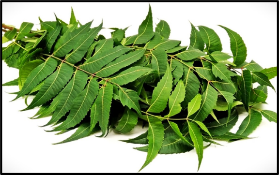
Figure 1: A. indica leaves
Anti-oxidants are a family of naturally occurring chemicals that prevent cellular damage caused due to the oxidation of other molecules [11]. Some of the major anti-oxidants found in plants are vitamins, polyphenols, and terpenoids groups [12]. These substances function by neutralising free radicals that cause oxidative stress and cellular damage, lowering lipid peroxidation, and scavenging reactive oxygen species (ROS). Both ethanolic and methanolic neem leaf extracts contain flavonoids like quercetin and quercetin-3-O-glucoside, as well as phenolic compounds like gallic acid, ellagic acid, and avicularin, which are major contributors to this activity [10]. Furthermore, limonoids like nimbin and azadirachtin, which are more prevalent in seeds, have also been found in leaves and support neem's cytoprotective and free radical scavenging properties [13]. The ability of neem-derived compounds like azadiradione to mimic antioxidant enzymes such as superoxide dismutase helps to neutralize oxidative stress, which in turn plays a crucial role in curbing inflammation establishing a functional link between neem’s anti-oxidant and anti-inflammatory properties [14].
Inflammation is the immune system's reaction to pathogens, damaged cells, toxic substances, or radiation. It works by eliminating harmful stimuli and beginning the healing process [15]. Some of the key anti-inflammatory phytochemicals are flavonoids, terpenoids, polyphenols, saponins, tannins, and alkaloids [16]. Most of which are found in neem leaf, making it a rich source of anti-inflammatory agents. Moreover, compounds like nimbolide, nimbidin, gedunin, and azadirachtin also give neem leaf its anti-inflammatory properties [17]. These substances function by blocking pathways like nuclear factor-κB (NF-κB) and cyclooxygenase-2 (COX-2) and downregulating inflammatory mediators like tumor necrosis factor-alpha (TNF-α), interleukin-1 beta, and interleukin-6 (IL-6) [18]. Gedunin and nimbolide are especially good at regulating immune cell reactions and oxidative stress. Nimbidin inhibits the activity of neutrophils and macrophages [19], whereas flavonoids scavenge ROS [9]. These components work together to give neem its capacity to lower both acute and chronic inflammation. Neem's strong anti-inflammatory properties are especially noteworthy because chronic inflammation is becoming more widely acknowledged as a major factor in the onset and advancement of metabolic diseases like diabetes.
Diabetes, which medical professionals frequently refer to as diabetes mellitus, is a collection of metabolic illnesses characterised by elevated blood glucose (blood sugar), either as a result of insufficient insulin secretion, improper insulin cellular response, or both [20]. Neem leaf's anti-diabetic properties are owing to the presence of flavonoids, triterpenoids, and glycosides, which work together to improve insulin sensitivity, encourage glucose absorption in peripheral tissues, and shield pancreatic β-cells from oxidative stress [21]. These substances help lessen postprandial glucose spikes by inhibiting important intestinal enzymes, including α-amylase and α-glucosidase [22]. Through processes including activation of glucose transporter 4 (GLUT4) and enhancement of insulin signalling molecules in skeletal muscle, studies show that neem leaf extracts dramatically reduce blood glucose levels and improve lipid profiles in diabetic animal models [21]. Neem's anti-oxidant qualities also aid in reducing oxidative stress, which is a significant cause of insulin resistance and problems from diabetes [22]. Beyond its role in metabolic regulation, neem's rich phytochemical composition provides potent anti-microbial defenses, addressing another critical aspect of human health by combating pathogenic microorganisms.
Worldwide, pathogenic microorganisms continue to remain the primary cause of sickness and mortality. As a result of their growing resistance to traditional anti-microbials, it is imperative to investigate plant-based substitutes, such as neem. Bioactive chemicals such as azadirachtin, quercetin, and β-sitosterol are responsible for the strong anti-bacterial action of neem leaves against pathogens like Staphylococcus aureus (S. aureus), Pseudomonas aeruginosa, and Candida albicans (C. albicans) [23]. By destroying cell membranes, preventing enzyme function, and obstructing DNA replication, these phytochemicals stop microorganisms from growing [24]. Limonoids such as azadirachtin exhibit broad-spectrum anti-bacterial and anti-fungal qualities, while the flavonoid quercetin strengthens anti-microbial activities by acting as a chelating agent and anti-oxidant [24]. A phytosterol called β-sitosterol helps pathogens destabilise their membranes. Neem leaf extracts have been shown in studies to be more efficient than bark or seed extracts at suppressing the growth of bacteria and fungi at concentrations as low as 500 µg/ml [23]. Minimum inhibitory concentrations (MICs) of 32 µg/ml against S. aureus and 64 µg/ml against Escherichia coli were reported in an in vitro study of methanolic leaf extract of A. indica grown in Sudan, indicating strong broad-spectrum anti-bacterial activity [25]. Neem has long been used to cure infections and fight off bacteria that are resistant to drugs because of the way these components work together.
The substantial anti-bacterial, anti-inflammatory, anti-diabetic, and anti-oxidant qualities of neem leaves are supported by their extensive phytochemical composition. Because of its wide range of medicinal effects, neem is a promising natural source for contemporary medicine that merits more investigation into its complete pharmacological potential.
REVIEW OF LITERATURE:
Historically, medicinal plants have been used to treat illnesses. For over 2,000 years, A. indica has been recognised as one of the most adaptable medicinal plants with a broad range of biological activities in India and its adjacent nations [26]. Meliaceae species A. indica and Melia azedarach (M. azedarach) are closely related [27]. The former is commonly referred to as Indian lilac or Indian neem (margosa tree), whereas the latter is called Persian lilac [27]. The evergreen tree, neem, is grown throughout the Indian subcontinent. Since ancient times, every part of the tree has been utilised as a home cure and traditional medicine to treat a variety of human illnesses [26]. Neem is increasingly recognized as a significant medicinal plant in modern medicine and is widely utilised in Ayurvedic, Unani, and Homoeopathic medicine [26]. The neem tree is known as “Sarbaroganibarini” because its Sanskrit name, “Arishtha,” means “reliever of sickness”. In India, the tree is still considered a “village dispensary” [28].
The tree can thrive on hard clay soils as well as sandy, stony, shallow soils, making it adaptable to a wide range of climates [29]. This adaptability contributes significantly to its widespread availability and accessibility, particularly in regions where other medicinal plants might struggle to thrive. The tree requires little water and lots of sunlight. It can flourish in a broad range of temperatures, from 0 to 49 ? [29]. Neem tree require a pH of 4 to 10 to develop, and they may also neutralise acidic soils owing to a unique calcium-mining characteristic. Neem is probably indigenous to drier regions of South Asia and the Indian subcontinent. Other South and Central American nations, as well as portions of Africa and the Caribbean, have been added. The Persian term “azaddhirakt,” which means “Noble tree,” is where the word “Azadirachta” originates [29].
The different parts of the Neem tree have a variety of practical and therapeutic uses. Rich in bioactive compounds like nimbin and quercetin, neem leaves are used to treat ringworm, eczema, wounds, dandruff, and disorders of the immune system [30]. Neem flowers are utilised in South Indian cooking and are also used to treat intestinal worms, nausea, and anorexia [31]. By keeping saliva's alkaline balance, combating bacteria, and healing swollen gums, neem twigs act as natural toothbrushes and improve oral health [32]. The bark has analgesic, anti-diabetic, and fever-reducing qualities due to its tannin and flavonoid content [33]. The insecticidal, anti-fungal, and anti-inflammatory properties of neem seeds and oil make them valuable in skincare, pest control, and pharmaceutical formulations [26]. All things considered neem as a priceless asset in both conventional and contemporary medicine.
Some major phytochemicals found in neem contribute to its extensive medicinal properties, particularly in its leaves. Neem leaves contain nimbin, nimbanene, nimbandiol, nimbolide, ascorbic acid, n-hexacosanol, and essential amino acids, which provide anti-microbial and immunomodulatory benefits [34]. Neem leaf extracts are useful for treating microbial infections because fresh neem leaves contain polyphenolic flavonoids, quercetin, and β-sitosterol, which are known to have strong anti-bacterial and anti-fungal properties [35]. Beyond the leaves, neem seeds contain azadirachtin and gedunin, which have significant anti-microbial and anti-malarial properties. Nimbolinin, salannin, and sodium nimbinate are additional important phytochemicals found in neem plants that all contribute to the plant's wide range of pharmacological actions [34]. Furthermore, because terpenoids and limonoids actively slow the progression of disease, neem bark and root extracts have shown anti-inflammatory, hypoglycemic, and anti-cancer properties [36]. These various bioactive substances, which are mostly present in neem leaves, emphasise the plant's potential as a potent natural remedy with a wide range of therapeutic uses.
Saponins are naturally occurring glycosidic compounds found in plants, known for their foaming properties and diverse biological activities [37]. Several studies showed the presence of saponins in neem in different extracts (methanol, water, ethyl acetate, benzene, toluene, acetone, butyl alcohol) [38,39]. Phenols are aromatic compounds with strong anti-oxidant qualities that are defined by one or more hydroxyl groups that are directly attached to a benzene ring [40]. The main phenolic compounds found in neem leaves are epicatechin, gallic acid, vanillic acid, coumaric acid, ferulic acid, and catechin [33]. These compounds greatly enhance the plant's anti-microbial and anti-oxidant properties.
Tannins are high-molecular-weight polyphenolic substances with potent anti-microbial and anti-oxidant qualities that can precipitate proteins [41]. Neem leaves contain tannins, including ellagitannins and derivatives of gallic acid, which have astringent and anti-bacterial properties [33]. Alkaloids are a broad class of naturally occurring organic compounds with nitrogen atoms that are mostly found in plants. They have a variety of strong physiological and pharmacological effects [42]. Several bioactive alkaloids, such as nimbidine, nimboline, and azadirachtin, are known to be present in neem leaves [26]. These alkaloids have shown significant anti-microbial, anti-malarial, and anti-inflammatory properties.
Plant-based flavonoids are a class of secondary metabolites of polyphenols that are well-known for their anti-inflammatory, anti-oxidant, and free-radical scavenging properties [43]. Quercetin, kaempferol, and rutin are among the flavonoids found in neem leaves that have been identified as contributing to their anti-bacterial and anti-oxidant qualities [10].
Table 1: Properties in phytochemicals
|
Phytochemical |
Examples in neem leaf |
Properties |
|
Saponins |
Triterpenoid saponins |
Anti-microbial and Anti-inflammatory [17] |
|
Phenols |
Gallic acid, ferulic acid, vanillic acid |
Anti-oxidant and Anti-diabetic [44] |
|
Tannins |
Ellagitannins, gallic acid derivatives |
Anti-microbial and Anti-oxidant [45] |
|
Alkaloids |
Nimbidine, nimboline, azadirachtin |
Anti-inflammatory and Anti-microbial [46] |
|
Flavonoids |
Quercetin, kaempferol, rutin |
Anti-oxidant and Anti-inflammatory [47] |
Neem leaf is recognized for its anti-oxidant capacity, which stems from its ability to scavenge free radicals and mitigate oxidative stress within biological systems. This anti-oxidant activity has been corroborated by various standardized assays, including the ferric reducing anti-oxidant power (FRAP) assay [48]. The FRAP assay quantifies the electron-donating capability of plant extracts, providing a direct measure of their anti-oxidant potential through the reduction of ferric ions. Research utilizing the FRAP assay has consistently demonstrated that neem leaf extracts exhibit significant reducing power, with observed activity increasing proportionally to concentration [49]. For instance, at a concentration of 800 μg/ml, neem leaf extracts demonstrated anti-oxidant activity of 57.52 and 57.87 μM in the FRAP assay, compared to 139.97 μM for ascorbic acid, a standard reference anti-oxidant [48]. The presence of phenolic compounds, flavonoids, and tannins in neem leaf extracts, as revealed through phytochemical screening, is strongly correlated with the observed FRAP activity, suggesting these bioactive compounds, functioning as electron donors, contribute significantly to overall anti-oxidant effect [50,49]. This reducing power is quantitatively assessed using FRAP assay. In contrast to DPPH assay, which measures free radical scavenging activity, the FRAP assay specifically evaluates the ability of anti-oxidants to reduce ferric ions, thereby providing a complementary perspective on the mechanisms underlying the anti-oxidant properties of neem leaf [49]. Notably, the FRAP assay results demonstrate high reproducibility across various extraction methodologies and concentrations, bolstering the conclusion that neem leaf is a reliable source of anti-oxidant compounds [49]. The observed anti-oxidant effects, as quantified by the FRAP assay, contribute to the broader protective mechanisms of neem leaf against oxidative stress, which is implicated in the pathogenesis of numerous chronic diseases [50]. This potent anti-oxidant capacity is particularly significant given oxidative stress's central role in the pathogenesis of numerous chronic diseases, including cardiovascular disorders and neurodegenerative conditions.
Neem leaf possesses notable anti-inflammatory properties, evidenced by its capacity to mitigate inflammation and modulate associated pathways within biological systems. Extensive research, utilizing both in-vitro and in-vivo models, has demonstrated the significant anti-inflammatory effects of neem leaf [51]. These effects are largely attributed to the presence of bioactive compounds, including nimbin, nimbolide, and various flavonoids, which act to modulate key inflammatory signaling cascades [50].In-vivo studies, particularly those employing animal models such as carrageenan-induced paw edema, have shown that neem leaf extracts can effectively reduce edema and inflammation, exhibiting efficacy comparable to that of conventional anti-inflammatory pharmaceuticals. Mechanistically, these anti-inflammatory effects are mediated through the inhibition of pro-inflammatory cytokines, such as IL-6 and TNF-α, alongside the suppression of COX-2 enzyme activity. Furthermore, neem leaf extracts have been shown to inhibit the NF-κB pathway, a critical regulator of inflammatory responses, thereby reducing the expression of genes involved in the inflammatory process [51]. Supporting these findings are studies demonstrating the protective effects of neem leaf extracts against oxidative damage at the cellular level, a phenomenon closely intertwined with inflammatory processes [50]. Emerging scientific evidence substantiates the anti-inflammatory properties of neem leaf, demonstrating a dose-dependent relationship wherein increased concentrations yield a more significant reduction in inflammatory markers. This corroborates the traditional application of neem leaf in managing inflammatory conditions, including arthritis and dermatitis [51]. The synergistic effect of its anti-oxidant and anti-inflammatory capabilities positions neem leaf as a potentially valuable agent in the management of chronic inflammatory diseases [51].
Neem leaf exhibits promising anti-diabetic properties through its capacity to modulate glucose metabolism, augment insulin sensitivity, and provide protection against diabetes-related complications. Evidence from both preclinical and clinical investigations highlights neem leaf's anti-diabetic potential, largely attributed to its high concentration of bioactive compounds, including flavonoids and polyphenols. These constituents are believed to safeguard pancreatic β-cells from oxidative stress, a critical element in the pathogenesis of diabetes [50,51].In-vivo studies employing animal models have demonstrated that neem leaf extracts can enhance glucose tolerance and insulin sensitivity, leading to a significant decrease in fasting blood glucose levels [51]. Further supporting its therapeutic role, the anti-oxidant activity of neem leaf, as evidenced by assays such as FRAP, suggests its ability to alleviate oxidative stress, a characteristic feature of diabetes and its sequelae [50,51]. Moreover, neem leaf extracts have been shown to promote glycogen synthesis in hepatic and muscle tissues, thereby contributing to the maintenance of stable blood glucose levels during periods of fasting [51]. Evidence suggests that neem leaf, by its capacity to normalize lipid profiles, specifically reduces levels of low-density lipoprotein (LDL) cholesterol while elevating high-density lipoprotein (HDL) cholesterol. These alterations in lipid metabolism are crucial in mitigating the risk of cardiovascular complications frequently associated with diabetes mellitus. Consequently, the traditional application of neem leaf in diabetes management is substantiated by findings derived from both preclinical animal models and clinical investigations. The mechanisms of action responsible for these beneficial effects are multifaceted, encompassing enhanced insulin sensitivity, improved glucose uptake by cells, and a protective effect on pancreatic beta cells, which are essential for insulin production [51]. The convergence of anti-oxidant and anti-diabetic properties positions neem leaf as a potentially valuable adjunctive therapy in the comprehensive management of diabetes.
Neem leaf exhibits significant anti-microbial properties, defined by its capacity to suppress the proliferation and viability of diverse pathogenic microorganisms, encompassing both bacteria and fungi. Its broad-spectrum anti-microbial activity has been extensively documented against a range of bacterial and fungal pathogens [50]. This bioactivity is primarily attributed to the presence of key bioactive compounds, notably polyphenols, flavonoids, and triterpenoids, as revealed through rigorous phytochemical analysis. Research indicates that neem leaf extracts demonstrate inhibitory effects against both Gram-positive (S. aureus) and Gram-negative (Escherichia coli) bacteria, as well as various fungal species. The anti-microbial efficacy displays a concentration-dependent relationship, wherein increased extract concentrations correlate with enhanced microbial growth. The underlying mechanisms of action involve the disruption of microbial cell membranes and the inhibition of biofilm formation, both of which are critical processes for pathogen survival and resistance. A strong correlation exists between the presence of polyphenols and flavonoids in neem leaf and its observed anti-oxidant and anti-microbial properties, lending scientific support to its traditional applications in wound healing and oral hygiene practices. The anti-microbial potential of neem leaf has been explored in various fields, including dentistry, food safety, and bacteriology [50].
Collectively, the extensive body of literature reviewed herein robustly supports neem’s multifaceted pharmacological profile, encompassing significant anti-oxidant, anti-inflammatory, anti-diabetic, and anti-microbial activities. While traditional knowledge has long revered neem for its medicinal attributes, modern science continues to validate and uncover the complex interactions of its diverse phytochemicals responsible for these profound effects. Despite this wealth of information affirming its therapeutic potential, further rigorous research is crucial to fully elucidate optimal dosages, long-term safety profiles, and precise mechanisms of action in human subjects. While the multifaceted properties of neem are increasingly recognized, a significant gap exists in the rigorous identification and validation of individual phytochemicals responsible for the observed biological activities. To address this critical need, the present study endeavors to systematically characterize the comprehensive phytochemical profile of neem leaf extract and subsequently evaluate its anti-oxidant, anti-inflammatory, anti-diabetic, and anti-microbial properties. This research provides a foundational framework for future investigations focused on elucidating the targeted therapeutic potential of specific bioactive compounds within neem leaf.
OBJECTIVES:
MATERIALS AND METHODS:
MATERIALS:
A. niger fungal culture, C. albicans fungal culture, E. faecalis bacterial culture, S. aureus bacterial culture, Sample - Azadirachta indica leaf, 96-well plate, Agar well cutter, Autoclave, Bacterial incubator, Beaker, Condenser, Cuvettes, Falcon tubes, Glass stirrer, Grinder, Heating mantle, Hot air oven, Laminar airflow, Petri plates, Pipette, Round bottom flask, Soxhlet Extractor, Spectrophotometer, Swab, Thimble, Water bath, Weighing scale, 4-nitrophenyl-β-D-glucopyranoside (SRL Pvt. Ltd.), Aluminium Chloride (Himedia Laboratories), Bovine serum albumin (SRL Pvt. Ltd.), Dilute hydrochloric acid (Medilise Chemicals), Distilled water , Ferric Chloride (Kanton Laboratories), Folin–Ciocâlteu Reagent (Medilise Chemicals), Gentamycin (American Pharma Remedies), Methanol (Nice Chemicals), Mueller-Hinton broth (Himedia Laboratories), Nutrient agar (Himedia Laboratories), Phosphate-buffered Saline (Himedia Laboratories), Potassium ferrocyanide (Kanton Laboratories), Potato dextrose agar (Himedia Laboratories), Sodium Carbonate (Nice Chemicals), Sodium Hydroxide (Kanton Laboratories), Sodium Nitrite (Kanton Laboratories), Sodium phosphate buffer (Kanton Laboratories), Sulfuric acid (Medilise Chemicals), Tap Water, Trichloroacetic acid (Medilise Chemicals), Vanillin in ethanol (Nice Chemicals), Wagner’s Reagent (Kanton Laboratories), α-glucosidase (SRL Pvt. Ltd.)
METHODS:
Sample Collection
Fresh leaves of A. indica were collected from a local area in Thiruvananthapuram. They were then washed multiple times with tap water and then with distilled water (DW). Then, A. indica leaves were dried in the hot air oven at 60 degrees Celsius (°C) for 6 hours (hrs). Then the leaves were powdered using a grinder.
Extraction
The powdered foliage of A. indica (25 g) was subsequently positioned within a thimble, which was subsequently affixed to a Soxhlet extractor apparatus. In the following step, 105 mL of methanol was introduced into a heating flask. The Soxhlet extractor apparatus was then meticulously assembled by placing it at the top of heating flask, with a condenser positioned above it. Following a duration of 12-14 hrs, a portion of the extract was collected within the heating flask and subsequently transferred into a falcon tube. Concurrently, the remaining portion underwent heating to yield a methanolic neem crude extract.
Phytochemical Analysis: Qualitative
Sample
The methanolic extract of A. indica leaves was subjected to standard qualitative phytochemical tests to detect the presence of major secondary metabolites, including saponins, phenolics, tannins, alkaloids, and flavonoids. The extract was prepared by dissolving 1 mg of dry neem extract in 1 mL of methanol for all tests.
Test for Saponins:
1 mL of the methanolic A. indica leaf extract was mixed with 1 mL of double - DW and vigorously shaken for 2 minutes (mins). The formation of a stable, persistent foam indicated the presence of saponins.
Test for Phenols:
To 1 mL of methanolic A. indica leaf extract, a few drops of 5% ferric chloride (FeCl3) solution were added. The development of a dark green or bluish-black colouration confirmed the presence of phenolic compounds.
Test for Tannins:
400 µL of methanolic A. indica leaf extract was mixed with 4 mL of 10% sodium hydroxide (NaOH) solution and shaken. Formation of a stable emulsion indicated the presence of tannins, particularly condensed tannins.
Test for Alkaloids:
To 1 mL of the A. indica leaf extract, 1-2 drops of Wagner’s reagent (iodine in potassium iodide) were added. The appearance of a reddish-brown precipitate indicated the presence of alkaloids.
Test for Flavonoids:
A few drops of 2% NaOH were added to 1 mL of methanolic A. indica leaf extract. This was followed by the addition of dilute hydrochloric acid (dil. HCl). A yellow colour appeared upon the addition of NaOH and disappeared after acidification, confirming the presence of flavonoids.
Phytochemical Analysis: Quantitative
Estimation of Saponins:
To the prepared methanolic A. indica leaf extract, 0.25 mL of 0.8% vanillin in ethanol was added, followed by 2.5 mL of 72% sulfuric acid (conc. H2SO4). The reaction mixture was incubated at 60?°C for 15 mins, then allowed to cool at room temperature (RT) for 5 mins. The absorbance was measured at 544 nm using a spectrophotometer.
Estimation of Phenols:
To the methanolic A. indica leaf extract, 2 mL of Folin-Ciocalteu (Fc) reagent was added, and the mixture was shaken thoroughly. Subsequently, 4 mL of 7.5% sodium carbonate (Na2CO3) solution was added, and the mixture was incubated at RT for 30 mins. After incubation, the mixture was shaken again. The absorbance was recorded at 760 nm using a spectrophotometer.
Estimation of Tannins:
To the methanolic A. indica leaf extract, 7.5 mL of double DW was added, followed by 0.5 mL of FC reagent and 1 mL of 35% Na2CO3 solution. An additional 1 mL of DW was added to the mixture, which was then shaken thoroughly and incubated in the dark for 30 mins at RT. The absorbance was measured at 700 nm using a spectrophotometer.
Estimation of Flavonoids:
To the methanolic A. indica leaf extract, 2 mL of 5% sodium nitrite solution (NaNO2) was added, and the mixture was shaken well. This was followed by the addition of 3 mL of 10% aluminium chloride solution and 2 mL of 1 M NaOH. The mixture was thoroughly mixed and allowed to stand for 5 mins. Finally, 2 mL of DW was added, and the solution was incubated at RT for 10 mins. The absorbance was determined at 510 nm using a spectrophotometer.
Anti-oxidant activity: FRAP assay
Varying concentrations of methanolic A. indica leaf extract (50, 100, 200, 350, and 500?µg/mL) were prepared. To each, 2.5?mL of sodium phosphate buffer and 2.5?mL of potassium ferrocyanide were added and mixed thoroughly. The mixtures were then incubated at 50?°C for 20 mins. After incubation, 2.5?mL of trichloroacetic acid was added and shaken well. From each tube, 2?mL of the supernatant was taken and mixed with 2?mL of distilled water, followed by 0.5?mL of ferric chloride. After 10 mins of incubation at RT, absorbance was recorded at 700?nm using a spectrophotometer.
Anti-inflammatory activity: Protein denaturation assay
Different concentrations of methanolic A. indica leaf extract (50, 100, 200, 350, and 500?µg/mL) were prepared. To each, 0.2?mL of 1% bovine serum albumin (BSA) and 1.8?mL of phosphate-buffered saline (PBS, pH 6.4) were added. The mixtures were incubated at RT for 20 mins, followed by heating at 70?°C for 5 mins. Absorbance was then measured at 660?nm using a spectrophotometer.
Anti-diabetic activity: α- glucosidase
Different concentrations of methanolic A. indica leaf extract (50, 100, 200, 350, and 500?µg/mL) were prepared. To each test tube, 10 µL α-glucosidase enzyme and 125 µL 0.1 M Sodium phosphate buffer (pH 6.9) were added. This mixture was then incubated at 37? for 20 mins. Post incubation, 20 µL of 4- 4-nitrophenyl-β-D-glucopyranoside (PNPG) was added. This mixture was then incubated at RT for 30 mins. Following this, the reaction was terminated by adding 50 µL of 0.1 M Na2CO3. The absorbance was then measured at 405 nm using a spectrophotometer.
Agar well diffusion
Inoculation
The fungi C. albicans and Aspergillus niger (A. niger) were cultivated in potato dextrose broth, whereas the bacteria S. aureus and Enterococcus faecalis (E. faecalis) were cultivated in nutrient broth. To ensure optimal growth, each microorganism is inoculated into its respective medium and incubated for 24 hrs at 37? under static conditions.
Plating
Post incubation, 120 mL each of nutrient agar (NA) and potato dextrose agar (PDA) are prepared for anti-microbial testing. The media is sterilized using an autoclave at 121?°C for 15 mins and poured into sterile Petri plates under aseptic conditions.
Swabbing
After 24 hrs of incubation, the bacterial cultures were aseptically swabbed onto the surface of nutrient agar plates, while fungal cultures are swabbed onto the PDA plates using sterile cotton swabs to ensure uniform distribution. Each plate is divided into five wells created using an agar well cutter.
The wells are loaded as follows:
Incubation
All inoculated plates were incubated at 37?°C for 24 hrs. After incubation, the anti-microbial activity is assessed by measuring the diameter of the zone of inhibition around each well.
Minimum Inhibitory Concentration (MIC)
Inoculation
A loop full of bacterial cultures of S. aureus and E. faecalis is inoculated in 10 ml of Mueller-Hinton Broth (MHB), respectively. Then incubated at 37? for 24 hrs.
Serial Dilution and MIC Assay
A 96-well microtiter plate was used to perform the MIC assay. Rows 1 to 6 were designated for serial dilutions of the test sample, while subsequent rows were assigned for positive control (PC), negative control (NC), and solvent control with bacteria (NEET). Each well, excluding the one labelled NEET, was filled with 100 µL of MHB. To initiate the dilution series, 100 µL of the test sample was added to wells in row 1, NC, and NEET. Serial two-fold dilutions were performed from wells 1 to 6 by transferring 100 µL from one well to the next and mixing thoroughly, discarding 100 µL from the final well to maintain equal volumes. Subsequently, 100 µL of the bacterial suspension was added to wells 1 - 6, PC, and NEET, resulting in a total volume of 200 µL per well.
Incubation and Measurement
The plate was covered with a sterile lid and incubated at 37?°C for 24 hrs. Following incubation, absorbance was measured at 600 nm using a microplate spectrophotometer. The MIC was defined as the lowest concentration of the test sample that resulted in a marked reduction in absorbance or the absence of visible turbidity, indicating inhibition of bacterial growth.
Minimum Bactericidal Concentration (MBC)
To determine the MBC, 10 µL aliquots were taken from wells at and above the MIC and plated onto sterile NA plates. The plates were then incubated at 37?°C for 24 hrs. MBC was defined as the lowest concentration of the sample at which no bacterial colony growth was observed, indicating complete bactericidal activity.
RESULTS
Qualitative phytochemical analysis of methanolic A. indica leaf extract
Qualitative phytochemical analysis (Fig:2) is performed on methanolic A. indica leaf extracts, and the results are depicted in Table 1.
|
Phytochemicals |
Presence / Absence |
|
Saponins |
+ |
|
Phenols |
+++ |
|
Tannins |
+++ |
|
Alkaloids |
- |
|
Flavonoids |
++ |
‘+++’ - high expression, ‘++’ - moderate expression, ‘+’ - low expression, ‘-’ – absent
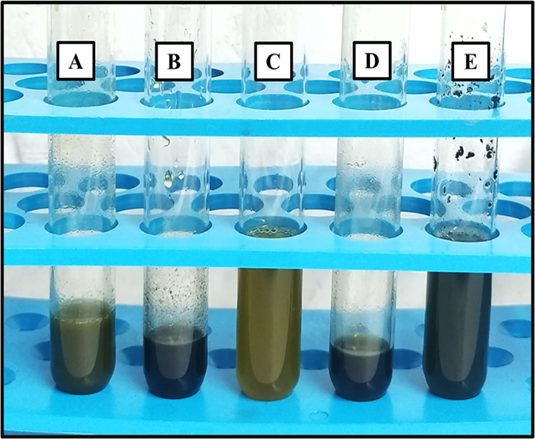
Figure 2 and Table 2: Qualitative analysis of phytochemicals in methanolic A. indica leaf extract (A- Saponins, B- Phenols, C- Tannins, D- Alkaloids, E- Flavonoids)
Quantitative phytochemical estimation of methanolic A. indica leaf extract
Estimation of Saponins
The estimation of saponins (Fig:3) was carried out by taking Diosgenin as a standard (Table:3; Fig:4). The equation used to quantify saponins in methanolic A. indica leaf extract is y = 0.0059x – 0.0318 from the standard curve of diosgenin (Table:4).
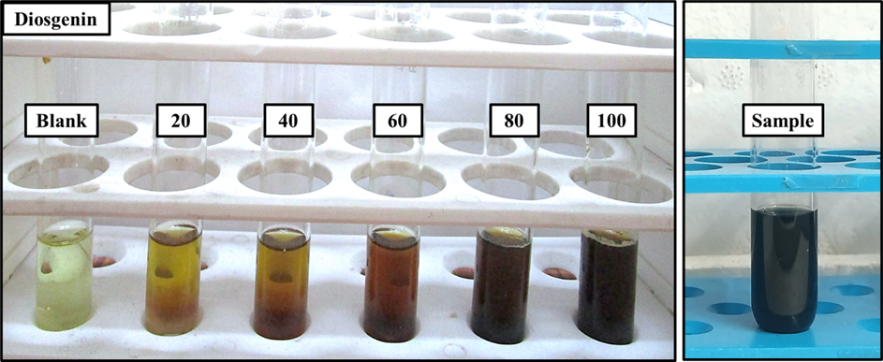
Figure 3: Quantitative estimation of saponins (standard - diosgenin, blank - methanol, and sample - methanolic A. indica leaf extract)
|
Concentration (µg/ml) |
Absorbance (at 544 nm) |
|
20 |
0.098 |
|
40 |
0.202 |
|
60 |
0.297 |
|
80 |
0.434 |
|
100 |
0.568 |
|
Sample |
3.000 |
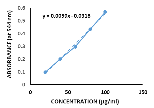
Table 3 and Figure 4: Absorbance values of diosgenin at different concentrations and the absorbance value of methanolic A. indica leaf extract & standard curve of diosgenin
Table 4: Quantity of saponins in methanolic A. indica leaf extract
|
Phytochemical |
Quantity |
|
Saponins |
513.864 µg/mL |
Estimation of Phenols
The estimation of phenols (Fig: 5) was carried out by taking gallic acid as the standard (Table:5 ; Fig:6). The equation used to quantify phenol in methanolic A. indica leaf extract is y = 0.0144x - 0.1095 from the standard curve of gallic acid (Table:6).
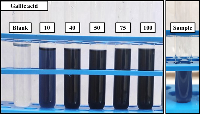
Figure 5: Quantitative estimation of phenols (standard - gallic acid, blank - methanol, and sample - methanolic A. indica leaf extract)
|
Concentration (µg/ml) |
Absorbance (at 760 nm) |
|
10 |
0.016 |
|
40 |
0.393 |
|
50 |
0.692 |
|
75 |
1.06 |
|
100 |
1.26 |
|
Sample |
0.821 |
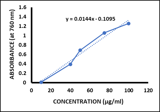
Table 5 and Figure 6: Absorbance values of gallic acid at different concentrations and the absorbance value of methanolic A. indica leaf extract & standard curve of gallic acid
Table 4: Quantity of saponins in methanolic A. indica leaf extract
|
Phytochemical |
Quantity |
|
Phenols |
64.618 µg/mL |
Estimation of Tannins
The estimation of tannins (Fig:7) was carried out by taking tannic acid as the standard (Table:7 ; Fig:8). The equation used to quantify tannin in methanolic A. indica leaf extract is y = 0.2011x + 0.0263 from the standard curve of tannic acid (Table:8).
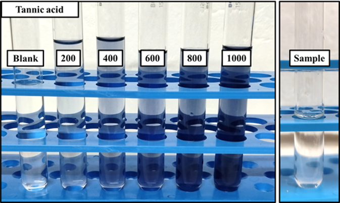
Figure 7: Quantitative estimation of tannins (standard - tannic acid, blank - methanol, sample - methanolic A. indica leaf extract)
|
Concentration (µg/ml) |
Absorbance (at 700 nm) |
|
200 |
0.223 |
|
400 |
0.431 |
|
600 |
0.636 |
|
800 |
0.828 |
|
1000 |
1.03 |
|
Sample |
0.891 |
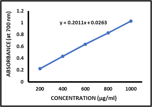
Table 8: Quantity of tannin in methanolic A. indica leaf extract
|
Phytochemical |
Quantity |
|
Tannins |
4.299 µg/mL |
Estimation of Flavonoids
The estimation of flavonoids (Fig:9) was carried out by taking Quercetin as standard (Table:9; Fig:10). The equation used to quantify flavonoids in methanolic A. indica leaf extract is y = 0.0023x + 0.226 from the standard curve of quercetin (Table:10).
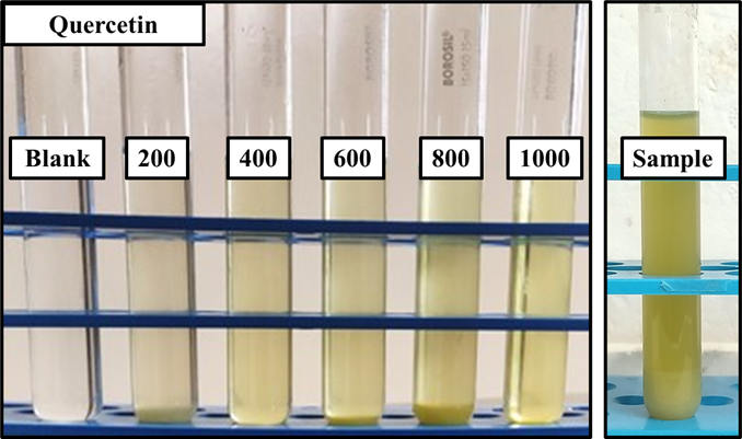
Figure 9: Quantitative estimation of flavonoids (standard - quercetin, blank - methanol and all the reagents, sample - methanolic A. indica leaf extract)
|
Concentration (µg/ml) |
Absorbance (at 510 nm) |
|
200 |
0.718 |
|
400 |
1.158 |
|
600 |
1.501 |
|
800 |
1.912 |
|
1000 |
2.591 |
|
Sample |
1.713 |
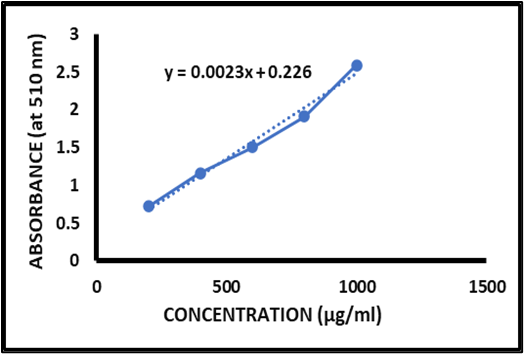
Table 9 and Figure 10: Absorbance values of quercetin at different concentrations and the absorbance value of methanolic A. indica leaf extract & standard curve of quercetin.
Table 10: Quantity of flavonoid in methanolic A. indica leaf extract
|
Phytochemical |
Quantity |
|
Flavonoids |
646.521 µg/mL |
Anti-oxidant activity of methanolic A. indica leaf extract
Anti-oxidant activity was determined using FRAP assay, and Ascorbic acid (Fig:11,12)was taken as standard (Table:11).
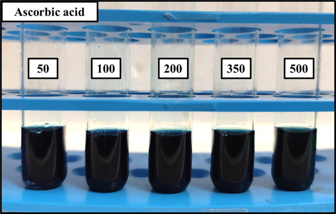
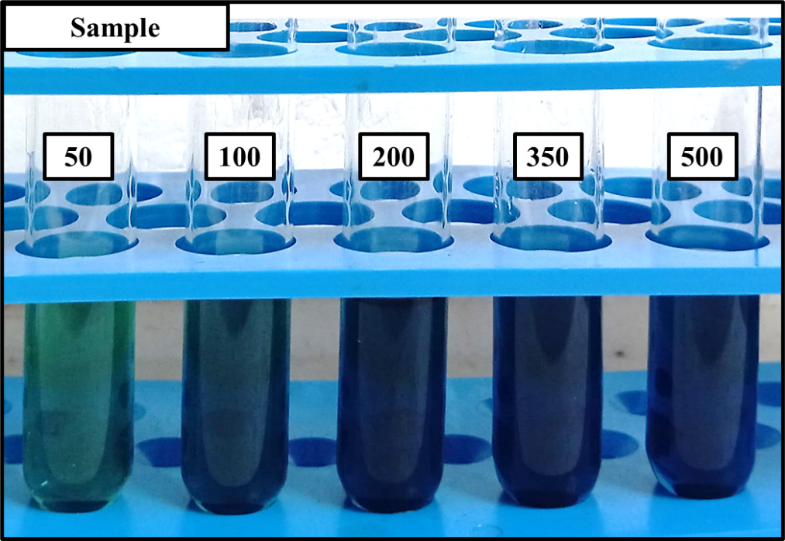
Figure 11: FRAP assay- Anti-oxidant activity of Ascorbic acid and A. indica leaf extract
Table 11: Absorbance of Ascorbic acid and Sample - methanolic A. indica leaf extract
|
Concentration (µg/ml) |
Absorbance of Ascorbic acid (at 700 nm) |
Absorbance of sample (at 700 nm) |
|
Control |
3.000 |
3.000 |
|
50 |
0.970 |
0.158 |
|
100 |
2.161 |
0.253 |
|
200 |
2.381 |
0.292 |
|
350 |
2.614 |
0.479 |
|
500 |
3.000 |
0.612 |
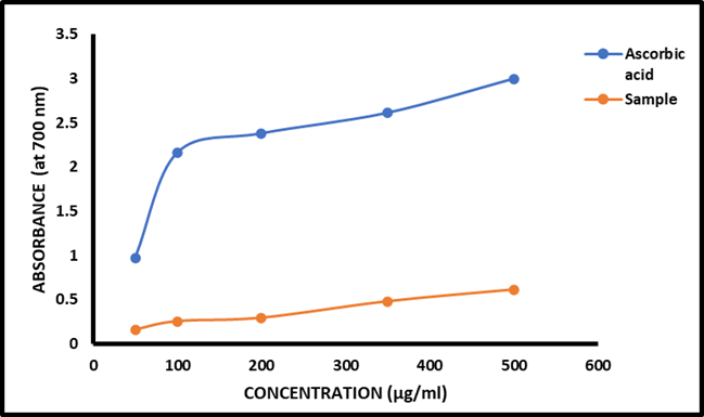
Figure 12: FRAP- Absorbance of Ascorbic acid and Sample - methanolic A. indica leaf extract
Anti-inflammatory activity of methanolic A. indica leaf extract
Anti-inflammatory property was determined using protein denaturation assay (Fig:13; Table:12), and diclofenac was taken as standard (Table:13; Fig:14).
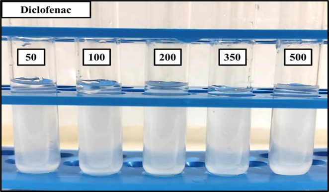
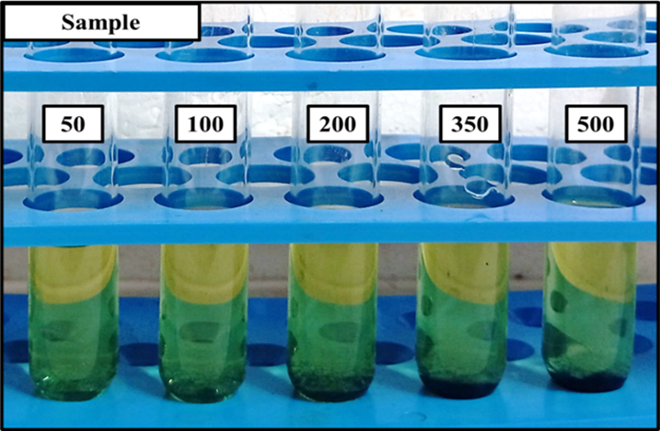
Figure 13: Protein denaturation assay- anti-inflammatory property of Diclofenac and methanolic A. indica leaf extract
Table 12: Absorbance of Diclofenac and Sample - methanolic A. indica leaf extract
|
Concentration (µg/ml) |
Absorbance of diclofenac (at 660 nm) |
Absorbance of sample (at 660 nm) |
|
Control |
0.401 |
0.401 |
|
50 |
1.167 |
1.338 |
|
100 |
0.762 |
1.229 |
|
200 |
0.695 |
0.778 |
|
350 |
0.525 |
0.624 |
|
500 |
0.456 |
0.470 |
Table 13: Percentage inhibition of Diclofenac and Sample - methanolic A. indica leaf extract
|
Concentration (µg/ml) |
% inhibition of Diclofenac |
% inhibition of Sample |
|
50 |
16.702 |
4.496 |
|
100 |
45.610 |
12.276 |
|
200 |
50.392 |
44.468 |
|
350 |
62.526 |
55.460 |
|
500 |
67.451 |
66.452 |
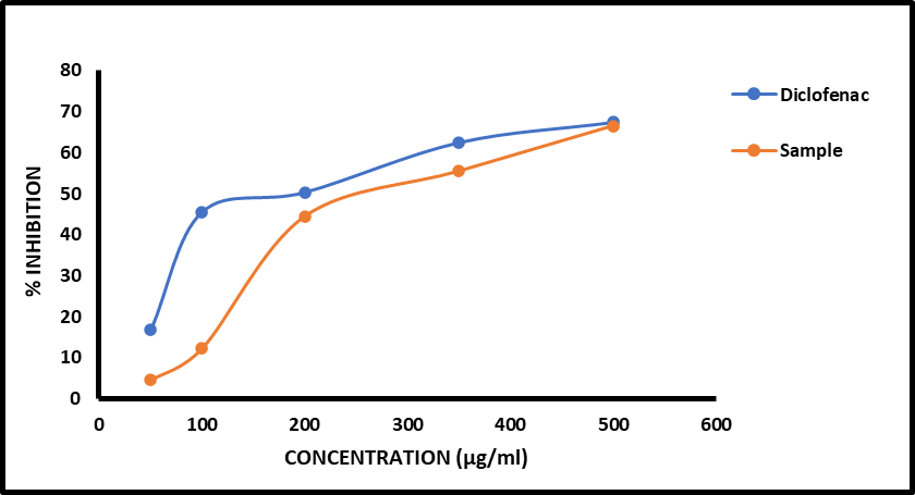
Figure 14: Percentage inhibition of Diclofenac and Sample - methanolic A. indica leaf extract
Anti-diabetic activity of methanolic A. indica leaf extract
Anti-diabetic activity was assessed through α-glucosidase assay (Fig:15; Table:14), and acarbose was taken as standard (Fig:16; Table:15).
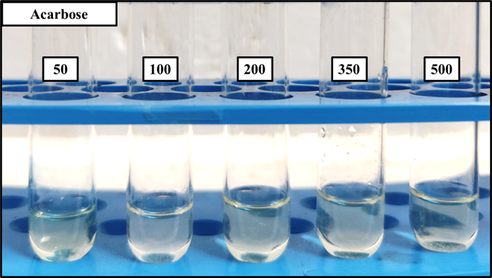
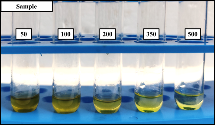
Figure 15: α-glucosidase assay - anti-diabetic property of Acarbose and methanolic A. indica leaf extract
Table 14: Absorbance of Acarbose and Sample - methanolic A. indica leaf extract
|
Concentration (µg/ml) |
Absorbance of acarbose (at 405 nm) |
Absorbance of sample (at 405 nm) |
|
Control |
1.776 |
1.776 |
|
50 |
1.119 |
1.218 |
|
100 |
0.320 |
0.887 |
|
200 |
0.219 |
0.632 |
|
350 |
0.148 |
0.498 |
|
500 |
0.043 |
0.373 |
Table 15: Percentage inhibition of Acarbose and Sample – Methanolic A. indica leaf extract
|
Concentration (µg/ml) |
% inhibition of Acarbose |
% inhibition of Sample |
|
50 |
36.993 |
31.418 |
|
100 |
81.981 |
50.056 |
|
200 |
87.668 |
64.414 |
|
350 |
91.666 |
71.959 |
|
500 |
97.578 |
78.997 |
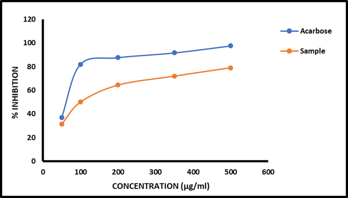
Figure 16: Percentage inhibition of Acarbose and Sample - methanolic A. indica leaf extract
Anti-microbial activity of methanolic A. indica leaf extract
Anti-microbial activity was assessed through agar well diffusion method. A. indica leaf extract demonstrated significant anti-bacterial properties against S. aureus and E. faecalis. However, no activity was seen against fungi like C. albicans and A. niger (Fig:17; Table:16).
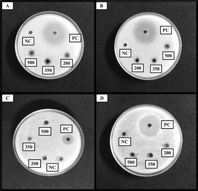
Figure 17: Anti-microbial activity of A. indica leaf extract. (A) against S. aureus, (B) E. faecalis, (C) C. albicans, (D) A. niger
Table 16: Zone of inhibition of methanolic A. indica leaf extract against different organisms
|
|
Zone of Inhibition (mm) |
|||
|
Concentration |
S. aureus |
E. faecalis |
C. albicans |
A. niger |
|
PC (Gentamycin) |
38 |
39 |
28 |
16 |
|
NC (Methanol) |
- |
- |
- |
- |
|
200 mg/mL |
9 |
- |
- |
- |
|
350 mg/mL |
10 |
- |
- |
- |
|
500 mg/mL |
11 |
11 |
- |
- |
Minimum Inhibition Concentration of methanolic A. indica leaf extract
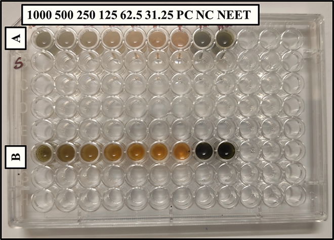
Figure 18: 96 microtiter plate - MIC of methanolic A. indica leaf extract on (A) S. aureus and (B) E. faecalis
Table 17: Absorbance of S. aureus and E. faecalis
|
Concentration (µL) |
Absorbance of S. aureus (at 600 nm) |
Absorbance of E. faecalis (at 600 nm) |
|
1000 |
0.398 |
0.741 |
|
500 |
0.022 |
0.069 |
|
250 |
0.286 |
0.057 |
|
125 |
0.021 |
0.641 |
|
62.5 |
0.802 |
0.706 |
|
31.25 |
0.707 |
0.762 |
|
PC |
0.802 |
0.852 |
|
NC |
0.261 |
0.261 |
|
NEET |
0.698 |
0.652 |
Minimum Bactericidal Concentration of methanolic A. indica leaf extract
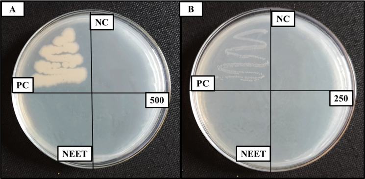
Figure 19: MBC of methanolic A. indica leaf extract - (A) S. aureus and (B) E. faecali
DISCUSSION:
A. indica leaf has long been recognized for its therapeutic potential, prompting this study to investigate its phytochemical composition and associated pharmacological properties. Specifically, the study aimed to elucidate the anti-oxidant, anti-inflammatory, anti-diabetic, and anti-microbial activities of the leaf extract. Qualitative phytochemical analysis of the methanolic A. indica leaf extract confirmed the presence of saponins, phenols, tannins, and flavonoids, while alkaloids were not detected. These findings partially concur with previous research by [52]; however, the aforementioned study also reported the presence of alkaloids, marking a point of divergence. Further quantitative analysis confirmed the presence of saponins, phenols, tannins, and flavonoids. The observed anti-oxidant activity is likely attributable to the presence of polyphenols, flavonoids, and limonoids. The extract exhibited a concentration-dependent anti-oxidant effect, with optimal activity observed at 500 µg/mL, as demonstrated by its ferric reducing ability, suggesting its capacity to mitigate oxidative stress. These findings are consistent with previous research by [53], who also demonstrated significant concentration-dependent anti-oxidant activity of A. indica leaf extracts using the FRAP assay. In their findings, the maceration aqueous leaf extract exhibited higher anti-oxidant activity (6.912±0.001 mg extract/mg AAE), followed by the maceration ethanolic leaf extract (6.449±0.001 mg extract/mg AAE), and the Soxhlet ethanolic seed extract showed lower activity (5.723±0.020 mg extract/mg AAE). The anti-inflammatory properties of A. indica leaf extract was thought to stem from the presence of nimbidin, nimbolide, and gedunin. The methanolic extract demonstrated significant BSA inhibition, ranging from 44.4% to 66.4%, with maximal inhibition occurring at 500µg/mL, supporting its potential as an anti-inflammatory agent. Specifically, the observed concentration-dependent inhibition of protein denaturation in the present study corroborates findings by [54], who similarly demonstrated notable anti-inflammatory effects of A. indica utilizing the protein denaturation assay. In terms of anti-diabetic activity, the presence of flavonoids, triterpenoids, and glycosides is believed to be responsible for the observed α-glucosidase inhibition, which ranged from 64% to 78%, particularly at concentrations between 200µg/mL and 500µg/mL. The observed inhibitory effect on α-glucosidase activity aligns with investigations by [55], who likewise reported significant anti-diabetic potential of A. indica leaf extract through α-glucosidase inhibitory activity with an IC50 value of 2.11 ± 0.05 µg/mL. This suggests that A. indica leaf extract can be effective at relatively low concentrations. Finally, the anti-microbial activity of A. indica leaf extract exhibited a concentration-dependent relationship, with greater activity observed at higher concentrations (500 µg/mL), showing inhibition of 11 mm. The extract exhibited a notably higher efficacy against bacteria as compared to fungi, demonstrating limited or no inhibitory effect on A. niger and C. albicans. This observation aligns with the findings of (Reddy et al., 2013), who similarly reported the absence of anti-microbial activity against A. niger and C. albicans at concentrations of 500 µg/mL in their investigation. The MIC values for S. aureus and E. faecalis were determined to be 500 µL and 250 µL, respectively, consistent with the results of [23]. The MBC results further underscore the potential of A. indica leaf as an anti-bacterial agent. Minimum fungicidal concentration (MFC) testing was not conducted due to the absence of fungal inhibition in the initial screening. While no significant anti-fungal activity was observed against C. albicans and A. niger in the present study, consistent with the findings of [23], it is notable that other investigations, such as those by [56] and [57], have reported varying degrees of anti-fungal efficacy for A. indica extracts, potentially due to the differences in extraction methods or fungal strains. The collective results presented here highlight the multifaceted therapeutic potential of A. indica leaf, warranting further investigation into its clinical applications.
CONCLUSION:
This study effectively assessed the different medicinal qualities of methanolic extract from A. indica leaves and described its phytochemical composition. Alkaloids were not present, but saponins, phenols, tannins, and flavonoids were confirmed by qualitative analysis. The precise concentrations of these substances were also ascertained through quantitative estimations. In terms of biological activity, we see a concentration-dependent anti-oxidant activity. At increasing concentrations, significant anti-inflammatory activity was observed. A potent concentration-dependent anti-diabetic activity was further demonstrated by methanolic A. indica leaf extract. The methanolic A. indica leaf extract demonstrated selective anti-bacterial activity in terms of anti-microbial properties, successfully preventing the growth of E. faecalis and S. aureus with established MIC values. In contrast, under the experimental conditions, no discernible anti-fungal activity was found against A. niger or C. albicans.
In summary, the methanolic leaf extract of A. indica possesses a rich phytochemical profile that correlates with its observed anti-oxidant, anti-inflammatory, anti-diabetic, and specific anti-bacterial properties. These findings confirm the presence of diverse bioactivities in the methanolic extract of A. indica, supporting its traditional applications.
ACKNOWLEDGMENT:
First and foremost, I would like to thank GOD almighty for his blessings and wisdom for helping this project to its successful completion.
I am indebted to my research guide, Dr. PARVATHY PRASAD, Managing Director, Bioroot Exploration Pvt Ltd, for her guidance, motivation, and advice during the project work. Her timely suggestion has enabled me to finish this project the best of my expectations.
I owe my sincere and deep sense of acknowledgement to Ms. AISWARYA SAMBASIVAN, Junior Research Associate, Bioroot Exploration India Pvt Ltd, Thiruvananthapuram, for her valuable advice, guidance, suggestions, and encouragement throughout the work. This project was indeed the outcome of her immense help and support.
Last but not the least a, special appreciation to my PARENTS, FRIENDS, and COLLEAGUES for their constant encouragement and patience during my study period. Besides this, several people have knowingly or unknowingly helped me in the successful completion of the project work.
REFERENCES

Ayaana Jethalia, Parvathy Prasad, Aiswarya Sambasivan, Therapeutic Potential of Azadirachta indica Leaf Extract, Int. J. of Pharm. Sci., 2025, Vol 3, Issue 12, 3856-3882. https://doi.org/10.5281/zenodo.18085652
 10.5281/zenodo.18085652