School of Pharmaceutical Sciences, Maharishi University of Information Technology, Noida, Uttar Pradesh, 201304, India
Microsponges, which are revolutionary polymer particles that were patented in 1987, have completely changed the way drugs are delivered. With sizes ranging from 5 to 300 microns, these microspheres with pores provide a distinct benefit when applied topically. By effectively enclosing a range of substances, such as fragrances, moisturizers, UV filters, and pharmaceutically active ingredients, they enhance the effectiveness, decrease irritation, and prolong the shelf life of the product. The present work addresses the advantages and disadvantages of microsponges, focusing on their drug-release mechanisms. Microsponge preparation methods are covered in detail, including solvent diffusion in quasi-emulsions, using porogens, and polymerization of liquid-liquid suspensions. To demonstrate the versatility of microsponge innovation, pH, temperature, solubility, and pressure-dependent drug release techniques are mentioned. Microsponges are used in a wide variety of dosage forms, such as those for the eyes, skin, and mouth. It is defined how they contribute to the healing of wounds in diabetes, psoriasis, acne, arthritis, colon cancer, and melanoma. Cosmetics like Salicylic Peel and Retin-A-micro, which use microsponge technology, show how effective this new method of drug delivery can be. Microsponge's versatility is demonstrated by research findings on their use in topical medicine and dermatology. Microsponge preparations have been successfully used to administer a variety of medications, such as voriconazole, diclofenac sodium, mupirocin, and nitrendipine. Compiling patents related to microsponges is a bonus of the paper, demonstrating how this field is continuously innovating and changing. Furthermore, microsponges have led to patented innovations, showcasing continual advancements in drug delivery. The array of patents underscores the ongoing commitment to refining microsponge technology, enhancing drug efficacy, and improving patient outcomes. The collective insights presented in this abstract emphasize the pivotal role of microsponges in revolutionizing drug delivery systems and advancing pharmaceutical science.
Microsponges are tiny, sponge-like polymer particles that have a spherical shape and are characterized by their porosity and permeability. This innovative solution offers a range of benefits including enhanced efficiency, reduced irritability, extended shelf life, increased formulation versatility, improved elegance, and enhanced aesthetic properties. Additionally, microsponges possess the unique capacity to absorb or retain various pharmaceutical active substances across a broad spectrum. [1,2] A range of dynamic substances such as moisturizers, fragrances, aromatic extracts, UV filters, and agents with anti-infective, anti-fungal, and anti-inflammatory properties can be captured within the permeable microspheres that constitute the microsponge. [3] The microsponge technology, initially patented by Advanced Polymer Systems, Inc. in 1987, was designed to regulate the release of active ingredients into the skin. By doing so, it reduces local cutaneous responses and decreases systemic exposure. This technology specifically benefits topical medication solutions. [4] The microcarriers mentioned in the text address the limitations of other similar carriers by offering advantages such as a faster release rate of medications, improved stability, and increased payload capacity. Additionally, they can be easily transformed into different forms, including liquids, gels, creams, and powders. [5] Microsponges, ranging in size from 5 to 300 microns, are currently the subject of extensive research in the realm of topical applications. These porous microspheres can remain on the skin's surface and facilitate the effective delivery of a wide range of medications for various topical conditions. [6,7]
2. ADVANTAGES OF MICROSPONGE [8]
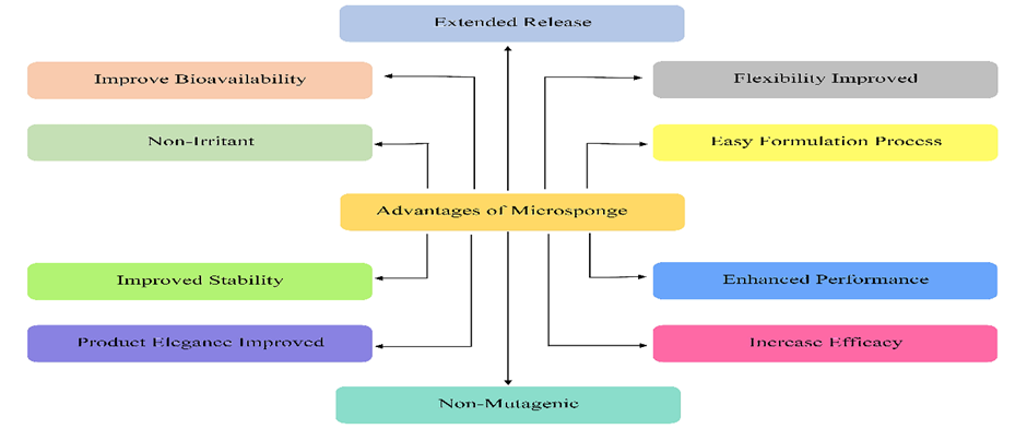
Fig. (1). Advantages of microsponge.
3. DISADVANTAGES OF MICROSPONGE [9]
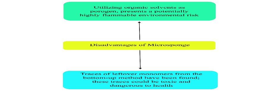
Fig. (2). Disadvantages of microsponge.
4. RELEASE MECHANISM OF MICROSPONGES
Various mechanisms are employed within microsponges to facilitate the release of drugs. Fig. (3)
4.1. pH: The pH-dependent release of drugs is facilitated by modifying the protective coating on the microsponges. [9]
4.2. Temperature: Temperature plays a significant role in the release of medication from the microsponge into the dermis. When the medication is too thick at room temperature, it cannot easily penetrate the microsponge. However, as the skin temperature increases, the flow rate and release rate of the active ingredients also increase. [9]
4.3. Solubility: When water is present, additives that can dissolve in water, like antiperspirants and antiseptics, will be released. Additionally, the diffusion mechanism can initiate the release of the drug. [9]
4.4. Pressure: Microsponges, when subjected to pressure during rubbing, have the potential to release drugs from the skin. The efficacy of drug release is directly influenced by the quality of the sponge. [9]
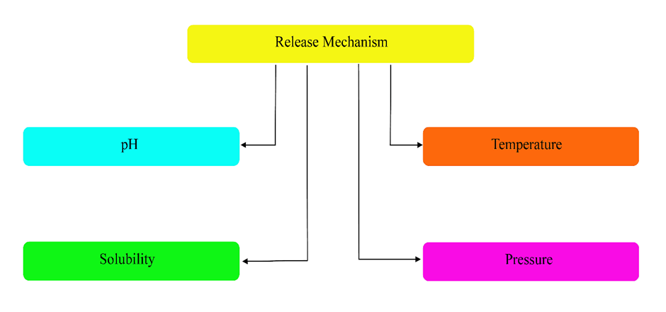
Fig. (3). Release mechanism of microsponges.
5. METHOD OF PREPARATION
In 1998, a method was introduced by Kawashima et al. to produce microspheres with a highly porous matrix. [10] The manufacturing process of microsponges is generally influenced by the physicochemical attributes of the drug and its solubility in the polymers used for encapsulation. Depending on the characteristics of the medication to be loaded, microsponges can be produced using two methods: (1) Polymerization of liquid-liquid suspensions and (2) Solvent diffusion in quasi-emulsions. [11]
Liquid-liquid suspension polymerization is the process of dissolving active substances (monomers) and additives (aqueous phase) in a solvent and then adding the active substance to the solvent. Adding a catalyst or increasing the temperature will initiate the polymerization process. The polymerization process continues to form a sphere-like form and reservoir-type system. The solvent is then evaporated to yield porous microspheres with a spherical shape. [12–14] Fig. (4)
To prepare microsponges using this method, the following steps are necessary: [15]
The process of quasi-emulsion solvent diffusion involves two distinct methods to create separate phases, known as the internal and external phases. Volatile solvents such as ethanol, acetone, or dichloromethane are commonly used for the internal phase, while the external phase is typically made up of aqueous polyvinyl alcohol (PVA) solution or water.
When these phases are mixed, quasi-emulsion globules are formed, which are in the process of becoming emulsion globules. These globules are then treated with a solvent to produce insoluble particles in the micrometer range. By adding 20% dichloromethane or triethyl citrate (TEC), the formulation gains flexibility. [16,17]
To extract the microsponges, the resulting microsponge emulsion is passed through filters, followed by a washing process. The vacuum-heated, separated, and cleaned microsponges are then dried for 24 hours at a temperature of 40 degrees Celsius. This method offers advantages over liquid-liquid suspension polymerization, as it reduces the exposure of the medication. [16,18,19] Fig. (4)
This technique entails dispersing a water-based phase within a solution of organic polymers, along with a thickening agent such as span, polyethyleneimine, and stearylamine. Subsequently, this water-in-oil emulsion is further dispersed within an external water-based phase containing PVA, resulting in a dual emulsion. The objective of this innovative method is to produce porous microspheres that can be regenerated. The advantage of this approach is its ability to encapsulate both water-soluble and water-insoluble medications, as well as thermolabile substances like proteins. Xanthan gum is also utilized as an emulsifier to stabilize the internal water-in-oil emulsion. [20–23] Fig. (4)
In this method, a substitute material (porogen) such as sodium bicarbonate or hydrogen peroxide was used instead of water in the internal hydrophilic phase of water in oil in water (w/o/w) emulsion. The porogen was mixed with a polymer mixture to create a consistent dispersion structure, which was then dissolved in a solution containing hydrophilic components. A catalyst was added to the emulsion, and the organic solvent was allowed to evaporate, resulting in the formation of microparticles. The introduction of the porogen created interconnected and evenly distributed pores ranging in size from 5 to 20 µm. [24] Fig. (4)
In contrast to the w/o/w method, a different approach was taken to create an oil-in-oil (o/o) emulsion. This involved using a volatile organic liquid as the internal phase, which was continuously stirred and allowed to slowly evaporate at a controlled pace. The process included mixing fixed mineral oil and dichloromethane with span as the external phase, polylactide glycolic acid as the polymer, and dichloromethane as the solvent for the internal phase. To form the microsponge, the internal phase was gradually added to the mixture while being consistently stirred for dispersion. The selection of the organic solvent and external phase depended on the physicochemical properties of the medicinal product and the polymer used to create the microsponges. This technique was utilized to produce Eudragit RS-100 microsponges containing hydroxyzine HCl, where liquid paraffin acted as the continuous medium and acetone functioned as the dispersing solvent. [25,26]
To produce porous microspheres, the gelation method was utilized, followed by the transformation into microspheres using lyophilization. In this process, the microspheres were immersed in a chitosan hydrochloride solution and then subjected to lyophilization, which rapidly removed the solvent, resulting in the formation of pores within the microspheres. However, a disadvantage of this technique is that the fast solvent elimination can lead to the formation of fractured or constricted microparticles. [27]
The initial report stated that lipid-bilayered mesoporous silica particles could be made using a vibrating orifice aerosol generator (VOAG). The process involved creating porous particles through surfactant templating in microdroplets generated by VOAG. To manufacture the core particles made of tetraethylorthosilicate, ethanol, water, and diluted hydrochloric acid were refluxed to create a stock solution. VOAG was then used to dilute the stock solution with a solvent containing surfactant and stir to produce uniform droplets. The resulting microspheres contained liposomes and could be used to target specific areas of the body for delivering active ingredients. [28]
The nanosponge creation technique involved modifying liquid-liquid suspension polymerization by using b-cyclodextrin (b-CD) as the monomer and diphenyl carbonate as the cross-linking agent. The size of the microparticles was controlled by heating and sonicating the reaction mixture. Once the reaction mixture cooled, the resulting product was transformed into coarse particles and cleaned with ethanol and distilled water. The porous microparticles made from cross-linked b-CD can effectively hold drugs. However, this method may have the disadvantage of potentially retaining dangerous residues from the cross-linking substance. [29]
The chitosan microspheres with pores were created using this method. Initially, a chitosan solution was subjected to sonication to generate bubbles. The resulting suspension of bubbles was then drawn into a syringe and pumped through a steel capillary using a syringe pump. Electrohydrodynamic atomization was then applied. The diameter of the capillary was carefully chosen to accommodate all the bubbles in the suspension as they passed through. The voltage used in the experiments depended on the amount of chitosan present in the solution, except for the case where the highest concentration was used and electrospraying became difficult. In all other cases, a stable cone-jet mode was achieved with different combinations of flow rate and applied voltage. Finally, a 4% w/v sodium hydroxide solution was employed to crosslink the chitosan microspheres. [30]
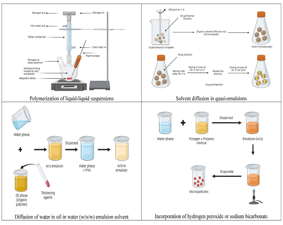
Fig. (4). Different methods of preparations.
6. APPLICATIONS OF MICROSPONGE
The microsponge can be utilized in oral medication form to enhance the release rate of poorly soluble drugs by trapping them within the pores of the microsponge. This can also improve the drug's availability and effectiveness while reducing any potential side effects. When the microsponge drug is taken orally, it may cause changes in pH, release the drug at a specific location, and prolong its retention in the stomach, especially for drugs primarily absorbed in the stomach or upper small intestine. Fig. (5) Floating dosage systems have a lower density compared to gastric fluid, allowing the medication to float on the stomach's contents and remain there for an extended period without disrupting the natural emptying process of the stomach. [31,32] Table 1
Table 1. The table below displays the various release profiles of different drug microsponges.
|
S. No. |
Drug |
Microsponge composition |
Release rate |
Reference |
|
1 |
Curcumin |
Ethylcellulose and Eudragit RS 100 |
88.4 – 90.8 % |
[33] |
|
2 |
Loratadine |
Ethylcellulose |
66.75 – 88.15 % |
[34] |
|
3 |
Cinnarizine |
Ethylcellulose |
57.9 – 88.7 % |
[35] |
|
4 |
Famotidine |
Eudragit RS 100 |
97.5 % |
[36] |
Furthermore, the utilization of microsponge technology shows potential in enclosing medications within a limited area and administering active components to the lower gastrointestinal tract in a regulated fashion. The primary rationale behind employing the microsponge system for delivering drugs to the colon is that active ingredients measuring less than 200 µm are easily assimilated by the macrophages present in colon tissues. This facilitates efficient targeted drug action at the intended site. [37]
It is widely believed that traditional topical medications only affect the outermost areas of the skin. When applied, these medications release their active ingredients, resulting in a concentrated cream that is quickly absorbed. However, by utilizing a microsponge drug delivery system, it is possible to prevent the excessive accumulation of active ingredients in the deeper layers of the skin, such as the epidermis and dermis. This technology not only reduces the risk of skin irritation caused by medication but also maintains effectiveness. Fig. (5) Examples of topical products that utilize this microsponge technology include Retin-A-micro for acne vulgaris and Retinol night cream for anti-wrinkle purposes. [38]
Water-soluble medications can be applied topically as ointments or aqueous suspensions, while water-insoluble medications can only be used as ointments or aqueous suspensions. The process of drug kinetics in the eye involves the medication crossing the blood-aqueous barrier and entering the anterior chamber. The medication is then eliminated from the body through aqueous humor turnover, moving from the anterior chamber to the Schlemm's canal and trabecular meshwork. Additionally, the medication is absorbed into the bloodstream through the blood-aqueous barrier, after being removed from the aqueous humor. Eventually, the drug molecule crosses the blood-retina barrier, allowing it to enter the posterior chamber of the eye. [39]
Microsponge technology has been utilized in cosmetics and dermatology to enhance the efficacy of drugs. By localizing the drug on the skin's surface and within the epidermis, microsponges help reduce both systemic and local cutaneous side effects. While dermatological products face stricter regulations, cosmetic products can be developed and brought to market more rapidly. Additionally, microsponge drug delivery systems offer the possibility of targeting specific areas of the skin, thereby minimizing absorption into the bloodstream. [38]
Skin-related psoriasis is a long-term inflammatory condition. It lowers the quality of life for those who are ill. An emulsion solvent diffusion method is used to make microsponge for the medication mometasone furoate. Psoriasis and other inflammatory disorders are treated with methotreasone furoate. Fig. (5) With an initial burst effect, the release profiles showed a biphasic release. After eight hours, 78–95% of the medication was released, with 29–36% of the medication being released in the first hour. [40] For effective topical treatment and to help with psoriasis, dithranol microsponge gel is encapsulated in a dendrimer. [41] The number of applications required for psoriasis treatment was decreased by using a microsponge gel containing clobetasol propionate. They found that, in contrast to the typical form, which lasts 2.5 hours, drug release might last up to 12 hours. [40]
Hydroquinone microsponges were developed by Grimes PE with 4% hydroquinone and 0.15 percent retinol to treat post-inflammatory hyperpigmentation (PIH) and melanoma. A hydroquinone-releasing microsponge was developed to deliver medication over an extended period with minimal irritation to the skin. At weeks 4, 8, and 12, there were statistically significant improvements in the intensity of pigmentation and symptoms of illness when compared to the baseline (p<0.001). With every visit, there was a significant improvement in both the lesion area and colorimetry measurements (p<0.001). [42]
A cream with prolonged release was created by Osmani et al. employing miconazole nitrate microsponge, an anti-acne agent. Using a quasi-emulsion solvent diffusion technique, the Eudragit RS-100 microsponge was produced. A cream was created and blended with a microsponge. Compared to conventional creams, which ran out of drugs after 4 hours and released only 83.09 percent, drug-loaded microsponges released 78.28% of the drug for up to 8 hours. [43] One of the most popular acne treatments is benzoyl peroxide. Jelvehgari and colleagues used ethylcellulose microsponges to release benzoyl peroxide into the skin. Studies show that drug release is greater in the first hour and remains stable for the next seven hours. [18]
Diclofenac administration via a microsponge has been researched for the treatment of arthritis. To create microsponge gels with diclofenac diethylamine that would have a prolonged release for the treatment of arthritis, Osmani et al. employed a quasi-emulsion solvent diffusion technique. They contrasted their results with the 1.16 percent w/w commercial Voltaren Emulgel. The microsponge-based gel released the medication over 8 hours, whereas the gel only released 81.11 percent of the medication in 4 hours. [43] In a different investigation, Hadi et al. created microsponge tablets with lornoxicam as the active ingredient to treat arthritis. Fig. (5) They discovered that the drug was released over an extended period, ranging from 86% to 96% to 12 hours. [44]
Eudragit RS100-based 5-Fluorouracil microsponge was developed by Gupta and his associates as a colon cancer treatment. The toxicity and prolonged release of oral medications can be reduced with the use of microsponge. 5-FU is useful in the treatment of a wide variety of solid tumor types. If 5-FU is more abundantly concentrated in tumors, it may be more efficacious. The process of creating a 5-FU microsponge involved oil-in-oil emulsion solvent diffusion. It was discovered that pure 5-FU releases in roughly 20 minutes, but the microsponge extends the release period to approximately 5 hours. It has been discovered that microsponge-loaded 5-FU outperforms 5-FU alone in terms of cell viability. [45]
Pandit and colleagues prepared microsponges loaded with nebivolol and encapsulated in gel to retain moisture in the wound during the final stages of healing. The antihypertensive drug nebivolol dilates blood vessels. In vitro tests have proven that approximately 80% of the drug is released after 8 hours. Due to the microsponge gel, the medicine is released at a slow rate. The porosity of the microsponge formulation enabled rapid wound healing in diabetic rats. [2]
Fig. (5). Different applications of microsponges.
The purpose of Microsponge is to enhance the delivery of medication in the most efficient way possible, while simultaneously improving product stability, reducing side effects, and modifying the release of the drug.
Table 2. The table below showcases some examples of marketed products that utilize Microsponge technology.
|
Sr. No. |
Ingredient |
Marketed product |
Activity |
Reference |
|
1 |
Tretinoin at concentrations of 0.1% and 0.04%, along with methyl methacrylate, forms the primary components of an aqueous gel base |
Retin-A-micro |
Wrinkle-reducing |
[15,46,47] |
|
2 |
Salicylic acid with a concentration of 20% |
Salicylic Peel 20 |
Anti-acne and hyperpigmentation |
[15,46,47] |
|
3 |
Dimethicone |
Ultra Guard |
Skin protectants |
[15,46,47] |
|
4 |
Salicylic acid with a concentration of 30% |
Salicylic Peel 30 |
Remove dead skin |
[15,46,47] |
|
5 |
Hydroquinone and retinol |
EpiQuin Micro |
Reduce skin irritation to the minimum level |
[15,46,47] |
|
6 |
Pure retinol and vitamin A |
Retinol 15-night cream |
Wrinkle-reducing |
[15,46,47] |
|
7 |
Benzoyl peroxide/methyl methacrylate/glycol |
NeoBenz Micro |
Fight against bacteria and promote the shedding of dead skin cells |
[15,46,47] |
|
8 |
Fluorouracil |
Carac cream |
Actinic keratoses |
[38] |
|
9 |
Retinol |
Line eliminator dual retinol facial treatment |
Wrinkle-reducing |
[38] |
|
10 |
Lactic acid |
Lactrex 12% moisturizing cream |
Moisturizer |
[38] |
|
11 |
Zinc gluconate |
Oil-free matte block SPF 20 |
Sunscreen |
[38] |
|
12 |
Glycolic acid |
Glycolic acid moisturizer SPF 15 |
Skin supplement |
[38] |
Throughout the year, a considerable quantity of research is conducted in the realm of microsponge drug formulation. This involves employing various preparation techniques to enhance medicinal products.
Table 3. Research studies were conducted on microsponges for topical administration.
|
Sr. No. |
Method of preparation |
Drug |
Observation |
Reference |
|
1 |
Quasi-emulsion solvent diffusion |
Nitrendipine |
A medicine with low water solubility was administered with a prolonged-release formulation. |
[48] |
|
2 |
Emulsion solvent diffusion |
Mupirocin |
The test showcased the stability and absence of skin irritation, as well as the long-lasting effectiveness in healing surgical wounds. |
[40] |
|
3 |
Double emulsification technique |
Diclofenac sodium |
The enhanced formulation provides increased advantages in terms of controlled absorption of diclofenac sodium into the skin. |
[21] |
|
4 |
Quasi-emulsion solvent diffusion |
Eberconazole nitrate |
The rat's skin remained comfortable throughout the controlled drug release, and the study revealed a fourfold increase in drug retention. |
[49] |
|
5 |
Oil-in-oil emulsion solvent diffusion |
Nebivolol |
Diabetic patients experience accelerated healing of wounds. |
[2] |
|
6 |
Quasi-emulsion solvent diffusion |
Oxiconazole nitrate |
The microsponge gel containing oxiconazole nitrate was developed to have controlled drug release, proving to be more advantageous compared to traditional therapy. |
[50] |
|
7 |
Quasi-emulsion solvent diffusion |
Terbinafine hydrochloride |
Reduced side effects are observed with controlled drug release, and less gel administration is needed. |
[51] |
|
8 |
Quasi-emulsion solvent diffusion |
Nystatin |
The results revealed that the gel with Nystatin-loaded microsponges exhibited a notably higher release of medication compared to the traditional Nystatin gel. |
[52] |
|
9 |
Quasi-emulsion solvent diffusion |
Voriconazole |
A voriconazole-loaded microsponge gel showed prolonged drug release. |
[53] |
|
10 |
Quasi-emulsion solvent diffusion |
Itraconazole |
It was discovered that itraconazole was released in a regulated manner when it was intended to be delivered as a medication using a microsponge. |
[54] |
|
11 |
Quasi-emulsion solvent diffusion |
5-Fluorouracil |
The level of skin irritation was significantly lower when compared to a 5-fluorouracil formulation that was sold commercially. |
[55] |
|
12 |
Quasi-emulsion solvent diffusion |
Lornoxicam |
As an anti-inflammatory drug, it was found that the medication loaded with microsponge was more effective. |
[56] |
|
13 |
Quasi-emulsion solvent diffusion |
Naringenin |
The microsponge gel loaded with naringenin demonstrated a triple improvement in drug penetration into the skin. This medication is utilized for the treatment of atopic dermatitis. |
[5] |
|
14 |
Quasi-emulsion solvent diffusion |
Nimesulide |
The prepared formulations exhibited a better-controlled release of pharmacological effects. |
[8] |
Table 4. A compilation of research studies conducted on the use of microsponge for dermatological administration.
|
Sr. No. |
Method of preparation |
Drug |
Observation |
Reference |
|
1 |
Quasi-emulsion solvent diffusion |
Ketoprofen |
Comparatively, it exhibits improved bioavailability in contrast to commercially sold ketoprofen tablets, as well as a delayed release of the drug. |
[57] |
|
2 |
Quasi-emulsion solvent diffusion |
Flurbiprofen |
The findings of this research indicate that the microsponge can compress and form a solid tablet that is both structurally robust and capable of sustaining the release of medication over an extended period. |
[58] |
|
3 |
Quasi-emulsion solvent diffusion |
Dicyclomine |
As the drug/polymer ratio increases, the drug content rises while the manufacturing yield and particle size decrease. |
[59] |
|
4 |
Quasi-emulsion solvent diffusion |
Paracetamol |
The results indicate that medications containing microsponge had a higher loading efficiency than medications utilizing alternative techniques for delivering microparticles. |
[60] |
|
5 |
Quasi-emulsion solvent diffusion |
Famotidine |
The microsponge loaded with fumonisin exhibited a reliable pattern of drug release. |
[43] |
|
6 |
Quasi-emulsion solvent diffusion |
Piroxicam |
A discovery was made that it is possible to create spherical, porous microsponges. These microsponges dissolved at a much faster rate compared to the pure piroxicam tablet. |
[61] |
|
7 |
Quasi-emulsion solvent diffusion |
Curcumin |
Over the course of eight hours, it was found that the prepared microsponge medication loaded in the gelatine capsule shell released 93.2% curcumin, but the capsule contained only 11.7% curcumin. This led to the discovery that the manufactured curcumin microsponge held more promise than the conventional curcumin formulation as a drug delivery system because it was able to provide an extended-release rate of the drug in oral treatment. |
[62] |
Table 5. Various Patents related to Microsponges.
|
S. No. |
Patent Number |
Inventors |
Filed Date |
Granted Date |
Published Date |
References |
|
1 |
US4690825A |
Richard Won |
1985 |
1987 |
1987 |
[63] |
|
2 |
US5100783A |
Frederick Cahn et al. |
1989 |
1992 |
1992 |
[64] |
|
3 |
US5135740A |
Katz M. et al. |
1989 |
1992 |
1992 |
[65] |
|
4 |
US5387411A |
Eric S. et al. |
1990 |
1995 |
1995 |
[66] |
|
5 |
US5849327A |
David L. et al. |
1996 |
1998 |
1998 |
[67] |
|
6 |
US5725869A |
Ray J. R. Lo |
1996 |
1998 |
1998 |
[68] |
|
7 |
US6228894B1 |
Marie A. Rinaldi et al. |
1999 |
2001 |
2001 |
[69] |
|
8 |
US6656517B2 |
Eugene T. Michal et al. |
2001 |
2003 |
2002 |
[70] |
|
9 |
US6501002B1 |
Donald C. Roe at el. |
2000 |
2002 |
2002 |
[71] |
|
10 |
US6403704B1 |
Isabelle Bara |
2000 |
2002 |
2002 |
[72] |
|
11 |
US6689345B2 |
Nathallie J. Lezer |
2002 |
2004 |
2004 |
[73] |
|
12 |
US7098315B2 |
Alfred Schaufler |
2002 |
2006 |
2006 |
[74] |
|
13 |
US7186687B2 |
Leonard M. Patt |
2005 |
2007 |
2007 |
[75] |
|
14 |
US7604814B2 |
Schaffner P. Carl et al. |
2006 |
2009 |
2009 |
[76] |
|
15 |
US7700124B2 |
Karine Loyen et al. |
2005 |
2010 |
2010 |
[77] |
|
16 |
US8449923B2 |
Gary S. et al. |
2012 |
2013 |
2013 |
[78] |
|
17 |
US10172292B2 |
Zador E. et al. |
2016 |
2019 |
2019 |
[79] |
|
18 |
US10839907B2 |
Tran V Hieu et al. |
2019 |
2020 |
2020 |
[80] |
CONCLUSION
In pharmaceutical delivery systems, microsponges demonstrate revolutionary technology because of their distinct qualities and broad range of applications. Many industries, which comprise dermatology, cosmetics, pharmaceuticals, and more, have been profoundly influenced by the invention of microsponges. According to published research, microsponges have several benefits, including higher efficacy, decreased irritability, longer lifespans, more formulation flexibility, better elegance, and better aesthetic qualities. There is a wide range of techniques available for preparing microsponges to meet certain pharmaceutical delivery requirements. These include liquid-liquid suspension polymerization, solvent diffusion in quasi-emulsions, water-in-oil-in-water emulsion solvent diffusion, lyophilization, the aerosol generator technique with a vibrating orifice, production with ultrasound assistance, and the atomization process using electrohydrodynamics. It is helpful to recognize the process of release mechanisms, such as temperature responsiveness, pressure impacts, pH-dependent release, and solubility aspects, to optimize patterns of drug release for various applications. To create formulations that specifically address drug distribution obstacles, this knowledge is essential. The delivery of a broad range of pharmaceutical ingredients using microsponges in oral, topical, ocular, cosmetic, and dermatological dosage forms highlights their adaptability and efficacy. Examples of medical conditions for which microsponges may be used to treat patients with better therapeutic results include psoriasis, melanoma, acne, arthritis, colon cancer, and diabetic wound healing. Further evidence of the commercial viability and approval of microsponge-based products in the pharmaceutical and cosmetic industries comes from their availability on the market. Retin-A micro, formulations of salicylic peel, and carac cream are a few salient examples. These goods highlight the impact of microsponge inventions on improving drug delivery efficacy by serving as an example of how the innovation was successfully transferred from study to commercialization. An exhaustive review of microsponges-related research papers and patents also highlights the field's continued inventiveness and intellectual contributions. Progress in microsponge innovation is evident in the ongoing investigation of new medications, formulations, and preparation techniques by researchers. In summary, the development of microsponges has proven to be a game-changer, offering answers to problems about medication administration and indicating that advances in the fields of cosmetic and pharmaceutical sciences are still to come. The future of targeted and controlled drug delivery systems is expected to be significantly shaped by microsponges as research into them continues.
LIST OF ABBREVIATIONS
CONSENT FOR PUBLICATION
Not applicable.
FUNDING
None.
CONFLICT OF INTEREST
The authors declare no conflict of interest, financial or otherwise.
ACKNOWLEDGEMENTS
Declared none.
REFERENCES

Mukul Nishad, Mansi Chauhan, Unlocking the Potential: Microsponge Technology Revolutionizing Drug Delivery Systems, Int. J. of Pharm. Sci., 2025, Vol 3, Issue 12, 3331-3348. https://doi.org/10.5281/zenodo.18016163
 10.5281/zenodo.18016163
