View Article

  • Vascular Cognitive Impairment and Dementia : From Pathophysiological Mechanisms to Emerging Therapeutic Strategies

  • Maharaja Agrasen School of Pharmacy, Maharaja Agrasen University, Baddi

Abstract

Vascular Cognitive Impairment and Dementia (VCID) is a major, potentially preventable cognitive decline that emerges from cerebrovascular dysfunction, influenced predominantly by aging and metabolic disorders. VCID represents a significant cause of dementia worldwide. VCID arises from heterogeneous cerebrovascular pathologies, including cerebral small and large vessel disease and chronic cerebral hypoperfusion. Therapeutic progress in VCID remains constrained by pathological heterogeneity, frequent overlap with mixed neurodegenerative dementia, limited availability of sensitive early biomarkers, and a persistent shortage of VCID-specific randomized trials validating disease-modifying interventionsThis review summarizes recent diagnostic concepts, mechanistic pathways, and preventive and potential therapeutic interventions in managing VCID. Key mechanisms in pathophysiology of VCID include vascular endothelium disruption and loss of blood–brain barrier integrity, which facilitate oxidative stress and neuroinflammation. These processes impair neurovascular coupling and drive the white-matter injury, disconnecting cortical and subcortical pathways, and subsequent cognitive dysfunction. Hypertension remains the most robust modifiable risk factor, with metabolic disorders, chronic kidney disease, aging, and stroke further accelerating vascular brain injury. Although current management largely focuses on vascular risk reduction, emerging disease-modifying approaches, including senolytics and mitochondrial boosters, necessitate the evaluation in clinical settings. Future progress in VCID management depends on detection of sensitive biomarkers, and phase III clinical trials targeting neurovascular repair.

Keywords

Vascular Cognitive Impairment and Dementia, Cerebrovascular dysfunction, Blood–brain barrier disruption, Neuroinflammation, Neurovascular coupling

Introduction

Dementia affects around 57 million people worldwide as of 2021, with the majority living in low- and middle-income parts of world. The number of dementia cases is predicted to raise to  152 million by 2050, with nearly 10 million new diagnoses are reported  each year. Dementia involves a broad spectrum of neurodegenerative and cerebrovascular pathologies, with Alzheimer’s disease being contributing to  two-thirds of dementia cases. India alone has  5.3 million dementia patients, and this figure is estimated  to double by 2035 due to rapid demographic aging [1][2]. Vascular Cognitive Impairment and Dementia (VCID) is  an umbrella term comprising  the full specturm of cognitive deficits arising from vascular brain injury, ranging from mild cognitive impairment (mVCI) to established dementia. Within this framework, vascular dementia (VaD) represents the most advanced manifestation of VCID, characterized by cognitive impairment severe enough to compromise daily functioning. Although VCI and VaD share a common vascular etiology, marked by cerebral infarction, hemorrhagic injury, small and large vessel pathology and chronic cerebral hypoperfusion, with difference in clinical severity and functional disability [3]. mVCI is objectively measurable cognitive decline that occurs in the absence of major functional disability and aligns with mild vascular neurocognitive disorder in DSM-5 and mild neurocognitive disorder due to vascular disease in ICD-11[4][5] .Conversely, major VCID, commonly referred to as vascular dementia, comprises conditions such as post-stroke dementia, subcortical ischemic vascular dementia, multi-infarct dementia, and mixed dementia, representing related yet heterogeneous conditions driven by overlapping cerebrovascular and neurodegenerative pathways [6][7] .

Despite recent advances, substantial knowledge gaps persist regarding the early detection of VCID and the development of effective disease-modifying therapies. Recent management of VCID largely relies on managing vascular risk factors and underscoring the urgent need for preventive interventions that can  reduce dementia burden, and improve and delay  long term cognitive progression.

This review aims to  outline  vascular dementia and VCID within current DSM-5 and ICD-11 diagnostic frameworks. It explores the underlying cerebrovascular and neuroinflammatory mechanisms driving cognitive impairment. The review also evaluates emerging preventive and therapeutic approaches for managing VCID. Clinical evidence from major intervention trials, including PROGRESS and SPRINT-MIND, are integrated to reform future strategies.

Table 1: Diagnostic framework for Vascular Cognitive Impairment and Dementia across DSM-5 and ICD-11

Clinical level

Diagnostic criteria

Core clinical description

Underlying cerebrovascular mechanism

Corresponding DSM-5/ICD-11 terminology

Non-dementia cognitive impairment

Mild vascular cognitive impairment

Cognitive decline detectable only on testing with preserved independence in daily activities

Chronic cerebral hypoperperfusion

DSM -5: Mild Vascular Neurocognitive Disorder

ICD-11 : Mild Neurocognitive Disorder due to Vascular Disease

Dementia level cognitive impairment

Post-stroke dementia

Cognition deterioration emerging with in six months following a stroke

Cerebral infarcts, large artery cerebrovascular disease

DSM-5: Major Vascular Neurocognitive Disorder

ICD-11: Dementia due to Cerebrovascular disease


Subcortical ischemic vascular dementia

Insidious cognitive decline dominated by executive dysfunction and psychomotor slowing

Cerebral small vessel disease with lacunes and white matter lesions

DSM-5: Major Vascular Neurocognitive Disorder

ICD-11: Dementia due to Cerebral Small Vessel Disease


Multi-infarct dementia

Progressive cognitive impairment linked to recurrent infarctions

Multiple cortical infarcts related to large vessel pathology

DSM-5: Major Vascular Neurocognitive Disorder

ICD-11: Dementia due to Cerebrovascular Disease


Mixed dementia

Cognitive impairment attributable to combined vascular and neurodegenerative pathology

Coexisting cerebrovascular and Alzheimer’s  like changes

DSM-5: Major Neurocognitive Disorder due to multiple etiologies

ICD-11 Mixed Dementia


MECHANISTIC BASIS OF  VCID

Cerebral small artery disease

Cerebral small vessel disease (CSVD) is  a key determinant in pathogenesis of VCID,  particularly subcortical ischemic vascular dementia [8]. Degenerative changes in cerebral blood vessels such as  arteriolosclerosis and lipohyalinosis causes endothelial dysfunction in small penetrating arterioles and capillaries, compromise cerebral blood flow  and neurovascular adaptability [9]. These alterations result in chronic cerebral hypoperfusion and increased susceptibility to ischemic injury within subcortical and deep white-matter regions [10]. These cumulative effects lead to disconnection of fronto-subcortical circuits, manifesting clinically as impairment in executive and cognitive  dysfunction [11].

Large vessel disease and Infarction

Cerebral large vessel disease (CLVD) contributes to vascular cognitive impairment through atherosclerotic stenosis and embolic occlusion of major cerebral arteries, culminating in  cortical and subcortical infarctions [12]. These vascular events cause abrupt reduction in regional cerebral perfusion and metabolic failure  causing irreversible cortical and subcortical neuronal loss [10]. Infarctions in cortical and subcortical regions disrupt connectivity across executive, and memory neural pathways [13]. In addition to focal tissue damage, large vessel infarction initiates secondary processes such as Wallerian degeneration and transneuronal diaschisis  which further  amplify cognitive deficits [14]. The cummulative  burden of recurrent and multifocal infarcts, specifically in the presence of coexisting small vessel disease, produces a  trajectory of cognitive decline and accelerates progression to vascular dementia.

Chronic Hypoperfusion

Chronic cerebral hypoperfusion induces cognitive decline through persistent impairment of neurovascular homeostasis rather than focal ischemic injury. Prolonged reductions in cerebral blood flow compromise oxygen and glucose delivery, leading to impaired mitochondrial respiration and subsequent metabolic energy failure, and increased production of reactive oxygen species within neurons and glia [15]. Hypoperfusion induced oligodendrocyte injury and subsequent axonal degeneration facilitate the breakdown of essential cortical and subcortical pathways, serving as the significant driver for slowed processing speed and executive dysfunction. [16]. In parallel to chronic hypoperfusion, disruption of the blood–brain barrier promotes neuroinflammation and the buildup of toxic metabolites, further exacerbate synaptic dysfunction and neuronal loss [17]. These interrelated pathways cognitive decline and potentiate the effects of coexisting infarction towards the VCID advancement.

Blood brain barrier dysfunction

Dysfunction of the blood–brain barrier (BBB) represents a primary mechanism connecting vascular pathology in the brain to cognitive decline. When  BBB integrity is compromised, it allows abnormal bidirectional movement of molecules between bloodstream and brain tissue. BBB breakdown arises from diverse contributing factors such as  damage to endothelial cells lining cerebral blood vessels, loss of supportive pericytes, and degradation of tight junction proteins that maintain BBB integrity [18]. These pathological changes in BBB results in the leakage of plasma proteins such as fibrinogen  and pro-inflammatory molecules into the brain interstitial space [19]. The entry of plasma-derived proteins and inflammatory molecules into interstitial space activates a series of pathogenic responses that exacerbate cerebral injury. Microglia, the brain's resident immune cells, become activated, while astrocytes enter a reactive state. Together, these processes disrupt the delicate signaling between neurons at synapses and threaten neuronal survival [20]

Neuroinflammation and Oxidative stress

Reactive oxygen species (ROS) are significant  drivers of cognitive decline in cerebrovascular disease. Vascular injury, chronic hypoperfusion and BBB dysfunction activate microglia and astrocytes, leading to sustained release of pro-inflammatory cytokines and reactive oxygen and nitrogen species [21]. Furthermore, ROS and proinflammatory cytokines together impair synaptic transmission by disrupting glutamate clearance, altering postsynaptic glutamate  receptor function, and destabilizing calcium homeostasis [22]. These events together suppresses  synaptic plasticity and long term potentiation  mediated by glutamate and  exaggerate  synaptic loss [23]. Oxidative stress damages mitochondrial proteins and mitochondrial DNA, causes inhibition of oxidative phosphorylation, and subsequent decrease in ATP production. Mitochondrial bioenergetics failure at synapses  induces diminished neurotransmitter release, further weakening neuronal communication [24]. Oxidative injury also affects oligodendrocytes, promoting demyelination and axonal dysfunction within cortical and subcortical neural pathways. Long standing  neuroinflammation  accelerate the advancement of VCID [25

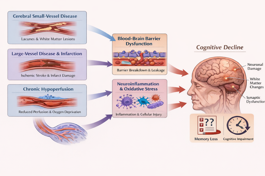
Fig 1. Pathophysiological changes in VCID

DETERMINANTS OF VCID

1. Hypertension

 Hypertension is a major preventable risk factor for VCID, as it induces sustained structural and functional damage to small and large cerebral blood  vessels. Several prospective cohort  and longitudinal studies indicate that hypertension is associated with a significant,  59 % increased risk of vascular dementia, compared with normotensives counterparts[26]. Persistent elevation of blood pressure promotes arteriolosclerosis and lipohyalinosis of small penetrating arteries, impair cerebral vessel autoregulation and decreases perfusion to subcortical and deeper white matter regions [27]. At the same time, hypertension accelerates atherosclerosis in the major cerebral arteries, heightening the incidence of cortical infarcts and the consequent stroke that impair cognition [28]. Hypertension induced mechanical stress on cerebral vessels disrupts endothelial signaling, causes impaired neurovascular coupling and  loss of BBB integrity, thereby accelerating progression across the VCID spectrum [29]

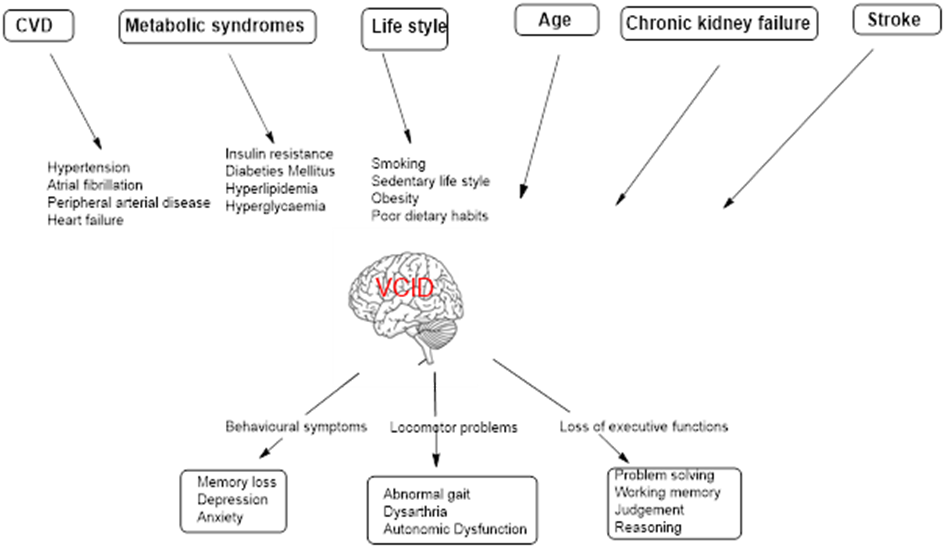
Fig 2. Manifestations of  VCID

2. Metabolic syndromes

Metabolic disorders including hyperlipidemia, insulin resistance and diabetes mellitus contribute to VCID through convergent vascular and cellular mechanisms. Hyperlipidemia accelerates atherosclerosis in large and medium cerebral arteries and promote lipid-mediated endothelial inflammation. Chronic hypoperfusion by the cerebral arteries increases the risk of cortical infarction. Elevated   circulating low-density lipoprotein (LDL), penetrates the arterial intima and undergoes oxidative changes, triggers endothelial dysfunction and inflammatory cell recruitment. Macrophage uptake of oxidized LDL leads to foam cell formation and fatty streak development, while sustained lipid-driven inflammation promotes plaque formation and progressive luminal narrowing [30].

Insulin resistance and diabetes mellitus expose cerebral microvessels to hyperglycemia, dyslipidemia, and oxidative stress. This carbohydrate and lipid mediated  injury stimulates excess extracellular matrix deposition, leading to basement membrane thickening in the endothelial cells and reduced capillary flexibility .Endothelial dysfunction further drive  capillary rarefaction and impaired NO mediated vasodilation [31].Advanced glycation end-product (AGEs) accumulate in cerebral blood vessels and activate RAGE (Receptors for  Advanced Glycation End products) mediated endothelial dysfunction [32]. These metabolic insults s amplify neuroinflammation, white-matter degeneration, and synaptic dysfunction in the cortical regions of brain, driving advancement from mild vascular cognitive deficits to established dementia [33].

3. Life Style

A sedentary lifestyle reduces skeletal muscle glucose uptake by decreasing insulin-stimulated GLUT4 translocation and mitochondrial  antioxidant defense, leading to inefficient glucose utilization and impaired mitochondrial bioenergetics  [34]. Physical inactivity also lowers muscle mass by decreasing muscle protein synthesis and activating proteolytic system. Concurrently, physical inactivity  promotes  lipid accumulation with in the skeletal muscle, generate lipotoxic intermediates such as DAG, ceramides, that activate serine kinases and  impair  insulin signaling  [35]. Obesity aggravates  insulin resistance by expanding adipose tissue, which releases excess free fatty acids into the circulation and drives  ectopic lipid accumulation  in liver and skeletal muscle [36]. These peripheral metabolic disturbances reduce  endothelial nitric oxide signaling [37], disrupt neurovascular coupling[38], and promote cerebral microvascular remodeling, culminating in chronic cerebral  hypoperfusion and BBB breakdown [39]. The resulting white-matter injury and synaptic energy failure weaken  cognitive pathways, thereby contributing towards VCID  continuum [40].

4. Age

Ageing promotes VCID through progressive structural and functional deterioration of the cerebral vasculature and neurovascular unit. With advancing age, cerebral arteries and arterioles undergo increased stiffness, collagen deposition, and loss of elastin, causing reduced vascular elasticity and impaired regulation of cerebral blood flow [41]. Age-related endothelial dysfunction diminishes nitric oxide bioavailability and further limits cerebral blood flow  during cognitive demand [42].Aging disrupts BBB integrity through oxidative stress, senescence of vascular cells, and neurovascular unit instability, leading to enhanced permeability of BBB.
Inflammatory ROS signaling promotes degradation of tight junction proteins such as occludin,.    Claudins and ZO-1, and consequent increase in  paracellular leakage. Simultaneously, pericyte loss and astrocyte end feet deattachment further weaken the BBB support
[43]. These vascular alterations promote chronic cerebral hypoperfusion, white-matter degeneration, and microinfarcts , which preferentially disrupt subcortical and cortical neural pathways, critical for cognition [44]. Together, age related vascular and neurovascular changes  contribute towards the advancement of VCID.

5. Chronic kidney failure

Chronic kidney failure (CKD) contributes to VCID through tightly linked vascular, metabolic, and inflammatory mechanisms.  Accumulation of uremic toxins in reduced renal clearance (e.g., indoxyl sulfate, p-cresyl sulfate)  induce systemic endothelial dysfunction  and impair cerebral autoregulation [45].  This disrupts tight junction proteins, increasing BBB permeability and allowing inflammatory mediators to enter the brain parenchyma. The ensuing microglial activation and neuroinflammatory signaling preferentially damage oligodendrocytes and axonal tracts. These processes accelerate white-matter injury and compromise subcortical neurons, promoting progression of  VCID [46]. Collectively, CKD driven vascular toxicity and systemic metabolic comorbidities converge to aggravate  cerebral small vessel dysfunction, BBB breakdown, and neuroinflammatory white-matter degeneration, thereby accelerating the trajectory of  VCID.

6. Stroke

Stroke induces VCID through an acute ischemic cascade of events followed by progressive cerebrovascular pathology. Diminished  cerebral blood flow  rapidly depletes ATP, disabling the energy-dependent Na?/K?-ATPase and collapsing transmembrane ion gradients. Loss of ionic homeostasis causes sustained membrane depolarization and reversal of glutamate transporters, while depolarization-induced vesicular release further elevates extracellular glutamate. Excess glutamate overactivates NMDA and AMPA receptors, driving pathological events [47]. In the peri ischemic region, partial energy failure activates mitochondrial apoptosis and death receptor signaling, resulting in delayed neuronal loss, specifically when lesions involve strategic cognitive regions such as the thalamus, frontal cortex, or limbic pathways. Long-term cognitive decline develops from the cumulative burden of recurrent cerebral infarction and progressive small vessel pathology. Together, these  excitotoxic and apoptotic processes, aggravate  vascular remodeling and disconnected neural pathways, ultimately drive  progressive evolution from poststroke cognitive impairment to established vascular dementia [48].

PREVENTIVE INTERVENTIONS FOR VASCULAR COGNITIVE IMPAIRMENT AND DEMENTIA

VCID might be prevented through two types of interventions. The first one is an early identification and medical treatment of cardiovascular conditions. An effective control of cardiovascular risk factors prevents VCID and may be more effective than therapeutic Intervetions [49]. Hypertension  is associated with  leukoaraiosis, or lesions in periventricular and subcortical white matter regions of brain [50]. Studies suggest that preserving ideal cardiovascular health in middle age contributes to better cognitive outcomes in older adulthood [51]. Another potentially important preventative intervention against VCID is to build neurocognitive reserve. The concept of neurocognitive reserve describes the ability to compensate age related brain pathology [52]. Neurocognitive reserve includes 1) Cerebral reserve related to structural differences in brain architechture in terms of neuronal and synaptic density  [53]; 2) Cognitive reserve is about differences in ability to make efficient use of existing brain resources when engaged in cognitive task [54];  3) Neural network maintenance  is the differential capacity to repair impaired  synapses, synaptogenesis, and neurogenesis [55]. Citicoline, an essential intermediate in the biosynthesis of structural phospholipids in cell membranes, has been reported to exhibit neuroprotective activity. Citicoline may provide neuroprotective benefits in VCID by enhancing phospholipid membrane repair, supporting cholinergic neurotransmission, and reducing oxidative stress. [56][57] ,  although  clinical benefits in ischemic stroke induced VCID, remains unexplored [58]. With growing insights into cognitive processes, boosting neurocognitive reserve through pharmacological strategies may emerge as a future therapeutic approach. Caution, however , needs to be taken, as the concept of neurocognition reserve implies both the chance to mitigate VCID by increasing the cerebral reserve and the danger of underestimating severity of cerebrovascular damage when just evaluating the VCID [52].

Collectively, evidence from observational studies, randomized controlled trials, and meta-analyses indicates that hypertension is a major modifiable driver of  VCID. Randomized evidence from trials including SPRINT-MIND and PRESERVE supports the cognitive relevance of intensive blood pressure lowering, even in older populations.
These interventions consistently slow white-matter hyperintensity (WMH) progression without demonstrating adverse effects on cerebral perfusion
[59]. More recent  analyses suggest that calcium channel blockers and angiotensin II receptor blockers may offer  additional protection against dementia beyond BP lowering alone [60]. Secondary analyses further indicate that angiotensin-converting enzyme inhibitors and calcium channel blockers may preferentially attenuate WMH progression [61]. Despite this, short term clinical investigations report no differential effects on cerebrovascular rreserve [62]. Taken together, the data suggest hypertension as a key modifiable determinant of VCID, while underscoring the need for adequately long duration clinical  trials to determine whether  antihypertensive agents provide cognitive protection beyond blood pressure lowering effects.

Beneficial effects of changing life styles and physical activity (diet, exercise, and environmental enrichment) on VCID and VaD have been reported [63][64]. In rats with 2-VO induced chronic cerebral hypoperfusion,  treadmill exercise for 4 weeks has been found to reduce cognitive decline by enhancing neurogenesis mediated by upregulation of BDNF expression [64]

Although elevated cholesterol has been implicated in cerebrovascular dysfunction and may contribute to VCID progression, the therapeutic effect of cholesterol reduction in VCID and VaD is  yet to be  determined. A recent Cochrane Database Review has indicated that statins do not prevent cognitive decline when given in late life to the high risk  people for vascular disease [65]. Clinically, antidiabetic therapy particularly metformin and long-term statins were able to demonstrate  a modest reduction in cognitive decline and dementia risk, especially among individuals with high vascular burden [66][67]. Clinical trials with improvement in cognition as the main outcome, show variable results, while VCID-specific randomized studies are comparatively lacking. [68].

CKD is increasingly recognized as a systemic vascular brain disorder, contributing to VCID. Large cohort studies show a graded increase in dementia risk with declining endothelial glomerular filtration rate (eGFR), independent of traditional vascular risk factors.[69]. ACE inhibitors and angiotensin receptor blockers (ARBs) mitigate Renin Angiotensin Aldosterone System (RAAS) driven endothelial dysfunction, microvascular injury, and resulting hypertension in CKD, which may moderately delay cognitive decline. In parallel, Sodium Glucose Cotransporter 2 (SGLT2 ) inhibitors provide renal protection by improving glomerular filtration rate, reducing albuminuria, and suppressing inflammatory and oxidative stress pathways, thereby delaying CKD advancement. Cardiovascular benefits of SGL2 and associated reductions in stroke risk may further  contribute to cognitive preservation [70].

Age is the dominant non-modifiable risk factor for VCID, acting through vascular stiffening, impaired autoregulation and  endothelial senescence induced decreased  nitric oxide signaling. Importantly, aging magnifies the deleterious vascular insults associated with  disorders  including stroke and CKD. Rapamycin and metformin has shown anti-aging neurovascular benefits in preclinical studies by improving cerebral perfusion, preservation of BBB integrity, and cognition [77]. Furthurmore, epidemiological evidence links metformin use with reduced cognitive decline and dementia risk, supporting potential translational relevance [70] [71][72]. Senolytics are the drugs that selectively eliminate senescent cells, that have permanently stopped dividing, and accumulated with age. Preclinical studies have demonstrated that targeting senescent cerebrovascular cells, improve BBB integrity, reduce neuroinflammation, and enhance cognitive performance in aged animal models highlighting potential vascular benefits of senolytic therapy in aging brain [73]. While preclinical evidence supports the neuroprotective role of senolytics, their therapeutic relevance in VCID remains untested in human trials. NAD? is Nicotinamide Adenine Dinucleotide (NAD?), is a vital cellular mitochondrial bioenergetics regulator. NAD? supplementation supports mitochondrial energetics and endothelial nitric oxide signaling in a rodent model of ageing,  thereby restoring neurovascular coupling and improving vasodilatory function. These vascular and mitochondrial  improvements are associated with better cognitive and motor coordination in preclinical aging models, although human cognitive evidence remains inconclusive [73] [74].

Stroke is the strongest clinical determinant of VCID with both symptomatic and silent infarcts causes irreversible neural network disconnection, chronic neuroinflammation, BBB integrity, and acceleration of cerebral small vessel disease. Experimental models provide evidence that ischemia initiates  neuroinflammation induced white-matter degenerative processes that drive later cognitive dysfunction [75]. In parallel, clinical data demonstrate that post-stroke VCID occurs in 30–50% of  stroke survivors, with risk amplified by recurrent strokes [76] . Supporting a preventative strategy, randomized evidence shows that antihypertensive and statin therapy can mitigate VCID development by limiting recurrent vascular injury and slowing cSVD progression. [77]. There is no FDA approved specific for post stroke VCID. Blood pressure management is the strongest pharmacological prevention strategy for stroke-induced VCID.A meta-analysis suggests that  memantidine is promising treatment for post stroke induced vascular cognitive impairment [78].

DISCUSSION

Overall, recent evidences supports risk modifying rather than disease modifying interventions with cognitive benefits, underscoring the need for VCID-focused trials. While preclinical evidence supports the neuroprotective role of senolytics, their therapeutic relevance in VCID remains unexplored in human clinical studies. Despite strong preclinical evidence for improved mitochondrial function with NAD? repletion, human cognitive benefits remain inconclusive and require further investigation. Hypertension promotes cerebrovascular injury through endothelial dysfunction and increases risk of clinical and covert stroke. Landmark clinical trials such as PROGRESS demonstrate that antihypertensive therapy reduces recurrent stroke and consequent cognitive decline, indicating that stroke prevention is a central mechanism by which blood pressure control mitigates vascular cognitive impairment. SPRINT-MIND study provides additional evidence that intensive blood pressure lowering lowers mild cognitive impairment risk, supporting the role of hypertension-driven neurovascular dysfunction as a key pathway linking stroke to dementia.

Blood pressure management represents one of the strongest modifiable strategies for preventing and slowing VCID progression. In PROGRESS, perindopril based therapy after stroke or transient ischemic attack reduced cognitive decline and dementia linked to recurrent cerebrovascular events, effective for  secondary prevention in high-risk populations [79]. Extending this evidence, SPRINT-MIND showed that intensive systolic BP control (<120 mmHg) significantly lowered mild cognitive impairment and effective for primary prevention in hypertensive adults, with sustained benefit confirmed in long-term follow-up analyses through seven years [59]. The China Rural Hypertension Control Project (CRHCP), a large-scale cluster-randomized trial involving over 30,000 participants, demonstrated that  blood pressure control  intervention significantly reduced the risk of dementia by 15% and cognitive impairment by 16% [80]. The SPRINT-MIND provided the proof  in a highly controlled environment, this Rural China study provided the "real-world relevance" in a general population with fewer medical resources. Mechanistic MRI substudies reinforce these clinical findings by showing attenuation of white-matter lesion progression and preservation of cerebral perfusion with intensive therapy, consistent with reduced small-vessel disease burden [59][81]. Collective real world evidences supports early and stable BP control as a cornerstone intervention for reducing VCID and vascular dementia [82].

CONCLUSION

VCID considerably impacts social life by impairing executive functions such as planning and communication, leads to social withdrawal and loss of independence. Primary risk factors are vascular including hypertension and stroke. Preventive strategies for VCID focuses on “what is good for heart is good for brain”, specifically intensive blood pressure control and regular exercise.There is an urgent need for clinical trial to approve new drugs for VCID.  

REFERENCES

  1. Dementia [Internet]. [cited 2026 Feb 5]. Available from: https://www.who.int/news-room/fact-sheets/detail/dementia
  2. Rodriguez JJL, Ferri CP, Acosta D, Guerra M, Huang Y, Jacob KS, et al. Prevalence of dementia in Latin America, India, and China: a population-based cross-sectional survey. Lancet [Internet]. 2008 [cited 2026 Feb 5];372(9637):464–74. Available from: https://pubmed.ncbi.nlm.nih.gov/18657855/
  3. Stephan BC, Matthews FE, Khaw KT, Dufouil C, Brayne C. Beyond mild cognitive impairment: vascular cognitive impairment, no dementia (VCIND). Alzheimers Res Ther [Internet]. 2009 [cited 2026 Feb 5];1(1):4. Available from: https://pubmed.ncbi.nlm.nih.gov/19674437/
  4. Vahia VN. Diagnostic and statistical manual of mental disorders 5: A quick glance. Indian J Psychiatry [Internet]. 2013 Jul [cited 2026 Feb 5];55(3):220. Available from: https://pmc.ncbi.nlm.nih.gov/articles/PMC3777342/
  5. ICD-11 for Mortality and Morbidity Statistics [Internet]. [cited 2026 Feb 5]. Available from: https://icd.who.int/browse/2025-01/mms/en#1365258270
  6. Kalaria RN. Neuropathological diagnosis of vascular cognitive impairment and vascular dementia with implications for Alzheimer’s disease. Acta Neuropathol [Internet]. 2016 May 1 [cited 2026 Feb 5];131(5):659–85. Available from: https://pubmed.ncbi.nlm.nih.gov/27062261/
  7. Skrobot OA, Black SE, Chen C, DeCarli C, Erkinjuntti T, Ford GA, et al. Progress toward standardized diagnosis of vascular cognitive impairment: Guidelines from the Vascular Impairment of Cognition Classification Consensus Study. Alzheimer’s Dement [Internet]. 2018 Mar 1 [cited 2026 Feb 5];14(3):280–92. Available from: /doi/pdf/10.1016/j.jalz.2017.09.007
  8. Pantoni L. Cerebral small vessel disease: from pathogenesis and clinical characteristics to therapeutic challenges. Lancet Neurol [Internet]. 2010 Jul [cited 2026 Jan 20];9(7):689–701. Available from: https://pubmed.ncbi.nlm.nih.gov/20610345/
  9. Iadecola C. The pathobiology of vascular dementia. Neuron [Internet]. 2013 Nov 20 [cited 2026 Feb 5];80(4):844–66. Available from: https://pubmed.ncbi.nlm.nih.gov/24267647/
  10. Hachinski VC, Iliff LD, Zilhka E, Boulay GH, McAllister VL, Marshall J, et al. Cerebral blood flow in dementia. Arch Neurol [Internet]. 1975 [cited 2026 Feb 5];32(9):632–7. Available from: https://pubmed.ncbi.nlm.nih.gov/1164215/
  11. O’Brien JT, Thomas A. Vascular dementia. Lancet (London, England) [Internet]. 2015 Oct 24 [cited 2026 Feb 5];386(10004):1698–706. Available from: https://pubmed.ncbi.nlm.nih.gov/26595643/
  12. Gorelick PB, Scuteri A, Black SE, Decarli C, Greenberg SM, Iadecola C, et al. Vascular contributions to cognitive impairment and dementia: a statement for healthcare professionals from the american heart association/american stroke association. Stroke [Internet]. 2011 Sep [cited 2026 Feb 8];42(9):2672–713. Available from: https://pubmed.ncbi.nlm.nih.gov/21778438/
  13. Román GC. Vascular dementia revisited: Diagnosis, pathogenesis, treatment, and prevention. Med Clin North Am [Internet]. 2002 [cited 2026 Feb 8];86(3):477–99. Available from: https://pubmed.ncbi.nlm.nih.gov/12168556/
  14. Stephan BC, Matthews FE, Khaw KT, Dufouil C, Brayne C. Beyond mild cognitive impairment: vascular cognitive impairment, no dementia (VCIND). Alzheimers Res Ther [Internet]. 2009 [cited 2026 Feb 8];1(1):4. Available from: https://pubmed.ncbi.nlm.nih.gov/19674437/
  15. Zlokovic B V. Neurovascular pathways to neurodegeneration in Alzheimer’s disease and other disorders. Nat Rev Neurosci [Internet]. 2011 Dec [cited 2026 Feb 8];12(12). Available from: https://pubmed.ncbi.nlm.nih.gov/22048062/
  16. Alafuzoff I, Gelpi E, Al-Sarraj S, Arzberger T, Attems J, Bodi I, et al. The need to unify neuropathological assessments of vascular alterations in the ageing brain. Multicentre survey by the BrainNet Europe consortium. Exp Gerontol [Internet]. 2012 Nov [cited 2026 Feb 8];47(11):825–33. Available from: https://pubmed.ncbi.nlm.nih.gov/22705312/
  17. Kitamura A, Saito S, Maki T, Oishi N, Ayaki T, Hattori Y, et al. Gradual cerebral hypoperfusion in spontaneously hypertensive rats induces slowly evolving white matter abnormalities and impairs working memory. J Cereb Blood Flow Metab [Internet]. 2016 Sep 1 [cited 2026 Feb 8];36(9):1592–602. Available from: https://pubmed.ncbi.nlm.nih.gov/26661170/
  18. Montagne A. Blood?Brain Barrier Dynamics in Vascular Dementia: Unravelling the Tripartite Crosstalk between the Endothelium, Pericytes, and Microglia. Alzheimer’s Dement [Internet]. 2025 Dec [cited 2026 Feb 8];20(Suppl 1):e087126. Available from: https://pmc.ncbi.nlm.nih.gov/articles/PMC11709832/
  19. Sharma C, Woo H, Kim SR. Addressing Blood-Brain Barrier Impairment in Alzheimer’s Disease. Biomedicines [Internet]. 2022 Apr 1 [cited 2026 Feb 8];10(4). Available from: https://pubmed.ncbi.nlm.nih.gov/35453494/
  20. Sweeney MD, Sagare AP, Zlokovic B V. Blood-brain barrier breakdown in Alzheimer disease and other neurodegenerative disorders. Nat Rev Neurol [Internet]. 2018 Mar 1 [cited 2026 Feb 8];14(3):133–50. Available from: https://pubmed.ncbi.nlm.nih.gov/29377008/
  21. Hosoki S, Hansra GK, Jayasena T, Poljak A, Mather KA, Catts VS, et al. Molecular biomarkers for vascular cognitive impairment and dementia. Nat Rev Neurol [Internet]. 2023 Dec 1 [cited 2026 Feb 8];19(12):737–53. Available from: https://research.monash.edu/en/publications/molecular-biomarkers-for-vascular-cognitive-impairment-and-dement/
  22. Zhang Y, Chu JMT, Wong GTC. Cerebral Glutamate Regulation and Receptor Changes in Perioperative Neuroinflammation and Cognitive Dysfunction. Biomolecules [Internet]. 2022 Apr 1 [cited 2026 Feb 8];12(4). Available from: https://pubmed.ncbi.nlm.nih.gov/35454185/
  23. Reis PA, Castro-Faria-Neto HC. Systemic Response to Infection Induces Long-Term Cognitive Decline: Neuroinflammation and Oxidative Stress as Therapeutical Targets. Front Neurosci [Internet]. 2022 Feb 18 [cited 2026 Feb 8];15. Available from: https://pubmed.ncbi.nlm.nih.gov/35250433/
  24. Aquilani R, Cotta Ramusino M, Maestri R, Iadarola P, Boselli M, Perini G, et al. Several dementia subtypes and mild cognitive impairment share brain reduction of neurotransmitter precursor amino acids, impaired energy metabolism, and lipid hyperoxidation. Front Aging Neurosci [Internet]. 2023 [cited 2026 Feb 8];15. Available from: https://pubmed.ncbi.nlm.nih.gov/37655338/
  25. Pansieri J, Hadley G, Lockhart A, Pisa M, DeLuca GC. Regional contribution of vascular dysfunction in white matter dementia: clinical and neuropathological insights. Front Neurol [Internet]. 2023 [cited 2026 Feb 8];14. Available from: https://pubmed.ncbi.nlm.nih.gov/37396778/
  26. Sharp SI, Aarsland D, Day S, Sønnesyn H, Ballard C. Hypertension is a potential risk factor for vascular dementia: systematic review. Int J Geriatr Psychiatry [Internet]. 2011 Jul [cited 2026 Feb 9];26(7):661–9. Available from: https://pubmed.ncbi.nlm.nih.gov/21495075/
  27. Wardlaw JM, Smith C, Dichgans M. Small vessel disease: mechanisms and clinical implications. Lancet Neurol [Internet]. 2019 Jul 1 [cited 2026 Feb 9];18(7):684–96. Available from: https://pubmed.ncbi.nlm.nih.gov/31097385/
  28. Seshadri S, Wolf PA. Lifetime risk of stroke and dementia: current concepts, and estimates from the Framingham Study. Lancet Neurol [Internet]. 2007 [cited 2026 Feb 9];6(12):1106–14. Available from: https://pubmed.ncbi.nlm.nih.gov/18031707/
  29. Qiu C, Winblad B, Fratiglioni L. The age-dependent relation of blood pressure to cognitive function and dementia. Lancet Neurol [Internet]. 2005 Aug [cited 2026 Feb 9];4(8):487–99. Available from: https://pubmed.ncbi.nlm.nih.gov/16033691/
  30. Smith EE, Aparicio HJ, Gottesman RF, Goyal MS, Greenberg SM, Schneider JA, et al. Vascular Contributions to Cognitive Impairment and Dementia in the United States: Prevalence and Incidence: A Scientific Statement From the American Heart Association. Stroke [Internet]. 2025 [cited 2026 Jan 28];56(10):e317–30. Available from: /doi/pdf/10.1161/STR.0000000000000494?download=true
  31. Lecordier S, Manrique-Castano D, El Moghrabi Y, ElAli A. Neurovascular Alterations in Vascular Dementia: Emphasis on Risk Factors. Front Aging Neurosci [Internet]. 2021 Sep 10 [cited 2026 Jan 28];13:727590. Available from: www.frontiersin.org
  32. Biessels GJ, Despa F. Cognitive decline and dementia in diabetes mellitus: mechanisms and clinical implications. Nat Rev Endocrinol 2018 1410 [Internet]. 2018 Jul 18 [cited 2026 Jan 28];14(10):591–604. Available from: https://www.nature.com/articles/s41574-018-0048-7
  33. Kennelly SP, Lawlor BA, Kenny RA. Blood pressure and the risk for dementia: a double edged sword. Ageing Res Rev [Internet]. 2009 Apr [cited 2026 Jan 28];8(2):61–70. Available from: https://pubmed.ncbi.nlm.nih.gov/19063999/
  34. Booth FW, Roberts CK, Laye MJ. Lack of exercise is a major cause of chronic diseases. Wiley Online Libr Booth, CK Roberts, MJ LayeComprehensive Physiol 2011•Wiley Online Libr [Internet]. 2012 Apr [cited 2026 Jan 29];2(2):1143–211. Available from: https://onlinelibrary.wiley.com/doi/abs/10.1002/cphy.c110025
  35. Houmard JA, Tanner CJ, Slentz CA, Duscha BD, McCartney JS, Kraus WE. Effect of the volume and intensity of exercise training on insulin sensitivity. journals.physiology.orgJA Houmard, CJ Tann CA Slentz, BD Duscha, JS McCartney, WE KrausJournal Appl Physiol 2004•journals.physiology.org [Internet]. 2004 Jan [cited 2026 Jan 29];96(1):101–6. Available from: https://journals.physiology.org/doi/abs/10.1152/japplphysiol.00707.2003
  36. Kahn S, Hull R, Nature KU, 2006 U. Mechanisms linking obesity to insulin resistance and type 2 diabetes. nature.comSE Kahn, RL Hull, KM UtzschneiderNature, 2006•nature.com [Internet]. 2006 [cited 2026 Jan 29];444(7121):840–6. Available from: https://www.nature.com/articles/nature05482
  37. Muniyappa R, Iantorno M, Quon MJ. An Integrated View of Insulin Resistance and Endothelial Dysfunction. Endocrinol Metab Clin North Am [Internet]. 2008 Sep [cited 2026 Jan 29];37(3):685. Available from: https://pmc.ncbi.nlm.nih.gov/articles/PMC2613319/
  38. Lecordier S, Manrique-Castano D, El Moghrabi Y, ElAli A. Neurovascular Alterations in Vascular Dementia: Emphasis on Risk Factors. Front Aging Neurosci [Internet]. 2021 Sep 10 [cited 2026 Jan 29];13:727590. Available from: www.frontiersin.org
  39. Coucha M, Abdelsaid M, Ward R, Abdul Y, Ergul A. Impact of Metabolic Diseases on Cerebral Circulation: Structural and Functional Consequences. Compr Physiol [Internet]. 2018 Apr 1 [cited 2026 Jan 29];8(2):773. Available from: https://pmc.ncbi.nlm.nih.gov/articles/PMC6586018/
  40. Muniyappa R, Sowers JR. Role of Insulin Resistance in Endothelial Dysfunction. Rev Endocr Metab Disord [Internet]. 2013 Mar [cited 2026 Feb 9];14(1):5. Available from: https://pmc.ncbi.nlm.nih.gov/articles/PMC3594115/
  41. Mitchell G, Buchem M Van, Sigurdsson S, Brain JG, 2011 U. Arterial stiffness, pressure and flow pulsatility and brain structure and function: the Age, Gene/Environment Susceptibility–Reykjavik study. Acad Mitchell, MA Van Buchem, S Sigurdsson, JD Gotal, MK Jonsdottir, Ó KjartanssonBrain, 2011•academic.oup.com [Internet]. 2011 [cited 2026 Jan 29];134(11):3398–407. Available from: https://academic.oup.com/brain/article-abstract/134/11/3398/314574
  42. Lipecz A, Csipo T, Tarantini S, Hand RA, Ngo BTN, Conley S, et al. Age-related impairment of neurovascular coupling responses: a dynamic vessel analysis (DVA)-based approach to measure decreased flicker light stimulus-induced retinal arteriolar dilation in healthy older adults. GeroScience [Internet]. 2019 Jun 15 [cited 2026 Jan 29];41(3):341. Available from: https://pmc.ncbi.nlm.nih.gov/articles/PMC6702523/
  43. Erdö F, Denes L, De Lange E. Age-associated physiological and pathological changes at the blood-brain barrier: A review. J Cereb Blood Flow Metab [Internet]. 2017 Jan 1 [cited 2026 Jan 29];37(1):4–24. Available from: https://scholar.google.com/scholar_url?url=https://journals.sagepub.com/doi/pdf/10.1177/0271678X16679420&hl=en&sa=T&oi=ucasa&ct=ufr&ei=0x97afb8K9DQ6rQPhK7SsAk&scisig=AHkA5jSu3ae00w-XYvglDZEsOqVN
  44. Hase Y, Horsburgh K, Ihara M, Kalaria RN. White matter degeneration in vascular and other ageing-related dementias. J Neurochem [Internet]. 2018 Mar 1 [cited 2026 Jan 29];144(5):617–33. Available from: /doi/pdf/10.1111/jnc.14271
  45. Lau W, Kalantar-Zadeh K, Nephron NV, 2015  undefined. The gut as a source of inflammation in chronic kidney disease. karger.comWL Lau, K Kalantar-Zadeh, ND VaziriNephron, 2015•karger.com [Internet]. [cited 2026 Jan 29]; Available from: https://karger.com/nef/article/130/2/92/211660
  46. Jing W, Jabbari B, Vaziri ND. Uremia induces upregulation of cerebral tissue oxidative/inflammatory cascade, down-regulation of Nrf2 pathway and disruption of blood brain barrier. Am J Transl Res [Internet]. 2018 [cited 2026 Jan 29];10(7):2137. Available from: https://pmc.ncbi.nlm.nih.gov/articles/PMC6079125/
  47. Rodríguez De Lores Arnaiz G, Graciela M, Ordieres L, De Biología Celular I, De Robertis NE. Brain Na+, K+-ATPase Activity In Aging and Disease. Int J Biomed Sci [Internet]. 2014 Jun 15 [cited 2026 Jan 30];10(2):85. Available from: https://pmc.ncbi.nlm.nih.gov/articles/PMC4092085/
  48. Lo EH, Dalkara T, Moskowitz MA. Mechanisms, challenges and opportunities in stroke. Nat Rev Neurosci [Internet]. 2003 [cited 2026 Jan 30];4(5):399–414. Available from: https://pubmed.ncbi.nlm.nih.gov/12728267/
  49. Rouch L, Cestac P, Hanon O, Cool C, Helmer C, Bouhanick B, et al. Antihypertensive drugs, prevention of cognitive decline and dementia: a systematic review of observational studies, randomized controlled trials and meta-analyses. SpringerL Rouch, P Cestac, O Hanon, C Cool C Helmer, B Bouhanick, B Chamontin, JF DartiguesCNS drugs, 2015•Springer [Internet]. 2015 [cited 2026 Jan 31];29(2):113–30. Available from: https://link.springer.com/article/10.1007/s40263-015-0230-6
  50. Sharp SI, Aarsland D, Day S, Sønnesyn H, Ballard C. Hypertension is a potential risk factor for vascular dementia: systematic review. Wiley Online Libr Sharp, D Aarsland, S Day, H SønnesynInternational J Geriatr psychiatry, 2011•Wiley Online Libr [Internet]. 2011 Jul [cited 2026 Jan 31];26(7):661–9. Available from: https://onlinelibrary.wiley.com/doi/abs/10.1002/gps.2572
  51. Reis JP, Loria CM, Launer LJ, Sidney S, Liu K, Jacobs DR, et al. Cardiovascular health through young adulthood and cognitive functioning in midlife. Wiley Online Libr Reis, C Loria, LJ Launer, S Sidney, K Liu, DR Jacobs Jr, N Zhu, DM Lloyd?Jones, K HeAnnals Neurol 2013•Wiley Online Libr [Internet]. 2013 Feb [cited 2026 Jan 31];73(2):170–9. Available from: https://onlinelibrary.wiley.com/doi/abs/10.1002/ana.23836
  52. Pinter D, Enzinger C, Neurology FFJ of, 2015 U. Cerebral small vessel disease, cognitive reserve and cognitive dysfunction. SpringerD Pinter, C Enzinger, F FazekasJournal Neurol 2015•Springer [Internet]. 2015 [cited 2026 Jan 31];262(11):2411–9. Available from: https://link.springer.com/article/10.1007/s00415-015-7776-6
  53. psychiatry MVC opinion in, 2008 U. Brain reserve and the prevention of dementia. journals.lww.comMJ Val Opin psychiatry, 2008•journals.lww.com [Internet]. 2008 [cited 2026 Jan 31];21(3):296–302. Available from: https://journals.lww.com/co-psychiatry/FullText/2008/05000/Brain_reserve_and_the_prevention_of_dementia.14.aspx
  54. Whalley LJ, Staff RT, Fox HC, Murray AD. Cerebral correlates of cognitive reserve. Elsevier [Internet]. 2016 Jan 30 [cited 2026 Jan 31];247:65–70. Available from: https://www.sciencedirect.com/science/article/pii/S092549271530127X
  55. Nyberg L, Lövdén M, Riklund K, … ULT in cognitive, 2012 U. Memory aging and brain maintenance. cell.comL Nyberg, M Lövdén, K Riklund, U Lindenberger, L BäckmanTrends Cogn Sci 2012•cell.com [Internet]. 2012 [cited 2026 Jan 31];16(5):292–305. Available from: https://www.cell.com/trends/cognitive-sciences/fulltext/S1364-6613(12)00083-6?large_figure=true
  56. Gareri P, Castagna A, Cotroneo AM, Putignano S, De Sarro G, Bruni AC. The role of citicoline in cognitive impairment: pharmacological characteristics, possible advantages, and doubts for an old drug with new perspectives. Taylor Fr Gareri, A Castagna, AM Cotroneo, S Putignano, G Sarro, AC BruniClinical Interv aging, 2015•Taylor Fr [Internet]. 2015 Sep 3 [cited 2026 Feb 3];10:1421–9. Available from: https://www.tandfonline.com/doi/abs/10.2147/CIA.S87886
  57. Alvarez-Sabín J, Ortega G, Jacas C, Santamarina E, Maisterra O, Ribo M, et al. Long-term treatment with citicoline may improve poststroke vascular cognitive impairment. karger.comJ Alvarez-Sabín, G Ortega, C Jacas, E Santamarina, O Maisterra, M Ribo, C MolinaCerebrovascular Dis 2013•karger.com [Internet]. 2013 [cited 2026 Feb 3];35:146–54. Available from: https://karger.com/ced/article-abstract/35/2/146/77643
  58. drugs PGC, 2014  undefined. Neuroprotective properties of citicoline: facts, doubts and unresolved issues. SpringerP GriebCNS drugs, 2014•Springer [Internet]. 2014 Mar [cited 2026 Feb 3];28(3):185–93. Available from: https://link.springer.com/article/10.1007/s40263-014-0144-8
  59. Williamson JD, Pajewski NM, Auchus AP, Bryan RN, Chelune G, Cheung AK, et al. Effect of Intensive vs Standard Blood Pressure Control on Probable Dementia: A Randomized Clinical Trial. JAMA [Internet]. 2019 Feb 12 [cited 2026 Feb 4];321(6):553–61. Available from: https://pubmed.ncbi.nlm.nih.gov/30688979/
  60. den Brok MGHE, van Dalen JW, Abdulrahman H, Larson EB, van Middelaar T, van Gool WA, et al. Antihypertensive Medication Classes and the Risk of Dementia: A Systematic Review and Network Meta-Analysis. J Am Med Dir Assoc. 2021 Jul 1;22(7):1386-1395.e15.
  61. Goldstein ED, Wolcott Z, Garg G, Navarro K, Delic A, Yaghi S, et al. Effect of Antihypertensives by Class on Cerebral Small Vessel Disease: A Post Hoc Analysis of SPRINT-MIND. Stroke. 2022 Aug 1;53(8):2435–40.
  62. Kopczak A, Stringer MS, van den Brink H, Kerkhofs D, Blair GW, van Dinther M, et al. Effect of blood pressure-lowering agents on microvascular function in people with small vessel diseases (TREAT-SVDs): a multicentre, open-label, randomised, crossover trial. Lancet Neurol. 2023 Nov 1;22(11):991–1004.
  63. Bayat M, Sharifi MD, Haghani M, Shabani M. An enriched environment elevates corticosteroid receptor levels in the hippocampus and restores cognitive function in a rat model of chronic cerebral hypoperfusion. Elsevier [Internet]. 2015 Oct 1 [cited 2026 Feb 3];119:34–40. Available from: https://www.sciencedirect.com/science/article/pii/S0091305712003577
  64. Bayat M, Sharifi M, Haghani M, Bulletin MSB research, 2015 U. Enriched environment improves synaptic plasticity and cognitive deficiency in chronic cerebral hypoperfused rats. Elsevier [Internet]. 2015 [cited 2026 Feb 3];119:34–40. Available from: https://www.sciencedirect.com/science/article/pii/S0361923015300411
  65. McGuinness B, Craig D, Bullock R, Passmore P. Statins for the prevention of dementia. cochranelibrary.comB McGuinness, D Craig, R Bullock, P PassmoreCochrane database Syst Rev 2009•cochranelibrary.com [Internet]. 2009 [cited 2026 Feb 3];(2). Available from: https://www.cochranelibrary.com/cdsr/doi/10.1002/14651858.CD003160.pub2/abstract
  66. Imfeld P, Bodmer M, Jick SS, Meier CR. Metformin, other antidiabetic drugs, and risk of Alzheimer’s disease: A population-based case-control study. J Am Geriatr Soc [Internet]. 2012 May 1 [cited 2026 Feb 3];60(5):916–21. Available from: /doi/pdf/10.1111/j.1532-5415.2012.03916.x
  67. Westphal Filho FL, Moss Lopes PR, Menegaz de Almeida A, Sano VKT, Tamashiro FM, Gonçalves OR, et al. Statin use and dementia risk: A systematic review and updated meta?analysis. Alzheimer’s Dement  Transl Res Clin Interv [Internet]. 2025 Jan 1 [cited 2026 Feb 3];11(1):e70039. Available from: https://pmc.ncbi.nlm.nih.gov/articles/PMC11736423/
  68. Linh TTD, Hsieh YC, Huang LK, Hu CJ. Clinical Trials of New Drugs for Vascular Cognitive Impairment and Vascular Dementia. Int J Mol Sci [Internet]. 2022 Oct 1 [cited 2026 Feb 3];23(19):11067. Available from: https://pmc.ncbi.nlm.nih.gov/articles/PMC9569827/
  69. Bugnicourt JM, Godefroy O, Chillon JM, Choukroun G, Massy ZA. Cognitive disorders and dementia in CKD: The neglected kidney-brain axis. J Am Soc Nephrol [Internet]. 2013 Feb 28 [cited 2026 Feb 3];24(3):353–63. Available from: https://journals.lww.com/jasn/fulltext/2013/03000/cognitive_disorders_and_dementia_in_ckd__the.7.aspx
  70. Subhani M, Sabani A, Omer O, Kheirelsid F, Cureus AS, 2025 U. Impact of Renin-Angiotensin-Aldosterone System Modulation on Cognitive and Neuropsychiatric Outcomes: A Systematic Review of Clinical and Mechanistic. cureus.comMK Subhani, A Sabani, O Omer, F Kheirelsid, AK Srour, O Omer Sr, FE KheirelsidCureus, 2025•cureus.com [Internet]. 2025 [cited 2026 Feb 3];17(7). Available from: https://www.cureus.com/articles/376096-impact-of-renin-angiotensin-aldosterone-system-modulation-on-cognitive-and-neuropsychiatric-outcomes-a-systematic-review-of-clinical-and-mechanistic-evidence.pdf
  71. Kodali M, Attaluri S, Madhu LN, Shuai B, Upadhya R, Gonzalez JJ, et al. Metformin treatment in late middle age improves cognitive function with alleviation of microglial activation and enhancement of autophagy in the hippocampus. Aging Cell [Internet]. 2021 Feb 1 [cited 2026 Feb 3];20(2). Available from: https://pubmed.ncbi.nlm.nih.gov/33443781/
  72. González Pérez N, Bellotto M, Porte Alcón S, Bentivegna M, Arcucci L, Vinuesa Á, et al. Metformin protects against insulin resistance-related dementia risk involving restored microglial homeostasis. Life Sci [Internet]. 2025 Oct 1 [cited 2026 Feb 3];378. Available from: https://pubmed.ncbi.nlm.nih.gov/40494433/
  73. Saliev T, Singh PB. Senotherapeutics for Brain Aging Management. Neurol Int 2025, Vol 17, [Internet]. 2025 Dec 15 [cited 2026 Feb 3];17(12). Available from: https://www.mdpi.com/2035-8377/17/12/204
  74. Szu J. Do NAD+ Boosters Work? What the Research Says About NR and NMN for Aging, Cognition & Muscle. gethealthspan.comJ Szugethealthspan.com [Internet]. [cited 2026 Feb 3]; Available from: https://www.gethealthspan.com/research/article/nad-boosters
  75. Iadecola C. The Pathobiology of Vascular Dementia. Neuron [Internet]. 2013 Nov 20 [cited 2026 Feb 14];80(4):844–66. Available from: https://pubmed.ncbi.nlm.nih.gov/24267647/
  76. Rost NS, Brodtmann A, Pase MP, van Veluw SJ, Biffi A, Duering M, et al. Post-stroke cognitive impairment and dementia. Circ Res [Internet]. 2022 Apr 15 [cited 2026 Feb 14];130(8):1252–71. Available from: /doi/pdf/10.1161/CIRCRESAHA.122.319951?download=true
  77. Pendlebury ST, Rothwell PM. Incidence and prevalence of dementia associated with transient ischaemic attack and stroke: analysis of the population-based Oxford Vascular Study. Lancet Neurol [Internet]. 2019 [cited 2026 Feb 4];18(3):248–58. Available from: https://www.thelancet.com/journals/laneur/article/PIIS1474-4422(18)30442-3/fulltext?uuid=uuid%3Ab5b14d8e-63fd-4971-b53d-3723075a70f6
  78. Li W, Liu X, Gao C, Li W, Liao X. Network meta-analysis of the efficacy of pharmacological treatments for post-stroke cognitive impairment and vascular cognitive impairment. Front Neurol [Internet]. 2025 Dec 1 [cited 2026 Feb 14];16:1683496. Available from: https://www.crd.york.ac.uk/PROSPERO/,
  79. MacMahon S, Neal B, Tzourio C, Rodgers A, Woodward M, Cutler J, et al. Randomised trial of a perindopril-based blood-pressure-lowering regimen among 6,105 individuals with previous stroke or transient ischaemic attack. Lancet (London, England) [Internet]. 2001 Sep 29 [cited 2026 Feb 14];358(9287):1033–41. Available from: https://pubmed.ncbi.nlm.nih.gov/11589932/
  80. Sun Y, Mu J, Wang DW, Ouyang N, Xing L, Guo X, et al. A village doctor-led multifaceted intervention for blood pressure control in rural China: an open, cluster randomised trial. Lancet [Internet]. 2022 May 21 [cited 2026 Feb 14];399(10339):1964–75. Available from: https://pubmed.ncbi.nlm.nih.gov/35500594/
  81. Dolui S, Detre JA, Gaussoin SA, Herrick JS, Wang DJJ, Tamura MK, et al. Association of Intensive vs Standard Blood Pressure Control With Cerebral Blood Flow: Secondary Analysis of the SPRINT MIND Randomized Clinical Trial. JAMA Neurol [Internet]. 2022 Apr 1 [cited 2026 Feb 14];79(4):380–9. Available from: https://pubmed.ncbi.nlm.nih.gov/35254390/
  82. Hakim AM. Small Vessel Disease. Front Neurol [Internet]. 2019 Sep 1 [cited 2026 Feb 12];10(SEP). Available from: https://pubmed.ncbi.nlm.nih.gov/31616367/
  83. Williamson JD, Pajewski NM, Auchus AP, Bryan RN, Chelune G, Cheung AK, et al. Effect of Intensive vs Standard Blood Pressure Control on Probable Dementia: A Randomized Clinical Trial. JAMA - J Am Med Assoc. 2019 Feb 12;321(6):553–61.
  84. Tarantini S, Valcarcel-Ares MN, Toth P, Yabluchanskiy A, Kiss T, Ballabh P, et al. Nicotinamide mononucleotide (NMN) supplementation rescues cerebromicrovascular endothelial function and neurovascular coupling responses and improves. Wiley Online Libr [Internet]. 2020 Apr [cited 2026 Feb 3];34(S1):1–1. Available from: https://faseb.onlinelibrary.wiley.com/doi/abs/10.1096/fasebj.2020.34.s1.05763
  85. Abdellatif M, Bugger H, … GKJ of L and, 2022  undefined. NAD+ and vascular dysfunction: from mechanisms to therapeutic opportunities. pmc.ncbi.nlm.nih.govM Abdellatif, H Bugger, G Kroemer, S SedejJournal Lipid Atheroscler 2022•pmc.ncbi.nlm.nih.gov [Internet]. [cited 2026 Feb 12]; Available from: https://pmc.ncbi.nlm.nih.gov/articles/PMC9133775/

Reference

  1. Dementia [Internet]. [cited 2026 Feb 5]. Available from: https://www.who.int/news-room/fact-sheets/detail/dementia
  2. Rodriguez JJL, Ferri CP, Acosta D, Guerra M, Huang Y, Jacob KS, et al. Prevalence of dementia in Latin America, India, and China: a population-based cross-sectional survey. Lancet [Internet]. 2008 [cited 2026 Feb 5];372(9637):464–74. Available from: https://pubmed.ncbi.nlm.nih.gov/18657855/
  3. Stephan BC, Matthews FE, Khaw KT, Dufouil C, Brayne C. Beyond mild cognitive impairment: vascular cognitive impairment, no dementia (VCIND). Alzheimers Res Ther [Internet]. 2009 [cited 2026 Feb 5];1(1):4. Available from: https://pubmed.ncbi.nlm.nih.gov/19674437/
  4. Vahia VN. Diagnostic and statistical manual of mental disorders 5: A quick glance. Indian J Psychiatry [Internet]. 2013 Jul [cited 2026 Feb 5];55(3):220. Available from: https://pmc.ncbi.nlm.nih.gov/articles/PMC3777342/
  5. ICD-11 for Mortality and Morbidity Statistics [Internet]. [cited 2026 Feb 5]. Available from: https://icd.who.int/browse/2025-01/mms/en#1365258270
  6. Kalaria RN. Neuropathological diagnosis of vascular cognitive impairment and vascular dementia with implications for Alzheimer’s disease. Acta Neuropathol [Internet]. 2016 May 1 [cited 2026 Feb 5];131(5):659–85. Available from: https://pubmed.ncbi.nlm.nih.gov/27062261/
  7. Skrobot OA, Black SE, Chen C, DeCarli C, Erkinjuntti T, Ford GA, et al. Progress toward standardized diagnosis of vascular cognitive impairment: Guidelines from the Vascular Impairment of Cognition Classification Consensus Study. Alzheimer’s Dement [Internet]. 2018 Mar 1 [cited 2026 Feb 5];14(3):280–92. Available from: /doi/pdf/10.1016/j.jalz.2017.09.007
  8. Pantoni L. Cerebral small vessel disease: from pathogenesis and clinical characteristics to therapeutic challenges. Lancet Neurol [Internet]. 2010 Jul [cited 2026 Jan 20];9(7):689–701. Available from: https://pubmed.ncbi.nlm.nih.gov/20610345/
  9. Iadecola C. The pathobiology of vascular dementia. Neuron [Internet]. 2013 Nov 20 [cited 2026 Feb 5];80(4):844–66. Available from: https://pubmed.ncbi.nlm.nih.gov/24267647/
  10. Hachinski VC, Iliff LD, Zilhka E, Boulay GH, McAllister VL, Marshall J, et al. Cerebral blood flow in dementia. Arch Neurol [Internet]. 1975 [cited 2026 Feb 5];32(9):632–7. Available from: https://pubmed.ncbi.nlm.nih.gov/1164215/
  11. O’Brien JT, Thomas A. Vascular dementia. Lancet (London, England) [Internet]. 2015 Oct 24 [cited 2026 Feb 5];386(10004):1698–706. Available from: https://pubmed.ncbi.nlm.nih.gov/26595643/
  12. Gorelick PB, Scuteri A, Black SE, Decarli C, Greenberg SM, Iadecola C, et al. Vascular contributions to cognitive impairment and dementia: a statement for healthcare professionals from the american heart association/american stroke association. Stroke [Internet]. 2011 Sep [cited 2026 Feb 8];42(9):2672–713. Available from: https://pubmed.ncbi.nlm.nih.gov/21778438/
  13. Román GC. Vascular dementia revisited: Diagnosis, pathogenesis, treatment, and prevention. Med Clin North Am [Internet]. 2002 [cited 2026 Feb 8];86(3):477–99. Available from: https://pubmed.ncbi.nlm.nih.gov/12168556/
  14. Stephan BC, Matthews FE, Khaw KT, Dufouil C, Brayne C. Beyond mild cognitive impairment: vascular cognitive impairment, no dementia (VCIND). Alzheimers Res Ther [Internet]. 2009 [cited 2026 Feb 8];1(1):4. Available from: https://pubmed.ncbi.nlm.nih.gov/19674437/
  15. Zlokovic B V. Neurovascular pathways to neurodegeneration in Alzheimer’s disease and other disorders. Nat Rev Neurosci [Internet]. 2011 Dec [cited 2026 Feb 8];12(12). Available from: https://pubmed.ncbi.nlm.nih.gov/22048062/
  16. Alafuzoff I, Gelpi E, Al-Sarraj S, Arzberger T, Attems J, Bodi I, et al. The need to unify neuropathological assessments of vascular alterations in the ageing brain. Multicentre survey by the BrainNet Europe consortium. Exp Gerontol [Internet]. 2012 Nov [cited 2026 Feb 8];47(11):825–33. Available from: https://pubmed.ncbi.nlm.nih.gov/22705312/
  17. Kitamura A, Saito S, Maki T, Oishi N, Ayaki T, Hattori Y, et al. Gradual cerebral hypoperfusion in spontaneously hypertensive rats induces slowly evolving white matter abnormalities and impairs working memory. J Cereb Blood Flow Metab [Internet]. 2016 Sep 1 [cited 2026 Feb 8];36(9):1592–602. Available from: https://pubmed.ncbi.nlm.nih.gov/26661170/
  18. Montagne A. Blood?Brain Barrier Dynamics in Vascular Dementia: Unravelling the Tripartite Crosstalk between the Endothelium, Pericytes, and Microglia. Alzheimer’s Dement [Internet]. 2025 Dec [cited 2026 Feb 8];20(Suppl 1):e087126. Available from: https://pmc.ncbi.nlm.nih.gov/articles/PMC11709832/
  19. Sharma C, Woo H, Kim SR. Addressing Blood-Brain Barrier Impairment in Alzheimer’s Disease. Biomedicines [Internet]. 2022 Apr 1 [cited 2026 Feb 8];10(4). Available from: https://pubmed.ncbi.nlm.nih.gov/35453494/
  20. Sweeney MD, Sagare AP, Zlokovic B V. Blood-brain barrier breakdown in Alzheimer disease and other neurodegenerative disorders. Nat Rev Neurol [Internet]. 2018 Mar 1 [cited 2026 Feb 8];14(3):133–50. Available from: https://pubmed.ncbi.nlm.nih.gov/29377008/
  21. Hosoki S, Hansra GK, Jayasena T, Poljak A, Mather KA, Catts VS, et al. Molecular biomarkers for vascular cognitive impairment and dementia. Nat Rev Neurol [Internet]. 2023 Dec 1 [cited 2026 Feb 8];19(12):737–53. Available from: https://research.monash.edu/en/publications/molecular-biomarkers-for-vascular-cognitive-impairment-and-dement/
  22. Zhang Y, Chu JMT, Wong GTC. Cerebral Glutamate Regulation and Receptor Changes in Perioperative Neuroinflammation and Cognitive Dysfunction. Biomolecules [Internet]. 2022 Apr 1 [cited 2026 Feb 8];12(4). Available from: https://pubmed.ncbi.nlm.nih.gov/35454185/
  23. Reis PA, Castro-Faria-Neto HC. Systemic Response to Infection Induces Long-Term Cognitive Decline: Neuroinflammation and Oxidative Stress as Therapeutical Targets. Front Neurosci [Internet]. 2022 Feb 18 [cited 2026 Feb 8];15. Available from: https://pubmed.ncbi.nlm.nih.gov/35250433/
  24. Aquilani R, Cotta Ramusino M, Maestri R, Iadarola P, Boselli M, Perini G, et al. Several dementia subtypes and mild cognitive impairment share brain reduction of neurotransmitter precursor amino acids, impaired energy metabolism, and lipid hyperoxidation. Front Aging Neurosci [Internet]. 2023 [cited 2026 Feb 8];15. Available from: https://pubmed.ncbi.nlm.nih.gov/37655338/
  25. Pansieri J, Hadley G, Lockhart A, Pisa M, DeLuca GC. Regional contribution of vascular dysfunction in white matter dementia: clinical and neuropathological insights. Front Neurol [Internet]. 2023 [cited 2026 Feb 8];14. Available from: https://pubmed.ncbi.nlm.nih.gov/37396778/
  26. Sharp SI, Aarsland D, Day S, Sønnesyn H, Ballard C. Hypertension is a potential risk factor for vascular dementia: systematic review. Int J Geriatr Psychiatry [Internet]. 2011 Jul [cited 2026 Feb 9];26(7):661–9. Available from: https://pubmed.ncbi.nlm.nih.gov/21495075/
  27. Wardlaw JM, Smith C, Dichgans M. Small vessel disease: mechanisms and clinical implications. Lancet Neurol [Internet]. 2019 Jul 1 [cited 2026 Feb 9];18(7):684–96. Available from: https://pubmed.ncbi.nlm.nih.gov/31097385/
  28. Seshadri S, Wolf PA. Lifetime risk of stroke and dementia: current concepts, and estimates from the Framingham Study. Lancet Neurol [Internet]. 2007 [cited 2026 Feb 9];6(12):1106–14. Available from: https://pubmed.ncbi.nlm.nih.gov/18031707/
  29. Qiu C, Winblad B, Fratiglioni L. The age-dependent relation of blood pressure to cognitive function and dementia. Lancet Neurol [Internet]. 2005 Aug [cited 2026 Feb 9];4(8):487–99. Available from: https://pubmed.ncbi.nlm.nih.gov/16033691/
  30. Smith EE, Aparicio HJ, Gottesman RF, Goyal MS, Greenberg SM, Schneider JA, et al. Vascular Contributions to Cognitive Impairment and Dementia in the United States: Prevalence and Incidence: A Scientific Statement From the American Heart Association. Stroke [Internet]. 2025 [cited 2026 Jan 28];56(10):e317–30. Available from: /doi/pdf/10.1161/STR.0000000000000494?download=true
  31. Lecordier S, Manrique-Castano D, El Moghrabi Y, ElAli A. Neurovascular Alterations in Vascular Dementia: Emphasis on Risk Factors. Front Aging Neurosci [Internet]. 2021 Sep 10 [cited 2026 Jan 28];13:727590. Available from: www.frontiersin.org
  32. Biessels GJ, Despa F. Cognitive decline and dementia in diabetes mellitus: mechanisms and clinical implications. Nat Rev Endocrinol 2018 1410 [Internet]. 2018 Jul 18 [cited 2026 Jan 28];14(10):591–604. Available from: https://www.nature.com/articles/s41574-018-0048-7
  33. Kennelly SP, Lawlor BA, Kenny RA. Blood pressure and the risk for dementia: a double edged sword. Ageing Res Rev [Internet]. 2009 Apr [cited 2026 Jan 28];8(2):61–70. Available from: https://pubmed.ncbi.nlm.nih.gov/19063999/
  34. Booth FW, Roberts CK, Laye MJ. Lack of exercise is a major cause of chronic diseases. Wiley Online Libr Booth, CK Roberts, MJ LayeComprehensive Physiol 2011•Wiley Online Libr [Internet]. 2012 Apr [cited 2026 Jan 29];2(2):1143–211. Available from: https://onlinelibrary.wiley.com/doi/abs/10.1002/cphy.c110025
  35. Houmard JA, Tanner CJ, Slentz CA, Duscha BD, McCartney JS, Kraus WE. Effect of the volume and intensity of exercise training on insulin sensitivity. journals.physiology.orgJA Houmard, CJ Tann CA Slentz, BD Duscha, JS McCartney, WE KrausJournal Appl Physiol 2004•journals.physiology.org [Internet]. 2004 Jan [cited 2026 Jan 29];96(1):101–6. Available from: https://journals.physiology.org/doi/abs/10.1152/japplphysiol.00707.2003
  36. Kahn S, Hull R, Nature KU, 2006 U. Mechanisms linking obesity to insulin resistance and type 2 diabetes. nature.comSE Kahn, RL Hull, KM UtzschneiderNature, 2006•nature.com [Internet]. 2006 [cited 2026 Jan 29];444(7121):840–6. Available from: https://www.nature.com/articles/nature05482
  37. Muniyappa R, Iantorno M, Quon MJ. An Integrated View of Insulin Resistance and Endothelial Dysfunction. Endocrinol Metab Clin North Am [Internet]. 2008 Sep [cited 2026 Jan 29];37(3):685. Available from: https://pmc.ncbi.nlm.nih.gov/articles/PMC2613319/
  38. Lecordier S, Manrique-Castano D, El Moghrabi Y, ElAli A. Neurovascular Alterations in Vascular Dementia: Emphasis on Risk Factors. Front Aging Neurosci [Internet]. 2021 Sep 10 [cited 2026 Jan 29];13:727590. Available from: www.frontiersin.org
  39. Coucha M, Abdelsaid M, Ward R, Abdul Y, Ergul A. Impact of Metabolic Diseases on Cerebral Circulation: Structural and Functional Consequences. Compr Physiol [Internet]. 2018 Apr 1 [cited 2026 Jan 29];8(2):773. Available from: https://pmc.ncbi.nlm.nih.gov/articles/PMC6586018/
  40. Muniyappa R, Sowers JR. Role of Insulin Resistance in Endothelial Dysfunction. Rev Endocr Metab Disord [Internet]. 2013 Mar [cited 2026 Feb 9];14(1):5. Available from: https://pmc.ncbi.nlm.nih.gov/articles/PMC3594115/
  41. Mitchell G, Buchem M Van, Sigurdsson S, Brain JG, 2011 U. Arterial stiffness, pressure and flow pulsatility and brain structure and function: the Age, Gene/Environment Susceptibility–Reykjavik study. Acad Mitchell, MA Van Buchem, S Sigurdsson, JD Gotal, MK Jonsdottir, Ó KjartanssonBrain, 2011•academic.oup.com [Internet]. 2011 [cited 2026 Jan 29];134(11):3398–407. Available from: https://academic.oup.com/brain/article-abstract/134/11/3398/314574
  42. Lipecz A, Csipo T, Tarantini S, Hand RA, Ngo BTN, Conley S, et al. Age-related impairment of neurovascular coupling responses: a dynamic vessel analysis (DVA)-based approach to measure decreased flicker light stimulus-induced retinal arteriolar dilation in healthy older adults. GeroScience [Internet]. 2019 Jun 15 [cited 2026 Jan 29];41(3):341. Available from: https://pmc.ncbi.nlm.nih.gov/articles/PMC6702523/
  43. Erdö F, Denes L, De Lange E. Age-associated physiological and pathological changes at the blood-brain barrier: A review. J Cereb Blood Flow Metab [Internet]. 2017 Jan 1 [cited 2026 Jan 29];37(1):4–24. Available from: https://scholar.google.com/scholar_url?url=https://journals.sagepub.com/doi/pdf/10.1177/0271678X16679420&hl=en&sa=T&oi=ucasa&ct=ufr&ei=0x97afb8K9DQ6rQPhK7SsAk&scisig=AHkA5jSu3ae00w-XYvglDZEsOqVN
  44. Hase Y, Horsburgh K, Ihara M, Kalaria RN. White matter degeneration in vascular and other ageing-related dementias. J Neurochem [Internet]. 2018 Mar 1 [cited 2026 Jan 29];144(5):617–33. Available from: /doi/pdf/10.1111/jnc.14271
  45. Lau W, Kalantar-Zadeh K, Nephron NV, 2015  undefined. The gut as a source of inflammation in chronic kidney disease. karger.comWL Lau, K Kalantar-Zadeh, ND VaziriNephron, 2015•karger.com [Internet]. [cited 2026 Jan 29]; Available from: https://karger.com/nef/article/130/2/92/211660
  46. Jing W, Jabbari B, Vaziri ND. Uremia induces upregulation of cerebral tissue oxidative/inflammatory cascade, down-regulation of Nrf2 pathway and disruption of blood brain barrier. Am J Transl Res [Internet]. 2018 [cited 2026 Jan 29];10(7):2137. Available from: https://pmc.ncbi.nlm.nih.gov/articles/PMC6079125/
  47. Rodríguez De Lores Arnaiz G, Graciela M, Ordieres L, De Biología Celular I, De Robertis NE. Brain Na+, K+-ATPase Activity In Aging and Disease. Int J Biomed Sci [Internet]. 2014 Jun 15 [cited 2026 Jan 30];10(2):85. Available from: https://pmc.ncbi.nlm.nih.gov/articles/PMC4092085/
  48. Lo EH, Dalkara T, Moskowitz MA. Mechanisms, challenges and opportunities in stroke. Nat Rev Neurosci [Internet]. 2003 [cited 2026 Jan 30];4(5):399–414. Available from: https://pubmed.ncbi.nlm.nih.gov/12728267/
  49. Rouch L, Cestac P, Hanon O, Cool C, Helmer C, Bouhanick B, et al. Antihypertensive drugs, prevention of cognitive decline and dementia: a systematic review of observational studies, randomized controlled trials and meta-analyses. SpringerL Rouch, P Cestac, O Hanon, C Cool C Helmer, B Bouhanick, B Chamontin, JF DartiguesCNS drugs, 2015•Springer [Internet]. 2015 [cited 2026 Jan 31];29(2):113–30. Available from: https://link.springer.com/article/10.1007/s40263-015-0230-6
  50. Sharp SI, Aarsland D, Day S, Sønnesyn H, Ballard C. Hypertension is a potential risk factor for vascular dementia: systematic review. Wiley Online Libr Sharp, D Aarsland, S Day, H SønnesynInternational J Geriatr psychiatry, 2011•Wiley Online Libr [Internet]. 2011 Jul [cited 2026 Jan 31];26(7):661–9. Available from: https://onlinelibrary.wiley.com/doi/abs/10.1002/gps.2572
  51. Reis JP, Loria CM, Launer LJ, Sidney S, Liu K, Jacobs DR, et al. Cardiovascular health through young adulthood and cognitive functioning in midlife. Wiley Online Libr Reis, C Loria, LJ Launer, S Sidney, K Liu, DR Jacobs Jr, N Zhu, DM Lloyd?Jones, K HeAnnals Neurol 2013•Wiley Online Libr [Internet]. 2013 Feb [cited 2026 Jan 31];73(2):170–9. Available from: https://onlinelibrary.wiley.com/doi/abs/10.1002/ana.23836
  52. Pinter D, Enzinger C, Neurology FFJ of, 2015 U. Cerebral small vessel disease, cognitive reserve and cognitive dysfunction. SpringerD Pinter, C Enzinger, F FazekasJournal Neurol 2015•Springer [Internet]. 2015 [cited 2026 Jan 31];262(11):2411–9. Available from: https://link.springer.com/article/10.1007/s00415-015-7776-6
  53. psychiatry MVC opinion in, 2008 U. Brain reserve and the prevention of dementia. journals.lww.comMJ Val Opin psychiatry, 2008•journals.lww.com [Internet]. 2008 [cited 2026 Jan 31];21(3):296–302. Available from: https://journals.lww.com/co-psychiatry/FullText/2008/05000/Brain_reserve_and_the_prevention_of_dementia.14.aspx
  54. Whalley LJ, Staff RT, Fox HC, Murray AD. Cerebral correlates of cognitive reserve. Elsevier [Internet]. 2016 Jan 30 [cited 2026 Jan 31];247:65–70. Available from: https://www.sciencedirect.com/science/article/pii/S092549271530127X
  55. Nyberg L, Lövdén M, Riklund K, … ULT in cognitive, 2012 U. Memory aging and brain maintenance. cell.comL Nyberg, M Lövdén, K Riklund, U Lindenberger, L BäckmanTrends Cogn Sci 2012•cell.com [Internet]. 2012 [cited 2026 Jan 31];16(5):292–305. Available from: https://www.cell.com/trends/cognitive-sciences/fulltext/S1364-6613(12)00083-6?large_figure=true
  56. Gareri P, Castagna A, Cotroneo AM, Putignano S, De Sarro G, Bruni AC. The role of citicoline in cognitive impairment: pharmacological characteristics, possible advantages, and doubts for an old drug with new perspectives. Taylor Fr Gareri, A Castagna, AM Cotroneo, S Putignano, G Sarro, AC BruniClinical Interv aging, 2015•Taylor Fr [Internet]. 2015 Sep 3 [cited 2026 Feb 3];10:1421–9. Available from: https://www.tandfonline.com/doi/abs/10.2147/CIA.S87886
  57. Alvarez-Sabín J, Ortega G, Jacas C, Santamarina E, Maisterra O, Ribo M, et al. Long-term treatment with citicoline may improve poststroke vascular cognitive impairment. karger.comJ Alvarez-Sabín, G Ortega, C Jacas, E Santamarina, O Maisterra, M Ribo, C MolinaCerebrovascular Dis 2013•karger.com [Internet]. 2013 [cited 2026 Feb 3];35:146–54. Available from: https://karger.com/ced/article-abstract/35/2/146/77643
  58. drugs PGC, 2014  undefined. Neuroprotective properties of citicoline: facts, doubts and unresolved issues. SpringerP GriebCNS drugs, 2014•Springer [Internet]. 2014 Mar [cited 2026 Feb 3];28(3):185–93. Available from: https://link.springer.com/article/10.1007/s40263-014-0144-8
  59. Williamson JD, Pajewski NM, Auchus AP, Bryan RN, Chelune G, Cheung AK, et al. Effect of Intensive vs Standard Blood Pressure Control on Probable Dementia: A Randomized Clinical Trial. JAMA [Internet]. 2019 Feb 12 [cited 2026 Feb 4];321(6):553–61. Available from: https://pubmed.ncbi.nlm.nih.gov/30688979/
  60. den Brok MGHE, van Dalen JW, Abdulrahman H, Larson EB, van Middelaar T, van Gool WA, et al. Antihypertensive Medication Classes and the Risk of Dementia: A Systematic Review and Network Meta-Analysis. J Am Med Dir Assoc. 2021 Jul 1;22(7):1386-1395.e15.
  61. Goldstein ED, Wolcott Z, Garg G, Navarro K, Delic A, Yaghi S, et al. Effect of Antihypertensives by Class on Cerebral Small Vessel Disease: A Post Hoc Analysis of SPRINT-MIND. Stroke. 2022 Aug 1;53(8):2435–40.
  62. Kopczak A, Stringer MS, van den Brink H, Kerkhofs D, Blair GW, van Dinther M, et al. Effect of blood pressure-lowering agents on microvascular function in people with small vessel diseases (TREAT-SVDs): a multicentre, open-label, randomised, crossover trial. Lancet Neurol. 2023 Nov 1;22(11):991–1004.
  63. Bayat M, Sharifi MD, Haghani M, Shabani M. An enriched environment elevates corticosteroid receptor levels in the hippocampus and restores cognitive function in a rat model of chronic cerebral hypoperfusion. Elsevier [Internet]. 2015 Oct 1 [cited 2026 Feb 3];119:34–40. Available from: https://www.sciencedirect.com/science/article/pii/S0091305712003577
  64. Bayat M, Sharifi M, Haghani M, Bulletin MSB research, 2015 U. Enriched environment improves synaptic plasticity and cognitive deficiency in chronic cerebral hypoperfused rats. Elsevier [Internet]. 2015 [cited 2026 Feb 3];119:34–40. Available from: https://www.sciencedirect.com/science/article/pii/S0361923015300411
  65. McGuinness B, Craig D, Bullock R, Passmore P. Statins for the prevention of dementia. cochranelibrary.comB McGuinness, D Craig, R Bullock, P PassmoreCochrane database Syst Rev 2009•cochranelibrary.com [Internet]. 2009 [cited 2026 Feb 3];(2). Available from: https://www.cochranelibrary.com/cdsr/doi/10.1002/14651858.CD003160.pub2/abstract
  66. Imfeld P, Bodmer M, Jick SS, Meier CR. Metformin, other antidiabetic drugs, and risk of Alzheimer’s disease: A population-based case-control study. J Am Geriatr Soc [Internet]. 2012 May 1 [cited 2026 Feb 3];60(5):916–21. Available from: /doi/pdf/10.1111/j.1532-5415.2012.03916.x
  67. Westphal Filho FL, Moss Lopes PR, Menegaz de Almeida A, Sano VKT, Tamashiro FM, Gonçalves OR, et al. Statin use and dementia risk: A systematic review and updated meta?analysis. Alzheimer’s Dement  Transl Res Clin Interv [Internet]. 2025 Jan 1 [cited 2026 Feb 3];11(1):e70039. Available from: https://pmc.ncbi.nlm.nih.gov/articles/PMC11736423/
  68. Linh TTD, Hsieh YC, Huang LK, Hu CJ. Clinical Trials of New Drugs for Vascular Cognitive Impairment and Vascular Dementia. Int J Mol Sci [Internet]. 2022 Oct 1 [cited 2026 Feb 3];23(19):11067. Available from: https://pmc.ncbi.nlm.nih.gov/articles/PMC9569827/
  69. Bugnicourt JM, Godefroy O, Chillon JM, Choukroun G, Massy ZA. Cognitive disorders and dementia in CKD: The neglected kidney-brain axis. J Am Soc Nephrol [Internet]. 2013 Feb 28 [cited 2026 Feb 3];24(3):353–63. Available from: https://journals.lww.com/jasn/fulltext/2013/03000/cognitive_disorders_and_dementia_in_ckd__the.7.aspx
  70. Subhani M, Sabani A, Omer O, Kheirelsid F, Cureus AS, 2025 U. Impact of Renin-Angiotensin-Aldosterone System Modulation on Cognitive and Neuropsychiatric Outcomes: A Systematic Review of Clinical and Mechanistic. cureus.comMK Subhani, A Sabani, O Omer, F Kheirelsid, AK Srour, O Omer Sr, FE KheirelsidCureus, 2025•cureus.com [Internet]. 2025 [cited 2026 Feb 3];17(7). Available from: https://www.cureus.com/articles/376096-impact-of-renin-angiotensin-aldosterone-system-modulation-on-cognitive-and-neuropsychiatric-outcomes-a-systematic-review-of-clinical-and-mechanistic-evidence.pdf
  71. Kodali M, Attaluri S, Madhu LN, Shuai B, Upadhya R, Gonzalez JJ, et al. Metformin treatment in late middle age improves cognitive function with alleviation of microglial activation and enhancement of autophagy in the hippocampus. Aging Cell [Internet]. 2021 Feb 1 [cited 2026 Feb 3];20(2). Available from: https://pubmed.ncbi.nlm.nih.gov/33443781/
  72. González Pérez N, Bellotto M, Porte Alcón S, Bentivegna M, Arcucci L, Vinuesa Á, et al. Metformin protects against insulin resistance-related dementia risk involving restored microglial homeostasis. Life Sci [Internet]. 2025 Oct 1 [cited 2026 Feb 3];378. Available from: https://pubmed.ncbi.nlm.nih.gov/40494433/
  73. Saliev T, Singh PB. Senotherapeutics for Brain Aging Management. Neurol Int 2025, Vol 17, [Internet]. 2025 Dec 15 [cited 2026 Feb 3];17(12). Available from: https://www.mdpi.com/2035-8377/17/12/204
  74. Szu J. Do NAD+ Boosters Work? What the Research Says About NR and NMN for Aging, Cognition & Muscle. gethealthspan.comJ Szugethealthspan.com [Internet]. [cited 2026 Feb 3]; Available from: https://www.gethealthspan.com/research/article/nad-boosters
  75. Iadecola C. The Pathobiology of Vascular Dementia. Neuron [Internet]. 2013 Nov 20 [cited 2026 Feb 14];80(4):844–66. Available from: https://pubmed.ncbi.nlm.nih.gov/24267647/
  76. Rost NS, Brodtmann A, Pase MP, van Veluw SJ, Biffi A, Duering M, et al. Post-stroke cognitive impairment and dementia. Circ Res [Internet]. 2022 Apr 15 [cited 2026 Feb 14];130(8):1252–71. Available from: /doi/pdf/10.1161/CIRCRESAHA.122.319951?download=true
  77. Pendlebury ST, Rothwell PM. Incidence and prevalence of dementia associated with transient ischaemic attack and stroke: analysis of the population-based Oxford Vascular Study. Lancet Neurol [Internet]. 2019 [cited 2026 Feb 4];18(3):248–58. Available from: https://www.thelancet.com/journals/laneur/article/PIIS1474-4422(18)30442-3/fulltext?uuid=uuid%3Ab5b14d8e-63fd-4971-b53d-3723075a70f6
  78. Li W, Liu X, Gao C, Li W, Liao X. Network meta-analysis of the efficacy of pharmacological treatments for post-stroke cognitive impairment and vascular cognitive impairment. Front Neurol [Internet]. 2025 Dec 1 [cited 2026 Feb 14];16:1683496. Available from: https://www.crd.york.ac.uk/PROSPERO/,
  79. MacMahon S, Neal B, Tzourio C, Rodgers A, Woodward M, Cutler J, et al. Randomised trial of a perindopril-based blood-pressure-lowering regimen among 6,105 individuals with previous stroke or transient ischaemic attack. Lancet (London, England) [Internet]. 2001 Sep 29 [cited 2026 Feb 14];358(9287):1033–41. Available from: https://pubmed.ncbi.nlm.nih.gov/11589932/
  80. Sun Y, Mu J, Wang DW, Ouyang N, Xing L, Guo X, et al. A village doctor-led multifaceted intervention for blood pressure control in rural China: an open, cluster randomised trial. Lancet [Internet]. 2022 May 21 [cited 2026 Feb 14];399(10339):1964–75. Available from: https://pubmed.ncbi.nlm.nih.gov/35500594/
  81. Dolui S, Detre JA, Gaussoin SA, Herrick JS, Wang DJJ, Tamura MK, et al. Association of Intensive vs Standard Blood Pressure Control With Cerebral Blood Flow: Secondary Analysis of the SPRINT MIND Randomized Clinical Trial. JAMA Neurol [Internet]. 2022 Apr 1 [cited 2026 Feb 14];79(4):380–9. Available from: https://pubmed.ncbi.nlm.nih.gov/35254390/
  82. Hakim AM. Small Vessel Disease. Front Neurol [Internet]. 2019 Sep 1 [cited 2026 Feb 12];10(SEP). Available from: https://pubmed.ncbi.nlm.nih.gov/31616367/
  83. Williamson JD, Pajewski NM, Auchus AP, Bryan RN, Chelune G, Cheung AK, et al. Effect of Intensive vs Standard Blood Pressure Control on Probable Dementia: A Randomized Clinical Trial. JAMA - J Am Med Assoc. 2019 Feb 12;321(6):553–61.
  84. Tarantini S, Valcarcel-Ares MN, Toth P, Yabluchanskiy A, Kiss T, Ballabh P, et al. Nicotinamide mononucleotide (NMN) supplementation rescues cerebromicrovascular endothelial function and neurovascular coupling responses and improves. Wiley Online Libr [Internet]. 2020 Apr [cited 2026 Feb 3];34(S1):1–1. Available from: https://faseb.onlinelibrary.wiley.com/doi/abs/10.1096/fasebj.2020.34.s1.05763
  85. Abdellatif M, Bugger H, … GKJ of L and, 2022  undefined. NAD+ and vascular dysfunction: from mechanisms to therapeutic opportunities. pmc.ncbi.nlm.nih.govM Abdellatif, H Bugger, G Kroemer, S SedejJournal Lipid Atheroscler 2022•pmc.ncbi.nlm.nih.gov [Internet]. [cited 2026 Feb 12]; Available from: https://pmc.ncbi.nlm.nih.gov/articles/PMC9133775/

Photo
Gaganjit Kaur
Corresponding author

Maharaja Agrasen School of Pharmacy, Maharaja Agrasen University, Baddi

Gaganjit Kaur, Vascular Cognitive Impairment and Dementia : From Pathophysiological Mechanisms to Emerging Therapeutic Strategies, Int. J. of Pharm. Sci., 2026, Vol 4, Issue 3, 323-340. https://doi.org/10.5281/zenodo.18871014

More related articles
Spectrophotometric Methods For Quantitative Estima...
Akash S Ingale, Vishwas S More, Suraj S Patil, Rahulsing U Khairn...
Multifactorial Pathway to Cardiovascular Disease E...
Abhijeet Welankiwar, Vaibhavi Tekade, ...
Ocular and Systemic Manifestations of Behçet’s ...
Khadeejathul Mauseera, Diya Anzita, M. Mallikarjuna Gouda , ...
Artificial Intelligence in Pharmaceutical Research: Current Applications, Limita...
Shifa Siddiqui, Aamir Patel, Vanshika Arvind Gujral, Tushar , Kiran Grewal, Deeksha D Ghatge, Ojasw...
A Review on Haematinic Chocolate: A Novel Nutraceutical Approach for Anemia Mana...
Tanuja Deshmukh , Shraddha Chavan , Anjali Sagare, Mayuri Bhosale , ...
Related Articles
Pharmacovigilance and the Process of ADR reporting and Monitoring in India ...
Pratiksha Salunkhe , Sunil Yadav, Harshada Jadhav, Dr. Shoheb Shaikh, ...
Isatin: A Multifunctional Heterocyclic Moiety With Promising Anti-Inflammatory A...
Anita Singh, Dona Adak, Kumud Kumari, Kajal Kumari, Shweta Pandey, ...
Spectrophotometric Methods For Quantitative Estimation Of Tramadol Hydrochloride...
Akash S Ingale, Vishwas S More, Suraj S Patil, Rahulsing U Khairnar , Tushar M Toke , Devendra D Tal...
More related articles
Spectrophotometric Methods For Quantitative Estimation Of Tramadol Hydrochloride...
Akash S Ingale, Vishwas S More, Suraj S Patil, Rahulsing U Khairnar , Tushar M Toke , Devendra D Tal...
Ocular and Systemic Manifestations of Behçet’s Disease: Exploring Pathogenesi...
Khadeejathul Mauseera, Diya Anzita, M. Mallikarjuna Gouda , ...