

We use cookies to ensure our website works properly and to personalise your experience. Cookies policy
Department of Pharmacy, MJP Rohilkhand University, Bareilly, 243006, Uttar Pradesh, India.
Diabetic kidney disease (DKD) is a progressive metabolic and microvascular disorder in which persistent metabolic derangements initiate a cascade of pathological events, ultimately predisposing the kidney to irreversible fibrotic remodeling. Beyond conventional manifestations of renal dysfunction, the transition towards renal fibrosis represents a critical pathological trajectory characterized by sustained inflammation, oxidative imbalance, cellular dysfunction, and excessive extracellular matrix accumulation. The intricate interplay of hyperglycaemia, hemodynamic alterations, renin–angiotensin–aldosterone system activation, and profibrotic signalling pathways orchestrates the progression of renal tissue injury towards fibrogenesis. This review provides an integrated appraisal of the epidemiological landscape and multifactorial risk determinants of DKD, delineates the underlying pathophysiological mechanisms driving renal fibrotic progression, and examines contemporary pharmacological interventions alongside emerging therapeutic modalities. Particular attention is directed towards therapeutic strategies aimed at interrupting pathogenic signalling networks and attenuating extracellular matrix deposition. A deeper understanding of the mechanisms linking diabetic injury to renal fibrogenesis may open new avenues for more effective therapeutic intervention and long-term preservation of renal integrity.
Diabetic kidney disease (DKD) is a major microvascular complication of diabetes mellitus and one of the leading causes of chronic kidney disease (CKD) and end-stage kidney disease worldwide. The continuously increasing prevalence of diabetes has substantially contributed to the global burden of DKD, making it a significant challenge for contemporary healthcare systems. DKD is a progressive and clinically heterogeneous disorder characterized by alterations in renal hemodynamics, glomerular architecture, and tubular–interstitial integrity, ultimately resulting in impaired renal function. Its clinical manifestations encompass albuminuria, glomerular hyperfiltration, mesangial expansion, glomerulosclerosis, and progressive decline in estimated glomerular filtration rate (eGFR). The increasing incidence of diabetes-associated renal complications and their substantial contribution to cardiovascular morbidity, mortality, and kidney failure underscore the growing epidemiological significance of DKD [1–3].
The susceptibility to DKD and its subsequent progression are governed by a multifaceted interaction of metabolic, hemodynamic, genetic, and environmental risk determinants. Persistent hyperglycaemia, prolonged duration of diabetes, hypertension, obesity, dyslipidemia, advanced age, smoking, and genetic predisposition are among the principal factors associated with increased renal vulnerability. In particular, sustained elevations in blood glucose and systemic blood pressure exert cumulative detrimental effects on glomerular filtration dynamics, vascular integrity, and renal cellular homeostasis. Furthermore, insulin resistance, altered lipid metabolism, and pre-existing cardiovascular abnormalities may intensify renal injury and accelerate disease progression. The coexistence of these risk factors creates a complex pathological milieu in which metabolic dysregulation and hemodynamic stress act synergistically to facilitate progressive renal structural and functional abnormalities [3,4].
At the molecular level, DKD develops through the convergence of several interconnected pathogenic mechanisms rather than a single isolated biochemical abnormality. Chronic hyperglycaemia induces excessive formation of advanced glycation end products (AGEs), activation of protein kinase C (PKC), increased polyol pathway flux, and enhanced generation of reactive oxygen species (ROS). These alterations promote oxidative stress, mitochondrial dysfunction, endothelial injury, and persistent inflammatory responses. Concurrently, activation of the renin–angiotensin–aldosterone system (RAAS) contributes to intraglomerular hypertension, altered renal autoregulation, and progressive glomerular damage. The sustained interaction between these metabolic and hemodynamic disturbances subsequently activates multiple intracellular signalling networks, including transforming growth factor-beta (TGF-β), Wnt/β-catenin, Notch, mitogen-activated protein kinase (MAPK), and phosphatidylinositol 3-kinase/protein kinase B (PI3K/Akt) pathways. These molecular events play important roles in cellular injury, inflammatory amplification, fibroblast activation, and excessive extracellular matrix (ECM) production [5–7].
Among the pathological consequences of progressive DKD, renal fibrosis represents a particularly important process associated with irreversible structural remodeling and deterioration of renal function. It is characterized by excessive deposition of collagen and other ECM components within the glomerular and tubulointerstitial compartments, resulting in glomerulosclerosis and tubulointerstitial scarring. TGF-β1 is a central profibrotic mediator that stimulates myofibroblast activation, enhances ECM synthesis, and disrupts the physiological balance between matrix production and degradation. Additionally, persistent oxidative stress, inflammatory cytokines, tubular epithelial dysfunction, and aberrant activation of intracellular signalling pathways collectively sustain fibrogenic responses. Consequently, the transition from chronic diabetic renal injury to progressive fibrotic remodeling represents a crucial pathological continuum underlying the advancement of DKD towards irreversible kidney damage [6,7].
The pharmacological management of DKD has undergone considerable advancement, with therapeutic approaches increasingly targeting both metabolic abnormalities and mechanisms directly associated with renal injury. Angiotensin-converting enzyme inhibitors (ACEIs) and angiotensin receptor blockers (ARBs) constitute established renoprotective interventions through suppression of RAAS activity and reduction of intraglomerular pressure. The introduction of sodium–glucose cotransporter-2 (SGLT2) inhibitors has further transformed DKD management by providing substantial kidney and cardiovascular protection through mechanisms extending beyond glucose lowering [8–10]. The present review provides an integrated overview of the epidemiological burden and major risk factors of DKD, explores the interconnected mechanisms driving its progression towards renal fibrosis, and examines established therapeutic interventions aimed at mitigating fibrotic progression and preserving renal function.
2. Epidemiology
DKD is predicted to afflict 30-40% of individuals in this population, making it the leading cause of CKD and end-stage renal failure (ESRD) worldwide [10]. In 2021, it was evaluated that nearly 537 million adults are struggling with diabetes, a figure predicted to rise to 783 million by the year 2045, significantly driven by the commonness of type 2 diabetes [11].
It affects around 50% of people with type 2 diabetes (T2DM) and roughly one-third of those with type 1 diabetes (T1DM) over their lifetime. It is among the most prevalent, demanding, and expensive long-term consequences associated with diabetes [12]. Approximately 20% of persons diagnosed with T2DM will have an eGFR of less than 60 mL/min/1.73 m2, whereas 30% to 50% will have increased urine albumin excretion. According to the UK Prospective Diabetes Study, after a median follow-up of 15 years, 28% of patients had an eGFR less than 60 mL/min/1.73 m2, and 28% had albuminuria [13]. For patients with T2DM aged 15 to 24, the lifetime risk of acquiring moderate albuminuria approaches 100% [14]. Albuminuria occurs at a rate of roughly 8% per year in T2DM, versus 2% to 3% in T1DM. The annual incidence of low eGFR is estimated to be between 2% and 4%, regardless of diabetes type [15]. The exact percentage of individuals with CKD attributable to diabetes remains unknown, because patients with diabetes may have multiple causes of CKD beyond diabetes itself, and kidney biopsies are infrequently conducted to confirm the specific diagnosis[16].
The prevalence of diabetes among people aged 20 to 79 was estimated to be 10.5% (536.6 million) worldwide in 2021 and is expected to increase to 12.2% (783.2 million) by 2045. The prevalence was highest among people aged 75–79 and was similar for both genders. The prevalence was higher in high-income nations (11.1%) than in low-income countries (5.5%) and in urban areas (12.1%) than in rural regions (8.3%). Middle-income nations are expected to have the largest relative increase in diabetes prevalence from 2021 to 2045 (21.1%), followed by high-income countries (12.2%) and low-income countries (11.9%) [12].
Over 80% of diabetic patients live in poor or middle-income nations. China currently has the highest number of diabetes cases worldwide, with 92 million affected persons, accounting for approximately one in every ten adults. Middle-income nations, notably those in Africa, are expected to experience the greatest relative increase in frequency between 2021 and 2045. T2DM's global epidemiology is gradually shifting from being mostly a chronic illness affecting middle-aged and older people to one that is becoming more common among younger populations, including young adults, adolescents, and children [17, 18].
Diabetics are twice as likely as non-diabetics to develop CKD. Despite advances in diabetes management that have reduced the likelihood of CKD development among diabetic patients, the combination of increased life expectancy and T1DM and T2DM has resulted in an increasing global prevalence of CKD [19,20]. The increased mortality rates linked with T1DM and T2DM primarily affect people with CKD [21]. As a result, diabetes individuals must prioritize CKD prevention and early identification.
3. Risk Factors for DKD
The development and progression of diabetic kidney disease are influenced by a complex interplay of metabolic, hemodynamic, genetic, demographic, and environmental factors, which collectively determine individual susceptibility and the extent of renal injury as shown in (Figure.1).
3.1 Metabolic Risk Factors Chronic
Prolonged hyperglycemia results in elevated glucose levels, leading to the production of AGEs, activation of pro-fibrotic pathways, and increased oxidative stress. These processes contribute to podocyte apoptosis, mesangial expansion, and glomerulosclerosis. Additionally, obesity significantly elevates systemic inflammation and insulin resistance, while hyperlipidemia correlates with increased lipid levels, including low density lipoprotein (LDL) and triglycerides. This condition promotes lipotoxicity in renal cells and accelerates fibrosis. Recent studies indicate that the accumulation of ectopic lipids in renal tissue negatively affects mitochondrial function and disrupts autophagy [22,23].
3.2 Hemodynamic Risk Factors
Chronic hypertension significantly contributes to glomerular hyperfiltration and mechanical stretching, which in turn stimulates the activation of the RAAS. This process accelerates nephron loss, posing serious risks to renal health. Recent research indicates that alterations in intra-renal hemodynamics, specifically vasodilation of afferent arterioles coupled with vasoconstriction of efferent arterioles, may occur even in the early stages of diabetes. This maladaptive hyperfiltration can manifest before the clinical onset of albuminuria, highlighting the critical need for early intervention and monitoring in affected populations [24,25].
3.3 Genetic and Epigenetic Risk Factors
Genetic susceptibility plays a significant role in the risk of developing DKD, particularly among individuals of African descent. Genome-wide association studies have identified two pivotal genes: APOL1 and ELMO1, which are strongly linked to this increased risk. Furthermore, epigenetic modifications such as alterations in DNA methylation and histone changes are crucial in understanding DKD. These epigenetic changes can "remember" prior exposure to hyperglycemia, leading to ongoing inflammation and fibrosis that persists even when glucose levels return to normal; this is termed metabolic memory. The implications of epigenetic imprinting are gaining traction, marking it as a vital area of exploration within precision medicine research [26,27,28].
3.4 Inflammatory and Immunological Risk Factors
Chronic low-grade inflammation significantly impacts endothelial function and podocyte integrity, primarily attributed to circulating cytokines such as interleukin-1β (IL-1β), tumor necrosis factor-α (TNF-α), and monocyte chemoattractant protein-1 (MCP-1). In hyperglycemic conditions, there is an observed increase in macrophage activation that exacerbates inflammation and nephropathy associated with T2D. Additionally, metabolic dysregulation in these individuals leads to impaired branched-chain amino acid (BCAAs) metabolism. The resultant deficiency in BCAA catabolism contributes to chronic inflammation and tissue damage, largely driven by oxidative stress in macrophages originating from branched-chain ketoacids (BCKA). These BCKA are implicated in inducing mitochondrial oxidative stress and creating a redox imbalance within both macrophages and diabetic tissues. This highlights the complex interplay between metabolic failures and inflammatory processes in the pathology of DKD [29,30].
3.5 Lifestyle and Environmental Risk Factors
The document discusses the impact of dietary patterns and lifestyle choices on the progression of CKD. It highlights that the consumption of processed foods and excessive alcohol intake significantly raises the risk of CKD progression. To mitigate these risks, clinically validated dietary guidelines for DKD recommend adhering to a diet that is low in protein, calories, salt, and fat. Additionally, the document emphasizes the importance of physical activity, noting that low levels of physical inactivity are associated with a more pronounced decline in eGFR in individuals with renal impairment. Furthermore, it illustrates that quitting smoking can lead to improved eGFR decline in non-diabetic CKD patients who are being treated with ACEI, thereby highlighting the interconnected lifestyle factors in managing kidney health [31,32].
3.6 Social and Demographic Determinants
Men diagnosed with T1D or T2D are at a greater risk of developing DKD compared to pre-menopausal women, with this gender-related disparity becoming more pronounced after 25 years of diabetes progression. The timing of diabetes onset plays a critical role; early onset of T1D during childhood offers some protective benefits for both genders, while puberty-related onset of either type of diabetes increases the likelihood of developing DKD. Furthermore, certain ethnic groups, particularly African Americans, Hispanics, and South Asians, exhibit a higher incidence of DKD, a situation often exacerbated by genetic predispositions and socioeconomic disparities. Specific socioeconomic factors such as limited healthcare access, educational barriers, and financial challenges contribute significantly to delays in diagnosis and medication adherence, resulting in a heightened burden of DKD globally [33,34].
3.7 Emerging Risk Factors
The gut microbiome significantly influences the progression of DKD through various mechanisms that affect systemic inflammation, insulin resistance, and renal damage. Key factors include gut permeability and microbial metabolites like short-chain fatty acids (SCFAs) and lipopolysaccharides (LPS). Specific bacterial taxa, particularly Alistipes and Subdoligranulum, are implicated in processes such as oxidative stress, inflammation, and cytokine signaling, all of which contribute to the worsening of DKD. In addition to gut microbiome dysbiosis, sleep disorders, notably obstructive sleep apnea and circadian rhythm abnormalities, have been identified as exacerbating factors for DKD. These conditions are associated with sympathetic overactivity and metabolic disruptions that further compromise kidney function. Furthermore, psychosocial stress, particularly related to anxiety, poses an additional risk for patients with DKD. Recent studies reveal that individuals with anxiety disorders tend to be younger and female, have a longer history of diabetes, and exhibit higher body mass index (BMI), systolic blood pressure (SBP), and albumin excretion rates (AER) compared to those without anxiety disorders. This highlights the complex interplay between psychological factors and physical health outcomes in diabetic patients [35,36,37].
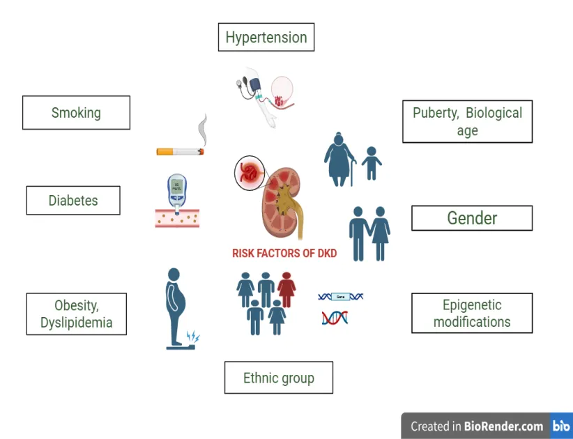
Figure 1: This Illustration Represents the Various Risk Factors Contributing to DKD, Encompassing Metabolic, Hormonal, Genetic, and Lifestyle Elements. It Emphasizes How Sex, Age, Ethnicity, Hypertension, Epigenetic Modifications, Obesity, Smoking, and Glycemic Control Influence Individual Susceptibility.
4. Pathophysiology
4.1 Glomerular Injury: The Starting Point
4.1.1 Podocyte Dysfunction and Loss
Podocytes are terminally differentiated, specialized glomerular visceral epithelial cells essential for maintaining the glomerular filtration barrier [38]. Podocyte injury has been identified as a pivotal event resulting in proteinuric kidney disease, glomerulosclerosis, and loss of renal function [39] Podocytes attach to the glomerular basement membrane (GBM) using α3β1 integrin and dystroglycans. The expression of α3β1 has been reported to be reduced in diabetic individuals , resulting in podocyte detachment from the GBM [40]. Elevated extracellular glucose causes cultured podocytes to undergo apoptosis, produce ROS, and activate proapoptotic p38 MAPK. Podocyte death precedes podocyte depletion, urinary albumin excretion (UAE), and mesangial matrix growth in murine T1 and T2D models, indicating that podocyte apoptosis/depletion represents a novel early pathomechanism leading to DKD. Hyperglycemia, ROS, podocyte death, podocyte depletion, and diabetes nephropathy are all mechanistically linked by nicotinamide adenine dinucleotide phosphate (NADPH) oxidase, a crucial modulator of podocyte apoptosis and subsequent DKD in vivo [41]
4.1.2 Mesangial Expansion
Mesangial expansion, a type of kidney fibrosis, is one of the numerous consequences of DKD. The abnormal proliferation of mesangial cells and the overproduction of matrix proteins are the hallmarks of mesangial expansion, a significant structural lesion. In DKD, mesangial growth plays a role in the development of renal failure[42]. Extensive research has been conducted on TGF-β role in matrix accumulation intracellular signaling pathways implicated and its potency as a profibrotic agent in other fibrotic illnesses. Ang II promotes matrix buildup indirectly by upregulating TGF-β. The activation of the renin-angiotensin system in the kidney causes an increase in Ang II . Ang II has two effects on mesangial cells: directly through Ang II receptors on mesangial cells and indirectly through changes in glomerular capillary pressure, both of which influence mesangial cell behavior. TGF-β overexpression activates Smad and non-Smad pathways, resulting in the accumulation of ECM proteins. TGF-β stimulates kinase receptors in the Smad pathway, causing phosphorylation and the synthesis of ECM proteins, while it can also activate non-Smad pathways, including MAPK, extracellular signal-regulated kinase / c-Jun N-terminal kinase (ERK/JNK), and phosphoinositide 3-kinase (PI3K), leading to greater accumulation of ECM proteins [43].
4.1.3 Endothelial Dysfunction
Elevated blood glucose levels reduce nitric oxide (NO) bioavailability and accelerate endothelin-1 release, resulting in capillary rarefaction, and increased glomerular permeability. Endothelial glycocalyx breakdown also leads to albuminuria [44].
4.2 Tubulointerstitial Injury: The Concealed Catalyst of Progression
4.2.1 Tubular Hypoxia
Renal tubular damage has been demonstrated to be caused by a number of causes, including hyperglycemia, lipid buildup, oxidative stress, hypoxia, RAAS, endoplasmic reticulum (ER) stress, inflammation, epithelial-mesenchymal transition (EMT), and programmed cell death [45]. The development of hypoxia is influenced by three interrelated factors: increased oxygen consumption, oxygen utilization disorder, and reduced oxygen supply. The kidneys consume 60% of their energy for sodium reabsorption, primarily through Na+/K+ ATPase activity, which is linked to glucose uptake in the proximal tubular epithelial cells via SGLT-2 transporters. In T2D, excessive glucose reabsorption raises oxygen consumption, worsening renal tubular hypoxia due to hyperfiltration and mitochondrial dysfunction. Additionally, diabetes often leads to systemic microangiopathy, characterized by vascular injury and reduced blood flow to the kidneys. Increased gluconeogenesis in diabetic kidneys also heightens oxygen and energy demand, further stressing the renal system and linking hypoxia to tubulointerstitial fibrosis via hypoxia inducible factor (HIF-1α) [46].
4.2.2 Tubular overload
Renal tubular and interstitial processes are greatly impacted by angiotensin II, a crucial effector molecule in the renin-angiotensin system (RAS). It controls a number of cytokines and chemokines that promote cellular proliferation, apoptosis, and monocyte/macrophage invasion. Through cytokines including TGF-β, osteopontin, TNF-α, secreted protein acidic and rich in cysteine (SPARC), and RANTES, the pathogenic effects of angiotensin II lead to tubulointerstitial fibrosis and inflammation [47]. Peritubular blood flow and oxygen delivery to the tubules are further hampered by glomerular lesions, interstitial vascular rarefaction, and changes in vasoactive factors like ANG II and/or NO, which impact postglomerular blood flow. Hypoxia can be made worse by impaired renal function, concomitant anemia, increased sodium transport load, and increased oxygen consumption in remaining nephrons [48]. According to the tubular theory of glomerular filtration, tubuloglomerular feedback causes supranormal glomerular filtration rate (GFR) by enhancing proximal tubule reabsorption through early tubular development and sodium-glucose cotransport. Early tubular cell proliferation takes place in a diabetic setting, but TGF-β and cyclin-dependent kinase inhibitors cause cell cycle arrest, which causes tubular hypertrophy and a phenotype resembling senescence. Atypical reactions, including the salt paradox in early diabetic kidneys, are explained by this growth phenotype; yet, tubulointerstitial damage and DKD may be encouraged by the active molecular pathways[49].
4.3 Hemodynamic Changes and Overactivation of the RAAS
4.3.1 Intra-renal hemodynamics
The RAAS, especially Ang II, which contracts both afferent and efferent arteries and raises intraglomerular pressure, causes tissue damage and fibrosis, and ultimately results in diabetic glomerulosclerosis, has a major impact on intra-renal hemodynamic changes. TGF-β activation intensifies the effects of endothelin-1 (ET-1), which also contributes to inflammation and fibrosis. In T2DM, elevated plasma ET-1 levels are related with endothelial dysfunction, which is linked to glomerular hyperfiltration in individuals with diabetes, according to recent clinical trials. [50].
4.3.2 Systemic Hypertension
Uncontrolled hyperglycemia leads to renal failure through biomolecule dysfunction and increased inflammatory cytokines. The renin-angiotensin system (RAS) regulates renal hemodynamics; in chronic hyperglycemia, local Ang II increases, causing tissue damage via oxidative stress and inflammation. This results in glomerular filtration barrier disruption, glomerular basement membrane hypertrophy, microvascular hyperpermeability, proteinuria, and decreased GFR. The reduced GFR falsely signals low blood pressure, stimulating RAS, causing chronic hypertension in DKD patients[51].
4.4 Oxidative Stress and Mitochondrial Dysfunction
4.4.1 Mitochondrial ROS
Diabetic individuals and animal models have increased oxidative stress, which is linked to hyperglycemia, altered lipoproteins, and glycation end products. ROS are mostly produced by dysfunctional mitochondria. Enzyme activity in the mitochondrial respiratory chain is hampered by elevated glucose and fat levels. Patients with diabetes have increased NADPH oxidase (NOX) activity, which is exacerbated by endothelial cells' hyperglycemia and hyperlipidemia. Numerous cardiovascular issues in diabetes are linked to this oxidative stress caused by increased ROS production [52].
4.4.2 Redox-sensitive Pathways
In addition to causing direct cell damage, hyperglycemia-induced ROS overproduction triggers the release of inflammatory mediators such TNF-α and IL-6. Mechanistically, ROS increase inflammation by activating nuclear factor kappa-light-chain-enhancer of activated B cells (NF-κB), a master transcriptional regulator of proinflammatory cytokine production. Additionally, ROS cause upregulation of MCP-1, which exacerbates damage by encouraging T-cell and macrophage infiltration into renal tissues. Through feedback processes, TNF-α modulates cellular proliferation/apoptosis and increases the formation of ROS. The renal microenvironment is disturbed by this ongoing interaction between OS and inflammation, which compromises physiological function[53].
4.5 Inflammation and Immune Dysregulation
Macrophages are important innate immune cells that polarize into M1 (pro-inflammatory) or M2 (anti-inflammatory/pro-fibrotic) phenotypes depending on their environment [54]. They are derived from circulating monocytes. M1 macrophages predominate in the early stages of diabetic kidney disease (DKD), producing ROS and pro-inflammatory cytokines that cause endothelial damage, podocyte death, and persistent inflammation. This starts a vicious cycle of inflammation and leukocyte recruitment. On the other hand, M2 macrophages are more prevalent in late-stage DKD. These macrophages promote fibrogenesis through TGF-β1-induced collagen production and myofibroblast activation, resulting in tubulointerstitial fibrosis and glomerulosclerosis. Renal stiffness in diabetes is significantly influenced by galectin-3 from M2 macrophages[55].
Different subsets of T lymphocytes, such as CD4+ helper T cells, CD8+ cytotoxic T cells, and Tregs, are produced from bone marrow and develop in the thymus. By controlling pathogen clearance and self-tolerance, these subsets preserve immunological equilibrium. Albuminuria levels are correlated with T cell presence in kidney interstitial spaces in T2D. While Th17 cells encourage glomerular injury through IL-17, Th1 cells cause inflammation through IFN-γ and TNF-α, which damages the kidneys. Because of the imbalance between pro-inflammatory Th1/Th17 cells and Tregs, hyperglycemia reduces the effectiveness of Tregs and lowers the production of TGF-β and IL-10, which increases renal inflammation [55,56,57].
4.6 Fibrosis: The Final Common Pathway
The last common pathway in the pathophysiology of DKD is renal fibrosis, which is brought on at least by changes in renal hemodynamics, anomalies in glucose metabolism and ischemia linked to elevated oxidative stress, inflammatory processes, and an overactive RAAS. Glomerular hyperfiltration leads to DKD, driven by hyperglycemia which causes afferent arteriolar dilation through vasoactive mediators like glucagon, insulin-like growth factor-1 (IGF-1), vascular endothelial growth factor (VEGF), NO, and prostaglandins. Additionally, early alterations in renal tubular function correspond to glycemic control, with increased glucose and sodium chloride reabsorption due to enhanced SGLT2 activity in proximal tubules, resulting in reduced sodium chloride delivery to the macula densa and subsequent afferent arteriolar dilation via tubuloglomerular feedback. Constriction of the efferent arteriole causes alterations in autoregulation and glomerular hypertension when local angiotensin II levels are high. By raising ROS, activating PKC, and encouraging pro-inflammatory signaling through AGE, hyperglycemia, insulin resistance, and compensatory hyperinsulinemia all simultaneously contribute to endothelial dysfunction. Vasoconstriction and vascular dysfunction result from this disruption of the balance of mediators produced by endothelial cells, particularly an increase in ET-1 release. Endothelin receptor A activation in the kidneys is associated with podocyte damage, oxidative stress, inflammation, vasoconstriction, and fibrosis as shown in (Figure.2)[58,59,60,61].
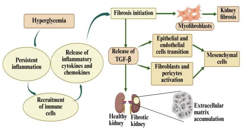
Figure 2: The figure demonstrates fibrotic progression in DKD. Persistent hyperglycaemia induces chronic inflammation and promotes the recruitment and activation of immune cells, resulting in the release of pro-inflammatory cytokines and chemokines. These inflammatory mediators initiate and perpetuate fibrogenic responses, including the activation of TGF-β signalling. Subsequently, epithelial and endothelial cell dysfunction and phenotypic transition, together with the activation of resident fibroblasts and pericytes, contribute to the expansion of mesenchymal and myofibroblast populations. Activated myofibroblasts promote excessive ECM synthesis and deposition, leading to progressive disruption of renal architecture and the transition from healthy renal tissue to a fibrotic kidney.
5. Therapeutic approaches
5.1 Lifestyle interventions
The onset and course of DKD are significantly influenced by both genetic susceptibility and bad lifestyle choices. It can be effectively slowed down with multidisciplinary treatment that includes medication therapy and lifestyle changes[62]. The effects of dietary and lifestyle changes on health were evaluated in eleven research. The BMI significantly decreased in every study. In patients with hyperfiltration, two studies found an increase in GFR, four found no change, and two found a reduction. Six trials showed a decrease in albuminuria, one showed no change, and five showed a decrease in proteinuria. Weight loss, particularly through surgery, improves proteinuria and albuminuria and restores normal GFR in obese patients with impaired renal function [63].
5.2 RAAS inhibitors
Current research reveals that drugs like ACE inhibitors (ACEIs) and angiotensin receptor blockers (ARBs) considerably decreasing the progression of proteinuria, and successfully lowering urine protein excretion thus, reducing the risk of renal impairment , providing renal protective effects particularly in individuals with both diabetes and hypertension. By adding additional RAAS inhibitors, such as aldosterone antagonists, or combining ACEIs and ARBs, blood pressure control and proteinuria reduction are improved, alleviating renal function. However, due to possible side effects such as hyperkalemia and abrupt renal failure, medical practitioners must closely monitor serum potassium levels and renal function and promptly modify treatment as necessary [64]. According to the IDNT research, Irbesartan offers renoprotective benefits regardless of the systolic blood pressure values reached in diabetic individuals[65].
5.3 SGLT2 inhbitors
SGLT2 inhibitors provide renoprotective benefits through the mechanisms that extend beyond simple glucose reduction by lowering hyperfilteration and improving metabolic control. reduce hyperfiltration and enhance metabolic regulation, providing reno-protective benefits through mechanisms that extend beyond mere glucose reduction. The CREDENCE and CANVAS trial provide evidence that Canagliflozin improve renal outcomes in DKD patients. Furthermore, the EMPA-REG OUTCOME trial demonstrated that Empagliflozin provide renal protection in patients of DKD. These findings collectively position SGLT2 inhibitors as an an essential part in preventing renal function decline in DKD patients [66,67].
5.4 ENDOTHELIN-1 ANTAGONISTS
ET-1 conveys signals via two separate receptors: endothelin A (ETA) and endothelin B (ETB). The selective antagonists of ETA are particularly appealing, as the ETB receptor facilitates the clearance of circulating ET-1 through the lungs. In contrast, blocking the ETA receptor can lead to a reduction in blood pressure, a decrease in fibrosis, and a mitigation of kidney inflammation. Furthermore, ETA receptor antagonists (ETA-RAs) have demonstrated effectiveness in lowering albuminuria and minimizing the risk of renal complications in individuals with DKD, although they are also linked to fluid retention. In the “Study Of diabetic Nephropathy with AtRasentan” (SONAR) trial, Atrasentan was found to significantly lower the risk of progression of DKD and kidney failure when compared to a placebo in patients diagnosed with DKD [68].
CONCLUSION
DKD remains a major cause of CKD and kidney failure worldwide, reflecting the cumulative effects of metabolic, hemodynamic, genetic, and environmental determinants. Its clinical course is heterogeneous: some individuals experience slowly progressive renal impairment, whereas others develop persistent albuminuria, declining glomerular filtration, and accelerated loss of kidney function. This variability underscores the importance of understanding DKD as a multifactorial disorder rather than a consequence of hyperglycaemia alone. The epidemiological burden is further amplified by the increasing prevalence of diabetes, ageing populations, coexisting hypertension, obesity, and disparities in access to preventive and renoprotective care. The transition from diabetic injury to renal fibrosis represents a pivotal pathological process underlying the progressive and often irreversible deterioration of renal architecture. Sustained hyperglycaemia, advanced glycation end-product formation, oxidative stress, altered intra-renal hemodynamics, and activation of the RAAS converge to promote glomerular and tubular injury. These disturbances engage interconnected inflammatory and profibrotic signalling networks, including TGF-β/Smad, Wnt/β-catenin, Notch, MAPK, and related pathways. Persistent activation of these mechanisms disrupts cellular homeostasis, promotes epithelial and endothelial dysfunction, stimulates myofibroblast activation, and enhances ECM synthesis. Consequently, glomerulosclerosis and tubulointerstitial fibrosis progressively compromise nephron integrity and contribute to declining renal function. The complexity and reciprocal interaction of these mechanisms also explain why targeting a single pathogenic pathway may be insufficient to halt disease progression. Therapeutic management has consequently evolved from conventional glycemic and blood-pressure control towards a more comprehensive, mechanism-informed strategy. Renin–angiotensin system inhibitors remain foundational interventions, while sodium–glucose cotransporter 2 inhibitors, glucagon-like peptide-1 receptor agonists, and the non-steroidal mineralocorticoid receptor antagonist finerenone have expanded the evidence-based approach to cardiorenal protection. These therapies provide benefits beyond their primary metabolic or hemodynamic effects and may influence pathways associated with inflammation, oxidative stress, and fibrotic remodeling. Nevertheless, residual risk persists despite contemporary multidrug treatment, highlighting the continuing need for improved therapeutic strategies and individualized disease management. Overall, DKD-associated renal fibrosis emerges from the sustained convergence of metabolic stress, hemodynamic dysregulation, inflammatory responses, and maladaptive tissue repair. Integrating epidemiological understanding with the molecular basis of fibrotic progression and current therapeutic evidence is essential for a coherent view of disease development and management. Continued translation of mechanistic insights into clinically validated interventions, alongside early recognition and sustained implementation of proven therapies, will be crucial to slowing renal decline, reducing the burden of kidney failure, and improving long-term outcomes for individuals living with DKD in diverse clinical settings.
7. Future Perspectives
Future research should prioritize the translation of mechanistic insights into therapies that directly attenuate renal fibrogenesis while preserving adaptive tissue-repair processes. Greater understanding of the interactions among metabolic stress, intra-renal hemodynamics, inflammation, and profibrotic signalling may reveal complementary targets for combination treatment. Longitudinal and well-powered clinical studies are needed to establish the durability of existing cardiorenal therapies, clarify their effects across diverse DKD populations, and evaluate emerging agents, including through pragmatic trials with longer follow-up periods across diverse healthcare settings. Precision-based treatment strategies, informed by disease stage, comorbidities, and treatment response, may ultimately improve therapeutic effectiveness and reduce residual risk.
REFERENCES


Aadya Misra, Dr. Shiv Dev Singh, Decoding Diabetic Kidney Disease: Epidemiological Trends, Risk Determinants, Fibrotic Pathogenesis, and Therapeutic Approaches, Int. J. of Pharm. Sci., 2026, Vol 4, Issue 11, 2012-2025. https://doi.org/10.5281/zenodo.24156259
 10.5281/zenodo.24156259