

We use cookies to ensure our website works properly and to personalise your experience. Cookies policy
1Student Wani College Of Pharmacy Ganegaon, Maharashtra.
2Assistant professor of Wani College Of Pharmacy Ganegaon, Maharashtra, Affiliated to Dr Babasaheb Aambedkar Technological University, Lonere, Raigad.
Oral ulceration is a common clinical problem with a broad differential diagnosis ranging from minor mechanical trauma and recurrent aphthous stomatitis (RAS) to infections, immune-mediated disorders, nutritional deficiencies, medication-related lesions and oral malignancy. Although many uncomplicated ulcers are self-limiting, persistent, recurrent, atypical or indurated lesions require systematic assessment so that significant local or systemic disease is not missed. This review summarizes the current approach to the diagnosis and management of mouth ulcers, with emphasis on clinical history, oral examination, classification according to duration and recurrence, identification of local and systemic causes, appropriate laboratory investigation, indications for biopsy and referral, and evidence-informed symptomatic treatment. RAS is discussed in detail because it is the most common recurrent ulcerative disorder. Management is individualized according to severity and underlying cause; general measures, pain control, topical corticosteroids and protective agents are commonly used for uncomplicated aphthous disease, while systemic therapies and specialist interventions are reserved for selected severe or refractory cases. Recent evidence also supports selected non-pharmacological approaches such as photobiomodulation/laser therapy for short-term pain and healing outcomes, although treatment parameters and long-term recurrence effects require further standardization. The review emphasizes that a non-healing oral ulcer should not automatically be labeled as aphthous and that biopsy or specialist assessment may be required when the clinical course is persistent or suspicious.
An oral ulcer is a localized loss of the oral epithelial surface with exposure of underlying connective tissue. Oral ulceration may occur as an isolated local event or as a manifestation of a broader inflammatory, infectious, hematologic, gastrointestinal, dermatologic or neoplastic disorder. The clinical appearance of different diseases can overlap considerably; therefore, diagnosis should not rely on ulcer appearance alone. History, duration, number, recurrence pattern, location, morphology, associated symptoms, medication exposure and systemic features all contribute to a structured diagnosis.
Recurrent aphthous stomatitis (RAS), commonly called canker sores, is the most frequent recurrent ulcerative disorder of the oral mucosa. Typical lesions are painful, shallow, round or oval ulcers with an erythematous halo and a yellow-grey fibrinous base. RAS is usually diagnosed clinically after other causes of recurrent ulceration have been considered. The disease is multifactorial and may be associated with local trauma, stress, nutritional deficiencies, immune factors, gastrointestinal disease and other systemic conditions. Contemporary management focuses on reducing pain, shortening the active episode, improving oral function and, where possible, reducing recurrence.
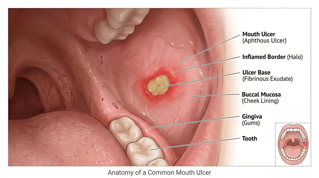
Figure 1: Mouth ulcer
A clinically important distinction is between an uncomplicated ulcer that is expected to heal and a lesion that is persistent, atypical, indurated, enlarging, recurrent in an unusual pattern or associated with systemic findings. Expert guidance recommends further investigation and consideration of biopsy when an oral ulcer remains unexplained, particularly when it persists beyond approximately two weeks or fails to respond to an appropriate initial approach.
Mouth ulcers, also referred to as oral ulcers, are common lesions of the oral mucosa characterized by localized loss or disruption of the epithelial lining, resulting in exposure of the underlying connective tissue. They may occur on the lips, buccal mucosa, tongue, gingiva, hard or soft palate, and floor of the mouth. Although many oral ulcers are self-limiting and heal within a few days to weeks, recurrent, persistent, unusually large, or painful ulcers may significantly affect eating, drinking, speaking, swallowing, and overall quality of life.
Oral ulceration can develop due to a wide range of local and systemic factors. Common local causes include accidental biting, sharp teeth, poorly fitting dentures, orthodontic appliances, thermal injury, chemical irritation, and excessive mechanical trauma. Recurrent aphthous stomatitis is another important cause and is characterized by recurrent episodes of painful ulcers, particularly on non-keratinized oral mucosa. Nutritional deficiencies involving iron, folate, vitamin B12, and other micronutrients may also contribute to recurrent ulceration.
In addition to local factors, oral ulcers can sometimes represent a manifestation of systemic diseases. Gastrointestinal disorders such as celiac disease and inflammatory bowel disease, hematological abnormalities, immune-mediated disorders, and certain infections may present with oral ulceration. Viral, bacterial, and fungal infections can also produce ulcerative lesions. Therefore, a detailed clinical history and appropriate examination are important for determining the probable cause.
Drug-associated oral ulceration should also be considered during diagnosis. Certain medicines may produce direct mucosal irritation, immune-mediated reactions, xerostomia, or other changes that predispose patients to ulcer formation. A complete medication history can therefore provide important information when evaluating recurrent or unexplained oral ulcers.
The clinical appearance of an ulcer provides valuable diagnostic information. Important features include the number of lesions, site, size, shape, border, depth, surrounding erythema, presence of induration, duration, recurrence pattern, and associated symptoms. A single acute ulcer following trauma may require only local supportive care, whereas multiple recurrent ulcers or ulcers associated with systemic symptoms may require laboratory investigations and specialist evaluation.
The management of mouth ulcers depends primarily on the underlying cause and severity of the lesion. Treatment may include removal of local irritants, maintenance of good oral hygiene, topical analgesics, antiseptic preparations, topical corticosteroids, protective oral gels or pastes, nutritional supplementation when a deficiency is confirmed, and treatment of an underlying systemic or infectious condition. In selected patients with severe or recurrent disease, systemic therapy or specialist-directed treatment may be required.
Early recognition of potentially serious lesions is particularly important. An ulcer that persists beyond the expected healing period, repeatedly occurs at the same site, becomes progressively larger, develops an indurated or irregular border, bleeds without an obvious cause, or is associated with a neck swelling, unexplained weight loss, or difficulty in swallowing requires further clinical assessment. Persistent oral ulceration may occasionally be associated with oral potentially malignant disorders or oral malignancy, making appropriate referral and, when indicated, biopsy essential.
Recent advances in oral healthcare have also encouraged the development of improved topical drug-delivery systems, including mucoadhesive gels, oral films, patches, lozenges, sprays, and in-situ gels. These systems are designed to prolong contact between the active ingredient and the ulcerated mucosa, provide local pain relief, and potentially improve therapeutic effectiveness. Natural products and herbal ingredients are also being investigated for their anti-inflammatory, antimicrobial, antioxidant, analgesic, and wound-healing properties.
Thus, diagnosis and management of mouth ulcers require an integrated approach involving clinical assessment, identification of causative factors, differential diagnosis, appropriate investigations, symptomatic treatment, and management of the underlying condition. A systematic understanding of oral ulceration can help healthcare professionals distinguish uncomplicated self-limiting lesions from ulcers requiring further investigation and specialized management.
Objectives
Clinical Significance of Mouth Ulcer
Most simple oral ulcers are benign and heal spontaneously, but oral ulceration has a wide differential diagnosis. A useful clinical framework is to classify the lesion by duration (acute versus chronic), number (solitary versus multiple), and pattern (first episode versus recurrent). An ulcer lasting less than two weeks is commonly considered acute; persistence for two weeks or more warrants increased diagnostic attention, particularly if there is no clear local cause.
Important associated symptoms include fever, malaise, weight loss, dysphagia, odynophagia, skin lesions, genital ulcers, ocular symptoms, gastrointestinal complaints, joint symptoms and recurrent infections. These findings may suggest an underlying systemic disorder rather than isolated idiopathic aphthosis.
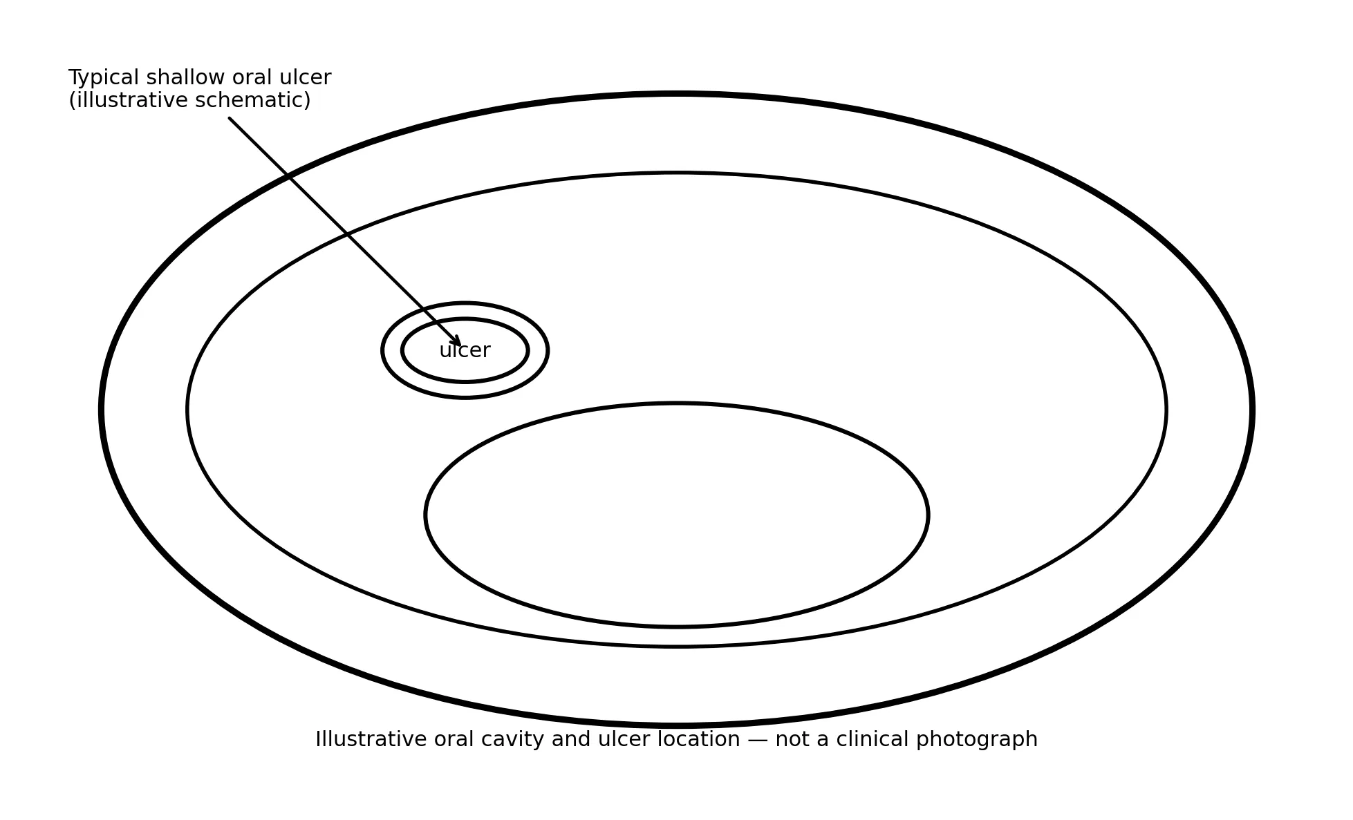
Figure 2: Illustrative schematic of an oral ulcer and its relationship to the oral cavity.
Illustrative Anatomy and Appearance
Classification of Mouth Ulcers
| Clinical category | Typical pattern | Examples | Diagnostic emphasis |
|---|---|---|---|
| Acute ulcer | Short duration; often resolves within days to <2 weeks | Trauma, burns, viral infection, acute mucositis | Trigger, infection, number and systemic symptoms |
| Chronic ulcer | Persistent, often ≥2 weeks | Chronic traumatic ulcer, immune-mediated disease, neoplasia | Induration, edge, base, growth and biopsy need |
| Recurrent ulcer | Repeated episodes with healing between attacks | RAS, systemic disease-associated aphthosis | Recurrence pattern and associated systemic features |
| Solitary ulcer | One dominant lesion | Traumatic ulcer, neoplasia, localized infection | Site, persistence and local irritant |
| Multiple ulcers | Several simultaneous lesions | RAS, viral disease, immune-mediated disease, drug reactions | Distribution, prodrome and systemic manifestations |
Recurrent Aphthous Stomatitis (RAS)
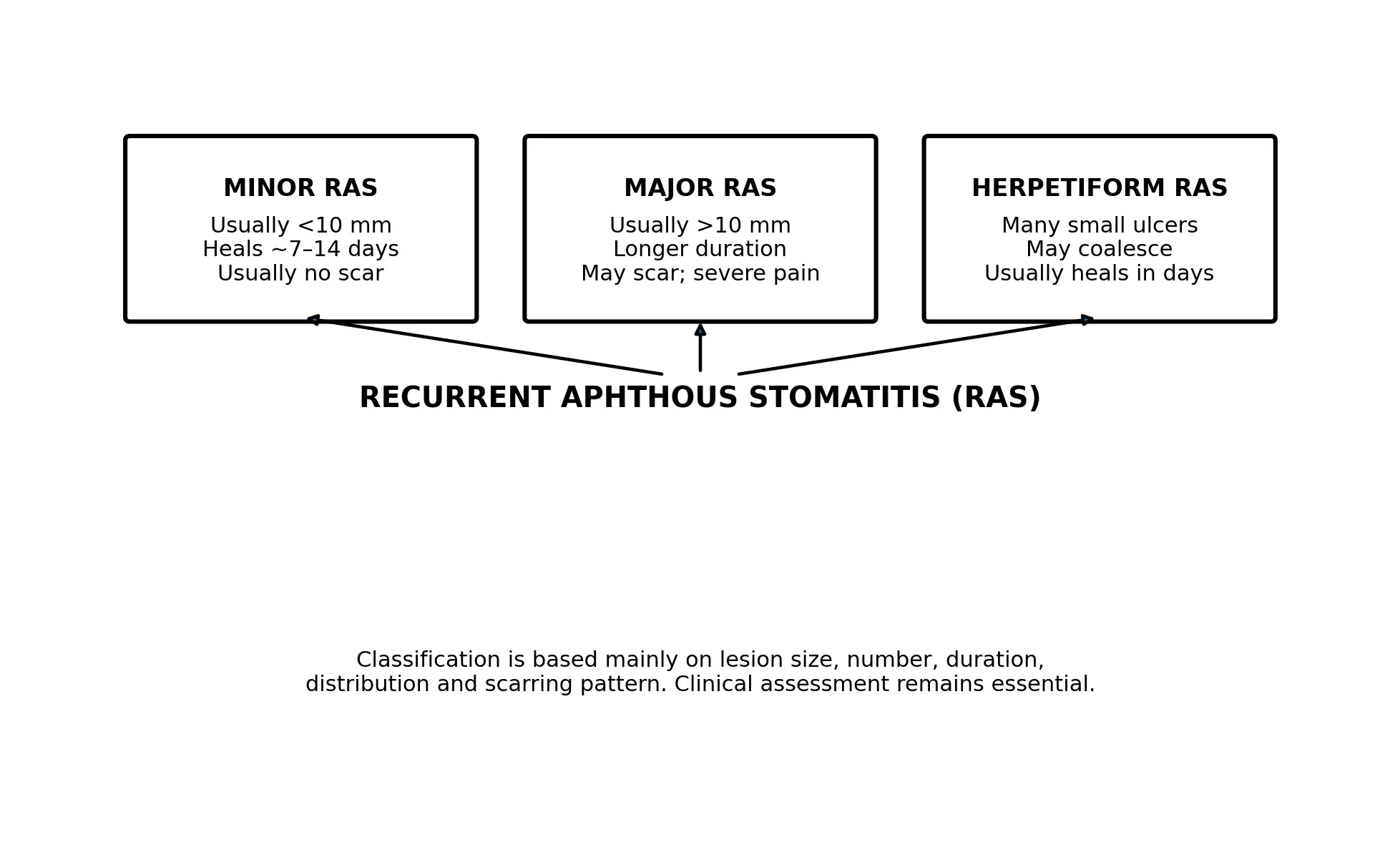
Figure 3: Original schematic classification of recurrent aphthous stomatitis (RAS).
RAS is a chronic, recurrent inflammatory condition characterized by episodic painful oral ulceration in the absence of a specific identifiable systemic cause. The disease is often diagnosed clinically. Three classical clinical patterns are recognized: minor, major and herpetiform aphthous ulcers. The distinction is based on lesion size, number, duration and tendency to scar.
1. Minor RAS : Minor aphthae are the commonest form. They are usually small, shallow and painful, commonly affecting non-keratinized mucosa such as the labial and buccal mucosa and ventral tongue. They generally heal without scarring. Symptoms can nevertheless interfere with eating, speaking and oral hygiene.
2. Major RAS : Major aphthae are larger and deeper, may persist for several weeks and may heal with scarring. Pain and functional impairment can be substantial. Severe or persistent major aphthae should prompt assessment for associated systemic disease and consideration of specialist management.
3. Herpetiform RAS : Herpetiform aphthae consist of numerous small ulcerations that can occur in clusters and occasionally coalesce into larger irregular lesions. Despite the name, they are not caused by herpes simplex virus. The clinical distinction from viral ulceration depends on history, distribution, associated vesicles and systemic findings.
Etiology and Risk Factors
| Cause/category | Examples / relevance |
|---|---|
| Local factors | Cheek or lip biting, sharp tooth/restoration, ill-fitting appliances, thermal injury, chemical irritation, aggressive brushing. |
| Nutritional factors | Iron deficiency, vitamin B12 deficiency, folate deficiency and other nutritional disturbances may be associated with recurrent ulceration. |
| Infectious causes | Herpes simplex virus, varicella-zoster virus, enteroviruses and selected bacterial/fungal infections can produce oral ulceration. |
| Immune/inflammatory disorders | Behçet disease, inflammatory bowel disease, celiac disease, lupus and other immune-mediated disorders may present with oral ulcers. |
| Hematologic disorders | Anemia, neutropenia, cyclic neutropenia and other blood disorders can be associated with recurrent or persistent ulcers. |
| Drug-related causes | Some medicines can induce mucositis, ulceration or lichenoid/immune-mediated reactions; a medication timeline is therefore important. |
| Psychological/physiological factors | Stress, sleep disturbance, hormonal variation and other patient-specific triggers may contribute in susceptible individuals. |
| Neoplastic causes | Persistent or indurated ulcers may represent oral squamous cell carcinoma or another neoplastic process and should not be assumed to be aphthous. |
Pathogenesis of Recurrent Aphthous Stomatitis
The exact pathogenesis of RAS remains incompletely understood. Current models support a multifactorial process involving genetic susceptibility, altered local immune responses, epithelial injury and inflammatory mediators. T-cell-mediated inflammation is an important component of lesion development. Local trauma may initiate ulceration in susceptible mucosa, while systemic factors such as hematinic deficiency, gastrointestinal disease, stress and immune dysregulation may modify severity or recurrence.
Because the biological mechanisms are heterogeneous, treatment is mainly directed toward controlling inflammation and pain and supporting mucosal healing rather than providing a universally curative therapy. Identification and correction of an associated systemic cause may reduce recurrence in selected patients.
Clinical Features to Record
Diagnostic Approach
Diagnosis begins with a careful history and complete oral examination. The clinician should inspect all oral mucosal surfaces, palpate suspicious lesions and examine regional lymph nodes when appropriate. The lesion should be documented with site, size and duration, and serial photographs may be useful in clinical practice when consent and appropriate privacy procedures are followed.
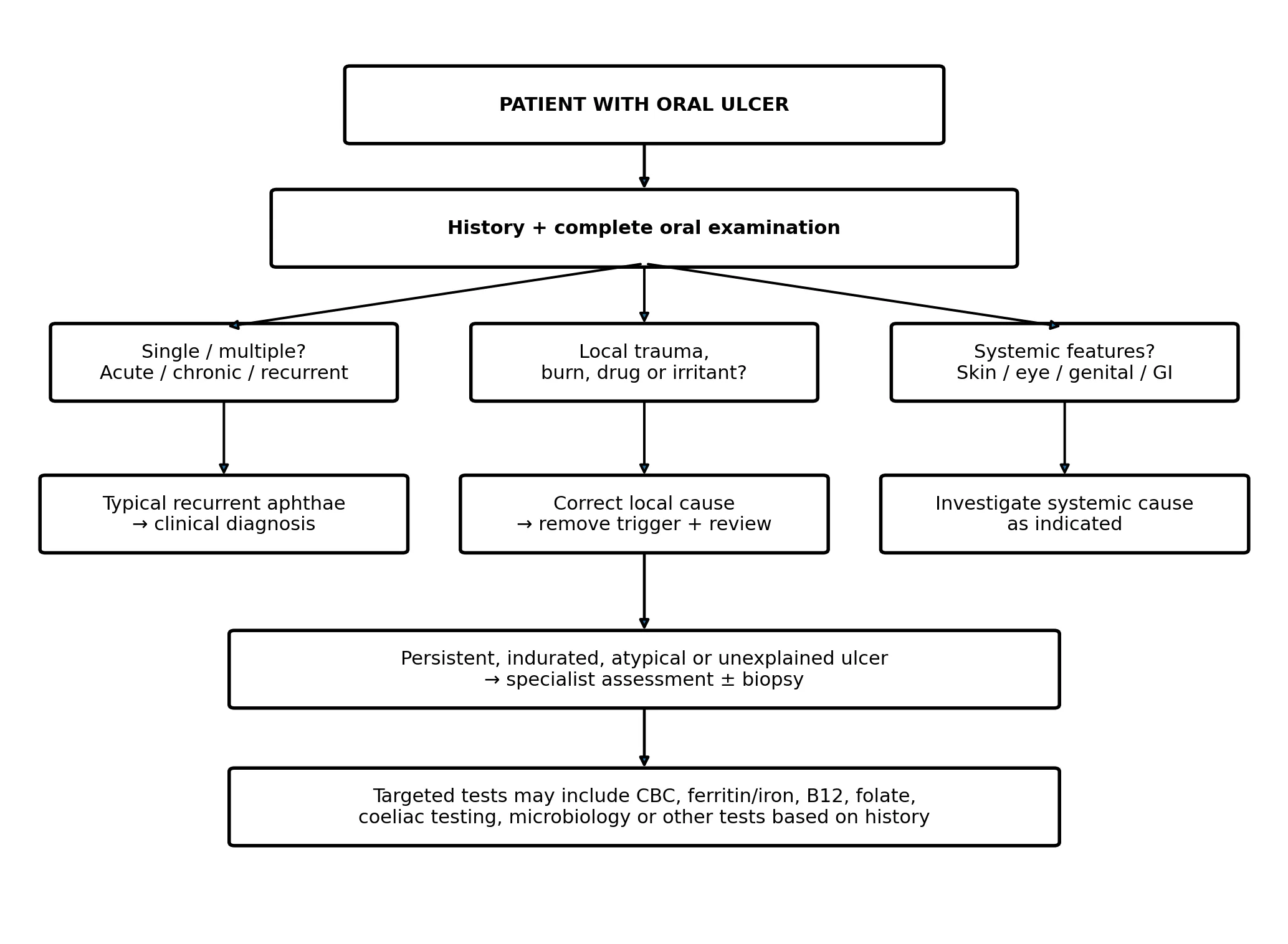
Figure 3: Structured diagnostic pathway for an oral ulcer. This is an original educational flowchart based on published diagnostic frameworks.
1. History : History should establish whether the lesion is acute, chronic or recurrent. Recurrent episodes with complete healing between attacks support aphthous disease, but recurrent ulcers can also accompany systemic disorders. The clinician should ask about trauma, dental appliances, new medicines, infections, nutritional status and systemic symptoms.
2. Oral Examination : Important examination findings include the ulcer's size, shape, depth, border, floor, surrounding mucosa and exact location. An ulcer with firm induration, rolled or irregular margins, unexplained bleeding, progressive enlargement, fixation or associated cervical lymphadenopathy deserves particular attention.
3. Laboratory Investigations : Laboratory testing should be targeted rather than automatic for every single self-limited ulcer. In patients with frequent, prolonged, large or otherwise atypical ulcers, reasonable investigations may include complete blood count, iron studies/ferritin, vitamin B12 and folate. Additional testing such as coeliac serology, inflammatory markers, microbiological testing or evaluation for gastrointestinal disease should be guided by the clinical history.
4. Biopsy : Biopsy is considered when the diagnosis remains uncertain, the ulcer persists despite appropriate management, the lesion is atypical or suspicious, or malignancy is part of the differential diagnosis. Expert consensus documents particularly emphasize biopsy consideration for unexplained ulcers lasting more than approximately two weeks or ulcers that do not respond to an initial treatment period. The biopsy site and technique depend on the clinical differential and should be selected by an appropriately trained clinician.
Differential Diagnosis
| Condition | Typical clues | Useful investigations | Management direction |
|---|---|---|---|
| Traumatic ulcer | Clear local mechanical/thermal/chemical trigger | Usually clinical; reassess after removing cause | Remove irritant; symptomatic care; reassess |
| RAS | Recurrent, painful, shallow ulcers; usually non-keratinized mucosa | Clinical; CBC/iron/B12/folate when indicated | Topical therapy; treat associated cause |
| HSV-related disease | Vesicles/ulcers, often clustered; systemic symptoms may occur | Viral testing in selected cases | Antiviral therapy when clinically indicated |
| Behçet disease | Oral ulcers plus genital, ocular, skin or vascular features | Clinical + specialist evaluation | Multidisciplinary management |
| Inflammatory bowel disease | Oral lesions with GI symptoms or other systemic findings | GI assessment, inflammatory tests, endoscopy as indicated | Treat underlying disease |
| Celiac disease | Recurrent aphthous-like ulcers with nutritional/GI features | Coeliac serology ± further evaluation | Dietary/systemic management |
| Drug-related ulceration | Temporal relationship to medicine; may have widespread mucosal involvement | Medication review; selected tests/biopsy | Prescriber review and supportive care |
| Oral lichen planus/immune mucositis | Erosions/ulcers with characteristic surrounding changes | Clinical ± biopsy/direct immunofluorescence depending on differential | Specialist-directed anti-inflammatory therapy |
| Oral squamous cell carcinoma | Persistent solitary ulcer, induration, irregularity, growth or nodes | Biopsy and histopathology | Urgent specialist/oncologic pathway |
Red-Flag Features
Management of Mouth Ulcer
Management should be individualized according to the cause, severity, duration, recurrence pattern and patient-specific risks. The main objectives are pain control, protection of the ulcer, promotion of healing, maintenance of nutrition and hydration, correction of precipitating factors, and treatment of an underlying systemic disorder when present.
1. General and Supportive Measures
2. Topical Pharmacological Therapy
For uncomplicated RAS, topical therapy is generally the initial pharmacological approach. Published reviews and consensus recommendations commonly discuss topical corticosteroids as an important first-line anti-inflammatory treatment. Examples used in clinical practice include triamcinolone acetonide dental paste and other topical corticosteroid preparations selected according to lesion site and patient factors. Topical anesthetics may provide short-term pain relief, while protective/barrier preparations can reduce mechanical irritation.
| Therapeutic group | Potential role | Important considerations |
|---|---|---|
| Topical corticosteroids | Reduce local inflammatory activity; commonly used for RAS | Use appropriate formulation and duration; assess for secondary candidiasis or other complications |
| Topical anesthetics/analgesics | Short-term reduction of pain | Avoid excessive use; consider swallowing/numbness risks |
| Antiseptic mouth rinses | May reduce microbial burden and support symptom control in selected patients | Not a substitute for diagnosis; avoid irritating preparations |
| Barrier/protective agents | Physically shield ulcerated mucosa and reduce mechanical pain | Useful as adjunctive therapy |
| Hyaluronic-acid based products | Support mucosal protection and may improve short-term symptoms/healing | Evidence varies by formulation |
3. Systemic Therapy : Systemic therapy is generally reserved for severe, frequent, major or refractory aphthous disease, or for ulceration associated with an underlying systemic condition. Published reviews describe systemic corticosteroids and selected immunomodulatory agents in specialist-managed cases. Thalidomide has demonstrated efficacy in severe RAS in systematic reviews, but its adverse-effect profile and strict safety considerations mean that it is not a routine treatment for ordinary mouth ulcers. Systemic therapy should therefore be individualized and supervised by an appropriate clinician.
4. Laser and Photobiomodulation : Photobiomodulation/low-level laser therapy has received increasing attention as an adjunctive approach for RAS. Recent systematic reviews and meta-analyses report reductions in pain and, in several trials, shorter healing time compared with control interventions. However, treatment devices, wavelengths, energy parameters and protocols vary between studies. Therefore, laser therapy should be regarded as a clinician-directed adjunct rather than a universal replacement for diagnosis and conventional treatment.
5. Management of Underlying Causes : When an underlying cause is identified, management should address that condition. Examples include correction of iron, folate or vitamin B12 deficiency when confirmed; removal of a traumatic dental source; review of a suspected medication reaction with the prescribing clinician; and specialist management of Behçet disease, inflammatory bowel disease, celiac disease, hematologic disorders or immune-mediated mucosal disease. Treating the underlying disorder may be more important than repeatedly treating the ulcer itself.
Management According to Severity
| Severity | Typical situation | General approach | Escalation |
|---|---|---|---|
| Mild / isolated | Single small ulcer with no red flags | Reassurance, trigger avoidance, oral hygiene, analgesia ± topical protective/corticosteroid therapy | Review if persistent |
| Moderate / recurrent | Several episodes, multiple lesions or significant pain | Topical anti-inflammatory therapy + pain control + assess triggers and nutritional/systemic factors | Investigate if frequent/prolonged |
| Severe / major | Large/deep ulcers, prolonged healing, major functional impairment | Specialist evaluation; topical plus selected systemic therapy | Oral medicine/dental/medical referral |
| Refractory / atypical | Persistent, indurated, unexplained or treatment-resistant lesion | Re-evaluate diagnosis; targeted tests and biopsy when indicated | Urgent specialist assessment |
Prevention of Recurrence in RAS
1. Identify personal triggers such as trauma, specific foods, stress or oral-care products.
2. Maintain good oral hygiene while minimizing mechanical trauma.
3. Address confirmed nutritional deficiencies rather than using indiscriminate supplementation.
4. Consider a sodium-lauryl-sulfate-free toothpaste in patients who report recurrent aphthae associated with conventional toothpaste.
5. Manage systemic disorders that may contribute to recurrent ulceration.
6. Use a symptom/ulcer diary when recurrence is frequent to identify patterns.
7. Seek specialist evaluation when ulcers become unusually frequent, large, prolonged or associated with systemic symptoms.
Role of the Pharmacist and Dental/Medical Team
Pharmacists can contribute by identifying potentially ulcerogenic medicines, checking for inappropriate self-medication, counselling on topical product use and recognizing red flags that warrant dental or medical referral. Dentists and oral medicine specialists play a central role in lesion examination, differential diagnosis, biopsy decisions and management of local causes. Physicians may be required when oral ulceration is a manifestation of hematologic, gastrointestinal, autoimmune, infectious or systemic disease. Multidisciplinary care is particularly valuable in severe or recurrent cases.
Recent Evidence and Emerging Approaches
Recent literature has expanded the therapeutic landscape beyond conventional topical corticosteroids. Systematic reviews have evaluated topical agents, hyaluronic acid, barrier preparations, laser/photobiomodulation, herbal and complementary interventions and selected systemic immunomodulators. The most consistent short-term evidence remains focused on pain reduction and acceleration of healing, while evidence for durable prevention of recurrence is less consistent. A 2025 umbrella review synthesized 41 reviews and concluded that topical corticosteroids and low-level laser therapy consistently improve short-term pain/healing outcomes, while systemic agents such as colchicine and thalidomide are more appropriate for selected refractory disease because of adverse-effect and evidence-quality considerations.
An important research gap is the lack of standardized treatment parameters for physical therapies and the heterogeneity of outcome measures used across clinical trials. Future studies should use standardized diagnostic criteria, validated pain scales, objective healing measures, longer follow-up and clear recurrence endpoints.
Patient Education
Patients should be advised that many simple mouth ulcers heal spontaneously, but persistent or unusual lesions should not be repeatedly self-treated without assessment. They should seek professional evaluation if an ulcer lasts around two weeks or longer, repeatedly returns with increasing severity, becomes hard or enlarges, is associated with unexplained bleeding or neck swelling, or occurs with systemic symptoms. Patients should also avoid deliberately applying caustic chemicals or unapproved substances to the ulcer because chemical injury can worsen mucosal damage.
Future Perspectives
1. Development of standardized diagnostic criteria for recurrent oral ulceration.
2. Large multicenter trials comparing topical corticosteroids, barrier agents, hyaluronic acid and photobiomodulation using common outcome measures.
3. Better biomarkers for distinguishing idiopathic RAS from systemic-disease-associated aphthosis.
4. Personalized treatment based on recurrence pattern, immune phenotype and nutritional status.
5. Long-term studies evaluating recurrence prevention rather than only short-term pain relief.
CONCLUSION
Mouth ulceration is a clinical sign rather than a single disease. The diagnostic process should begin with careful characterization of duration, number, recurrence, site, morphology and associated systemic features. Recurrent aphthous stomatitis is common and is usually managed with supportive care, pain control and topical anti-inflammatory treatment, while severe or refractory disease may require specialist-directed systemic therapy. The presence of a persistent, indurated, atypical or unexplained ulcer changes the diagnostic priority: malignancy and other significant causes must be excluded rather than assuming aphthous disease. Targeted laboratory testing can identify nutritional or systemic contributors in selected patients, and biopsy is an important diagnostic tool when the clinical course is suspicious or unexplained. Current evidence supports several adjunctive approaches, including photobiomodulation, but treatment should remain individualized and diagnosis should always precede repeated symptomatic therapy.
REFERENCES

Hasim Inamdar, Vaishnvi Kalamkar, Diagnosis and Management of Mouth Ulcer, Int. J. of Pharm. Sci., 2026, Vol 4, Issue 11, 2095-2105. https://doi.org/10.5281/zenodo.24215727
 10.5281/zenodo.24215727