

We use cookies to ensure our website works properly and to personalise your experience. Cookies policy
Department of Pharmacology, Sarojini Naidu Vanita Pharmacy Maha Vidyalaya (Co-Ed.), Tarnaka, Hyderabad, Telangana, India-500017.
Drug-induced peptic ulcer (DIPU) is an important gastrointestinal disorder that commonly develops due to the prolonged or excessive use of ulcerogenic medications, particularly non-steroidal anti-inflammatory drugs (NSAIDs), aspirin, corticosteroids, antiplatelet agents, and anticoagulants. These drugs cause gastric mucosal damage by inhibiting cyclooxygenase (COX) enzymes, reducing protective prostaglandin synthesis, increasing oxidative stress, and promoting inflammation. Experimental animal models, especially rats and mice, are widely used to investigate the mechanisms of ulcer development and evaluate the gastroprotective effects of different therapeutic agents. Various drugs, including indomethacin, aspirin, ibuprofen, diclofenac, ketorolac, and meloxicam, are commonly employed to induce experimental gastric ulcers. The severity of gastric damage is assessed through macroscopic, biochemical, and histopathological examinations using parameters such as ulcer score, ulcer index, ulcer area, gastric volume, gastric pH, and gastric wall mucus. Biochemical investigations also include oxidative stress markers such as malondialdehyde (MDA), reduced glutathione (GSH), superoxide dismutase (SOD), and catalase (CAT), along with inflammatory mediators such as nitric oxide (NO), prostaglandin E? (PGE?), TNF-?, IL-1?, and IL-6. In recent years, medicinal plants, phytochemicals, probiotics, antioxidants, and synthetic compounds have gained considerable attention for their potential gastroprotective properties. Future research should focus on improving experimental ulcer models, understanding chronic gastric injury and healing mechanisms, and exploring advanced techniques such as gastric organoids and computational models. Overall, experimental animal studies provide valuable information about drug-induced gastric injury and contribute to the development
A common gastrointestinal condition called peptic ulcer disease (PUD) is defined by a rupture in the duodenum or stomach mucosa that penetrates the muscularis mucosa. Drug-induced peptic ulcers have gained importance due to the widespread use of non-steroidal anti-inflammatory drugs (NSAIDs), aspirin, corticosteroids, antiplatelet agents, anticoagulants, and other ulcerogenic medications, even though the prevalence of Helicobacter pylori-associated ulcers has decreased (1)
NSAIDs are the most common medicine that causes stomach ulcers. Both topical irritation of the stomach epithelium and systemic suppression of cyclooxygenase (COX)-1 and COX-2 enzymes, which results in decreased prostaglandin synthesis, are responsible for their ulcerogenic effects. Depletion of prostaglandins increases vulnerability to oxidative stress and inflammation, decreases mucosal blood flow, inhibits mucus and bicarbonate secretion, and delays epithelial healing. Therefore, long-term NSAID use may cause bleeding, perforation, stomach erosions, and peptic ulcers, especially in older patients and those with a history of gastrointestinal disorders (2,3)
In order to assess the gastroprotective effectiveness of new treatment medicines and to comprehend the pathophysiology of drug-induced peptic ulcers, experimental animal models are essential. Because they offer dependable and repeatable models that closely mimic human stomach ulcers, rats and mice are the most widely utilized laboratory animals (4) NSAID-induced models (indomethacin, ibuprofen, aspirin, diclofenac, and naproxen), ethanol-induced ulcers, stress-induced ulcers, pyloric ligation, and acetic acid-induced chronic ulcers are only a few of the ulcer induction techniques that have been discovered. These models make it easier to study oxidative stress, inflammatory reactions, ulcer healing processes, and stomach mucosal damage (5)
Experimental ulcers are evaluated using both macroscopic and microscopic methods, such as ulcer index, gastric acidity, gastric mucus content, histopathological analysis, and biochemical measurements of oxidative stress markers such as catalase (CAT), superoxide dismutase (SOD), reduced glutathione (GSH), and malondialdehyde (MDA). Additionally, measures of inflammatory cytokines including interleukin-1β (IL-1β), interleukin-6 (IL-6), and tumor necrosis factor-alpha (TNF-α) are commonly employed to elucidate the molecular mechanisms underlying gastrointestinal injury. (6)
Finding safer pharmaceuticals and natural compounds that can prevent or lessen drug-induced stomach injury without affecting the therapeutic efficacy of NSAIDs has been the subject of extensive research in recent years. Through antioxidant, anti-inflammatory, anti-apoptotic, and cytoprotective processes, a variety of medicinal herbs, phytochemicals, antioxidants, probiotics, and synthetic compounds have shown notable gastroprotective effect in animal experiments (2,4,7) Thus, experimental animal models are still essential for the preclinical assessment of new anti-ulcer treatments and continue to offer insightful information on the mechanisms behind drug-induced peptic ulcers.
2.Epidemiology
Peptic ulcer disease (PUD) has been linked to drug-induced peptic ulcer (DIPU), especially in affluent nations where the prevalence of Helicobacter pylori infection has decreased while the use of ulcerogenic drugs has increased. The epidemiology of PUD has shifted toward medication-associated diseases due to the extensive long-term use of drugs that can harm the gastroduodenal mucosa brought on by the rising frequency of chronic musculoskeletal disorders, cardiovascular diseases, and an aging population (1,11)
Among all the drug classes, NSAID-induced PUD is still a major global public health issue. According to studies, the prevalence of stomach ulcers among NSAID users ranges from 10% to 30% worldwide, with older people and those with comorbidities bearing a greater burden (8) Gastrointestinal bleeding, perforation, and obstruction are complications linked to NSAID-induced PUD and significantly increase morbidity and death (9) According to a systemic assessment, the annual incidence of PUD complications among NSAID users varies from 3.77 to 14.0 cases per 100,000 people for perforation and from 19.5 to 57.0 cases per 100,000 people for bleeding (10)
Drug-induced peptic ulcer disease is less commonly caused by other drugs. Corticosteroids by themselves only slightly raise the risk of ulcers, but when taken in conjunction with NSAIDs, they greatly increase the incidence of ulcers. Similarly, rather than causing ulcers to form, antiplatelet medications like clopidogrel and anticoagulants mainly raise the risk of gastrointestinal bleeding. Additionally, selective serotonin reuptake inhibitors (SSRIs) raise the risk of upper gastrointestinal bleeding, particularly when used with aspirin or NSAIDs (12)
The strongest predictors of drug-induced peptic ulcers and ulcer complications, according to epidemiological studies, are advanced age, prior peptic ulcer disease, high-dose or multiple NSAID therapy, concurrent use of corticosteroids, anticoagulants, antiplatelet agents, or SSRIs, and Helicobacter pylori infection. According to a meta-analysis, individuals who are exposed to both NSAIDs and H. pylori infection are substantially more likely to develop peptic ulcer disease than those who are only exposed to one of these factors (3,10,12)
3. Drugs that causing peptic ulcers (13,24,37,41,45,53,57,59)
| S.no | Drug class | Drug |
|---|---|---|
| 1. | Non-steroidal anti-inflammatory drugs | Indomethacin,Aspirin,Ibuprofen, Diclofenac, Ketorolac, Meloxicam |
| 2. | Antiplatelet drugs | Aspirin, Clopidogrel |
| 3. | Antihypertensive agent | Reserpine |
| 4. | Glucocorticoid | Predsisolone,Dexamethasone |
| 5. | Anti-coagulants | Warfarin,Heparin |
| 6. | Antineoplastic drugs | 5-Fluorouracil, Methotrexate |
| 7. | Immunosuppressants | Mycophenolate, Azathioprine |
| 8. | Bisphosphonates | Alendronate, Risedronate |
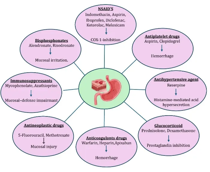
Figure 1: Major Mechanisms of Drug-Induced Peptic Ulcer (DIPU) and Representative Ulcerogenic Drugs
4. Experimental Animal models on Drug induced peptic ulcer
4.1 Indomethacin induced ulcer
Cyclooxygenase (COX) enzymes, which are involved in the manufacture of prostaglandins, are inhibited by indomethacin, an NSAID. Reduced prostaglandin production may exacerbate inflammation and weaken the stomach's protective mucosal barrier, which could result in the development of ulcers (13)
Rats are given 18–60 mg/kg of indomethacin orally or injection. The duration of the investigation is 7–14 days (13,14,15). Indomethacin is injected or given orally to mice at a dose of 18–40 mg/kg, and the study is carried out for 7–14 days (16,17). Indomethacin is injected or given orally to guinea pigs at a dose of 10–100 mg/kg, and the study lasts for seven to fourteen days (18, 19). Indomethacin is given orally or subcutaneously to rabbits at a dose of 20–75 mg/kg, and the trial lasts for seven to fourteen days (20,21).
4.2 Aspirin induced ulcer
Nonsteroidal anti-inflammatory medicines (NSAIDs) like aspirin are frequently prescribed to individuals with peripheral artery disease, coronary heart disease, or cerebrovascular disease.(22) However, research has demonstrated that aspirin exacerbates gastrointestinal (GI) damage by preventing the synthesis of prostaglandins, causing direct cytotoxicity, and producing microvascular damage along with the activation of neutrophils that generate an excess of free radicals obtained from oxygen. Lipid peroxidation and tissue damage are brought on by free radicals.(23)
A popular experimental model for researching NSAID-induced gastrointestinal damage and assessing possible gastroprotective and anti-ulcer medications is aspirin-induced peptic/gastric ulceration. Numerous experimental animals, including rats, mice, guinea pigs, rabbits, and, less frequently, other species, have been used to study it. Rats are the most commonly used of these. The medicine is given orally to rats and mice at a dose of 150–300 mg/kg, and the trial lasts for seven to fourteen days.(24, 25, 26, 27, 28, 29) The drug is given orally to guinea pigs at a dose of 200–500 mg/kg for a duration of 7–14 days.(30, 31) In rabbits, the dose is given orally for 30 days at a level of 150–400 mg/kg.(32, 33, 34).
4.3 Ibuprofen induced ulcer
Ibuprofen's ulcerogenic effect is mainly caused by its inhibition of cyclooxygenase (COX) enzymes, especially COX-1, which reduces the production of protective gastric prostaglandins like PGE? and PGI?. Prostaglandins normally preserve the integrity of the gastric mucosa by promoting mucus and bicarbonate secretion, preserving mucosal blood flow, promoting epithelial restoration, and restricting excessive gastric acid secretion. Therefore, the stomach mucosal defense mechanism is weakened and the mucosa is more vulnerable to damage caused by acid and pepsin when these protective prostaglandins are reduced.(35)
Oxidative stress is also linked to stomach damage caused by ibuprofen. Lipid peroxidation and cellular membrane damage can result from increased reactive oxygen species (ROS) generation. Ibuprofen administration has been linked to increased lipid peroxidation, increased membrane permeability, decreased antioxidant enzyme activity, and increased myeloperoxidase (MPO) activity in experimental rat investigations. These alterations play a role in the formation of stomach ulcers, inflammation, and epithelial damage.(36)
Ibuprofen is given orally or by injection to rats and mice at a dose of 200–400 mg/kg for seven to fourteen days.(37,38,39)
4.4 Diclofenac induced ulcer
A common non-steroidal anti-inflammatory medication (NSAID) for its analgesic and anti-inflammatory properties is diclofenac. On the other hand, erosions, peptic ulcers, and damage to the stomach mucosa might result from prolonged or high-dose administration. Diclofenac-induced stomach damage is mostly linked to the loss of beneficial prostaglandins, suppression of cyclooxygenase (COX) enzymes, and other local irritating effects on the gastric mucosa.(40) Rats and mice are given diclofenac orally or by injection at a dose of 10–100 mg/kg, and the trial is carried out for 7–14 days.(41,42,43)
4.5 Ketorolac induced ulcer
Strong NSAIDs like ketorolac are mostly used to treat pain. However, it can result in peptic ulcers and damage to the stomach mucosa. Because ketorolac inhibits COX-1, less protective prostaglandins are produced. This impairs the gastrointestinal mucosal barrier, reduces mucus and bicarbonate secretion, and increases the stomach's susceptibility to gastric acid and digesting enzyme damage. Additionally, ketorolac may induce stomach ulcers via a mitochondrial mechanism. Pyruvate dehydrogenase (PDH), a crucial enzyme that links glycolysis to the Krebs cycle, can be inhibited by it. This disrupts the generation of energy in the mitochondria, lowers the conversion of pyruvate to acetyl-CoA, and may lower ATP levels. Additionally, it may enhance the production of reactive oxygen species (ROS), which can cause oxidative stress and severe harm to the cells lining the stomach.(44)
In rats, the drug is given orally at a dose of 5–50 mg/kg (45,46) The medication is given orally to mice in doses of 10–30 mg/kg (47,48). 0.5% of opthalmic solution of ketorolac is administered in dogs for 28days (49).
4.6 Meloxicam induced ulcer
Meloxicam primarily causes gastrointestinal ulcers by blocking COX enzymes, which lowers PGE? production, mucus, bicarbonate secretion, and blood flow to the stomach mucosa.
This makes the stomach mucosal barrier weaker and makes the stomach more vulnerable to damage from pepsin and acid.Additionally, meloxicam increases oxidative stress by raising lipid peroxidation and lowering antioxidants like SOD and GSH.These consequences lead to erosion, bleeding, damage to the stomach epithelium, and eventually the development of ulcers.(50)
Rats are given the drug orally for 28 days at a dose of 3.75–30 mg/kg (50). The medication is given orally and subcutaneously to mice at a dose of 10–20 mg/kg for 7 - 14 days (51,52).
4.7 Reserpine induced ulcer
Reserpine is a vesicular monoamine transporter (VMAT) inhibitor that causes sympathetic nerve terminals to lose catecholamines and other monoamines. It has long been used to induce experimental gastrointestinal ulcers in rats, especially as a model of stomach mucosal damage brought on by stress. Thru disruptions in autonomic regulation, stomach secretion, mucosal blood flow, and de-granulation of gastric mast cells as a result of histamine release, which is thot to be regulated by the cholinergic system, reserpine treatment causes gastric mucosal ulcers. (53)
Rats are given intraperitoneal and subcutaneous doses of 5-8 mg/kg of reserpine (54,55,56)The drug is given intraperitoneally and subcutaneously to mice at a dose of 5–10 mg/kg (57) and the trial lasts for 7–14 days.
4.8 Prednisolone induced ulcer
A synthetic glucocorticoid with strong anti-inflammatory and immunosuppressive properties is prednisolone. Experimental investigations have shown that repeated or high-dose prednisolone administration can cause stomach mucosal lesions, despite the fact that corticosteroids are often less ulcerogenic than NSAIDs. The primary cause of prednisolone-induced gastric damage is disruption of the stomach mucosa's natural defensive and repair processes. Prednisolone 50 mg/kg administered subcutaneously once daily for four days caused stomach lesions in rats that were not fasting in a traditional rat paradigm (58)
4.9 5-Fluorouracil induced ulcer
By preventing DNA synthesis and the proliferation of rapidly dividing gastric epithelial cells, 5-fluorouracil (5-FU) can cause damage to the gastric mucosa by decreasing epithelial regeneration and repair. Additionally, it reduces the synthesis of mucus and mucin in the stomach, which weakens the gastrointestinal barrier and exposes the mucosa to more gastric acid. Furthermore, 5-FU can lower blood flow to the stomach mucosa, which can hinder the availability of nutrients and oxygen needed for mucosal healing and defense (59). When 5-FU is given to rats at doses of 50–500 mg/kg (60,61), it increases the mucosal susceptibility to damage, leading to erosion and ulceration as well as delayed healing of stomach ulcers.The medication is given intraperitoneally (i.p.) to mice at a dose of 50–450 mg/kg (62).
4.10 Mycophenolate induced ulcer
Mycophenolate mofetil (MMF) is transformed into mycophenolic acid (MPA), which prevents the fast division of gastrointestinal epithelial cells and inhibits inosine monophosphate dehydrogenase. Mucosal regeneration and repair may be hampered by this. Additionally, MMF causes a local inflammatory response that results in oxidative/inflammatory tissue damage due to elevated myeloperoxidase (MPO) activity and nitric oxide (NO). Mucosal erosion and ulceration may develop as a result of the ensuing epithelium damage, inflammation, and poor repair. Rather than a repeatable isolated gastric ulcer model, the best evidence in rats is for intestinal (especially jejunal and ileal) harm (63). The drug is given orally, intraperitoneally, in rat at a dose of 10-300 mg/kg (64,65). Drug doses in mice range from 20 to 500 mg/kg (66).
4.11 Alendronate induced ulcer
When alendronate comes into direct touch with the stomach mucosa, it acts as a topical irritant that damages the epithelium and disrupts the mucosal barrier. It increases the mucosa's susceptibility by reducing the transmucosal potential differential. Then, alendronate increases oxidative stress by lowering antioxidant defenses like SOD and GSH and increasing lipid peroxidation/MDA and MPO activity. Additionally, it inhibits the protective NO/cGMP/KATP pathway by lowering eNOS and iNOS expression, which lowers nitric oxide (NO) production. Antral ulceration and stomach mucosal damage are the results of the ensuing oxidative and inflammatory damage (67,68). Rats are given the medication orally at a dose of 20–700 mg/kg (69).
| Drug | Animals | Dose(mg/kg/day) | Route | Duration |
|---|---|---|---|---|
| Indomethacin | Rats | 18–60 mg/kg | P.O , I.P | 7-14 days |
| Mice | 18–40 mg/kg | P.O, I.P | 7-14 days | |
| Guinea pig | 10–100 mg/kg | P.O, I.P | 7-14 days | |
| Rabbit | 20–75 mg/kg | P.O , S.C | 7-14 days | |
| Aspirin | Rats | 150–300 mg/kg | P.O | 7-14 days |
| Mice | 150–300 mg/kg | P.O | 7-14 days | |
| Guinea pig | 200–500 mg/kg | P.O | 7-14 days | |
| Rabbit | 150–400 mg/kg | P.O | 30 days | |
| Ibuprofen | Rat | 200–400 mg/kg | P.O, I.P | 7-14 days |
| mice | 200–400 mg/kg | P.O, I.P | 7-14 days | |
| Diclofenac | Rat | 10–100 mg/kg | P.O, I.P | 7-14 days |
| mice | 10–100 mg/kg | P.O, I.P | 7-14 days | |
| Ketorolac | Rat | 5-50mg/kg | P.O | 28 days |
| Mice | 10-30mg/kg | P.O | 28days | |
| Dog | 0.5% Opthalmic solution | Opthalmic | 28 days | |
| Meloxicam | Rat | 3.75–30 mg/kg | P.O | 28 days |
| Mice | 10–20 mg/kg | P.O , S.C | 7-14 days | |
| Reserpine | Rat | 5-8 mg/kg | P.O , S.C | 7-14 days |
| mice | 5–10 mg/kg | P.O , S.C | 7-14 days | |
| Prednisolone | rat | 50 mg/kg | S.C | 4 days |
| Rat | 0.21–0.25 ml/kg of 30% (low dose) to 80% (high dose) | Topical | 7-14 days | |
| Mice | 0.1 ml of 20% | Topical | 7-14 days | |
| Rabbit | 0.2 ml of 60% | Topical | 30 days | |
| 5-Fluorouracil | Rat | 50-500mg/kg | I.P | 7-14 days |
| Mice | 50-450mg/kg | I.P | 7-14 days | |
| Mycophenolate | Rat | 10-300mg/kg | I.P | 7-14 days |
| Mice | 20-500mg/kg | I.P | 7-14 days | |
| Alendronate | Rat | 20-700mg/kg | P.O | 7-14 days |
5. Biomarkers of Peptic ulcer
5.1 Macroscopic biomarkers
The following are Ulcer Scores (51)
0 = lesions are absent
1 = tiny, spherical hemorrhagic areas
2 = Lesions <2 mm
3 = 2–3 mm lesions
4 = lesions 3–4 mm
5 indicates lesion > 4 mm
The ulcer index, the percentage protective ratio, and the percentage curative ratio are, respectively, given by the following equation:
Ulcer Index = Total ulcer score
No. of animals ulcerated
Protection inhibition (%) = [(UA control − UA treated) / UA control] × 100
5.2 Gastric secretion biomarkers
Acidity (meq/l/100 g) was calculated by the equation (acidity = VolumeNaOH ×NormalityNaOH × 100/0.1).
Gastric wall mucus: Gastric wall mucus was determined using the Alcian blue binding method. Briefly, the glandular part of the stomach was separated after opening it along the greater curvature and weighed. The tissue was then immersed in 10 mL of 0.1% (w/v) Alcian blue solution prepared in 0.16 M sucrose and buffered with 0.05 M sodium acetate. The pH of the solution was adjusted using hydrochloric acid (HCl), and the tissue was allowed to remain in the dye solution for 2 hours. Following incubation, the tissue was washed twice with 10 mL of 0.25 M sucrose solution for 15 and 45 minutes, respectively, to eliminate unbound dye. The mucus-bound Alcian blue was subsequently extracted using 10 mL of 0.5 M magnesium chloride (MgCl?), with the mixture being shaken for one minute at 30-minute intervals over a period of 2 hours. An equal volume of diethyl ether was then added, and the mixture was shaken thoroughly before centrifugation at 3000 rpm for 10 minutes. The absorbance of the separated aqueous layer was measured at 580 nm against a magnesium chloride blank using a spectrophotometer. The concentration of Alcian blue was determined with the help of a standard calibration curve, and the gastric wall mucus content was expressed as micrograms of Alcian blue per gram of glandular stomach tissue (µg/g).(60)
5.3 Oxidative strees biomarkers :
5.4 Inflammatory biomarkers:
5.5 Pro Inflammatory cytokines:
Table 3: Changes in biomarkers commonly reported in experimental Drug-Induced Peptic ulcer (DIPU).
| Biomarker | Change in DIPU | Range in DIPU animal studies | Ref |
|---|---|---|---|
| Ulcer score | Increased | 29.0 + 1.7 | 50 |
| Ulcer index | Increased | 22.57 + 6.86 mm | 65 |
| Ulcer area | Increased | mm2 | 53,54,55 |
| Gastric volume | Increased | 4.69 + 0.37 mL | 56,57 |
| Gastric pH | Decreased | 2.78+0.25 pH | 55,56,57,59 |
| Free acidity | Increased | 9.6 + 0.6 mEq/L | 55,58,59 |
| Total Acidity | Increased | 20.8 + 2.5 mEq/L | 58,59 |
| Gastric wall mucus | Decreased | 100.33 + 0.76 µg | 60 |
| MDA | Increased | 59.73 + 11.20 nmol/mg tissue | 14,55 |
| GSH | Decreased | 22.18 + 5.42 mmol/mg tissue | 37 |
| SOD | Decreased | 1.003 + 0.19 U/mg tissue | 38 |
| CAT | Decreased | 14.29 + 0.48 µmol/L | 38 |
| Nitric oxide | Decreased | 8.02 + 2.75 nmol/mg tissue | 58,61,63 |
| PGE? | Decreased | 72.50 + 0.593 ng/mg protein | 62,63 |
| TNF-α | Increased | 796.5 + 13.7 | 58,63 |
| IL-1β | Increased | - | 58,63 |
| IL-6 | Increased | - | 58,63 |
6. Future Perspective on drug induced peptic ulcer in experimental animals.
6.1 Development of Advanced Animal Models
Future research should focus on developing advanced experimental animal models that closely resemble clinical drug-induced peptic ulcers. Most conventional studies use a single ulcer-inducing drug, such as indomethacin or aspirin, to produce acute gastric injury. However, future studies should investigate combination-drug models, such as NSAIDs with corticosteroids or antiplatelet agents, to understand the effects of multiple medications on gastric mucosal damage. Such models may provide better insights into drug interactions and ulcer complications.(7)
6.2 Investigation of Chronic Ulcer Formation and Healing
Most experimental studies focus on acute gastric ulcer formation. Future investigations should emphasize chronic ulcer development, delayed healing, recurrence, and tissue regeneration. Acetic acid-induced chronic gastric ulcer models in rats can be used to investigate ulcer healing, angiogenesis, mucosal regeneration, and recurrence. Researchers should also examine the effects of prolonged NSAID exposure on matrix metalloproteinases (MMPs), growth factors, and tissue remodeling.(7)
6.3 Identification of Novel Molecular and Biochemical Biomarkers
Future studies should investigate additional molecular pathways involved in drug-induced gastric injury. Apart from conventional oxidative stress markers such as MDA, GSH, SOD, and CAT, researchers should evaluate inflammatory cytokines, apoptosis-related proteins, and angiogenic factors. Particular attention should be given to NF-κB, Nrf2, COX-1, COX-2, caspases, VEGF, and matrix metalloproteinases. These investigations may help identify new therapeutic targets for gastroprotection.(84)
6.4 Development of Gastric Organoid and Stomach-on-a-Chip Models
Gastric organoids and stomach-on-a-chip technologies may provide additional platforms for investigating drug-induced gastric mucosal injury. These models can help study epithelial barrier damage, cellular responses, and drug toxicity before conducting animal experiments. In the future, combining these techniques with rat and mouse models may improve the reliability of preclinical research and reduce unnecessary animal use.(85)
6.5 Translational Research and Development of Safer Gastroprotective Agents
Future investigations should focus on translating experimental findings into clinically useful gastroprotective treatments. Newly developed compounds should be evaluated for their ability to prevent gastric mucosal injury without interfering with the therapeutic activity of NSAIDs. Standardized experimental protocols, reproducible results, and comprehensive toxicological evaluations will be important for successful translation into clinical applications.(84)
CONCLUSION
Drug-induced peptic ulcer is an important gastrointestinal disorder commonly associated with the use of NSAIDs and other ulcerogenic medications. Experimental animal models, particularly rats and mice, play a significant role in understanding the mechanisms involved in gastric mucosal damage and evaluating the gastroprotective potential of various therapeutic agents. Different ulcer induction models, including indomethacin, aspirin, ibuprofen, diclofenac, ketorolac, and other drugs, help researchers investigate the pathophysiology of gastric ulceration. Assessment of macroscopic, biochemical, and histopathological parameters provides valuable information regarding ulcer severity, oxidative stress, inflammation, and mucosal protection. Recent studies have highlighted the potential of medicinal plants, phytochemicals, antioxidants, probiotics, and synthetic compounds in preventing and treating gastric ulcers. Future research should focus on developing advanced experimental models, identifying novel molecular biomarkers, investigating chronic ulcer healing, and exploring alternative technologies such as gastric organoids. Overall, experimental animal studies provide a strong foundation for discovering safer and more effective gastroprotective agents and translating preclinical findings into potential clinical applications.
REFERENCES

Savanth Swetha, Nabila Kulsum Abdul Aleem, Dr. Venu Talla, Drug Induced Peptic Ulcer in Experimental Animals, Int. J. of Pharm. Sci., 2026, Vol 4, Issue 11,
 10.5281/zenodo.24156023