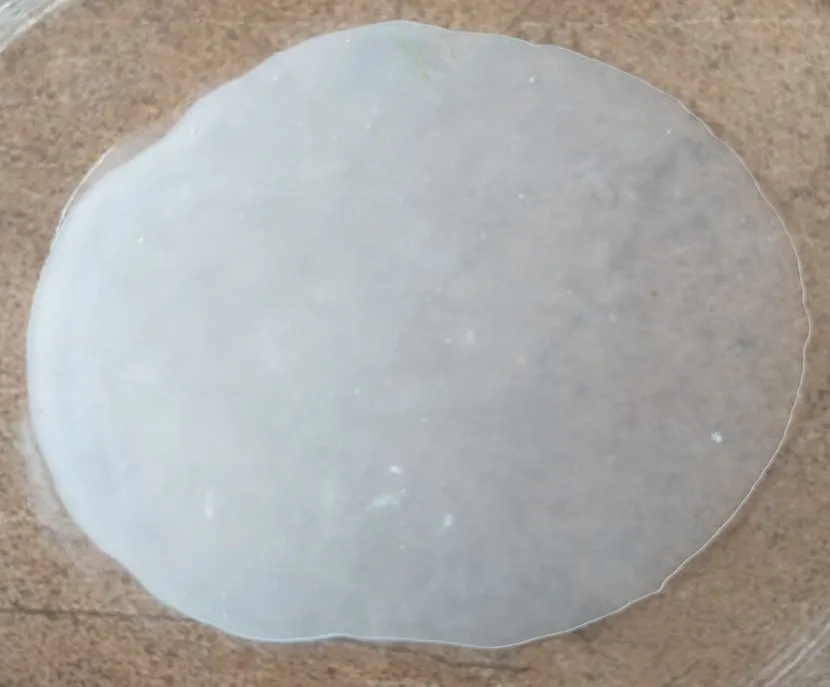
View Article

Abstract

This study aimed to develop and evaluate a topical gel containing Terbinafine Hydrochloride and aloe vera by dispersion method. The combination of antifungal activity with aloe vera soothing and moisturizing properties may provide an effective topical treatment for fungal infections. Terbinafine Hydrochloride was characterized for organoleptic properties, melting point (197°C), ?max (282.1 nm), partition coefficient (2.23 ± 0.003), solubility, and FTIR compatibility, confirming purity and absence of drug–excipient interactions. Three formulations (F1–F3) containing different concentrations of Carbopol 934 were prepared and evaluated for appearance, pH, drug content, spreadability, grittiness, skin irritation, washability, and in vitro drug diffusion. All gels were white and homogeneous; F1 was fluid and runny, F2 was smooth and semisolid, and F3 was slightly thick and viscous. The pH values were 6.44 ± 0.03, 6.53 ± 0.02, and 6.55 ± 0.03 for F1, F2, and F3, respectively, indicating skin compatibility. Drug content was 84.38 ± 0.5%, 98.58 ± 0.2%, and 87 ± 0.2%, respectively, with F2 highest. In vitro drug release was 79.50%, 94.49%, and 91.30% for F1, F2, and F3, respectively, with F2 showing the highest release after 8 hrs. F1 and F2 showed no grittiness or irritation and were easily washable, whereas F3 showed slight grittiness and moderate washability without irritation. Spreadability was 22.83 ± 0.020, 18.06 ± 0.010, and 15.23 ± 0.020, respectively. Overall, F2 could serve as an effective formulation for soothing, moisturizing property and effective treatment of fungal infections.

Keywords

Terbinafine Hydrochloride, Aloe vera, Gel formulation, Drug content, In vitro drug release

Introduction

× Popup Image

SKIN

The skin is the largest organ of the human body and serves as a dynamic interface between the individual and the external environment. Its major role is to provide a protective barrier against various environmental stressors, including ultraviolet (UV) radiation, chemical and physical agents, and pathogenic microorganisms. In addition to its protective function, the skin plays an important role in preventing excessive water loss, maintaining thermoregulation, and facilitating tissue repair and regeneration following injury. 1

Skin is composed of an upper layer (the epidermis), and a lower layer (the dermis) which is divided by a membrane in the basement.

Epidermis: The epidermis is composed of five distinct layers arranged from the outermost to the innermost region: the stratum corneum, stratum lucidum, stratum granulosum, stratum spinosum, and stratum basale. The stratum corneum forms the outermost protective layer of the epidermis. The stratum lucidum is a thin, translucent layer found primarily in areas of thick skin, such as the palms of the hands and soles of the feet. The stratum granulosum and stratum spinosum occupy the intermediate regions, whereas the stratum basale constitutes the deepest layer and contains epidermal stem and progenitor cells responsible for continuous epidermal renewal.

Dermis: The dermis is located beneath the epidermis and is primarily composed of connective tissue rich in collagen fibers. It is structurally divided into two regions: the superficial papillary dermis, which contains relatively fine collagen fibers and forms dermal papillae, and the deeper reticular dermis, which consists predominantly of thicker and more densely organized collagen fibers. Together, these layers provide structural strength, elasticity, and support to the overlying epidermis.

Hypodermis or Subcutaneous Tissue: The hypodermis, also referred to as the subcutaneous tissue, is composed primarily of loose connective tissue and elastin fibers. It plays several important physiological roles, including energy and nutrient storage, mechanical protection against physical impact, and thermal insulation. The hypodermal layer is particularly well developed in regions such as the hands, buttocks, and plantar surfaces of the feet. With advancing age, progressive loss or atrophy of subcutaneous tissue occurs, which contributes to reduced skin thickness and the development of fine lines and wrinkles.

In addition to its structural functions, the skin constitutes an important component of the immune system and contains a diverse population of both innate and adaptive immune cells. These include mast cells, macrophages, natural killer (NK) cells, specialized antigen-presenting cells (APCs), epidermal dendritic cells (EDCs), commonly known as Langerhans cells, and dermal dendritic cells (DCs), also referred to as interstitial or migratory dendritic cells. Furthermore, the cutaneous immune environment contains αβ T cells, γδ T cells, and B cells, which collectively contribute to immune surveillance and defense of the skin.
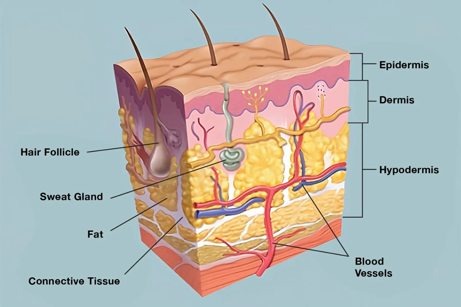
Fig. No. 1: Skin Anatomy

Following disruption or impairment of the cutaneous barrier due to trauma, a coordinated repair process is initiated to restore the structural integrity and physiological functions of the skin. In cases of minor injury, this reparative process generally requires approximately one week for completion. 2

Functions of the Skin

  • Protection: The skin forms a relatively impermeable barrier that protects underlying tissues from external environmental influences and minimizes excessive loss of water from the body.
  • Thermoregulation: The skin, together with its appendages such as hair, plays a fundamental role in maintaining body temperature by regulating heat exchange between the body and its surrounding environment.
  • Sensation: A dense network of sensory nerve endings within the skin detects a wide range of external stimuli, including touch, pressure, temperature, and pain. These sensory signals are rapidly transmitted to the central nervous system, enabling appropriate physiological and reflex responses.
  • Water and electrolyte homeostasis: The barrier properties of the skin are essential for maintaining fluid balance by limiting trans epidermal water loss and thereby contributing to the regulation of water and electrolyte levels within the body.
  • Absorption: The skin has the capacity to facilitate the penetration of certain substances, including water-soluble compounds. However, substances with lipophilic characteristics generally penetrate the skin more efficiently than highly hydrophilic compounds.
  • Social expression: Particularly in the facial region, the skin contributes significantly to appearance, facial expression, communication, and social interaction. These functions play an important role in interpersonal recognition and social behavior.
  • Vitamin D synthesis: The skin is a major site of vitamin D synthesis, particularly following exposure to ultraviolet radiation from sunlight. Under normal physiological conditions, cutaneous production represents an important source of vitamin D for the body. 3

Skin Surface pH and Cutaneous Homeostasis

Healthy skin is characterized by a mildly acidic surface environment, with the physiological pH of the skin generally ranging from approximately 4 to 6. In contrast, the deeper internal tissues maintain a relatively neutral to mildly alkaline environment, resulting in a distinct pH gradient between the stratum corneum and the underlying epidermal and dermal layers. This acidic surface environment, commonly known as the acid mantle, has traditionally been regarded as an important defense mechanism that limits the colonization and proliferation of potentially pathogenic microorganisms.

However, accumulating evidence suggests that the biological significance of skin pH extends beyond antimicrobial protection. The activity of several enzymes involved in the synthesis, organization, and maintenance of the epidermal barrier is strongly influenced by the local pH environment. Consequently, preservation of the appropriate acidic conditions at the skin surface is increasingly recognized as an essential factor in maintaining epidermal barrier function, tissue integrity, and overall cutaneous homeostasis. 4

FUNGAL INFECTION

Fungal infections of the skin are among the most common dermatological disorders. They are caused by microscopic fungi that colonize and invade epithelial tissues. The fungal kingdom comprises various organisms, including yeasts, molds, rusts, and mushrooms. Excessive growth or proliferation of pathogenic fungi may result in symptomatic infections affecting the skin, oral mucosa, vagina, and gastrointestinal tract. 5

Types of fungal infections:

  • Cutaneous fungal infections: These primarily affect the skin and may involve areas such as the feet and hands, including infections commonly referred to as ringworm.
  • Mucosal fungal infections: These involve mucous membranes and commonly include vaginal candidiasis.
  • Systemic fungal infections: These occur when fungi disseminate beyond localized tissues and enter the bloodstream, potentially resulting in systemic infection.

Candida is a common fungal pathogen that can cause infections of the skin and mucosal surfaces in humans. Although Candida species are part of the normal human microbiota and may colonize mucosal sites without causing disease, their excessive proliferation or invasion can lead to opportunistic infections. Among these species, Candida albicans is one of the most clinically significant pathogens affecting humans. In certain circumstances, C. albicans can cause serious infections, including ocular infections that may adversely affect eye health. 6

ANTIFUNGAL AGENTS

An antifungal agent is a therapeutic compound that targets and inhibits fungal pathogens while causing minimal toxicity to the host.

Commonly used antifungal agents include Amphotericin B, Fluconazole, Terbinafine hydrochloride, and Amorolfine. 7

Classification and Mechanism of Action of Antifungal Agents:

  1. Polyene antifungals: These agents interact with ergosterol present in the fungal cell membrane, leading to membrane disruption and increased permeability, which ultimately compromises cellular integrity.
  2. Azole antifungals: Azoles inhibit the enzyme lanosterol 14α-demethylase, thereby interfering with ergosterol biosynthesis. This disruption impairs the normal development and structural integrity of the fungal cell membrane.
  3. Echinocandins: These antifungal agents inhibit the synthesis of β-(1,3)-D-glucan, an essential component of the fungal cell wall. Consequently, inhibition of glucan production weakens the cell wall and compromises its structural stability.
  4. Allylamines: These antifungal agents target and inhibit squalene epoxidase, a key enzyme involved in the ergosterol biosynthetic pathway. Inhibition of this enzyme disrupts ergosterol synthesis and consequently affects the integrity and function of the fungal cell membrane.
  5. Pyrimidine analogues: These agents interfere with fungal nucleic acid metabolism through the formation of active metabolites, resulting in inhibition of DNA and RNA synthesis and thereby restricting fungal growth and proliferation.

Mechanisms of Antifungal Resistance:

  1. Efflux pump overexpression: Fungal cells may increase the activity or expression of efflux transporters, promoting the removal of antifungal agents from the cell and consequently reducing their intracellular concentrations.
  2. Alteration of drug targets: Genetic mutations or other modifications in the molecular targets of antifungal drugs, including lanosterol 14α-demethylase and β-(1,3)-D-glucan synthase, can decrease drug–target interaction and thereby reduce antifungal efficacy.
  3. Modifications in the ergosterol biosynthetic pathway: Modification or upregulation of alternative steps in the ergosterol biosynthetic pathway enables fungi to compensate for drug-induced inhibition.
  4. Biofilm formation: The development of fungal biofilms creates a protective extracellular environment that can restrict antifungal drug penetration, promote cellular persistence, and contribute to reduced susceptibility to antifungal therapy. 8

PHARMACEUTICAL GEL

Gels are semisolid formulations designed for application to the skin or accessible mucous membranes. They consist of two interpenetrating phases, in which colloidal particles, referred to as gelling agents or gelators, are uniformly dispersed within a solvent to form a three-dimensional polymeric network.

Gel formulations are generally prepared by incorporating a gelling agent into an aqueous, organic, or inorganic solvent system. The gelling agents may be derived from natural, synthetic, or semisynthetic polymers, as well as from low-molecular-weight compounds (Fig. 2).
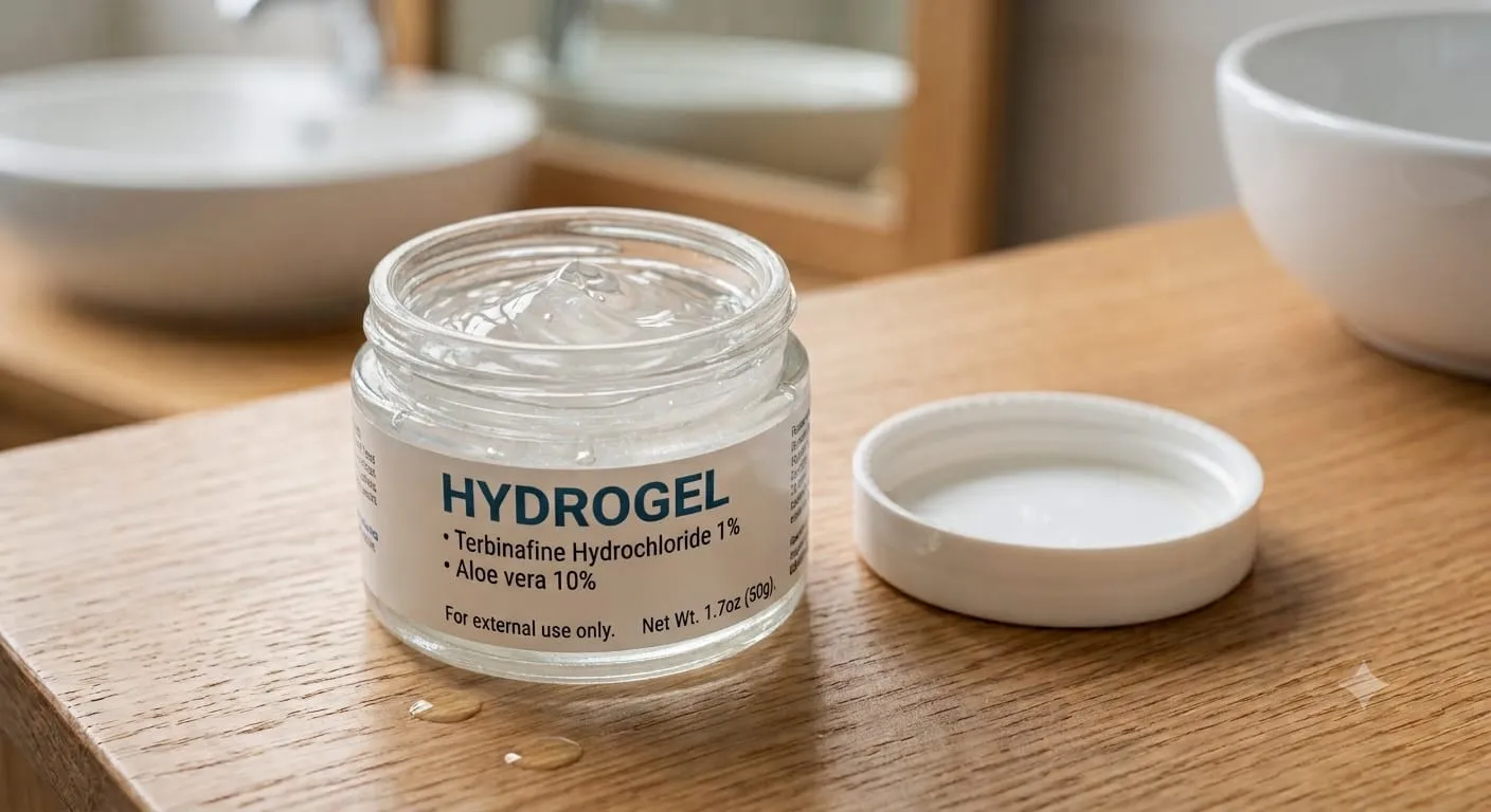
 

Fig. No. 2: Pharmaceutical Hydrogel

Advantages of Gel Formulations

  • Gel formulations are relatively simple to prepare compared with other semisolid dosage forms.
  • Gels have an elegant and non-greasy appearance, which makes them suitable and acceptable for topical application.
  • Gels can be formulated to provide controlled drug release by incorporating and entangling suitable polymers within the formulation.
  • They possess good adhesive properties, allowing them to remain at the site of application for a longer period.
  • Many gel formulations exhibit good biodegradability and biocompatibility.
  • Gels generally have a longer residence time at the site of application compared with several other topical dosage forms.
  • They can maintain their physical characteristics under various mechanical and environmental stress conditions.
  • Gels may form a protective film or layer over the application site, thereby providing protection from external influences.
  • They are generally easy to wash off and can be formulated to be non-toxic and well tolerated.
  • Gels exhibit good spreadability and can provide a cooling sensation due to the evaporation of volatile solvents present in the formulation.
  • Properly formulated gels may have fewer long-term stability problems compared with certain other semisolid preparations.
  • Gel systems are versatile drug delivery vehicles and can be designed to incorporate both hydrophilic and hydrophobic drugs.

Disadvantages of Gel Formulations

  • Gel formulations may exhibit a relatively delayed onset of action and are generally associated with sustained drug release.
  • Certain gelling agents, polymers, or other excipients incorporated into the formulation may cause irritation at the site of application.
  • The high water content of many gel formulations can provide suitable conditions for microbial and fungal proliferation, potentially affecting their stability and safety.
  • Syneresis, characterized by the expulsion of liquid from the gel network, may occur during storage and can adversely affect the physical stability of the formulation.
  • Evaporation of the solvent from the formulation may cause dehydration and drying of the gel, resulting in changes in its consistency and performance.
  • In some gel systems, strong covalent interactions within the polymeric network may restrict drug diffusion and lead to entrapment of the drug within the gel matrix.
  • Flocculation may occur in certain gel formulations, potentially resulting in structural instability and deterioration of the formulation.
  • The rheological characteristics of gels may be altered by environmental conditions such as temperature, humidity, and other external factors, thereby affecting their consistency and application properties.
  • Under certain conditions, precipitation or salting-out of the gelling agents may occur, which can negatively influence the physical stability and uniformity of the formulation.
  • Some drug molecules may undergo degradation within gel formulations due to interactions with polymers or other components present in the formulation.

Classification

  1. Hydrogels: Hydrogels are three-dimensional polymeric networks characterized by a high water content, which may constitute up to approximately 90% of the total gel composition. Their interconnected polymeric structure enables them to absorb and retain substantial quantities of water while maintaining their structural integrity. Common examples of hydrogels include gelatin and cellulose derivatives.
  2. Organogels: Organogels are also known as oleaginous gels, are semisolid systems composed of both polar and nonpolar components, with a predominance of the nonpolar fraction. These gels may incorporate up to approximately 35% water and can exhibit swelling upon hydration. 9

Common examples include Olag gels and dispersions of metallic stearates in oils.

  1. Xerogels: Xerogels are solid, porous gel materials produced by the gradual drying of a gel at room temperature under conditions that allow unrestricted shrinkage. Upon heating to elevated temperatures, these materials undergo viscous sintering, resulting in the progressive collapse of the porous structure and its transformation into a dense, glass-like material.

Tragacanth ribbons are an example of a xerogel.

Uses of Gels

  • Gels are widely utilized as topical drug delivery systems for the localized administration of therapeutic agents to the skin, mucous membranes, and ocular tissues.
  • In pharmaceutical manufacturing, gels can function as granulating agents for tablets, protective colloids in suspension formulations, and viscosity-enhancing agents in oral liquid preparations and suppository bases.
  • Gels are commonly incorporated into shampoos, dentifrices, and various skin- and hair-care formulations due to their suitable consistency, spreadability, and stability.
  • They can be employed as lubricating agents for catheters, facilitating smooth insertion and minimizing friction.
  • Sodium chloride (NaCl) gels are used as conductive media in electrocardiographic (ECG) procedures to improve electrical contact between the electrodes and the skin.
  • Phosphoric acid gels and sodium fluoride gels are used in dental care formulations, particularly for dental treatment and preventive oral healthcare applications. 10

Formulation Considerations for Pharmaceutical Gels

  1. Selection of vehicle or solvent: Purified water is commonly employed as the primary solvent in gel formulations. The incorporation of co-solvents may be considered to enhance the solubility of the active pharmaceutical ingredient and/or facilitate its penetration through the skin. Commonly used co-solvents include alcohols, glycerol, propylene glycol, and polyethylene glycol 400.
  2. Incorporation of buffers: Buffer systems are incorporated into aqueous and hydroalcoholic gel formulations to maintain the desired pH and thereby improve formulation stability. However, the solubility of buffer salts may decrease in hydroalcoholic vehicles; therefore, appropriate consideration should be given to their selection and concentration. Phosphate and citrate buffer systems are commonly employed.
  3. Use of preservatives: Preservatives are incorporated into gel formulations to prevent microbial growth and maintain product stability during storage. However, interactions between preservatives and hydrophilic polymers used as gelling agents can reduce the concentration of free preservative available for antimicrobial activity. Consequently, the initial concentration of the preservative may need to be optimized to compensate for such interactions. Parabens and phenolic compounds are among the preservatives commonly used in pharmaceutical gels.
  4. Incorporation of antioxidants: Antioxidants may be included to enhance the chemical stability of active pharmaceutical ingredients that are susceptible to oxidative degradation. The selection of an antioxidant depends largely on the nature of the vehicle used for gel preparation. Since most pharmaceutical gels are predominantly aqueous, water-soluble antioxidants such as sodium metabisulfite and sodium formaldehyde sulfoxylate are generally preferred.

Methods of gel preparation

Gels can be prepared by following methods:

  1. Thermal Changes - Gel formation can occur through changes in temperature, particularly in solvated polymers or lyophilic colloidal systems. Many hydrophilic polymers exhibit greater solubility and hydration at elevated temperatures. Upon cooling, the degree of hydration decreases, promoting intermolecular interactions and resulting in gel formation. Thus, cooling a concentrated hot polymer solution can lead to the formation of a gel. This method is commonly applicable to materials such as gelatin, agar, sodium oleate, guar gum, and certain cellulose derivatives. However, thermal gelation is not universally applicable. For example, some cellulose ethers exhibit solubility in water due to hydrogen bonding between the polymer and water molecules. An increase in temperature can disrupt these hydrogen bonds, thereby reducing polymer solubility and inducing gel formation.
  2. Flocculation - In the flocculation method, gel formation is achieved by adding an appropriate amount of a precipitating agent to a colloidal system. The quantity of the precipitant must be sufficient to induce a flocculated state but insufficient to cause complete precipitation of the dispersed phase. Rapid and uniform mixing is essential to prevent localized areas of high precipitant concentration.

For example, solutions of ethyl cellulose or polystyrene in benzene can undergo gelation following rapid mixing with an appropriate quantity of a non-solvent, such as petroleum ether. Although the addition of salts to hydrophobic colloidal systems generally promotes coagulation, gel formation is less commonly observed. Gels produced through flocculation typically exhibit thixotropic behavior.

  1. Chemical Reaction - Chemical gelation involves the formation of a three-dimensional gel network through a chemical interaction between the reactants present in the system. The nature and concentration of the reactants play an important role in determining gel formation and structure.

For instance, aluminium hydroxide gel can be prepared through the reaction of an aluminium salt with sodium carbonate in an aqueous medium. Increasing the concentration of the reactants can promote the development of a more pronounced gel structure. 11

  1. Fusion technique - In the fusion method, the drug, gelling agent, other formulation components, and vehicle are combined and heated with continuous mixing. The mixture is maintained at an elevated temperature until a uniform, semi-solid gel consistency is obtained.
  2. Cold method - In this method, all formulation ingredients except the active pharmaceutical ingredient are heated and mixed thoroughly. Afterward, the mixture is allowed to cool to an appropriate temperature, followed by incorporation of the drug. The formulation is then mixed adequately until a homogeneous gel is formed.
  3. Dispersion method - The dispersion method involves gradual incorporation of the gelling agent into water with continuous mixing and allowing it to hydrate or swell adequately. The drug is separately dissolved or dispersed in the appropriate medium and subsequently incorporated into the hydrated polymer. Where required, the pH of the formulation is adjusted using a suitable buffer solution to obtain the desired gel characteristics. 12

ALOE VERA

Aloe vera is widely recognized for its therapeutic and medicinal properties and has been used in traditional medicine for thousands of years. Historical evidence indicates that its therapeutic use dates back approximately 6,000 years to ancient Egypt, where it was revered as the “plant of immortality.” Due to its reported wound-healing and growth-promoting properties, Aloe vera is also commonly referred to as a “healing plant” 13

Aloe barbadensis Mill., widely referred to as Aloe vera, is a succulent, fleshy xerophytic plant that was traditionally classified within the family Liliaceae. However, based on more recent taxonomic classifications, the species is placed under the family Aloaceae. 14

Aloe vera is a significant medicinal plant with considerable commercial demand worldwide. It is widely traded in the medicinal and pharmaceutical markets, particularly for its use as a flavouring agent in liquid preparations and as a major source of aloin, which constitutes approximately 4.5–25% of its composition. 15

Synonyms and Nomenclature

Aloe barbadensis Miller has been described under several synonymous names, including Aloe chinensis Bak., Aloe elongata Murray, Aloe indica Royle, Aloe officinalis Forsk.

Taxonomic Description

Aloe vera is a perennial succulent herb characterized by a short, sessile stem and a relatively shallow root system. Its thick, fleshy leaves are arranged in a basal rosette and possess serrated margins. The leaves are generally pea-green in colour and measure approximately 30–50 cm in length, with a basal width of nearly 10 cm. The plant produces bright yellow, tubular flowers arranged in axillary spikes, with flower stalks measuring approximately 25–35 cm in length. The stamens commonly extend beyond the perianth tube, and the fruits contain numerous seeds.

Active Constituents

The leaves of Aloe vera consist of three distinct layers. The outermost layer, or protective epidermal region, is approximately 15–20 cells thick and is involved in the synthesis of carbohydrates and proteins. The biological activity of Aloe vera is attributed to a diverse range of phytoconstituents, including anthraquinones, chromones, polysaccharides, and enzymes. Anthraquinones and chromones have been associated with several pharmacological effects, including anticancer, anti-inflammatory, and laxative or evacuant activities. In addition to these organic constituents, Aloe vera gel has also been reported to contain various mineral elements, including aluminium (Al), boron (B), barium (Ba), calcium (Ca), iron (Fe), magnesium (Mg), sodium (Na), phosphorus (P), and silicon (Si). 16

Biological activities of Aloe vera leaf gel

  • Anti-diabetic effects - In a study involving streptozotocin-induced diabetic rats, oral administration of the alcohol-insoluble residue extract of Aloe vera gel significantly lowered fasting blood glucose levels, hepatic transaminase activities, and plasma and tissue concentrations of cholesterol, triglycerides, free fatty acids, and phospholipids. Furthermore, treatment with the extract resulted in a significant elevation of plasma insulin levels.
  • Immunomodulatory effects - Several studies have reported that polysaccharides present in Aloe vera gel exhibit immunomodulatory properties. These effects are thought to be mediated primarily through the activation of macrophages, resulting in enhanced nitric oxide production, secretion of cytokines such as tumor necrosis factor-alpha (TNF-α), interleukin-1 (IL-1), interleukin-6 (IL-6), and interferon-gamma (IFN-γ), as well as increased expression of cell-surface markers.
  • Anti-inflammatory effects - Aloe vera gel has been reported to attenuate inflammation induced by agents that promote prostaglandin synthesis and enhance leukocyte infiltration. However, its anti-inflammatory activity appears to be comparatively limited against inflammation triggered by allergic mechanisms.
  • Anti-oxidant effects - Aloe vera gel contains antioxidant enzymes, including glutathione peroxidase and superoxide dismutase, along with phenolic compounds, which may contribute to its overall antioxidant activity.
  • Wound-Healing Effects - Wound healing is a complex biological process that restores tissue integrity following injury. Previous studies have demonstrated that aloe vera gel may promote wound healing by enhancing tissue repair following both topical and systemic administration.
  • Anti-cancer Effects - The anti-cancer potential of Aloe vera has been primarily attributed to two major bioactive fractions, namely glycoproteins, particularly lectins, and polysaccharides. Among these, polysaccharides such as acemannan have been extensively investigated for their potential antitumor properties using various in vitro models and experimental animal systems. Several studies have reported that Aloe vera gel and its bioactive constituents may exert antitumor effects, as evidenced by reductions in tumor burden and tumor size, increased tumor necrosis, and improved survival rates. 17

TERBINAFINE HYDROCHLORIDE

Terbinafine hydrochloride (TFH) is an allylamine antifungal compound chemically designated as (2E)-6,6-dimethylhept-2-en-4-yn-1-yl(naphthalen-1-ylmethyl) amine hydrochloride. It has a molecular formula of C₂₁H₂₆NCl and a molecular weight of 327.89 g/mol. TFH exhibits a melting point in the range of 195–198 °C. It is freely soluble in methanol and methylene chloride, soluble in ethanol, and slightly soluble in water. 18

Due to its highly lipophilic nature, the compound exhibits a strong tendency to accumulate in the skin, nails, and adipose tissues. It may also interact with or persist in microbial populations within the duodenum and in tissues associated with gastric viral infections. 19

It has a pKa value of 7.1 and it exhibits high therapeutic efficacy in the treatment of dermatophyte infections. 20

Terbinafine hydrochloride is administered through both topical and oral routes for the management of a wide range of fungal infections, with particular efficacy against Candida species. 21

The drug demonstrates potent fungicidal activity against dermatophytes, molds, fungi, and certain yeast species. Owing to its high efficacy against dermatophytes, terbinafine is considered a drug of choice for the treatment of dermatophyte-associated nail infections. Its antifungal activity is primarily mediated through the inhibition of squalene epoxidase, an essential enzyme involved in an early stage of fungal ergosterol biosynthesis. Inhibition of this enzyme results in the intracellular accumulation of squalene, which contributes to disruption of fungal cell integrity and ultimately produces a fungicidal effect. 22

The gel dosage form is non greasy, easy to apply, and provides good patient acceptability. The combination of Terbinafine Hydrochloride and Aloe vera results in a synergistic therapeutic effect. Therefore, the development of a Terbinafine Hydrochloride and Aloe vera gel is a promising strategy for the effective management of fungal infections.

MATERIALS AND METHODS

MATERIALS USED IN GEL FORMULATION:

Glycerin (glycerol) is a naturally occurring, three-carbon polyol with prominent humectant properties. It is widely utilized as a pharmaceutical carrier and excipient in the formulation of various medicinal products. 23

Carbopol 934 was utilized as the primary gelling agent to impart viscosity and maintain the consistency of the gel formulation. Methyl paraben was incorporated as preservatives to enhance the stability of the prepared formulation. Propylene glycol served as a penetration enhancer and contributed to improved skin hydration, Triethanolamine was employed to adjust the pH of the formulation. Distilled water was used as the vehicle and served as the aqueous medium for gel preparation. 24

Table No. 1: Formulation Ingredients and their roles

Sr. No

Ingredients

Role

1

Terbinafine Hydrochloride

Antifungal Drug

2

Aloe vera

Soothing agent and moisturizer to reduce irritation often associated with fungal infections

3

Glycerine

Humectant

4

Carbopol 934

Gelling agent providing viscosity

5

Methyl paraben

Preservative

6

Propylene glycol

Penetration enhancer

7

Triethanolamine

pH adjuster

8

Distilled water

Solvent for preparation

METHADOLOGY:

  1. PREFORMULATION STUDIES OF TERBINAFINE HYDROCHLORIDE

Preformulation testing is a crucial precursor in drug development, involving the thorough evaluation of a candidate drug's physicochemical characteristics prior to compounding. A comprehensive understanding of these properties informs rational formulation design, identifies potential development barriers, and indicates whether molecular modifications are necessary. Ultimately, these investigations serve as an essential foundation for establishing safe, stable, and therapeutically effective dosage forms.

  1. Organoleptic Characteristics – The drug sample was characterized for the physical characterization like color, appearance and odor.
  2. Melting point – The melting point of the solid is defined as the temperature at which solid and liquid are at equilibrium at a total pressure.

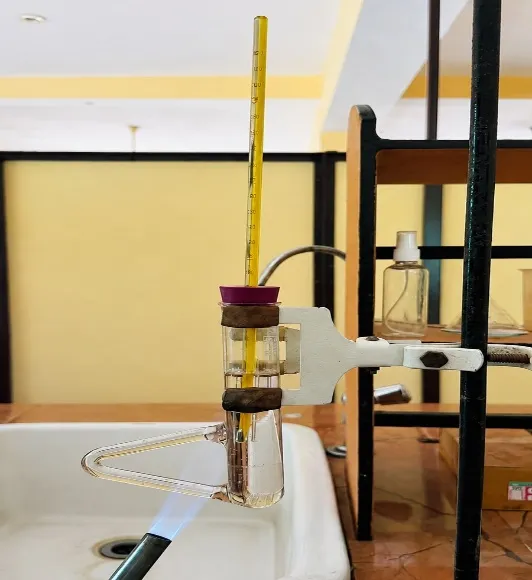
The melting point of the drug sample was determined using a standard capillary melting point apparatus. Briefly, a small quantity of the drug was introduced into a thin-walled capillary tube (10–15 mm in length, ~1 mm internal diameter, sealed at one end). The sample-loaded capillary was mounted in the apparatus alongside a calibrated thermometer and subjected to controlled, uniform heating. The temperature range spanning the initial onset to the complete liquefaction of the sample was recorded as the melting point.

  1. UV Spectrophotometry - UV–visible spectrophotometry is a widely employed analytical technique for obtaining structural information about drug molecules, particularly the characteristics of their chromophoric groups. When exposed to ultraviolet or visible radiation, molecules absorb light at specific wavelengths corresponding to the electronic transitions occurring within the molecular structure. The resulting UV spectrum is commonly represented as a plot of absorbance against wavelength. The maximum absorption wavelength (λ max) of Terbinafine hydrochloride was determined using a double-beam UV–visible spectrophotometer (Shimadzu UV-1800, Japan). Terbinafine solutions prepared in methanol at concentrations of 2 and 60 µg/mL were scanned over the wavelength range of 200–400 nm to identify the characteristic absorption maximum.

Preparation of Calibration Curve

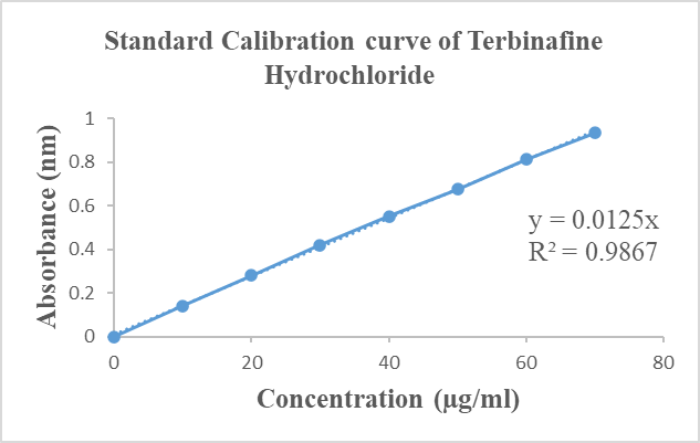
The standard stock solution of Terbinafine (100µg/ml) was prepared in methanol. This solution was diluted with methanol, to obtain various dilutions from 0.4-3.6µg/ml & 4-32µg/ml. Absorbance of these solutions was recorded at 223 & 282nm against methanol as blank using UV-visible spectrophotometer and standard curve was plotted against concentration. From the calibration curve intercept, slope, straight line equation and correlation coefficient were obtained.

  1. Solubility Study – The spontaneous interaction of two or more substances to form a homogeneous molecular dispersion is known as solubility.

The solubility of the drug was evaluated by preparing saturated solutions in different solvents. An excess amount of the drug was added to thoroughly cleaned culture tubes containing 5 mL of the respective solvents. The tubes were tightly sealed and agitated using a water-bath shaker at 25°C for 24 h to achieve equilibrium. Following incubation, the samples were centrifuged at 15,000 rpm, and the resulting supernatants were carefully collected. The supernatants were subsequently filtered, appropriately diluted with the respective solvent, and analyzed spectrophotometrically to determine the drug concentration.

  1. Partition Coefficient of Drug - Partition coefficient (oil/water) is a measure of a drug’s lipophilicity/hydrophilicity and an indication of drug’s ability to cross cell membranes. It is defined as the ratio of unionized drug distributed between the organic and aqueous phases at equilibrium. Partition coefficient provides a means of characterizing the lipophilic/hydrophilic nature of the drug. Drugs having values of P much greater than 1 are classified as lipophilic, where as those with values much less than 1 are indicative of a hydrophilic drug. The partition coefficient is commonly determined using an oil phase of n-octanol and water.

In the case n-octanol and water:

P o/w = C n-octanol/C water

The partition coefficient of Terbinafine hydrochloride was determined using the shake-flask method. An excess quantity of the drug was added to a 10 ml solvent system comprising n-octanol and water in a 1:1 ratio. The mixture was equilibrated for 24 h to allow the drug to distribute between the two phases. Following equilibration, the n-octanol and aqueous layers were carefully separated and centrifuged at 15,000 rpm for 15 min. The absorbance was taken in UV spectrophotometer at the respective λ max after proper dilution.

  1. Fourier transmission Infra-Red Spectroscopy - Fourier-transform infrared (FT-IR) spectroscopy was employed to investigate the structural characteristics of Terbinafine hydrochloride and to assess potential drug–excipient interactions. The potassium bromide (KBr) disc method was used for sample preparation, as KBr exhibits negligible absorption in the fundamental infrared region, thereby allowing the characteristic absorption bands of the sample to be observed. FT-IR spectra of the pure drug and the physical mixture containing the drug and excipients were recorded and compared to evaluate possible interactions between Terbinafine hydrochloride and the selected excipients.

For the preparation of the KBr discs, approximately 1 mg of Terbinafine hydrochloride or the drug–excipient physical mixture was thoroughly blended with 100 mg of spectroscopic-grade KBr. The KBr was previously dried under an infrared lamp to minimize moisture interference. The resulting mixture was compressed under hydraulic pressure to form a transparent disc. The prepared disc was then placed in the FT-IR sample chamber, and spectra were recorded over the wavenumber range of 4000–400 cm⁻¹. The obtained spectra were analyzed for any significant changes or shifts in characteristic absorption bands, which could indicate possible drug–excipient interactions. 25

  1. PREPARATION OF TERBINAFINE HYDROCHLORIDE AND ALOE VERA GEL
  • Step 1: Prepare the Carbopol dispersion by sprinkle Carbopol 934 slowly into the required quantity of distilled water. Allow it to swell for 2 hours to overnight.
  • Step 2: Dissolve 0.5 gm Terbinafine Hydrochloride in 5ml of Propylene glycol and mix thoroughly. Then dissolve 0.2 gm methyl paraben and dissolve adequately.
  • Step 3: Mix 10 ml of glycerine with 10 ml of Aloe vera gel /extract.
  • Step 4: Add the drug solution slowly to the carbopol dispersion with gentle stirring, then add the glycerine/aloe vera mixture and continue stirring until uniformity occurs.
  • Step 5: Add Triethanolamine slowly while stirring the solution gently until the desired viscosity and pH of 6 to 7.5.
  • Step 6: The whole admixture was stirred for 30 min continuously at 1500 RPM. The sparkling clear gel was formed and stored for evaluation. 26

Table No. 2: Formulation of Terbinafine hydrochloride and Aloe vera Gel

Ingredients

GF 1

GF 2

GF 3

Terbinafine Hydrochloride

0.5 gm

0.5 gm

0.5 gm

Carbopol 934

0.25 gm

0.5 gm

0.75 gm

Propylene glycol

5 ml

5 ml

5 ml

Aloe vera

10 ml

10 ml

10 ml

Glycerine

10 ml

10 ml

10 ml

Triethanolmine

q.s

q.s

q.s

Methyl paraben

0.2 gm

0.2 gm

0.2 gm

Distilled water

q.s

q.s

q.s

  1. EVALUATION OF GEL
  1. Physical Appearance: The prepared Terbinafine hydrochloride hydrogel formulations were examined visually to assess their color, uniformity, and consistency.
  2. Drug Content Uniformity: The drug content of the prepared gel was determined by accurately weighing 10 mg of the formulation and diluting it with 10 mL of a suitable solvent. The resulting solution was filtered, and its absorbance was measured spectrophotometrically at 281 nm. The drug content was calculated using the regression equation derived from the corresponding calibration curve
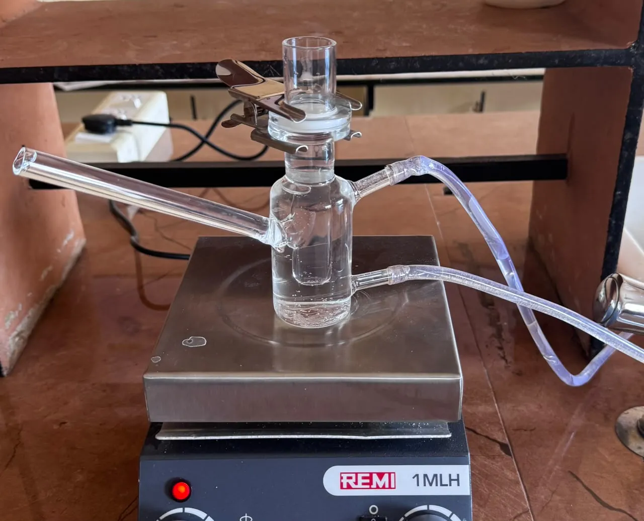
  3. In vitro Drug Diffusion Study: The in vitro diffusion profile of Terbinafine hydrochloride from the different gel formulations was evaluated using a Franz diffusion cell equipped with a cellophane membrane as the diffusion barrier. The membrane was immersed in the receptor compartment containing 40 ml of phosphate buffer (pH 7.4) and allowed to equilibrate for 24 h at 37±2oC. The diffusion cell was assembled with the membrane separating the donor and receptor compartments and placed on a magnetic stirrer.

Approximately 2 gm of each gel formulation was uniformly applied to the donor compartment. The receptor medium was continuously stirred at 50 rpm throughout the experiment. Aliquots of 5 mL were withdrawn from the receptor compartment at predetermined intervals of 10, 20, 30, 60, 90, 120, 180, 240, 300, 360, 420, and 480 min. An equal volume of fresh phosphate buffer was added after each sampling to maintain the sink conditions and constant receptor volume. The absorbance of the collected samples was measured spectrophotometrically at 281 nm using a UV–Visible spectrophotometer. The cumulative percentage release of Terbinafine hydrochloride from each gel formulation was calculated. The resulting release data were subjected to linear regression analysis to determine the drug-release kinetics according to zero-order and first-order models. 27

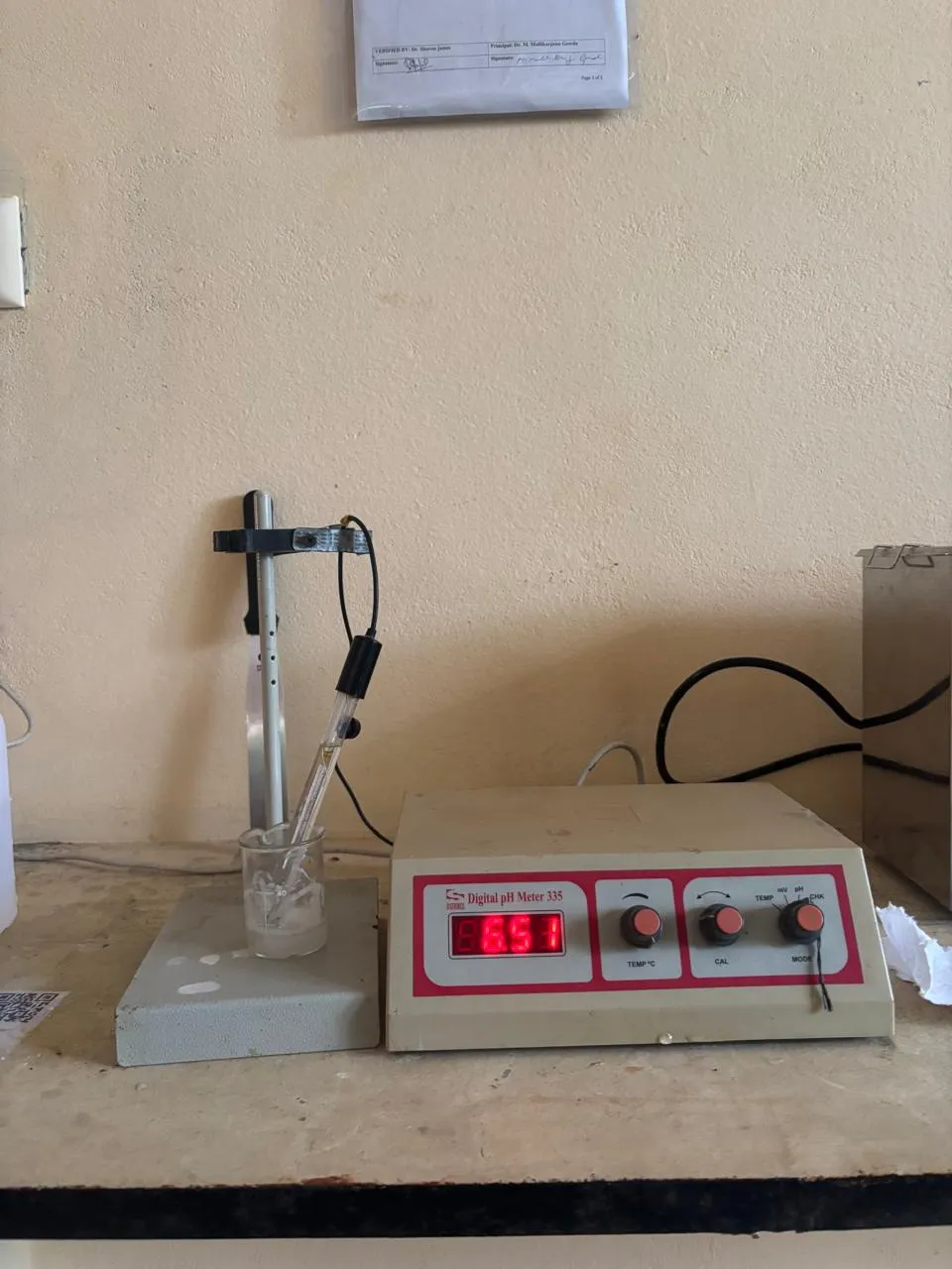
  1. pH Determination: The pH of the prepared gel formulation was determined using a calibrated digital pH meter at room temperature. Approximately 1 gm of the gel was dispersed in distilled water, and the pH electrode was immersed in the resulting dispersion. The pH value was recorded once a stable reading was obtained. The measured pH was evaluated to ensure that it fell within a suitable range for scalp application, thereby minimizing the risk of skin irritation and discomfort.
  2. Grittiness Test: The gel was evaluated manually between the fingers to assess the presence of coarse particles or gritty texture. An acceptable formulation was expected to exhibit a smooth and homogeneous texture without any detectable particulate matter.
  3. Skin Irritation Test: The prepared gel formulation was applied to a small, defined area of the skin and monitored for any signs of redness, itching, swelling, or other irritation over a specified observation period. The absence of such adverse skin reactions indicated that the formulation was well tolerated and suitable for topical application.
  4. Washability Evaluation: The gel was applied to the skin and rinsed with tap water to assess its removal properties. Ease of cleansing and the absence of residual stickiness or greasy film were qualitatively evaluated to confirm acceptable washability. 28
  5. Spreadability Determination: Spreadability was evaluated using the conventional parallel-plate technique. Briefly, 1 gm of the gel formulation was placed between two horizontal glass plates. A standard weight of 200 gm was applied to the upper plate for 60 seconds, after which the spread diameter of the gel mass was recorded. Spreadability was quantified using the following equation:

S=W× d/T

Where S=Spreadability (g.mm/s)

W=Weight applied on the sample(g)

T=Time(s)

d=Diameter(mm) 29,30

RESULTS

PREFORMULATION STUDIES

  1. Preformulation Studies of Terbinafine Hydrochloride:
  1. Organoleptic properties: Terbinafine hydrochloride was examined for its organoleptic properties which are shown in table 3.

Table No. 3: Organoleptic properties of Terbinafine Hydrochloride

Sr. No

Properties

Inference

01

Color

White  or  almost white

02

Odor

odorless

03

Form

crystalline


  1. Melting point: A sharp and well-defined melting point indicates the purity of the drug and supports for formulation development. Melting point was determined as shown in table 4.


Table No. 4: Melting point of Terbinafine Hydrochloride
Drug

Reference melting point

Observed melting point

Terbinafine Hydrochloride

195-1980c

1970c


Fig. No. 3: Melting point of Terbinafine Hydrochloride

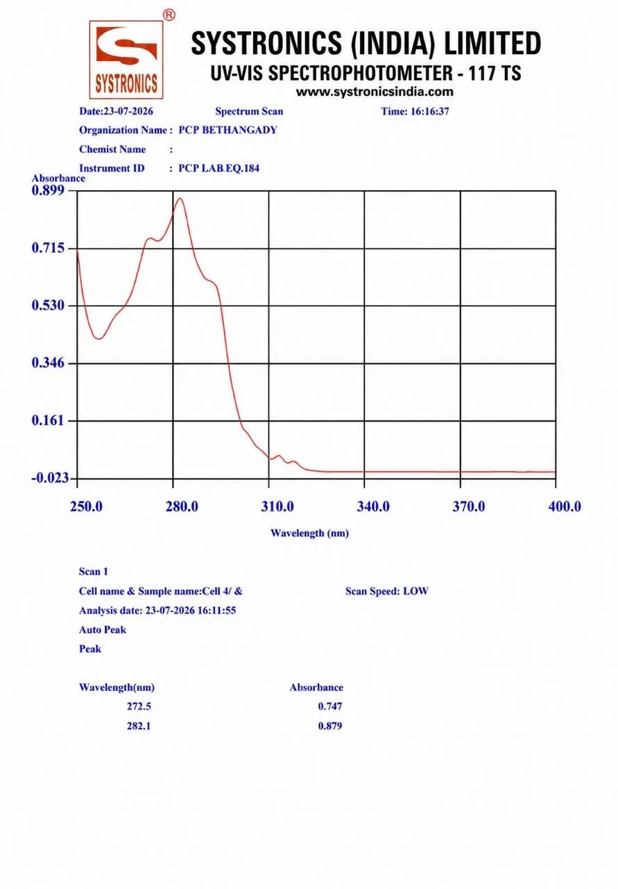
  1. UV-Visible Spectroscopy study: Determination of Absorption Maxima of Terbinafine Hydrochloride in Methanol. Absorption Maxima of Terbinafine Hydrochloride in Methanol are shown in Fig. No. 4


Fig. No. 4: Absorption maxima of Terbinafine Hydrochloride in Methanol

Preparation of Calibration Curve:

Table No. 5: Calibration Curve of Terbinafine Hydrochloride

Sr. No.

Concentration (µg/ml)

Absorbance (nm)

01

0

0.000

02

10

0.140

03

20

0.280

04

30

0.420

05

40

0.552

06

50

0.675

07

60

0.811

08

70

0.934


Fig. No. 5: Graph of Calibration Curve of Terbinafine Hydrochloride in Methanol

  1. Solubility studies: Analysis of the drug was carried out on UV Spectrophotometer at 282 nm. Solubility of Terbinafine Hydrochloride has been shown in the table 6.

Table No. 6: Solubility studies of Terbinafine Hydrochloride

Sr. No

Solvent

Solubility in(mg/ml)

(mean ± SD)

Solubility

01

Ethanol

0.894 ± 0.285

Soluble

02

Phosphate Buffer 7.4

0.159 ± 0.285

Sparingly soluble

  1. Partition coefficient determination: Partition coefficient of Terbinafine Hydrochloride was determined using n-octanol and water. Log P greater than one indicates that the drug is lipophilic in nature, whereas those with partition coefficient less than one are indicative of a hydrophilic drug. This indicate the lipophilicity and purity of drug.

Table No. 7: Partition coefficient determination of Terbinafine Hydrochloride

Partition coefficient

Solvent system

Log P value

Terbinafine Hydrochloride

n-octanol : water

2.23 ± 0.003

  1. FTIR Analysis

Fig. No. 6 (a): FTIR peak of Terbinafine Hydrochloride

Fig. No. 6 (b): FTIR peak of Aloe vera

Fig. No. 6 (c): FTIR peak of the physical mixture of the formulation components

Table No. 8: FTIR peak of Terbinafine Hydrochloride

Sr. No

Functional group

Standard frequency(cm-1)

Observed            frequency(cm-1)

01

Aliphatic C-H                       Stretching

3000 - 2850

2962.66

02

Protonated amine (N-H+) Stretching

2500 - 2400

2438.02

03

CC /combination region

2400 - 2200

2376.30

04

C-N Stretching

1300 - 1200

1255.66

05

Fingerprint region

600 - 500

536.21

Table No. 9: FTIR peak of Aloe Vera

Sr. No

Functional group

Standard frequency(cm-1)

Observed            frequency(cm-1)

01

O-H Stretching

3200 - 3600

3282.84

02

N-H Stretching

3200 - 3500

3219.19

03

Aliphatic  C-H Stretching

2850 - 3000

2962.66

04

C-H Stretching

2700 - 2850

2791.00

05

Combination region

2300 - 2400

2349.30

06

C=C Stretching

1600 - 1650

1629.85

07

C-N Stretching

1200 - 1300

1269.16

08

C-O Stretching

1000 - 1150

1041.56

09

Finger print region

500 - 550

532.35

Table No. 10: FTIR peak of the physical mixture of the formulation components

Sr. No

Functional group

Standard frequency(cm-1)

Observed            frequency(cm-1)

01

OH-Stretching

3600 - 3200

3294.42

02

COO- assymetric Stretching

1650 - 1600

1635.64

03

COO- symmetric Stretching

1450 - 1400

1433.11


04

C-O-C Stretching

1260 - 1200

1224.80

05

Finger region vibration

580 - 550

570.93

The compatibility between Terbinafine Hydrochloride, Aloe vera and Physical mixture of the formulation components was evaluated using FTIR peak matching method. The FTIR spectrum of pure Terbinafine Hydrochloride showed characteristics absorption peaks at 2962.66 cm-1 (Aliphatic C-H Stretching), 2438.02 cm-1 (N-H+ Stretching), 2376.30 cm-1 (CC/ combination region), 1255.66 cm-1 (C-N Stretching), 536.21 cm-1 (Fingerprint region). The FTIR spectrum of Aloe vera showed characteristics absorption peaks at 3294.42 cm-1 (O-H Stretching), 570.93 cm-1 (Fingerprint region vibration), 1635.64 cm-1 (COO- assymetric Stretching), 1433.11 cm-1 (COO- symmetric Stretching), 1224.80 cm-1 (C-O-C Stretching). The FTIR spectrum of physical mixture showed characteristics absorption peaks at 2962.66 cm-1 (Aliphatic C-H Stretching), 3219.19 cm-1 (N-H Stretching), 2349.30 cm-1 (combination region), 1269.16 cm-1 (C-N Stretching), 532.35 cm-1 (Fingerprint region), 3282.84 cm-1 (O-H Stretching), 1629.85 cm-1 (C=C Stretching), 1041.56 cm-1 (C-O Stretching). No significant shifts or disappearance of peaks were observed, indicating no significant chemical interaction between Terbinafine Hydrochloride, Aloe vera and the excipients. Thus, they were found to be compatible for Gel formulation.
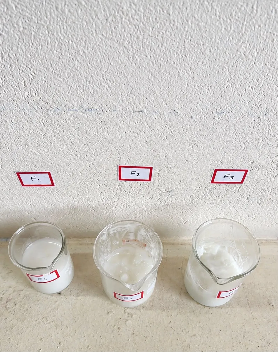
  1. Preparation of Terbinafine Hydrochloride Gel:


Fig. No. 7: Preparation of Terbinafine Hydrochloride Gel

  1. Evaluation Parameters of Terbinafine Hydrochloride Gel:
  1. Physical appearance of Gel:

Table No. 11: Physical appearance of Gel

Sr. NO

Parameters

F1

F2

F3

01

Color

White

White

White

02

Homogeneity

Homogenous

Homogenous

Homogenous

03

Consistency

Very fluid /                 runny in      consistency

Smooth, Semisolid in consistency

Slightly thick and viscous

consistency

  1. Drug content:

Table No. 12: Drug content of all formulations

Sr. No

Formulation

Drug content(%) ± SD

01

F1

84.38 ± 0.5

02

F2

98.58 ± 0.2

03

F3

87 ± 0.2

The drug content of the prepared gel formulations was determined to evaluate the uniformity of drug distribution within the formulations. The drug content was found to be 84.38 ± 0.5% for F1, 98.58 ± 0.2% for F2, and 87.00 ± 0.2% for F3. Among the three formulations, F2 showed the highest drug content (98.58 ± 0.2%), indicating better drug uniformity and incorporation compared with F1 and F3.

  1. In vitro drug diffusion study:

Table No. 13: Cumulative amount of Terbinafine Hydrochloride diffused (%) from all the formulations through cellophane membrane using Franz diffusion cell

Time (hrs)

%CDR

%CDR

%CDR


F1

F2

F3

0

0

0

0

1

5.94

9.15

9.31

2

16.00

20.31

20.00

3

27.00

36.22

32.00

4

39.00

52.26

45.00

5

51.00

65.19

58.00

6

62.00

77.68

70.00

7

71.00

88.69

82.00

8

79.50

94.49

91.30


Fig. No. 8: In vitro drug release of F2 formulation
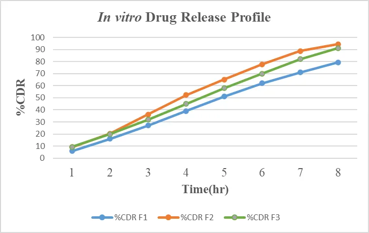
 

Fig. No. 9: Cumulative amount of Terbinafine Hydrochloride permeated (%).

The in vitro drug release profile of Terbinafine Hydrochloride through cellophane membrane was represented in fig. no. 9 and table no. 13. The data was also compared for release pattern and % drug release after 8 hours from all the three formulations. The F2 formulation showed consistent and maximum drug release compared to the other formulation. The Drug release of F2 formulation after 8 hours were found to be higher.

  1. pH Determination:

Table No. 14: pH value for the formulations

Sr. No

Formulation

Observation

01

F1

6.44 ± 0.03

02

F2

6.53 ± 0.02

03

F3

6.55 ± 0.03


Fig. No. 10: Determination of pH

The pH of all the prepared gel formulations was determined. Formulation F1 showed a pH of 6.44 ± 0.03, F2 showed 6.53 ± 0.02, and F3 showed 6.55 ± 0.03. The pH values of all formulations were found to be close to the normal physiological pH of the skin, indicating that the prepared gel formulations are likely to be non-irritant and suitable for topical application.

Determination of Grittiness, Skin irritation, Washability tests:

Table No. 15: Determination of Grittiness, Skin irritation, Washability tests

Sr. No

Parameters

F1

F2

F3

5.

Grittiness test

No grittiness

observed

No grittiness

observed

Slight grittiness

observed

6.

Skin irritation

test

No irritation

observed

No irritation

observed

No irritation

observed

7.

Washability

test

Easily washable

with water

Easily washable

with water

Moderately

washable

  1. Determination of Spreadability:

Table No. 16: Spreadability of formulations

Sr. No

Formulation

Spreadability

(g.cm/sec) ± SD

01

F1

22.83 ± 0.020

02

F2

18.06 ± 0.010

03

F3

15.23 ± 0.020


Fig. No. 11: Spreadability of F2 formulation

The spreadability values of F1, F2, and F3 were found to be 22.83 ± 0.020, 18.06 ± 0.010, and 15.23 ± 0.020 g·cm/sec, respectively. Among the three formulations, F1 exhibited the highest spreadability, followed by F2 and F3. The higher spreadability of F1 indicates that it can be easily and uniformly spread over the skin. Although F1 showed the highest spreadability, F2 exhibited an optimum spreadability, which indicates that the gel can be easily applied and distributed over the skin while maintaining adequate consistency. Therefore, considering the overall formulation characteristics, F2 was selected as the optimized formulation.

CONCLUSION:

The present study focused on the formulation and evaluation of Terbinafine Hydrochloride and aloe vera gel formulation using dispersion approach for the effective management of fungal infections. Preformulation and compatibility studies confirmed the suitability of both active ingredients. The combination of antifungal activity of Terbinafine Hydrochloride with the soothing, moisturizing properties of aloe vera can provide an effective dosage form for topical treatment of fungal infections. Three formulations (F1, F2 and F3) were developed using different concentrations of carbopol 934 as gelling agent and evaluated for various parameters. Among the 3 developed gel formulations, F2 was found to be the optimized formulation. The most important advantage of F2 is its drug content of 98.58 ± 0.2 % which is higher than F1 and F3, which indicate the better incorporation and uniform distribution of Terbinafine Hydrochloride in the gel. Although F1 exhibited greater spreadability than F2, F2 still showed satisfactory spreadability. F2 had a smooth, semisolid and homogenous consistency with no grittiness, no skin irritation and easy washability. Its pH of 6.53 ± 0.02 is closer to normal skin pH range indicating better skin compatibility. F2 also had a consistant and higher in vitro drug release. Hence, it can be concluded that Terbinafine Hydrochloride- aloe vera gel formulation can be successfully developed as a stable and effective dosage form.

REFERENCES

  1. Honari G. Skin structure and function. In: Honari G, Andersen R, Maibach HI, editors. Sensitive skin syndrome. 2nd ed. Boca Raton (FL): CRC Press; 2017. p. 16-22. doi:10.1201/9781315121048-3.
  2. Bragazzi NL, Sellami M, Salem I, Conic R, Kimak M, Pigatto PD, Damiani G. Fasting and its impact on skin anatomy, physiology, and physiopathology: A comprehensive review of the literature. Nutrients. 2019 Jan 23;11(2):249.
  3. McKnight G, Shah J, Hargest R. Physiology of the skin. Surgery (Oxford). 2022 Jan 1;40(1):8-12.
  4. Ali SM, Yosipovitch G. Skin pH: from basic science to basic skin care. Acta dermato-venereologica. 2013 Jan 10;93(3):261-7.
  5. Jamagondi LN, Katte AS, Rumane MB, Pathan M, Kale AR. Formulation and evaluation of topical antifungal gel containing itraconazole. American Journal of PharmTech Research. 2019;9(02):333-8.
  6. Shiva K, Mandal S, Kumar S. Formulation and evaluation of topical antifungal gel of fluconazole using aloe vera gel. Int J Sci Res Develop. 2021;1: 187-93.
  7. Dixon DM, Walsh TJ. Antifungal Agents. In: Baron S, editor. Medical Microbiology. 4th edition. Galveston (TX): University of Texas Medical Branch at Galveston; 1996. Chapter 76. Available from: https://www.ncbi.nlm.nih.gov/books/NBK8263/
  8. Singh M, Rathour A, Dey U, Kumar Gaur P, Kumar V, Modi K, Abdul A. Antifungal agents: a comprehensive review of mechanisms and applications. J Popul Ther Clin Pharmacol. 2022;29(04):1343-58.
  9. Un Nabi SA, Sheraz MA, Ahmed S, Mustaan N, Ahmad I. Pharmaceutical gels: a review. RADS J. Pharm. Pharm. Sci. 2016;4: 40-8.
  10. Bhuyan C, Saha D, Rabha B. A brief review on topical gels as drug delivery system. J. Pharm. Res. Int. 2021;33: 344-57.
  11. Bisht P, Badola A. Gel as a topical drug delivery system: a review. JETIR. 2019;6(4):321-30.
  12. Sharma U, Arjariya S, Chouksey R, Sharma N. A review: formulation and evaluation of pharmaceutical gel. Journal of Pharmaceutical Negative Results. 2022;13.
  13. Al-Nima A. Preparation and evaluation of topical gel containing Aloe vera exudate. Iraqi Journal of Pharmacy. 2021;18(2):39-52.
  14. Sharma P, Kaur R. A comprehensive review of aloe vera: Composition, properties, processing, and applications. The Natural Products Journal. 2025 Jan 15.
  15. Pounikar YO, Jain PU, Khurana NA, Omray LK, Patil S, Gajbhiye A. Formulation and characterization of Aloe vera cosmetic herbal hydrogel. International Journal of Pharmacy and Pharmaceutical Sciences. 2012;4(4):85-6.
  16. Sahu PK, Giri DD, Singh R, Pandey P, Gupta S, Shrivastava AK, Kumar A, Pandey KD. Therapeutic and medicinal uses of Aloe vera: a review. Pharmacology & Pharmacy. 2013 Nov 8;4(8):599-610.
  17. Hamman JH. Composition and applications of Aloe vera leaf gel. Molecules. 2008 Aug 8;13(8):1599-616.
  18. Kanakapura B, Penmatsa VK. Analytical methods for determination of terbinafine hydrochloride in pharmaceuticals and biological materials. Journal of Pharmaceutical Analysis. 2016;6(3):137-49.
  19. Dewangan A. Development and evaluation of terbinafine hydrochloride for tablet formulation.
  20. Tanrıverdi ST, Özer Ö. Novel topical formulations of Terbinafine-HCl for treatment of onychomycosis. European Journal of Pharmaceutical Sciences. 2013 Mar 12;48(4-5):628-36.
  21. Arpa MD, Ünükür MZ, Erim ÜC. Formulation, characterization and in vitro release studies of terbinafine hydrochloride loaded buccal films. Journal of Research in Pharmacy. 2021 Sep 1;25(5):667-80.
  22. Pal P, Mishra SD. Formulation development and evaluation of antifungal proniosomal gel for topical application. World J Pharm Res. 2017;6: 656-74.
  23. Stout EI, McKessor A. Glycerin-based hydrogel for infection control. Advances in wound care. 2012 Feb;1(1):48-51.
  24. Sneha Sonarkhan*, Shravani Asamwar, Gajanan Gujatwar, Nilesh Chachda, Design And Development Of Topical Gel Formulation From Leaf Extract Of Passiflora Incarnata, Int. J. of Pharm. Sci., 2026, Vol 4, Issue 7, 3776-3785. https://doi.org/10.5281/zenodo.21438909
  25. Kumar A, Soni A, Jamwal S, Thakur S, Kumari K. Preformulation study of terbinafine for novel drug delivery system formulation. International Journal of Research and Analytical Reviews. 2020;7(3):34-2.
  26. Aswal DS, Biswkarma VK, Wadhawan S, Kumar P. ALOE VERA GEL CONTAINING FLUCONAZOLE SOLID DISPERSION: FORMULATION AND EVALUATION FOR TOPICAL ANTIFUNGAL ACTION.
  27. Datir SK, Patil PB. Formulation and evaulation of Terbinafine Hydrochloride Topical Hydrogel. International journal of Recent Development in Engineering and Technology. 2026; 15(4):1444-70.
  28. Bombatkar PS, Wavhale M, Pohekar A. Formulation and evaulation of Herbal Aloe vera Hair gel using Carbopol 934. TIJER-International Research Journal. 2026;13(5):544-50.
  29. Bakhrushina EO, Anurova MN, Zavalniy MS, Demina NB, Bardakov AI, Krasnyuk II. Dermatologic gels spreadability measuring methods comparative study. International Journal of Applied Pharmaceutics. 2022;14(1):164-8.
  30. Al-Barghouthy EY, Hamed S, Mehyar GF, AlKhatib HS. Comparative Evaluation of Spreadability Measurement Methods for Topical Semisolid Formulations/A Scoping Review. Gels. 2025 12;11(12):1006

Reference

  1. Honari G. Skin structure and function. In: Honari G, Andersen R, Maibach HI, editors. Sensitive skin syndrome. 2nd ed. Boca Raton (FL): CRC Press; 2017. p. 16-22. doi:10.1201/9781315121048-3.
  2. Bragazzi NL, Sellami M, Salem I, Conic R, Kimak M, Pigatto PD, Damiani G. Fasting and its impact on skin anatomy, physiology, and physiopathology: A comprehensive review of the literature. Nutrients. 2019 Jan 23;11(2):249.
  3. McKnight G, Shah J, Hargest R. Physiology of the skin. Surgery (Oxford). 2022 Jan 1;40(1):8-12.
  4. Ali SM, Yosipovitch G. Skin pH: from basic science to basic skin care. Acta dermato-venereologica. 2013 Jan 10;93(3):261-7.
  5. Jamagondi LN, Katte AS, Rumane MB, Pathan M, Kale AR. Formulation and evaluation of topical antifungal gel containing itraconazole. American Journal of PharmTech Research. 2019;9(02):333-8.
  6. Shiva K, Mandal S, Kumar S. Formulation and evaluation of topical antifungal gel of fluconazole using aloe vera gel. Int J Sci Res Develop. 2021;1: 187-93.
  7. Dixon DM, Walsh TJ. Antifungal Agents. In: Baron S, editor. Medical Microbiology. 4th edition. Galveston (TX): University of Texas Medical Branch at Galveston; 1996. Chapter 76. Available from: https://www.ncbi.nlm.nih.gov/books/NBK8263/
  8. Singh M, Rathour A, Dey U, Kumar Gaur P, Kumar V, Modi K, Abdul A. Antifungal agents: a comprehensive review of mechanisms and applications. J Popul Ther Clin Pharmacol. 2022;29(04):1343-58.
  9. Un Nabi SA, Sheraz MA, Ahmed S, Mustaan N, Ahmad I. Pharmaceutical gels: a review. RADS J. Pharm. Pharm. Sci. 2016;4: 40-8.
  10. Bhuyan C, Saha D, Rabha B. A brief review on topical gels as drug delivery system. J. Pharm. Res. Int. 2021;33: 344-57.
  11. Bisht P, Badola A. Gel as a topical drug delivery system: a review. JETIR. 2019;6(4):321-30.
  12. Sharma U, Arjariya S, Chouksey R, Sharma N. A review: formulation and evaluation of pharmaceutical gel. Journal of Pharmaceutical Negative Results. 2022;13.
  13. Al-Nima A. Preparation and evaluation of topical gel containing Aloe vera exudate. Iraqi Journal of Pharmacy. 2021;18(2):39-52.
  14. Sharma P, Kaur R. A comprehensive review of aloe vera: Composition, properties, processing, and applications. The Natural Products Journal. 2025 Jan 15.
  15. Pounikar YO, Jain PU, Khurana NA, Omray LK, Patil S, Gajbhiye A. Formulation and characterization of Aloe vera cosmetic herbal hydrogel. International Journal of Pharmacy and Pharmaceutical Sciences. 2012;4(4):85-6.
  16. Sahu PK, Giri DD, Singh R, Pandey P, Gupta S, Shrivastava AK, Kumar A, Pandey KD. Therapeutic and medicinal uses of Aloe vera: a review. Pharmacology & Pharmacy. 2013 Nov 8;4(8):599-610.
  17. Hamman JH. Composition and applications of Aloe vera leaf gel. Molecules. 2008 Aug 8;13(8):1599-616.
  18. Kanakapura B, Penmatsa VK. Analytical methods for determination of terbinafine hydrochloride in pharmaceuticals and biological materials. Journal of Pharmaceutical Analysis. 2016;6(3):137-49.
  19. Dewangan A. Development and evaluation of terbinafine hydrochloride for tablet formulation.
  20. Tanr?verdi ST, Özer Ö. Novel topical formulations of Terbinafine-HCl for treatment of onychomycosis. European Journal of Pharmaceutical Sciences. 2013 Mar 12;48(4-5):628-36.
  21. Arpa MD, Ünükür MZ, Erim ÜC. Formulation, characterization and in vitro release studies of terbinafine hydrochloride loaded buccal films. Journal of Research in Pharmacy. 2021 Sep 1;25(5):667-80.
  22. Pal P, Mishra SD. Formulation development and evaluation of antifungal proniosomal gel for topical application. World J Pharm Res. 2017;6: 656-74.
  23. Stout EI, McKessor A. Glycerin-based hydrogel for infection control. Advances in wound care. 2012 Feb;1(1):48-51.
  24. Sneha Sonarkhan*, Shravani Asamwar, Gajanan Gujatwar, Nilesh Chachda, Design And Development Of Topical Gel Formulation From Leaf Extract Of Passiflora Incarnata, Int. J. of Pharm. Sci., 2026, Vol 4, Issue 7, 3776-3785. https://doi.org/10.5281/zenodo.21438909
  25. Kumar A, Soni A, Jamwal S, Thakur S, Kumari K. Preformulation study of terbinafine for novel drug delivery system formulation. International Journal of Research and Analytical Reviews. 2020;7(3):34-2.
  26. Aswal DS, Biswkarma VK, Wadhawan S, Kumar P. ALOE VERA GEL CONTAINING FLUCONAZOLE SOLID DISPERSION: FORMULATION AND EVALUATION FOR TOPICAL ANTIFUNGAL ACTION.
  27. Datir SK, Patil PB. Formulation and evaulation of Terbinafine Hydrochloride Topical Hydrogel. International journal of Recent Development in Engineering and Technology. 2026; 15(4):1444-70.
  28. Bombatkar PS, Wavhale M, Pohekar A. Formulation and evaulation of Herbal Aloe vera Hair gel using Carbopol 934. TIJER-International Research Journal. 2026;13(5):544-50.
  29. Bakhrushina EO, Anurova MN, Zavalniy MS, Demina NB, Bardakov AI, Krasnyuk II. Dermatologic gels spreadability measuring methods comparative study. International Journal of Applied Pharmaceutics. 2022;14(1):164-8.
  30. Al-Barghouthy EY, Hamed S, Mehyar GF, AlKhatib HS. Comparative Evaluation of Spreadability Measurement Methods for Topical Semisolid Formulations/A Scoping Review. Gels. 2025 12;11(12):1006

Photo
Bhanunandana G.S.
Corresponding author

Graduate student, Prasanna college of Pharmacy, Laila, Belthangady (po), Dakshina Kannada, Karnataka, India - 574214

Photo
Shashank K.
Co-author

Graduate student, Prasanna college of Pharmacy, Laila, Belthangady (po), Dakshina Kannada, Karnataka, India - 574214

Photo
Salman Paris
Co-author

Graduate student, Prasanna college of Pharmacy, Laila, Belthangady (po), Dakshina Kannada, Karnataka, India - 574214

Photo
Alan Joseph
Co-author

Graduate student, Prasanna college of Pharmacy, Laila, Belthangady (po), Dakshina Kannada, Karnataka, India - 574214

Photo
Mahammad Shaz
Co-author

Graduate student, Prasanna college of Pharmacy, Laila, Belthangady (po), Dakshina Kannada, Karnataka, India - 574214

Photo
Dr. M. Mallikarjuna Gouda
Co-author

Principal, Department of Pharmaceutics, Prasanna college of pharmacy, Laila, Belthangady (po), Dakshina Kannada, Karnataka, India-574214

Bhanunandana G.S.*, Shashank K., Salman Paris, Alan Joseph , Mahammad Shaz, Dr. M. Mallikarjuna Gouda , Formulation And Evaluation Of An Antifungal Gel Containing Aloe Vera And Terbinafine Hydrochloride Prepared By The Dispersion Method, Int. J. of Pharm. Sci., 2026, Vol 4, Issue 10, 3244-3271. https://doi.org/ 10.5281/zenodo.23560726

More related articles
In SILICO Molecular Docking Analysis of Pyrazole D...
B. Vasantha Rajan, S. Vaani Priya Lakshmi, P. Divakar, E. Gopina...
Enhancing Chemotherapy Response in Brain Cancer Th...
Ananthi Saravanan, Dhanush I., Yokesh P., Kesavan N., Uma Shankar...
Biosimilars: Development, Regulatory Landscape, Ch...
P. Khedkar, S. D. Mhaske, Y. M. Charade, H. Usendi, S. Sheikh, S....
Formulation and Evaluation of Microemulsion Based Gel of Alverine Citrate...
Tamilarasan S., Perumal P., Krishnamoorthy B., Elangovan S....
Intraoperative Lactate as a Predictor of Complications in Major Surgery...
Esteban Diaz Rivadeneira, Alder Gomez Angulo, Jhon Alvarez Molina, Julian Amaya Perez, Ana Carranza ...
Pharmaceutico Analytical and Experimental Study of Amalakyadi Churna in Jwara wi...
Dr. Shantala Patil, Dr. C . Shristy Shetty, Dr. Rachana C....
More related articles
In SILICO Molecular Docking Analysis of Pyrazole Derivatives as Potential Anti-I...
B. Vasantha Rajan, S. Vaani Priya Lakshmi, P. Divakar, E. Gopinath...
Enhancing Chemotherapy Response in Brain Cancer Through Combination of Morinda C...
Ananthi Saravanan, Dhanush I., Yokesh P., Kesavan N., Uma Shankar M., S. Dinesh D....
Biosimilars: Development, Regulatory Landscape, Challenges, and Future Perspecti...
P. Khedkar, S. D. Mhaske, Y. M. Charade, H. Usendi, S. Sheikh, S. Dhabale...