

We use cookies to ensure our website works properly and to personalise your experience. Cookies policy
Anuradha College of Pharmacy, Chikhli, Maharashtra, India.
The present review focuses on the green synthesis of silver nanoparticles using plant extracts. Silver nanoparticles are widely studied because of their strong antimicrobial activity and their use in dressings, coatings and other biomedical products, but the physical and chemical methods of their synthesis need costly equipment or toxic chemicals. In green synthesis, the phytochemicals of plant extracts, such as polyphenols, flavonoids, terpenoids and proteins, reduce silver ions to silver nanoparticles and also cap and stabilise them, in a simple, low-cost and environmentally friendly one-step process. The review describes the methods of synthesis, the mechanism of green synthesis, the factors affecting it, the profile of Neem (Azadirachta indica) and Tulsi (Ocimum sanctum) leaves and of silver nitrate, the procedure of synthesis, the techniques of characterisation, the evaluation of the antibacterial activity by the agar well diffusion method, and the important reports on the subject. The review indicates that plant extracts can be effectively used for the preparation of silver nanoparticles, which show surface plasmon resonance at about 400 to 450 nm and antibacterial activity, while further work on safety, characterisation and stability is required.
Nanotechnology deals with the design, production and use of materials whose dimensions lie in the nanometre range, generally between 1 and 100 nm. At this size a material has a very large surface area compared with its volume, and its optical, electrical, chemical and biological properties can differ greatly from those of the same material in bulk form. Because of these properties, nanoparticles are being used in medicine, drug delivery, diagnostics, cosmetics, textiles, food packaging and environmental treatment.[1,2]
Among the metal nanoparticles, silver nanoparticles (AgNPs) are the most widely studied because of their strong antimicrobial activity. They have been reported to act against bacteria, fungi and some viruses, and they are used in wound dressings, coatings for medical devices, water purification and personal care products.[3,4] Their antibacterial action is explained by several mechanisms: attachment to the bacterial cell wall and membrane, release of silver ions, interaction with sulphur-containing proteins and enzymes, and generation of reactive oxygen species.[4,5,6,7]
Silver nanoparticles also show a characteristic colour, because the free electrons on the particle surface oscillate with the incident light (surface plasmon resonance), which gives an absorption band in the visible region and makes the particles easy to detect by UV-visible spectroscopy.[8]
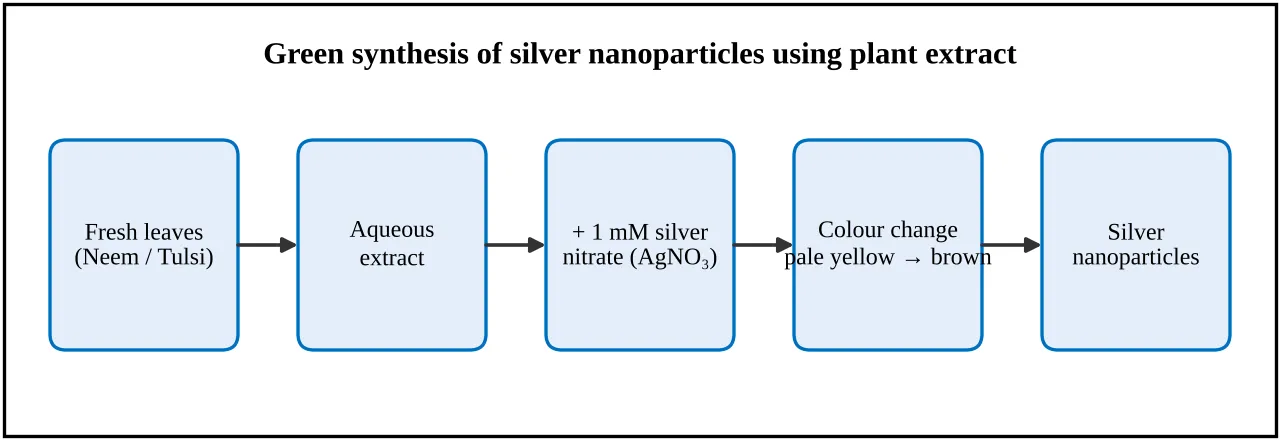
Figure 1: Green Synthesis of Silver Nanoparticles Using Plant Extract
Methods of Synthesis of Nanoparticles
Nanoparticles can be prepared by physical, chemical and biological methods, each of which has its own advantages and drawbacks.[1,2] The methods are compared in the table below.
Table: Methods of Synthesis of Silver Nanoparticles[1,2,9]
| Sr. No. | Method | Examples | Limitations |
|---|---|---|---|
| 1 | Physical | Evaporation-condensation, laser ablation, ball milling | Need costly equipment and high energy |
| 2 | Chemical | Reduction of a silver salt with sodium borohydride, citrate or other reducing agents, with a stabiliser | Use of toxic chemicals; hazardous waste |
| 3 | Biological (green) | Reduction by plant extracts, bacteria, fungi or algae | Variable composition of the biological material; control of size needs study |
Green Synthesis Using Plant Extracts
Green synthesis uses biological materials in place of hazardous chemicals. Plant extracts are particularly convenient because they are easily available, safe to handle and need no special culture conditions, which makes the process faster and simpler than synthesis with microorganisms.[1,2,10,11] Plant extracts contain polyphenols, flavonoids, terpenoids, alkaloids, proteins and sugars. These phytochemicals reduce the silver ions (Ag+) of silver nitrate to neutral silver atoms (Ag0), and the same or other molecules from the extract adsorb on the particle surface and act as capping and stabilising agents that prevent aggregation.[1,2,12]
The process can be pictured in three steps: first the silver ions are reduced by the phytochemicals and the atoms collect to form small nuclei; then the nuclei grow into nanoparticles; and finally the particles are capped by plant molecules and become stable in suspension. The first visible sign is a change of colour of the reaction mixture from pale yellow or colourless to yellowish-brown or dark brown. The method is simple, inexpensive, mostly one-step, can be done at room temperature and is environmentally friendly.[1,10]
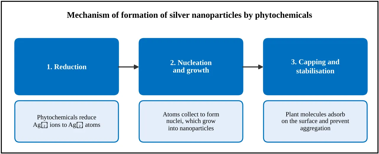
Figure 2: Mechanism of Formation of Silver Nanoparticles by Plant Phytochemicals
Factors Affecting the Synthesis
Table: Factors Affecting the Green Synthesis of Silver Nanoparticles[2,12]
| Sr. No. | Factor | Effect |
|---|---|---|
| 1 | Type and concentration of extract | Decides which and how much reducing and capping phytochemical is available |
| 2 | Concentration of silver nitrate | Too low gives few particles; too high may cause aggregation and a precipitate |
| 3 | Ratio of extract to silver nitrate | Affects the rate of reduction, the size and the stability |
| 4 | Temperature | Higher temperature generally speeds up the reaction |
| 5 | pH | Changes the reducing power of the phytochemicals and the charge on the particles |
| 6 | Reaction time | Colour and absorption intensity increase until the reaction is complete |
| 7 | Light | Silver salts are light-sensitive; reactions are usually protected from strong light |
Profile of Materials
The plants, the silver salt and the nanoparticles are described here, because their properties decide how the synthesis is carried out. Neem and Tulsi are two easily available plants that have been used for the green synthesis of silver nanoparticles.[13,14]
Neem (Azadirachta indica)
Table: Profile of Neem[13,15]
| Attribute | Details |
|---|---|
| Biological source | Fresh or dried leaves of Azadirachta indica A. Juss. |
| Family | Meliaceae |
| Common names | Neem, Kadunimb |
| Part used | Leaves |
| Main constituents | Limonoids (such as azadirachtin, nimbin), flavonoids, polyphenols, tannins, glycosides |
| Known uses | Traditional antibacterial, antifungal, anti-inflammatory and skin uses |
| Relevance | Neem leaf broth has been used to reduce silver and gold ions to nanoparticles |
Tulsi (Ocimum sanctum)
Table: Profile of Tulsi[14,15]
| Attribute | Details |
|---|---|
| Biological source | Fresh or dried leaves of Ocimum sanctum L. (also called Ocimum tenuiflorum) |
| Family | Lamiaceae |
| Common names | Tulsi, Holy basil |
| Part used | Leaves |
| Main constituents | Volatile oil rich in eugenol, flavonoids, phenolic acids such as rosmarinic acid, ursolic acid |
| Known uses | Traditional use for cough, fever and as an antimicrobial and adaptogen |
| Relevance | Tulsi leaf extract has been used to prepare silver nanoparticles with antimicrobial activity |
Silver Nitrate and Silver Nanoparticles
Table: Profile of Silver Nitrate
| Attribute | Details |
|---|---|
| Formula and molecular weight | AgNO3; 169.87 g/mol |
| Description | Colourless crystalline solid; dissolves freely in water |
| Behaviour | Light-sensitive; darkens on exposure to light and organic matter; strong oxidising agent |
| Role | Source of silver ions (Ag+), which are reduced to silver atoms (Ag0) |
| Handling | Stains skin and clothing black; irritant to eyes and skin; silver waste must be collected, not drained |
The weight of silver nitrate needed for a 1 mM solution is worked out as follows: one mole is 169.87 g, so 1 mM (0.001 mole per litre) needs 0.16987 g per litre, which is about 0.017 g in 100 mL.
Silver nanoparticles are particles of metallic silver, usually 1 to 100 nm in size. In suspension they appear yellow to brown, depending on the size, shape and concentration, and they show a surface plasmon resonance absorption band, which for spherical silver particles generally lies in the region of about 400 to 450 nm.[8] Their antibacterial action makes them useful for dressings, coatings and other biomedical uses.[3,4]
Role of Plant Phytochemicals
Table: Role of Phytochemicals in the Green Synthesis[1,2,12]
| Sr. No. | Phytochemical class | Likely role |
|---|---|---|
| 1 | Polyphenols and flavonoids | Donate electrons and reduce Ag+ to Ag0; also adsorb on the particle surface |
| 2 | Terpenoids and essential-oil components | Reduction and capping |
| 3 | Alkaloids and glycosides | Possible reducing and stabilising action |
| 4 | Proteins and amino acids | Capping and stabilising the particles |
| 5 | Reducing sugars | Reduction of silver ions |
Method of Synthesis of Silver Nanoparticles Using Plant Extract
The procedure generally followed for the synthesis of silver nanoparticles with a leaf extract is described step by step below.[1,2,12]
Step 1: Collection and Washing of Plant Material
Fresh, healthy leaves are collected, authenticated by a botanist or pharmacognosist, and washed thoroughly with tap water and then with distilled water to remove dust.[15]
Step 2: Preparation of Plant Extract
About 10 g of finely chopped leaves are boiled in 100 mL of distilled water for 10 to 15 minutes. The mixture is cooled to room temperature and filtered through Whatman No. 1 filter paper, and the filtrate is used as the aqueous extract, preferably fresh.[13,14]
Step 3: Preparation of Silver Nitrate Solution
A 1 mM solution of silver nitrate is prepared by dissolving about 0.017 g of silver nitrate in 100 mL of distilled water. The solution is kept in an amber-coloured bottle, because it is light-sensitive, and is prepared fresh for each experiment.
Step 4: Mixing of Extract and Silver Nitrate
Ninety millilitres of the silver nitrate solution are taken in a clean conical flask, and 10 mL of the plant extract (a ratio of about 1:9) are added slowly with constant stirring on a magnetic stirrer. The mixture is kept at room temperature or warmed gently to about 60 °C and protected from strong light.
Step 5: Observation of Colour Change
A change in colour from pale yellow or colourless to yellowish-brown or dark brown shows the reduction of silver ions and the formation of nanoparticles. It usually occurs within minutes to a few hours. Two controls are run in parallel: silver nitrate without extract, and extract without silver nitrate, and neither should show the brown colour.
Step 6: UV-Visible Spectroscopic Analysis
The reaction mixture is diluted suitably and scanned between 300 and 700 nm. The appearance of an absorption band at about 400 to 450 nm indicates the surface plasmon resonance of the silver nanoparticles.[8] A sharp, narrow band suggests small and uniform particles, whereas a broad band or a band at a longer wavelength suggests larger or aggregated particles.
Step 7: Optimisation of Reaction Conditions
One variable is changed at a time, while the others are kept constant, and the effect is judged from the colour and from the intensity and position of the UV-visible peak (see the table below).[2,12]
Step 8: Recovery and Purification
The reaction mixture is centrifuged at about 10,000 rpm for 15 minutes, the supernatant is discarded and the pellet is resuspended in distilled water and centrifuged again. The washing is repeated two or three times to remove free extract and unreacted silver ions.
Step 9: Drying and Storage
The final pellet is dried at about 50 to 60 °C in an oven, powdered gently and stored in a closed, light-protected container.
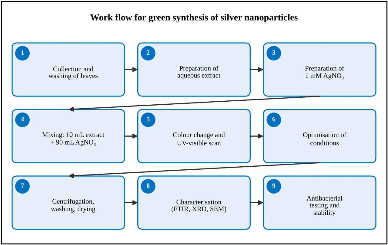
Figure 3: Work Flow for Green Synthesis of Silver Nanoparticles
Table: Variables Studied for the Optimisation of the Synthesis[2,12]
| Sr. No. | Variable | Levels commonly studied | Reference value |
|---|---|---|---|
| 1 | Ratio of extract to silver nitrate (v/v) | 1:9, 2:8, 3:7 | 1:9 |
| 2 | Silver nitrate concentration | 0.5 mM, 1 mM, 2 mM | 1 mM |
| 3 | Temperature | Room temperature, about 60 °C | Room temperature |
| 4 | pH of the mixture | About 4, 7 and 9 | Natural pH |
| 5 | Reaction time | 0, 15, 30, 60 minutes and up to 24 hours | Until the colour is stable |
Preliminary Phytochemical Screening of the Extract
Table: Phytochemical Tests for the Plant Extract[15]
| Sr. No. | Constituent | Test | Positive indication |
|---|---|---|---|
| 1 | Phenolic compounds and tannins | 5% ferric chloride test; lead acetate test | Blue-black or green colour; white precipitate |
| 2 | Flavonoids | Shinoda test; alkaline reagent test | Pink to magenta colour; yellow colour fading on acid |
| 3 | Alkaloids | Mayer's, Dragendorff's and Wagner's tests | Cream, orange to reddish-brown precipitate |
| 4 | Saponins | Foam (froth) test | Persistent foam |
| 5 | Terpenoids | Salkowski test | Reddish-brown ring at the interface |
| 6 | Carbohydrates | Molisch's test | Purple ring at the junction |
| 7 | Proteins | Biuret test | Violet colour |
Characterisation of Silver Nanoparticles
The nanoparticles formed are confirmed and characterised by one or more of the techniques given below. UV-visible spectroscopy is the simplest and is available in most college laboratories, while the other techniques need special facilities.[2,12]
Table: Techniques Used for the Characterisation of Silver Nanoparticles[2,8]
| Sr. No. | Technique | Information obtained |
|---|---|---|
| 1 | Visual observation | Colour change, formation of a precipitate (simplest first indication) |
| 2 | UV-visible spectroscopy | Surface plasmon resonance peak at about 400 to 450 nm |
| 3 | FTIR spectroscopy | Functional groups of the extract compounds present on the particle surface |
| 4 | X-ray diffraction (XRD) | Crystalline nature of the silver; estimate of crystallite size |
| 5 | SEM or TEM | Shape and size of the particles |
| 6 | Particle size and zeta potential | Average size, distribution and surface charge (stability) |
Evaluation of Antibacterial Activity
The antibacterial activity of the nanoparticles is commonly tested by the agar well diffusion method.[16,17] Sterile nutrient agar plates are seeded with a standardised suspension of a Gram-positive organism (for example Staphylococcus aureus) and a Gram-negative organism (for example Escherichia coli). Wells of about 6 mm are cut with a sterile borer and filled with a fixed volume of the test samples, the plates are incubated at 37 °C for 24 hours, and the diameter of the zone of inhibition is measured in millimetres. Each test is done in triplicate.
Table: Arrangement of Wells in the Agar Well Diffusion Test[16,17]
| Sr. No. | Well | Content |
|---|---|---|
| 1 | Test | Silver nanoparticle suspension made with the plant extract |
| 2 | Control 1 | Plant extract alone |
| 3 | Control 2 | 1 mM silver nitrate solution |
| 4 | Negative control | Distilled water |
| 5 | Positive control | A standard antibiotic of known activity |
The stability of the nanoparticle suspension is checked by storing it in amber bottles at room temperature and in a refrigerator, and by recording the colour, any sedimentation and the UV-visible spectrum at fixed intervals. A stable suspension keeps its colour and absorption peak, whereas aggregation shows as loss of colour, a shifted or broadened peak or a deposit.
Review of Literature
Iravani (2011) reviewed the green synthesis of metal nanoparticles using plants, compared plant-based synthesis with physical and chemical methods, and discussed the role of plant metabolites in reducing metal ions and stabilising the particles.[1]
Mittal et al. (2013) reviewed the synthesis of metallic nanoparticles using plant extract and described the possible mechanisms of formation, the plant parts used, the factors that control the size and shape, and the applications of the nanoparticles.[2] Kumar and Yadav (2009) reviewed the plant-mediated synthesis of silver and gold nanoparticles and their applications.[10]
Ahmed et al. (2016) reviewed the plant extract mediated synthesis of silver nanoparticles for antimicrobial applications, and summarised the plants used, the methods of characterisation and the antimicrobial activity of the nanoparticles.[12] Makarov et al. (2014) and Kharissova et al. (2013) reviewed the green synthesis of metal nanoparticles using plants and the greener approaches to nanoparticle synthesis.[9,11]
Shankar et al. (2004) reported the rapid synthesis of gold, silver and bimetallic gold-core silver-shell nanoparticles using neem (Azadirachta indica) leaf broth, which showed that simple neem leaf broth can reduce metal ions.[13] Singhal et al. (2011) prepared silver nanoparticles using Ocimum sanctum (Tulsi) leaf extract and screened their antimicrobial activity.[14]
Rai et al. (2009) described silver nanoparticles as a new generation of antimicrobials, and discussed their activity against bacteria and fungi, their mode of action and their potential biomedical uses.[3] Morones et al. (2005) studied the bactericidal effect of silver nanoparticles, and Sondi and Salopek-Sondi (2004) used Escherichia coli as a model Gram-negative bacterium to study the antimicrobial effect of silver nanoparticles; Feng et al. (2000) studied the mechanism of the antibacterial effect of silver ions on E. coli and S. aureus.[5,6,7]
Prabhu and Poulose (2012) reviewed the mechanism of the antimicrobial action, the methods of synthesis, the medical applications and the toxicity effects of silver nanoparticles, and are a reminder that safety must be considered together with activity.[4] Mulvaney (1996) described the surface plasmon spectroscopy of nanosized metal particles, which is the basis of the use of UV-visible spectroscopy for the detection of silver nanoparticles.[8]
Balouiri et al. (2016) reviewed the methods for the in vitro evaluation of antimicrobial activity, including agar diffusion, and Bauer et al. (1966) described the standardised single disk method for antibiotic susceptibility testing, which is the basis of diffusion tests in which the zone of inhibition is measured.[16,17]
Research gap
Many plants have been used to make silver nanoparticles, but the reaction conditions, the extract strength and the methods of testing differ between studies, so the plants cannot easily be compared with each other. Neem and Tulsi have each been reported separately.[13,14] Studies that run both plants through the same simple procedure, vary the main reaction conditions systematically and test the product against the same organisms are few.
Safety and Waste Disposal
Silver nitrate stains skin and clothing and irritates the eyes and skin, so gloves, a laboratory coat and safety glasses should be worn. Silver-containing solutions and nanoparticles should not be poured into the drain, and should be collected in a labelled container for heavy-metal waste. Bacterial cultures must be handled with aseptic technique, and used plates and cultures must be autoclaved before disposal. Since the toxicity of silver nanoparticles has been reported,[4] nanoparticles prepared in a student laboratory are meant for laboratory study only and should not be used on humans or animals.
CONCLUSION
Green synthesis of silver nanoparticles with plant extracts such as Neem and Tulsi is a simple, low-cost, one-step and environmentally friendly alternative to the physical and chemical methods. The phytochemicals of the extract act as both reducing and capping agents, and the formation of the nanoparticles can be easily followed by the colour change and by the surface plasmon resonance band at about 400 to 450 nm in the UV-visible spectrum. The extract-to-silver nitrate ratio, the silver nitrate concentration, the temperature, the pH and the reaction time are the main variables that decide the quality of the product.
The nanoparticles show antibacterial activity, which can be tested by the agar well diffusion method, and they have potential uses in dressings, coatings and other biomedical products. However, the reaction conditions and the testing methods differ between studies, the size, shape and crystallinity need special facilities for confirmation, and the toxicity of the nanoparticles must be studied before any use in humans. Further work with other plants, more organisms, determination of the minimum inhibitory concentration, cytotoxicity testing and long-term stability studies is required.
REFERENCES

Ramankit Jaiswal, Mahesh Padghan, Kshitij Shirsat, Kunal Khandeshe, Manoj Dakhore, Krushna Sharma, K. Biyani, Green Synthesis of Silver Nanoparticles Using Plant Extracts: A Review, Int. J. of Pharm. Sci., 2026, Vol 4, Issue 10, 3404-3412. https://doi.org/10.5281/zenodo.23609735
 10.5281/zenodo.23609735