

We use cookies to ensure our website works properly and to personalise your experience. Cookies policy
1,2MBBS, Third Year, Department of Pharmacology, fergana medical institute of public health, Uzbekistan.
3Assistant Professor Department of Pharmacology, fergana medical institute of public health, Uzbekistan.
Immune checkpoint inhibitors (ICIs) have reshaped the treatment of many solid malignancies, producing durable remissions in patients who previously had few options. Yet most patients either never respond (primary resistance) or relapse after an initial benefit (acquired resistance). Resistance is not a single phenomenon. It arises from a layered set of tumor-intrinsic programs, an immunosuppressive and often physically exclusionary tumor microenvironment (TME), progressive dysfunction of tumor-reactive T cells, and host-level factors such as the gut microbiome, systemic inflammation and prior therapies. This review integrates current knowledge of these mechanisms, following the sequence of the cancer-immunity cycle, and distinguishes resistance that is already present before treatment from resistance that evolves under therapeutic pressure. We then examine biomarkers that attempt to anticipate resistance, including PD-L1 expression, mismatch-repair status, tumor mutational burden, interferon-gamma signatures, HLA genotype and spatial immune architecture, and discuss why no single marker is sufficient. Strategies to overcome resistance are surveyed, from neoadjuvant timing and rational drug combinations to cellular therapy, personalized vaccines and microbiome modulation, together with the clinical evidence supporting or limiting each. Finally, we outline translational obstacles, including trial design, heterogeneity, and the need for dynamic biomarkers, and propose directions for future work. A mechanism-guided, biomarker-driven approach is likely to be required to extend the benefit of immunotherapy to a larger proportion of patients.
The idea that the immune system can recognize and eliminate cancer is old, but only in the last fifteen years has it produced a reliable class of therapies. Blocking inhibitory receptors on T cells, chiefly cytotoxic T-lymphocyte antigen 4 (CTLA-4) and programmed cell death protein 1 (PD-1), or the PD-1 ligand PD-L1, releases restraints that normally prevent excessive immune activity and that tumors exploit to avoid destruction. The first randomized evidence that this strategy prolongs survival came from ipilimumab in metastatic melanoma [6], and early studies of PD-1 and PD-L1 antibodies soon showed objective responses across melanoma, lung cancer and kidney cancer [7]. Since then, checkpoint blockade has become a standard component of care in a long list of tumor types, alone or together with chemotherapy, targeted agents or other immunotherapies.
Despite these advances, the proportion of patients who benefit remains modest. One analysis estimated that, in 2018, roughly 44% of patients with cancer in the United States were eligible for checkpoint inhibitor therapy, while only about 13% were likely to respond [11]. Even among responders, a meaningful fraction relapse, sometimes after years of disease control. The gap between eligibility and benefit is the central clinical problem of the field, and it explains why the study of resistance has become as important as the development of new agents.
Resistance to immunotherapy differs conceptually from resistance to cytotoxic or kinase-directed therapy. A cytotoxic drug or kinase inhibitor acts directly on the tumor cell, so resistance usually reflects a change in the cancer cell's drug handling or target. Immunotherapy instead depends on a multicellular process: tumor antigens must be generated and presented, T cells must be primed, must traffic to the tumor, infiltrate it, recognize cancer cells, and kill them while surviving in a hostile environment. A defect at any of these steps can produce failure, and different patients, or different lesions in the same patient, may fail for different reasons [5,12]. Reviews of the topic increasingly emphasize this multidimensional character [1-4].
This review follows the structure of the field's current thinking. After a brief overview of immunotherapy modalities (Section 2), we define primary, adaptive and acquired resistance and summarize their main mechanisms (Section 3). We then discuss the immunosuppressive TME (Section 4), tumor-intrinsic programs (Section 5), and host and systemic influences (Section 6). Section 7 evaluates biomarkers, Sections 8 and 9 examine therapeutic strategies and combinations, and Section 10 weighs the clinical evidence and translational challenges. Section 11 outlines future perspectives before concluding in Section 12.
2. Overview of Cancer Immunotherapy
2.1 Immunological basis of checkpoint blockade
The adaptive antitumor response is commonly described as a cycle: dying tumor cells release antigens, dendritic cells capture and present them, T cells are primed in lymphoid tissue, activated cells travel through the circulation and enter the tumor, recognize antigen on cancer cells, and kill them, releasing more antigen and perpetuating the cycle. Checkpoints operate at different stages. CTLA-4 competes with the costimulatory receptor CD28 for B7 ligands and chiefly restrains T cell priming, whereas PD-1 mainly dampens effector function in peripheral tissues after repeated antigen exposure. Tumor cells and infiltrating myeloid cells can express PD-L1, and early work showed that this expression can induce apoptosis of activated T cells and protect tumors from immune attack [8]. Antibodies that block these interactions therefore act at different points, which provided a rationale for combining them (Figure 1).
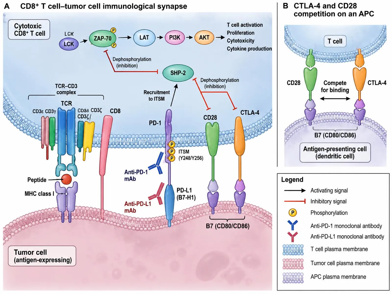
Figure 1. Molecular architecture of the CD8⁺ T cell–tumor cell immunological synapse and the site of action of immune checkpoint inhibitors. (A) Recognition of tumor peptide–MHC class I by the TCR–CD3 complex and CD8 co-receptor triggers the LCK→ZAP-70→LAT→PI3K→AKT activation cascade. Engagement of PD-1 by PD-L1 phosphorylates the PD-1 ITSM (Y248/Y256), recruiting the phosphatase SHP-2, which dephosphorylates ZAP-70 and CD28 and attenuates effector signaling; anti-PD-1 and anti-PD-L1 monoclonal antibodies disrupt this interaction. (B) On antigen-presenting cells, CTLA-4 competes with the costimulatory receptor CD28 for B7 ligands (CD80/CD86), restraining T cell priming. Black arrows, activation; red T-bars, inhibition; P, phosphorylation.
2.2 Clinical development of checkpoint inhibitors
Ipilimumab was the first checkpoint inhibitor to demonstrate a survival benefit, extending median overall survival in previously treated metastatic melanoma compared with a peptide vaccine control [6]. Antibodies against PD-1 and PD-L1 followed, with objective responses reported across several tumor types in first-in-human studies [7]. In advanced melanoma, the combination of nivolumab and ipilimumab produced a three-year survival rate of 58%, compared with 52% for nivolumab alone and 34% for ipilimumab alone [9]. In non-squamous non-small cell lung cancer, adding pembrolizumab to platinum-pemetrexed chemotherapy raised the 12-month overall survival rate from about 49% to about 69%, and the benefit was seen across PD-L1 expression categories [10]. These and many other trials established checkpoint blockade in lung cancer, melanoma, renal cell carcinoma, urothelial carcinoma, head and neck cancer, hepatocellular carcinoma and several others.
2.3 Other immunotherapeutic modalities
Checkpoint inhibitors are only one branch of immunotherapy. Adoptive cell transfer, in which tumor-infiltrating lymphocytes (TILs) are expanded ex vivo and reinfused after lymphodepletion, has shown efficacy in melanoma, including in patients whose disease progressed after anti-PD-1 therapy [38]. Personalized neoantigen vaccines aim to prime T cells against mutations unique to the patient's tumor, and early studies in melanoma showed that vaccination could generate neoantigen-specific T cells [52]. Cytokine therapy, oncolytic viruses, bispecific engagers, agonists of innate immune sensors, and chimeric antigen receptor (CAR) T cells constitute further approaches, though outside hematologic malignancy the latter have so far had limited success in solid tumors. Because these modalities act through different biology, they offer opportunities for use in patients whose tumors resist checkpoint blockade, and they are discussed in that context in Sections 8 and 9.
2.4 Patterns of response
Immunotherapy responses have features that differ from those of cytotoxic treatment. Responses can be delayed, can be preceded by apparent radiographic growth, and can persist after treatment is stopped. These features complicate the definition of resistance, since early progression may be true failure, a pseudoprogression driven by immune infiltration, or, in a small minority of patients, an accelerated growth pattern termed hyperprogression [53,54]. Conventional response criteria were therefore adapted to account for atypical kinetics, and the distinction between primary and acquired resistance is made with reference to the time course and depth of initial benefit, as described next.
3. Mechanisms of Primary and Acquired Resistance
3.1 Definitions
Although terminology varies between reports, three categories are widely used [5,12]. Primary (intrinsic) resistance describes tumors that fail to respond from the outset and show no meaningful disease control. Adaptive resistance refers to the tumor's reactive upregulation of immune-evasion programs in response to an ongoing immune attack, the best-known example being interferon-driven induction of PD-L1. Acquired resistance describes disease that initially responded, or was controlled for a clinically meaningful period, and then progressed during continued therapy. Because these labels are applied inconsistently, comparisons across studies must take account of the criteria used, such as the minimum duration of initial benefit.
The frequency of acquired resistance varies. In melanoma, a subset of patients with an initial response progress later, and in lung cancer acquired resistance is common among those who respond for only a limited time [12]. Because tumor evolution under immune pressure involves both genetic and non-genetic changes, resistance may be heterogeneous even within a single patient, with some lesions regressing while others grow.
3.2 Failure to generate or present antigen
The immune system can only attack what it can see. Tumors with few mutations, or with mutations that generate few immunogenic peptides, have less to offer T cells. In non-small cell lung cancer, higher nonsynonymous mutation burden was associated with improved response to PD-1 blockade [27], and in colorectal and other cancers deficiency in DNA mismatch repair, which produces very high mutation loads, predicts benefit [28]. Conversely, tumors with low mutational load and little neoantigen expression are less likely to respond. Even when neoantigens exist, their presentation depends on the major histocompatibility complex (MHC) class I pathway. Loss of beta-2 microglobulin (B2M), a component of MHC class I, has been documented in patients who developed resistance to checkpoint blockade [16,24]. Impaired HLA class I antigen processing and presentation was also identified as a mechanism of acquired resistance in lung cancer [25]. At the host level, HLA class I genotype itself matters: patients heterozygous at all HLA class I loci showed better survival after checkpoint blockade than those homozygous at one or more loci, presumably because they can present a wider repertoire of peptides [26].
3.3 Defective interferon-gamma signaling
Interferon-gamma (IFN-γ), released by activated T cells, is both a mediator of tumor killing and a signal that induces antigen-presentation machinery and PD-L1. Loss-of-function mutations in the IFN-γ receptor pathway therefore allow tumor cells to ignore a T cell attack. Whole-exome sequencing of paired biopsies from melanoma patients who relapsed on PD-1 blockade revealed loss-of-function mutations in JAK1 or JAK2, together with a B2M mutation in another patient [16]. JAK1/2 mutations have also been linked to primary resistance, including in tumors with high mutational burden [17]. In patients treated with anti-CTLA-4, loss of IFN-γ pathway genes was enriched in non-responders [18]. Functional genomic screens have broadened this picture, identifying genes whose loss alters tumor sensitivity to T cell killing [19], and showing for example that loss of the chromatin regulator PBRM1 can in some models sensitize tumors to IFN-γ [20]. Such findings highlight that the relationship between interferon signaling and resistance is not uniform: complete loss of signaling can abolish T cell sensitivity, whereas chronic, sustained signaling can drive a multigenic resistance program, as discussed in Section 5 (Figure 2).
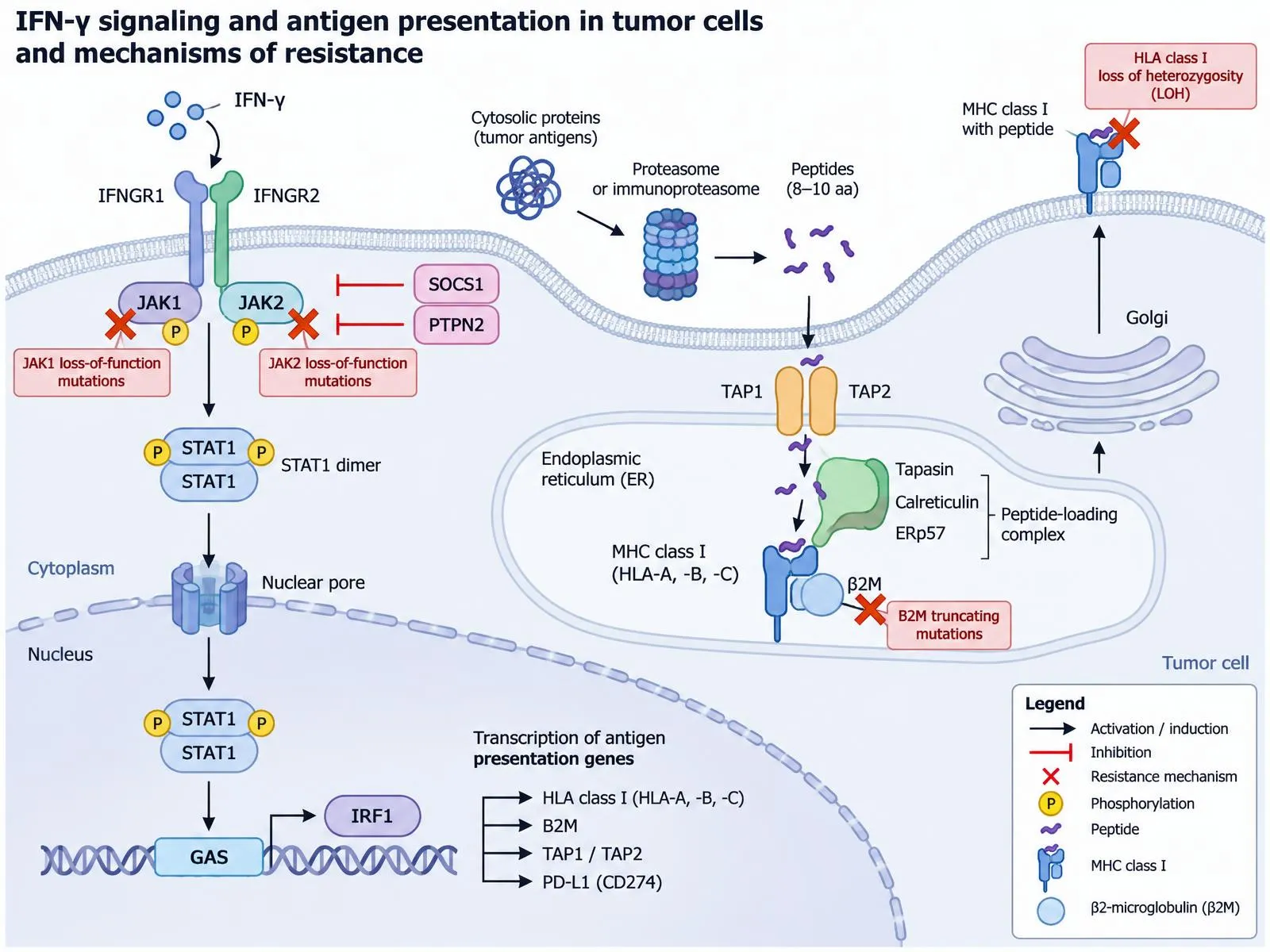
Figure 2. Interferon-γ signaling and the MHC class I antigen-processing pathway, with genetic lesions that confer resistance to checkpoint blockade. IFN-γ binds IFNGR1/IFNGR2, activating JAK1/JAK2, which phosphorylate STAT1; STAT1 homodimers translocate to the nucleus, bind GAS elements and induce IRF1, driving transcription of HLA class I, B2M, TAP1/TAP2 and PD-L1 (CD274). Proteasome-derived peptides are transported by TAP into the endoplasmic reticulum, loaded onto MHC class I/β2M by the peptide-loading complex (tapasin, calreticulin, ERp57) and trafficked via the Golgi to the cell surface. SOCS1 and PTPN2 provide negative feedback. Red crosses mark resistance mechanisms: JAK1/JAK2 loss-of-function mutations, B2M truncating mutations and HLA class I loss of heterozygosity.
3.4 Oncogenic signaling that shapes immune exclusion
Certain oncogenic pathways influence the immune landscape. Activation of WNT/β-catenin signaling in melanoma was associated with absence of a T cell infiltrate, via failure to recruit CD103-positive dendritic cells [22]. Loss of the tumor suppressor PTEN, which activates PI3K signaling, was linked to reduced T cell infiltration and poorer outcomes of immunotherapy in melanoma models and patient samples [21]. In KRAS-mutant lung adenocarcinoma, co-occurring STK11/LKB1 alterations are associated with resistance to PD-1 inhibitors [23]. These examples show that tumor genotype can pre-determine the immune phenotype, and that a given immunotherapy may be ineffective if the tumor's own signaling excludes T cells.
3.5 Dysfunction and exhaustion of T cells
Even when T cells reach tumor, they may be dysfunctional. Persistent antigen exposure drives T cells into a state termed exhaustion, characterized by sustained expression of multiple inhibitory receptors and reduced cytokine production and proliferative capacity [41]. Importantly, exhausted T cells are heterogeneous. A subset with progenitor-like features and expression of the transcription factor TCF1 retains the capacity to proliferate and to respond to PD-1 blockade, whereas terminally exhausted cells do not [42]. Epigenetic studies indicate that exhausted T cells acquire stable chromatin changes that are not reversed by PD-1 blockade, which limits the durability of reinvigoration [43]. Thus, the balance between progenitor and terminally exhausted cells in a tumor may determine whether blockade is effective, and acquired resistance may reflect progressive exhaustion or loss of the progenitor reservoir.
3.6 Compensatory immune checkpoints
Blocking one checkpoint can lead to compensatory upregulation of others. In preclinical models and in patient samples, adaptive resistance to PD-1 blockade was accompanied by increased expression of TIM-3 on T cells, and TIM-3 blockade restored sensitivity in mouse models [40]. Such compensatory mechanisms provide a rationale for dual checkpoint blockade, exemplified by the clinical benefit of adding the LAG-3 inhibitor relatlimab to nivolumab in melanoma [57], discussed in Section 9.
3.7 Summary of mechanisms
Table 1 summarizes the principal categories of resistance, their typical manifestation, and representative supporting evidence. The categories are not mutually exclusive; a tumor often exhibits several simultaneously, and different mechanisms may predominate in primary and acquired settings.
Table 1: Principal Mechanisms of Resistance to Immune Checkpoint Blockade, with Representative Evidence and Therapeutic Implications.
| Category | Representative mechanism | Typical setting | Key supporting evidence | Potential therapeutic implication |
|---|---|---|---|---|
| Antigen deficiency | Low mutational or neoantigen burden; mismatch-repair proficiency | Primary | [27,28,29,30] | Vaccines, cell therapy, agents that enhance antigen release |
| Antigen presentation defect | B2M loss; HLA class I downregulation or loss of heterozygosity | Primary and acquired | [16,24,25,26] | NK-cell or CD4 T cell-directed strategies; epigenetic agents |
| Interferon pathway defect | JAK1/JAK2 loss-of-function; IFN-γ receptor pathway loss | Primary and acquired | [16,17,18,19] | Strategies independent of IFN-γ signaling |
| Chronic interferon signaling | Multigenic resistance program, expression of alternative inhibitory ligands | Adaptive and acquired | [50,51] | Targeting downstream inhibitory ligands; modulating signaling duration |
| Oncogenic signaling and immune exclusion | WNT/β-catenin activation; PTEN loss; STK11/LKB1 alteration | Primary | [21,22,23] | Pathway inhibitors combined with ICIs |
| T cell exclusion and stromal barriers | TGF-β-driven stroma; abnormal vasculature; VEGF signaling | Primary | [35,36,37,39] | TGF-β or VEGF blockade; stromal modulation |
| T cell exhaustion | Loss of TCF1-positive progenitors; epigenetic fixation | Acquired | [41,42,43] | Early treatment, cell therapy, combination with other checkpoint inhibitors |
| Alternative checkpoints | TIM-3, LAG-3 upregulation | Adaptive and acquired | [40,57] | Dual checkpoint blockade |
| Suppressive cells | Regulatory T cells, tumor-associated macrophages, myeloid-derived suppressor cells | Primary and acquired | [44,45] | Cell depletion or reprogramming |
| Host factors | Gut microbiome composition, antibiotic exposure | Primary | [46,47,48,49] | Microbiome modulation, fecal microbiota transplantation |
4. Tumor Microenvironment and Immune Suppression
4.1 Immune phenotypes: inflamed, excluded and desert
The TME is not simply a backdrop. Its composition predicts and shapes the response to immunotherapy. Three broad phenotypes are commonly distinguished. Inflamed tumors contain T cells within the tumor parenchyma, frequently along with signs of interferon activity and PD-L1 expression, and they respond most often to checkpoint inhibition. Immune-excluded tumors have T cells that accumulate at the periphery or in the stroma but do not penetrate cancer cell nests. Immune-desert tumors lack T cells altogether, reflecting failure of priming or trafficking [37]. An early landmark study demonstrated that pretreatment density of CD8-positive T cells at the invasive tumor margin was associated with response to pembrolizumab in melanoma, and that responders showed increased proliferation of intratumoral cytotoxic T cells after treatment [32]. This supports the principle that checkpoint blockade works largely by reinvigorating a pre-existing response, and that tumors without such a response are poor candidates for monotherapy.
4.2 Physical and stromal barriers
In immune-excluded tumors, the stroma can act as a barrier. Analysis of urothelial cancer treated with atezolizumab showed that T cell exclusion was associated with transforming growth factor beta (TGF-β) signaling in fibroblasts and with a lack of response, and that combining TGF-β blockade with anti-PD-L1 in mouse models reduced stromal reactivity and promoted T cell penetration [35]. A parallel study in a genetically defined model of colorectal cancer metastasis found that TGF-β drove immune evasion by limiting the effector differentiation of T cells, and that blocking TGF-β signaling unleashed cytotoxic T cell activity and, together with anti-PD-1, eradicated established metastases [36]. Beyond TGF-β, dense extracellular matrix, cancer-associated fibroblasts, and abnormal tumor vasculature can restrict T cell access. Aberrant vessels with low expression of adhesion molecules impede lymphocyte extravasation, and vascular endothelial growth factor (VEGF) has been shown to promote this phenotype and to upregulate inhibitory receptors on CD8-positive T cells [39]. These observations underpin the rationale for combining checkpoint blockade with anti-angiogenic agents, discussed later.
4.3 Immunosuppressive cell populations
Several cell types actively suppress antitumor immunity. Regulatory T cells (Tregs) restrain effector function through cytokines, metabolic competition and inhibition of antigen-presenting cells. The role of PD-1 on these cells is notable. In patients treated with PD-1 blockade, the balance of PD-1 expression between effector T cells and Tregs in the tumor predicted outcome; when PD-1 was highly expressed on Tregs, blockade could enhance their suppressive activity and was associated with hyperprogression [44]. This work provides a mechanistic explanation for why PD-1 blockade, in some contexts, may paradoxically accelerate tumor growth.
Tumor-associated macrophages (TAMs) and myeloid-derived suppressor cells (MDSCs) form a second major axis. TAMs, which can constitute a large part of the tumor mass, often adopt an alternatively activated phenotype that supports angiogenesis, tissue remodeling and immune suppression, and a variety of strategies to deplete, reprogram or block their recruitment are being tested [45]. MDSCs suppress T cells through arginase, nitric oxide, reactive oxygen species and other mediators. Because these myeloid populations are diverse and plastic, targeting them has proven difficult, and clinical results to date have been modest.
4.4 Metabolic constraints and soluble factors
Tumors create a metabolically hostile environment characterized by hypoxia, low glucose, acidity and accumulation of immunosuppressive metabolites. T cells compete with cancer cells for nutrients, and adenosine generated by the CD39 and CD73 ectoenzymes can dampen effector function. Tryptophan catabolism by indoleamine 2,3-dioxygenase 1 (IDO1) was an attractive target, but a large randomized phase 3 trial of the IDO1 inhibitor epacadostat combined with pembrolizumab in melanoma failed to improve outcomes over pembrolizumab alone [58], a result that highlights the gap between preclinical rationale and clinical benefit and the importance of target engagement and patient selection.
Soluble mediators also shape the TME. Besides TGF-β and VEGF, cytokines such as interleukin-10 and a variety of chemokines modulate T cell recruitment. Epigenetic silencing of T helper 1-type chemokines in tumor cells has been proposed to limit T cell infiltration and may be reversible with epigenetic therapy, pointing to non-genetic mechanisms that may be pharmacologically accessible [13] (Figure 3).
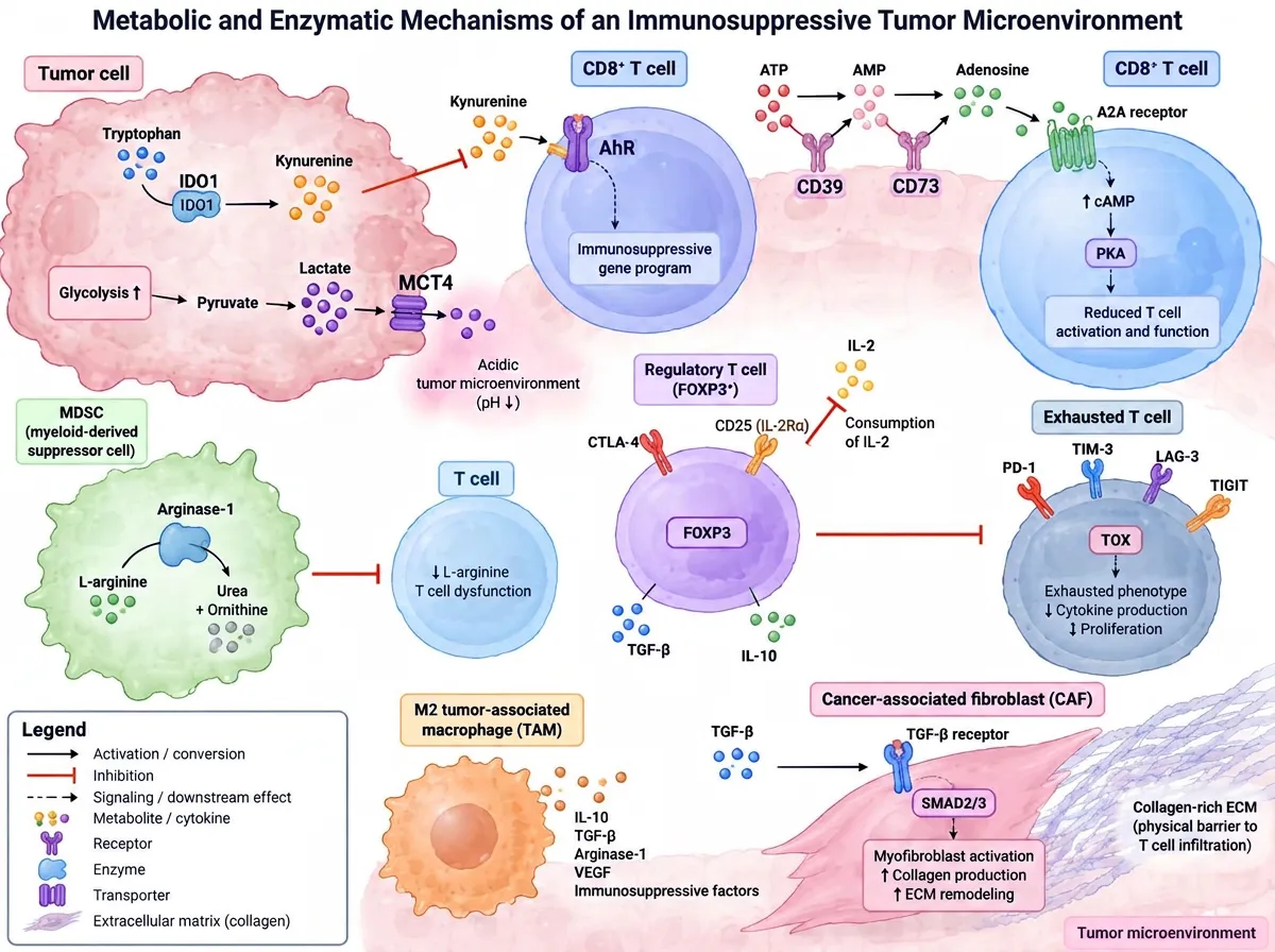
Figure 3. Metabolic, enzymatic and cellular mechanisms of immunosuppression in the tumor microenvironment. Tumor IDO1 converts tryptophan to kynurenine, which activates AhR in T cells; ectonucleotidases CD39 and CD73 generate adenosine that signals through the A2A receptor (↑cAMP/PKA) to suppress CD8⁺ T cell function; glycolytic tumor cells export lactate via MCT4, acidifying the milieu; MDSC-derived arginase-1 depletes L-arginine. FOXP3⁺ regulatory T cells consume IL-2 via CD25 and secrete TGF-β and IL-10; M2 tumor-associated macrophages release IL-10, TGF-β, arginase-1 and VEGF. TGF-β/SMAD2/3 signaling in cancer-associated fibroblasts builds a collagen-rich extracellular matrix that excludes T cells. Chronically stimulated T cells adopt a TOX-driven exhausted state co-expressing PD-1, TIM-3, LAG-3 and TIGIT.
4.5 Tertiary lymphoid structures and B cells
Organized immune aggregates within tumors, known as tertiary lymphoid structures (TLS), have emerged as a feature associated with response. In melanoma, B cell signatures and TLS were enriched in responders to checkpoint blockade, and TLS presence was associated with improved survival [55,56]. These structures may serve as local sites of antigen presentation and T cell activation, offering a counterbalance to systemic priming defects. Although the finding is correlative, it suggests that the TME contributes to response not only through T cell numbers, but through spatial organization and cell-cell interaction, and it encourages the development of spatial biomarkers.
4.6 Implications
The TME thus mediates resistance through at least four routes: exclusion of T cells from tumor nests, active suppression of those that enter, metabolic limitation of their function, and absence of the organized structures that sustain immune responses. These routes are partly independent of tumor genotype and can therefore evolve during treatment. Therapeutic strategies must consequently be matched to the dominant barrier in a given tumor, an idea that motivates the biomarker approaches described in Section 7.
5. Tumor-Intrinsic Mechanisms of Resistance
5.1 Immunoediting and clonal selection
The interaction between tumor and immune system is dynamic. Tumor cells that survive immune pressure are, by definition, those best able to evade it, so ongoing immune attack selects for resistant clones. This process, long described as cancer immunoediting, helps explain why some tumors lose neoantigens or antigen-presentation components over time [14]. In patients receiving nivolumab, serial biopsies showed that tumors evolved under treatment, with changes in the neoantigen landscape and in the composition of the T cell compartment, and that clonal neoantigen loss could accompany progression [34]. Acquired resistance can thus be viewed as Darwinian selection of tumor subclones that are invisible or insensitive to the existing response, and it implies that intratumoral heterogeneity is a risk factor for relapse.
5.2 Loss of antigenicity
Antigen loss can take several forms: deletion or silencing of the genes encoding immunodominant neoantigens, loss of whole chromosomal regions carrying such genes, or global reduction in antigen presentation. HLA loss of heterozygosity is one route, and B2M loss is another [24,25]. Because MHC class I loss also removes the ligand that inhibits natural killer (NK) cells, tumors with such defects may become susceptible to NK-mediated killing, an observation that has driven interest in strategies that engage NK cells or that are independent of MHC class I. Epigenetic silencing of antigen-processing genes can contribute and is potentially reversible, which distinguishes it from permanent genetic loss.
5.3 Altered interferon responsiveness and chronic interferon signaling
Section 3 noted that loss of IFN-γ signaling renders tumor cells insensitive to T cells. The opposite situation, prolonged exposure to interferon, can also be harmful. Studies in murine models showed that persistent interferon signaling in tumor cells activates a transcriptional program that upregulates multiple inhibitory ligands and that is associated with resistance to checkpoint blockade, and that this program was also detectable in patient tumors [50]. The effect depends on the duration and context of signaling. In vivo genome-wide screening identified deletion of the phosphatase PTPN2, a negative regulator of interferon signaling, as a way to sensitize tumors to immunotherapy by enhancing interferon-dependent effects on antigen presentation and growth inhibition [51]. These data indicate that the pathway is finely tuned and that both too little and too much signaling may be unfavorable, so that therapeutic modulation must be precise.
5.4 Oncogenic pathways and metabolic reprogramming
As outlined in Section 3, WNT/β-catenin activation, PTEN loss and STK11/LKB1 mutation are associated with immune exclusion or resistance [21-23]. In addition, signaling through the MAPK pathway can reduce antigen expression and increase production of immunosuppressive factors, a basis for combining MAPK inhibitors with immunotherapy in BRAF-mutant melanoma. In KRAS-mutant lung cancer with STK11/LKB1 alterations, tumors exhibit low PD-L1 expression and a poorly inflamed microenvironment, features that together contribute to primary resistance [23]. Intrinsic metabolic reprogramming, notably high glycolytic flux that depletes glucose and generates lactate, is another way that tumor cells constrain T cells. These findings suggest that oncogenic drivers, rather than being irrelevant to immunotherapy, are among its principal determinants.
5.5 Epigenetic regulation
Chromatin regulators and DNA methylation influence both antigen presentation and T cell susceptibility. The PBRM1 finding mentioned above is an instance: functional loss of this subunit of a chromatin remodeling complex influenced how tumor cells responded to T cell-derived cytokines [20]. Epigenetic silencing of chemokine genes has been proposed to limit T cell trafficking into tumors [13], and epigenetic modifiers such as inhibitors of histone methyltransferases or DNA methyltransferases have been proposed to restore antigen presentation and chemokine expression. Because epigenetic states are reversible, they are considered attractive therapeutic targets, although achieving selectivity and tolerability in combination with immunotherapy remains challenging.
5.6 Genomic instability and tumor mutational features
High mutational burden is associated with response across many but not all tumor types. The KEYNOTE-158 analysis showed that patients with tumors harboring a high tissue tumor mutational burden had a higher objective response rate to pembrolizumab than those with lower burden, leading to a tissue-agnostic indication [29]. However, subsequent analyses showed that the predictive power of mutational burden varies considerably between cancer types, and that high burden does not reliably predict benefit in all of them [30]. The likely explanation is that mutation count is a crude proxy for the quantity that matters, namely the number and clonality of neoantigens that are actually presented and recognized. Tumors with high burden but subclonal neoantigens, or with defects in presentation, may still resist.
6. Host and Systemic Factors Influencing Response
6.1 The gut microbiome
Of the host factors, the intestinal microbiome has received the most attention. In epithelial tumors, antibiotic use around the time of checkpoint blockade was associated with reduced clinical benefit, and the abundance of the bacterium Akkermansia muciniphila was enriched in responders; moreover, transferring fecal material from responders into germ-free mice improved the antitumor effect of PD-1 blockade [46]. In melanoma, patients whose stool contained a more diverse microbiome, with enrichment of bacteria in the Ruminococcaceae family, had better responses to PD-1 therapy, and favorable immune profiles in the tumor microenvironment were noted [47]. Two small clinical trials subsequently showed that fecal microbiota transplantation (FMT) from responders, combined with anti-PD-1, could restore response in a minority of patients with PD-1-refractory melanoma, with changes in the recipient microbiome and in immune gene expression [48,49]. Although these results are encouraging, they come from small, uncontrolled studies. Taxa associated with response differ across cohorts, and diet, geography, and sequencing methods confound interpretation. Large randomized trials will be needed before microbiome modulation can be considered established.
6.2 Medications, comorbidity and general health
Several systemic factors have been linked to outcome. Antibiotics and acid-suppressing drugs may act through the microbiome [46]. Corticosteroids, often needed to manage immune-related adverse events or cancer-related symptoms, can suppress T cell activity, although steroids given for adverse events after an initial response appear to have a smaller impact than steroids at baseline. Performance status, tumor burden, and systemic inflammatory markers such as neutrophil-to-lymphocyte ratio and lactate dehydrogenase correlate with outcome in numerous series. A high tumor burden may reflect a larger population of suppressive cells and a lower ratio of reinvigorated T cells to tumor mass. These associations are generally prognostic rather than mechanistic, but they matter for patient selection and for stratification in trials [1,3].
6.3 Genetic background of the host
Germline variation also influences response. As noted, HLA class I diversity is associated with survival after checkpoint blockade [26]. Variation in genes related to immune regulation and in the capacity to mount an inflammatory response has been proposed as another contributor, though evidence is less mature. Age, sex and obesity have all been reported to influence response and toxicity, sometimes in counterintuitive directions, and these observations remain active research areas. Whether differences reflect true biology, differences in treatment exposure, or confounding by other variables is not settled.
6.4 Systemic immunity and lymphoid organs
Because T cell priming occurs in lymphoid tissue, systemic immune competence matters. Antitumor responses may be limited if tumor-draining lymph nodes are involved or have been surgically removed, or if systemic immunosuppression, whether caused by disease or therapy, impairs naive T cell pools. Conversely, the presence of tertiary lymphoid structures and B cell activity within tumors may help compensate [55,56]. These issues are only beginning to be integrated into clinical practice, but they suggest that the host response should be considered a whole-body phenomenon.
6.5 Hyperprogressive disease
A small subset of patients experience a marked acceleration of tumor growth soon after starting PD-1 or PD-L1 inhibitors. In an early series, around 9% of patients treated with anti-PD-1 or anti-PD-L1 therapy met criteria for hyperprogression, which was associated with older age and poor outcome [53]. A study in advanced non-small cell lung cancer reported a higher incidence of hyperprogression with checkpoint inhibitors (about 14%) than with single-agent chemotherapy (about 5%), arguing that the phenomenon is not merely a reflection of aggressive disease [54]. Proposed mechanisms include expansion of PD-1-positive Tregs [44], oncogenic signaling, and altered myeloid activity. Its definition varies between reports, and prospective biomarkers are lacking. It nonetheless illustrates that resistance can sometimes be accompanied by active harm.
7. Biomarkers for Predicting Immunotherapy Resistance
7.1 Principles
An ideal biomarker would identify, before treatment, which patients will not benefit, and would signal early in treatment when resistance is emerging. Since response depends on several mechanisms at once, single analytes have limited predictive value, and composite approaches are being explored. Reviews consistently note that no current biomarker is both sensitive and specific enough to be used in isolation [1,2,4].
7.2 PD-L1 expression
PD-L1 expression assessed by immunohistochemistry was the first biomarker used to select patients, and higher expression is associated with greater likelihood of response in several diseases. However, its limitations are well recognized: expression is heterogeneous and dynamic, different assays and scoring systems are used for different drugs, and patients with low or no expression can still respond. In non-squamous lung cancer, the benefit of adding pembrolizumab to chemotherapy was observed across PD-L1 categories [10], which illustrates why PD-L1 alone is an incomplete guide. It remains useful as one input, particularly for monotherapy decisions.
7.3 Mismatch-repair deficiency and tumor mutational burden
Tumors with mismatch-repair deficiency (dMMR) or microsatellite instability carry a high mutational load and are strongly responsive to PD-1 blockade. In the initial study, immune-related objective response rates were 40% in dMMR colorectal cancer and 0% in mismatch-repair-proficient colorectal cancer [28]. This marker is therefore among the most robust predictors of benefit, and the converse, mismatch-repair proficiency in colorectal cancer, predicts primary resistance to monotherapy. Tumor mutational burden is a broader measure; high burden was associated with response in lung cancer [27] and was approved as a tissue-agnostic marker following KEYNOTE-158 [29]. As discussed above, it performs unevenly between cancer types [30], and assay standardization and threshold selection remain problems.
7.4 Interferon-gamma and inflammation signatures
Gene expression signatures that capture interferon-gamma activity and T cell inflammation have shown predictive value. An 18-gene signature reflecting IFN-γ-related and T cell-inflamed biology predicted response to pembrolizumab across multiple tumor types [31]. Such signatures reflect pre-existing immunity and thus overlap with the immunohistochemical finding that CD8-positive T cells at the tumor margin predict benefit [32]. A transcriptional signature associated with innate resistance to anti-PD-1 therapy, comprising features such as mesenchymal transition, angiogenesis and wound healing, was identified in melanoma [33]. These signatures broadly capture the inflamed versus non-inflamed dichotomy but do not identify the specific barrier, which limits their usefulness for choosing a second-line strategy.
7.5 Genomic markers of resistance
Direct genomic correlates of resistance include loss-of-function alterations in JAK1, JAK2 and B2M [16,17,24], as well as alterations in STK11/LKB1 [23] and PTEN [21]. Their prevalence is low, so they explain only a minority of resistance, but they are informative when present. HLA class I genotype, including heterozygosity and loss of heterozygosity, is another candidate that can be derived from the same sequencing data [26]. Serial sampling during treatment can show how these features change, as in studies of paired pre- and on-treatment biopsies [34].
7.6 Tumor microenvironment and spatial biomarkers
Quantification of immune cells, including their spatial distribution relative to tumor cells, has emerged as a powerful approach. Beyond the CD8-based assessment at the invasive margin [32], the presence of tertiary lymphoid structures and B cells has been associated with response [55,56]. Multiplexed imaging and spatial transcriptomics can capture this information at high resolution, though standardization and cost currently limit adoption. Stromal TGF-β signatures correlated with T cell exclusion and poor response in urothelial cancer [35] and are a candidate to identify patients who might benefit from TGF-β blockade, once such agents are validated.
7.7 Circulating and microbiome-based markers
Blood-based assays are attractive because they permit repeated sampling. Circulating tumor DNA, circulating immune cell populations and systemic inflammatory indices have all been investigated as dynamic markers of response and early indicators of resistance [2,4]. The microbiome, discussed above, may provide predictive information, but taxa associated with response differ across studies and prospectively validated tests are not available [46,47]. These approaches are promising but remain largely investigational.
7.8 Toward composite and dynamic biomarkers
Because resistance is multifactorial, integrated models that combine tumor genomics, transcriptional signatures, spatial immune features, host genetics and clinical variables are likely to outperform single markers. Early multi-feature models have been reported, but they require validation in independent prospective cohorts. Equally important are on-treatment markers, since early changes in circulating tumor DNA or in T cell clonal dynamics might identify patients who are failing to respond before radiographic progression. This has particular value for deciding when to escalate therapy.
8. Strategies to Overcome Resistance
8.1 Matching strategy to mechanism
A central principle is that strategies should address the dominant mechanism of resistance in a given tumor. Patients whose tumors are inflamed but progress after initial benefit may need a different checkpoint or an agent that restores T cell function. Patients with immune-excluded tumors may need an agent that alters the stroma or vasculature. Those with immune-desert tumors may require an intervention that generates or supplies a T cell response, such as vaccination or cell therapy. This framework, although conceptually appealing, is only partially realized in practice because mechanisms are rarely determined before treatment selection.
8.2 Optimizing the timing of immunotherapy: the neoadjuvant setting
Giving immunotherapy before surgery, while the primary tumor and its draining lymph nodes are still in place, may produce a broader and more durable response because antigen supply is maximal. In resectable non-small cell lung cancer, adding nivolumab to neoadjuvant chemotherapy raised the pathological complete response rate to about 24%, compared with about 2% with chemotherapy alone, and improved event-free survival [15]. Similarly impressive results have been reported in selected tumors with high sensitivity, notably mismatch-repair-deficient locally advanced rectal cancer, where treatment with PD-1 blockade produced clinical complete responses in all of the first twelve patients studied, allowing some to avoid chemoradiotherapy and surgery [60]. These data suggest that the clinical setting, as well as the drug, affects the likelihood of resistance, and that treatment of earlier-stage disease may circumvent some mechanisms that arise in advanced disease.
8.3 Targeting alternative immune checkpoints
Given the evidence that other inhibitory receptors are upregulated when PD-1 is blocked [40], a logical approach is to inhibit several checkpoints at once. The combination of nivolumab and ipilimumab improved outcomes over either alone in advanced melanoma [9], and the combination of relatlimab, which targets LAG-3, with nivolumab produced a longer progression-free survival than nivolumab alone in previously untreated advanced melanoma, roughly ten months versus under five months [57]. Agents against TIM-3, TIGIT and others are in development. Combination checkpoint inhibition often increases toxicity, so improved selection of patients is important.
8.4 Counteracting immune exclusion
In tumors where T cells are excluded, strategies that normalize the stroma or vasculature are attractive. Blockade of TGF-β combined with anti-PD-L1 improved outcomes in preclinical models [35,36], and clinical development of such combinations is ongoing. Anti-angiogenic therapy, discussed in Section 9, is the most advanced approach clinically. Targeting oncogenic drivers that cause exclusion, for example inhibition of PI3K signaling in PTEN-deficient tumors [21], or modulation of WNT signaling [22], is a conceptually related approach. These strategies remain largely at an early stage of clinical evaluation.
8.5 Modulating suppressive cells and metabolism
Depletion of Tregs, blockade of macrophage recruitment or survival signals, and reprogramming of myeloid cells have all been tested, with mixed results [44,45]. Efforts to inhibit immunosuppressive metabolic pathways have been disappointing; the failure of the IDO1 inhibitor epacadostat in a large phase 3 melanoma trial is the most prominent example [58]. Possible reasons include incomplete target inhibition, redundancy of metabolic pathways, and failure to select patients likely to benefit. The experience argues for biomarker-guided early-phase studies that confirm pharmacodynamic effects before large trials.
8.6 Epigenetic and interferon-pathway modulation
Because some resistance mechanisms are epigenetic and reversible, agents that alter chromatin or DNA methylation are being combined with checkpoint inhibitors, with the aim of restoring antigen presentation, chemokine expression and T cell function [13]. Modulating interferon signaling is another approach: deletion or inhibition of PTPN2 sensitized tumors in preclinical models [51], and understanding the duration-dependent effects of interferon may allow timing or dosing strategies that avoid triggering the inhibitory program [50]. Translating these ideas into the clinic requires a careful therapeutic window.
8.7 Adoptive cell therapy and neoantigen vaccines
When a tumor lacks an effective T cell response, one option is to provide the cells directly. In a randomized phase 3 trial in advanced melanoma, TIL therapy improved progression-free survival compared with ipilimumab (median about 7 months versus 3 months), including in many patients previously treated with anti-PD-1 [38]. This demonstrates that tumors resistant to PD-1 blockade can remain susceptible to T cell therapy, and that resistance is often due to inadequate T cell numbers or function rather than irreversible tumor insensitivity. Personalized neoantigen vaccines represent another approach: in an early melanoma study, vaccination elicited neoantigen-specific T cells, and most of the small number of patients treated remained free of recurrence for a prolonged period, although the study was uncontrolled [52]. Both approaches are resource-intensive, and in the case of TIL therapy entail toxicities related to lymphodepletion and interleukin-2, which limits their use.
8.8 Microbiome modulation
As noted, FMT from responders overcame resistance to PD-1 blockade in a subset of patients with refractory melanoma [48,49]. Other approaches include defined bacterial consortia, dietary modification and avoidance of unnecessary antibiotics. The most pragmatic measure available today is likely to be careful antibiotic stewardship in patients receiving immunotherapy [46].
9. Combination Therapy Approaches
9.1 Checkpoint inhibitors with chemotherapy
Chemotherapy can kill tumor cells in an immunogenic fashion, release antigen, and deplete suppressive cells, which provides a rationale for combining it with checkpoint blockade. The most successful example is first-line lung cancer, where adding pembrolizumab to platinum-based chemotherapy improved overall survival in non-squamous disease regardless of PD-L1 expression [10]. In the neoadjuvant setting, the addition of nivolumab to chemotherapy increased pathological complete responses in resectable lung cancer [15]. The mechanisms of benefit likely include increased antigen availability and relief of chemotherapy-induced lymphodepletion effects, but the optimal sequencing, dosing and choice of chemotherapy backbone remain incompletely defined.
9.2 Checkpoint inhibitors with anti-angiogenic therapy
Because VEGF contributes to abnormal vasculature, impaired T cell infiltration and upregulation of inhibitory receptors [39], blocking VEGF is expected to improve immunotherapy. In unresectable hepatocellular carcinoma, atezolizumab plus bevacizumab improved overall survival and progression-free survival compared with sorafenib, an outcome that established the combination as a standard first-line option [59]. Similar principles underlie combinations of checkpoint inhibitors with VEGF receptor tyrosine kinase inhibitors in other tumors. The success of these combinations supports the notion that modifying the vascular and stromal environment can convert some poorly infiltrated tumors into responsive ones, although it is difficult to separate the contribution of direct antitumor activity from that of immune modulation.
9.3 Dual immune checkpoint blockade
Combining anti-CTLA-4 with anti-PD-1 therapy provided higher response and survival rates in melanoma than either agent alone, at the price of greater toxicity [9]. Adding the LAG-3 inhibitor relatlimab to nivolumab also improved progression-free survival in melanoma [57], consistent with the concept that compensatory checkpoints contribute to resistance [40]. Not all combinations are successful, and the benefit has been greatest in melanoma, with more variable results elsewhere. Rational selection of the partner checkpoint, informed by the expression pattern of inhibitory receptors in a patient's tumor, might improve the benefit-to-risk ratio.
9.4 Checkpoint inhibitors with targeted therapy
Several oncogenic pathways that produce resistance can be inhibited with approved or investigational drugs, so combining pathway inhibitors with immunotherapy is attractive. Examples include inhibition of PI3K in PTEN-deficient tumors [21], and inhibition of the MAPK pathway in BRAF-mutant melanoma. Early combinations have often met toxicity problems, because overlapping adverse effects limit dosing, and schedule-dependent effects may be important. Sequential rather than concurrent approaches, and intermittent dosing, are being explored. The failure of epacadostat combined with pembrolizumab [58] also serves as a caution that a mechanistically attractive partner does not guarantee benefit.
9.5 Combination with cellular therapy and vaccines
In tumors with insufficient T cell responses, vaccination or adoptive cell transfer may supply what is missing, and checkpoint blockade may then sustain the transferred or newly primed cells. Personalized neoantigen vaccines were tested with this logic [52], and TIL therapy has shown benefit in patients who had progressed on PD-1 blockade [38]. The interplay between such cells and the TME, including the presence of suppressive cells and stromal barriers, will likely determine efficacy, and combinations with agents that modify these barriers are logical next steps. For solid tumors in general, the challenge of identifying shared or individual targets with sufficient specificity remains considerable.
9.6 Combination with radiotherapy, local therapies and microbiome modulation
Local therapies such as radiotherapy can release antigen, induce interferon signaling and alter the vasculature, thereby potentially converting a poorly inflamed tumor into a more responsive one. Evidence for systemic (abscopal) effects is mostly anecdotal, and randomized trials have produced mixed results; dose, fractionation and timing are important variables. Approaches that modulate the microbiome, including FMT [48,49], offer another combination avenue with relatively low toxicity, although the field lacks randomized evidence.
9.7 Principles for designing combinations
The number of possible combinations is far greater than can be tested clinically. Prioritization should be guided by mechanism, by biomarker evidence that a target is present and that the drug engages it, and by early readouts of pharmacodynamic effect. Combination trials also need appropriate controls: a combination should be compared to the best available single-agent or standard therapy, not merely to a weaker control, so that the contribution of each component is clear. Several negative phase 3 trials in recent years, in which a promising partner failed to improve on a checkpoint inhibitor alone, show the cost of omitting these steps [58].
10. Clinical Evidence and Translational Challenges
10.1 Summary of clinical evidence
The clinical data reviewed above can be summarized as follows. Checkpoint inhibitors have proven survival benefit across multiple tumor types, and combination regimens, particularly those combining checkpoint inhibitors with chemotherapy, anti-angiogenic agents or a second checkpoint inhibitor, have improved outcomes in defined settings [9,10,57,59]. Neoadjuvant immunotherapy yields high pathological response rates in some cancers [15], and dramatic responses in mismatch-repair-deficient disease [60]. Cellular therapy can induce responses in tumors resistant to PD-1 blockade [38]. By contrast, strategies targeting immunosuppressive metabolism have been disappointing [58], and results of microbiome and vaccine approaches are preliminary [48,49,52]. Most evidence for specific resistance mechanisms is still correlative, derived from small series of patients, retrospective biopsies or preclinical models.
10.2 Limitations of the preclinical models
Many mechanistic insights come from syngeneic mouse tumors, which are often highly immunogenic, grow quickly, and have an immune system that differs from that of humans. Human tumors are slow-growing, genetically heterogeneous, and exposed to prior therapy and a lifetime of immune and microbial exposures. These differences may explain why interventions that cure mice often fail in patients. Improved models, including humanized mice, patient-derived organoids with immune components and genetically engineered models that better reflect human tumor evolution, are needed. Functional genomic screens in vivo have yielded useful candidate targets [19,51], but validation in human settings is a necessary next step.
10.3 Heterogeneity and sampling
Resistance can differ between patients, between lesions in the same patient, and across time. A single baseline biopsy captures only a fraction of this variation and may mislead. Studies of paired samples showed that tumors change under treatment [16,34], which argues for repeated sampling, either through serial biopsies or through less invasive approaches such as circulating tumor DNA. Practical barriers, including the risk and cost of biopsy, the limited tissue available, and the lack of standardized assays, restrict the implementation of such strategies in routine care.
10.4 Trial design and endpoints
Several features of immunotherapy complicate trial design. Delayed separation of survival curves violates the proportional hazards assumption typically used in analysis, and the plateau in survival seen in responders requires different statistical approaches. Response criteria must account for pseudoprogression and atypical kinetics. Definitions of primary and acquired resistance are inconsistent between studies, hindering comparison [5,12]. Trials of drugs in patients who have progressed on a checkpoint inhibitor need to define precisely what resistance means and to stratify by mechanism where possible, otherwise heterogeneous populations dilute any effect. Enrichment strategies based on biomarkers, adaptive designs and window-of-opportunity studies that obtain tissue before and after short exposure can accelerate learning, and they are increasingly used.
10.5 Toxicity, cost and access
Effective combinations often increase immune-related adverse events, which can be severe and may cause permanent damage or necessitate stopping treatment. The balance between efficacy and tolerability is a particular concern for combination checkpoint blockade [9]. Costs of checkpoint inhibitors, cellular therapy and personalized vaccines are high, and disparities in access mean that the global benefit is limited, especially in low- and middle-income settings [11]. Practical considerations such as manufacturing time for personalized products can also exclude patients with rapidly progressing disease.
10.6 Integrating mechanistic knowledge into practice
Translating the understanding of resistance into routine practice requires an infrastructure that does not yet exist in most centers: rapid genomic and transcriptomic profiling, standardized spatial analysis, access to a broad range of clinical trials, and decision support that integrates multiple data types. Collaborative consortia that share well-annotated, longitudinally sampled cohorts would help, as would the standardization of how resistance is defined and reported [1-4]. Without such efforts, much of the mechanistic knowledge will remain confined to research settings.
11. Future Perspectives
11.1 Single-cell and spatial technologies
Single-cell sequencing has already revealed substantial heterogeneity within T cell populations, including the distinction between progenitor and terminally exhausted states that determine responsiveness to PD-1 blockade [42]. Applied longitudinally and combined with spatial profiling, these technologies can map how cell states and cell-cell interactions evolve under treatment and may identify the cellular circuits that distinguish responders from non-responders. As costs fall, derived signatures may become clinically feasible biomarkers.
11.2 Artificial intelligence and multimodal integration
Machine learning is well suited to integrating the high-dimensional data discussed above: histology images, genomics, transcriptomics, radiomics, clinical variables and microbiome profiles. Models trained on such data could predict response and resistance more accurately than single markers and could suggest which combination is most appropriate for a patient. The main challenges are the need for large, diverse and well-annotated datasets, rigorous external validation, and transparent models that clinicians can interpret. Bias in training data may produce models that perform poorly in underrepresented populations.
11.3 Dynamic monitoring
On-treatment monitoring with circulating tumor DNA, circulating T cell clones and imaging may allow early detection of resistance and timely switching of therapy. Peripheral blood analyses that reflect tumor-specific T cell dynamics could be especially useful, since they sample the whole body. Prospective trials in which treatment is adapted according to such markers, for example by escalating therapy early in patients whose markers do not change, are required to demonstrate clinical utility [2,4].
11.4 Next-generation therapeutics
The pipeline includes new checkpoint and costimulatory targets, bispecific antibodies that combine two immune-modulating activities, engineered cytokines with reduced toxicity, improved TIL products and engineered T cell receptors targeting shared neoantigens, and mRNA-based personalized vaccines. Therapies that engage NK cells or myeloid cells may overcome the limitations of T cell-based approaches in tumors with antigen presentation defects. Agents that reprogram the TME, including modulators of TGF-β, macrophage function and the vasculature, will be tested more systematically as biomarker tools mature [35,36,45].
11.5 The microbiome and host modulation
If the early FMT findings are confirmed in randomized trials [48,49], microbiome-directed therapy could become a relatively simple way to improve the odds of response. Defined bacterial consortia are likely to replace crude fecal preparations because of safety and reproducibility. In parallel, a better understanding of how diet, antibiotics, host genetics and the immune system interact would help in designing interventions and in counseling patients [46,47].
11.6 Earlier intervention
The success of neoadjuvant immunotherapy suggests that moving treatment earlier in the disease course, when tumors are less heterogeneous and the immune system is less compromised, may prevent resistance from arising [15,60]. Studies that use pathological response to adapt subsequent therapy are in progress. In the longer term, the possibility of intercepting premalignant lesions or minimal residual disease with immunotherapy is an exciting but speculative prospect.
CONCLUSION
Resistance to immunotherapy in solid tumors is a multifactorial problem that arises from defects in antigen generation and presentation, altered interferon signaling, oncogenic pathways that exclude or inactivate T cells, an immunosuppressive and physically restrictive TME, progressive T cell exhaustion, and host factors including the microbiome. These mechanisms interact and evolve, so that a patient may have primary resistance through one route and acquired resistance through another. Existing biomarkers capture only some of this complexity, and no single test can reliably predict resistance.
Therapeutic progress has come from rational combinations, earlier use of immunotherapy, cellular therapy and, more tentatively, microbiome modulation. At the same time, several failures have shown that mechanistic plausibility must be backed by target engagement, patient selection and carefully designed trials. The priorities for the field are clear: standardize the definitions of resistance, develop composite and dynamic biomarkers, build better preclinical models, and design trials that match therapy to mechanism. If these goals can be met, the benefit of immunotherapy may extend to many of the patients who currently do not respond.
REFERENCES



Prathipa Kannan, Thrisha Thimmaraj, Shivprasad Dhage, Immunotherapy Resistance in Solid Tumors: Mechanisms and Therapeutic Strategies, Int. J. of Pharm. Sci., 2026, Vol 4, Issue 10, 3647-3665. https://doi.org/10.5281/zenodo.23639874
 10.5281/zenodo.23639874