

We use cookies to ensure our website works properly and to personalise your experience. Cookies policy
Department of Pharmacology, Sarojini Naidu Vanita Pharmacy Maha Vidyalaya (Co-Ed.), Tarnaka, Hyderabad, Telangana, India-500017.
Ulcerative Colitis (UC) is a chronic, relapsing disease that includes mucosal inflammation, abnormal immune response and oxidative stress. There are a number of therapeutic strategies for the treatment of UC, which include amino salicylates, corticosteroids, immunomodulatory agents, biologicals, and small molecule drugs; however, therapy resistance, recurrence and adverse effects resulting from prolonged use still pose a challenge. In this regard, it becomes essential to discover new drugs that possess both anti-inflammatory and antioxidative properties. Usnic acid is a dibenzofuran compound isolated mainly from lichens that has drawn a lot of interest owing to its various pharmacological activities. This review highlights the use of usnic acid in ulcerative colitis treatment, specifically discussing its origin, chemical structure, pharmacological actions, mechanism of action and safety. Its anti-inflammatory activity has been proved by experimental studies on the modulation of inflammatory mediators, which include TNF-?, IL-1?, IL-6, NO, and inflammatory signalling pathways such as NF-?B and TLR4. The antioxidant property is due to the enhancement of SOD and GSH activity, and reduction of MDA, MPO and lipid hydroperoxides levels. Moreover, its immunomodulatory actions and protective effects on the intestinal barrier have been considered. However, incomplete data on its pharmacokinetics, insufficient experimental results related to disease-specificity and problems with the toxicity especially hepatotoxicity are the main challenges. It is necessary to carry out additional research to confirm its therapeutic effectiveness, mechanism of action, optimum dose and safety profile.
Definition and Overview of Inflammatory Bowel Disease (IBD)
Inflammatory Bowel Disease is an inflammatory condition of unknown cause that involves the destruction of the intestinal mucosa. Inflammatory bowel disease (IBD) includes Crohn’s disease (CD) and ulcerative colitis (UC). Crohn’s disease and ulcerative colitis are chronic and recurrent inflammatory disorders of the gastrointestinal tract with their own pathological and clinical features [1].
Ulcerative Colitis (UC), which is a type of Inflammatory Bowel Disease (IBD), is an inflammatory condition that usually affects the colon and rectum but, in some cases, can affect the terminal ileum through backwash ileitis [2].
Epidemiology and Prevalence of UC
Over the last two decades, UC has emerged as a serious global health threat, indicative of the evolving nature of its epidemiology [3]. Between 2010 and 2019, Japan witnessed an increased annual prevalence per 100,000 people from 5 to 98. At the same time, the prevalence in the US increased from 158 to 233. While it appears that the incidence of UC is levelling off for all age groups in the US, the incidence rate is still increasing for pediatric and adolescent populations (4).
Major Clinical Symptoms
Symptoms usually include bloody diarrhoea, but can also include other GI symptoms, as well as systemic inflammation signs and other complications such as sclerosing cholangitis and arthritis [2].
Risk Factors and Disease Complications
UC has multiple etiologies in that there are different interactions between the environment, genetics, immunity, and microbes [2]. Consumption of particular diets and certain life styles is associated with increased chances of having UC through influencing the microbiome and causing the body’s immune system to react to the antigenic response. (5) Susceptibility to genetic factors is essential for the occurrence of UC. Genetic alterations in immune-related, epithelial barrier-related and host-microorganism interaction-related genes could predispose to the disease. Environmental factors such as smoking, diet, antibiotics and changes in the composition of the gut microbiome could be involved in the onset of UC. Severe and long-term UC increases the likelihood of developing colorectal cancer. The complications could also be extraintestinal and include PSC, arthritis, uveitis and skin problems [6].
Limitations of Existing UC Treatments and Need for Exploring Natural Compounds Such as Usnic Acid
However, even though they have their advantages, such therapies often possess limited effectiveness and may cause negative side effects, particularly during prolonged usage [7]. Although much progress has been made in treating UC, treatment resistance, especially resistance to anti-TNF drugs, is still an issue [8,9]. The long-term monitoring of the disease poses another problem because of the inadequacy of the current biomarkers in terms of specificity and sensitivity [10].
The standard treatment for UC is primarily directed at controlling inflammation and ensuring remission; yet, some individuals might not have effective therapeutic outcomes, suffer from a relapse, or experience side effects. Such limitations justify the need for exploration of new therapeutic targets. The importance of natural products lies in the fact that some of them have antioxidant and anti-inflammatory activity and might affect the signalling pathways associated with gut inflammation. Usnic acid has shown biological activity in experimental inflammatory models, and therefore, it could become an interesting object for further investigation. Yet, it requires disease-specific studies and has hepatotoxicity [11].
Aim and Scope of the Review
The review provides an analysis of the therapeutic potential of usnic acid for ulcerative colitis by discussing its anti-inflammatory, antioxidative, and protective functions. In addition, the pathogenesis of UC, mechanisms of action, current treatment methods, pharmacological characteristics, studies performed, safety issues, and gaps in knowledge regarding TNBS colitis are considered.
2. Overview of Ulcerative Colitis
2.1. Pathophysiology of UC
Abnormal Immune Responses
Inflammation that arises in association with ulcerative colitis might be due to the immune dysregulation associated with T cell-mediated mechanisms and other inflammatory cells [12]. The inflammation response involves CD4 cells, neutrophils, and macrophages entering the lamina propria along with the secretion of cytokines [13].
However, the anti-inflammatory cytokines function as inhibitors of the inflammation response process, thus stopping the exaggeration thereof that could have an unwanted effect on the process of inflammation [14]. The sources provide evidence of the immune system inclination to maintain balance between the values of biochemical markers of inflammation [15,16].
Genetic and Environmental Factors
UC has multiple etiologies in that there are different interactions between the environment, genetics, immunity, and microbes [2]. It is not known yet what the actual cause is but the essential pathogenic mechanisms include intestinal epithelial barrier defects, abnormal immune response, inflammatory mediator release and gut flora imbalances [17].
Intestinal Inflammation
While the exact etiology remains to be identified, the key pathogenic pathways involve intestinal barrier defects, abnormal immune function, the production of inflammatory molecules, and alterations in the gut microbiome [17]. Individuals suffering from ulcerative colitis often have altered composition of gut microorganisms with low diversity of microbes and increased presence of inflammatory bacteria [18].
The disruption of gut microbiota balance by means of dysbiosis causes UC as it affects the integrity of the intestinal barrier causing epithelial hyperpermeability and allows pathogenic agents to get into the intestine [19]. Lower amounts of beneficial bacteria of the gut microbiome can negatively affect the regulatory function of T-cells and cause more pronounced pro-inflammatory Th2 and Th17 reactions [20]. Excess of potentially pathogenic bacteria, including Enterobacteriaceae and Proteobacteria, causes inflammation due to LPS-activation of Toll-like receptors (TLRs) [21,22].
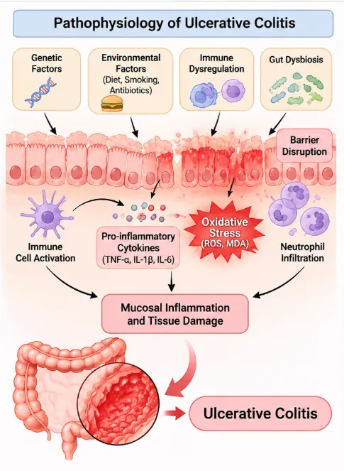
Figure 1: Pathophysiology of Ulcerative Colitis (UC)
2.2. Inflammatory Mediators
TNF-α
In patients of UC, the gut mucosa shows raised levels of IL-1β, IL-6, TNF-α, and cytokines of Th1, Th2, and Th17 [23]. TNF-α has the ability to recruit immune cells, activate cytokines, and induce apoptosis of epithelial cells and damage of tight junctions [24]. TNF-α is a pro-inflammatory cytokine which is synthesized by the innate immunity of IBD [25]. There have been studies that revealed how an increase in this pro-inflammatory cytokine is associated with the pathogenesis of IBD [26-27].
NF-κB Signalling Pathway
The nuclear factor kappa B (NF-κB) pathway is a key pathway involved in mediating inflammation in UC. In normal conditions, NF-κB is kept inactive in the cytoplasm by binding with inhibitory proteins referred to as IκBs. In response to inflammatory stimuli, IκB kinase (IKK) is activated resulting in the phosphorylation and degradation of IκBs. This results in the translocation of NF-κB, especially the p65/p50 subunit complex, to the nucleus, where it regulates the transcription of pro-inflammatory genes. The pro-inflammatory genes include cytokines, and other inflammatory mediators including TNF-α, IL-1β, IL-6, and iNOS. Increased levels of NF-κB activity have been observed in mucosal colonic biopsies obtained from UC patients [6].
2.3. Oxidative Stress in Ulcerative Colitis (UC)
Reactive Oxygen Species (ROS)
Once activated, neutrophils produce enzymes, ROS, and cytotoxic agents that can further damage the mucosa [28].
Lipid Peroxidation
Malondialdehyde (MDA) produced by lipid peroxidation is one of the most common indicators of oxidative stress in UC. Overproduction of reactive oxygen species results in the oxidative damage of membrane lipids, which leads to the production of MDA and cell membrane damage. Higher rates of erythrocyte lipid peroxidation were found in patients with UC than in controls [29].
Antioxidant Defence Mechanisms
Antioxidant defense system of the gut which includes SOD, CAT, GPx and GSH helps protect cells from ROS. Changes have been noted in these antioxidants in UC. An Indian study found that there was an increase in lipid peroxidation, CAT and SOD concentrations along with a decrease in GSH concentration in UC patients [48]. Another study has shown the presence of increased myeloperoxidase activity and reduced glutathione transferase concentration in colonic mucosa [11].
2.4. Dysfunction of the Intestinal Barrier Function
The Intestinal Epithelial Cells
In UC, the dysfunctional barrier due to the epithelial cell damage facilitates the translocation of more antigens and pathogens through the lamina propria and activates immune cells [30]. For people suffering from UC, poor function of goblet cells leads to reduced production of mucin-2 and hence a weak mucus barrier [31].
Tight Junction Proteins (ZO-1, Occludin and Claudins)
In UC, tight junction modifications, particularly involving the claudin-2 upregulation, heighten paracellular permeability, promoting the infiltration of antigens, toxins, and bacteria into the lamina propria [31]. Tight junction proteins such as occludin and ZO-1 help in maintaining the integrity of the intestinal epithelial barrier as well as controlling permeability. Lower levels of occludin and reduced ZO-1 expression in proximity to the areas of neutrophil migration have also been observed during active IBD [32].
Increased Intestinal Permeability
In UC, the weakened barrier, resulting from epithelial cell damage, allows more antigens and pathogens to breach the lamina propria, where they engage with immune cells, triggering their activation [30]. In UC, tight junction modifications, particularly involving the claudin-2 upregulation, heighten paracellular permeability, promoting the infiltration of antigens, toxins, and bacteria into the lamina propria [31]. The disruption of gut microbiota balance by means of dysbiosis causes UC as it affects the integrity of the intestinal barrier causing epithelial hyperpermeability and allows pathogenic agents to get into the intestine [19].
Mucosal Damage and Inflammation
In UC, the weakened barrier, resulting from epithelial cell damage, allows more antigens and pathogens to breach the lamina propria, where they engage with immune cells, triggering their activation [30]. After being recruited, neutrophils release proteases, reactive oxygen species, and cytotoxic molecules, which may exacerbate mucosal injury [28].
3. Current Therapeutic Approaches for UC
Conventional drugs like corticosteroids, aminosalicylates, and immunosuppressants are essential in managing the disease and reducing any inflammatory response.(33)
5-Aminosalicylic Acid (5-ASA)
5-Aminosalicylic acid (5-ASA), mesalamine is one of the most commonly prescribed drugs to treat mild to moderate forms of ulcerative colitis. 5-ASA is locally acting in the intestines and plays an anti-inflammatory role by affecting inflammatory mediators and signaling pathways. It exists in several preparations (tablets, enemas, and suppositories) both for oral and rectal administration. (34)
Corticosteroids
Induction of remission in moderate to severe UC and in cases where 5-ASA does not work can be done using corticosteroids. Drugs like prednisolone and budesonide help inhibit gene expression and reduce the levels of pro-inflammatory cytokines. However, corticosteroids should not be used for maintenance because of their side effects, such as osteoporosis, hyperglycemia, and hypertension. (34)
Immunomodulators
These drugs are immunomodulators that can control the irregular response of the immune system and prevent UC flare-ups. They are especially beneficial for those patients who are dependent on corticosteroids. Nevertheless, their slow onset makes it difficult to utilize them in induction therapy. Potential side effects include bone marrow depression, liver toxicity, and increased susceptibility to infections [34].
Biological Therapies
Biosimilars have been employed in the case of moderate to severe UC, when conventional therapy fails. The mode of action of the biosimilars is aimed at targeted inflammation pathways such as TNF-α (infliximab and golimumab), α4β7 integrin (vedolizumab), and IL-12/IL-23 (ustekinumab). The biosimilars aid in the induction and maintenance of remission. (34)
Small-Molecule Therapies
Oral small molecule drugs provide more options for moderate to severe UC patients. Janus Kinase Inhibitors like tofacitinib and upadacitinib are drugs that inhibit cytokine signaling inside cells, whereas S1P receptor modulators like ozanimod and etrasimod are involved in lymphocyte trafficking. These drugs are administered orally and are modern therapies for UC [34].
Limitations and Adverse Effects of Existing Treatments
However, even though they have their advantages, such therapies often possess limited effectiveness and may cause negative side effects, particularly during prolonged usage [7]. Although much progress has been made in treating UC, treatment resistance, especially resistance to anti-TNF drugs, is still an issue [8,9]. The long-term monitoring of the disease poses another problem because of the inadequacy of the current biomarkers in terms of specificity and sensitivity [10].
Need for Novel Therapeutic Agents
Even with current treatments, there are cases of poor efficacy or unwanted side effects in UC patients. It is thus important to look into the benefits of natural substances that are antioxidant and anti-inflammatory in nature [34].
4. Usnic Acid: An Overview
4.1. Source and Origin
Lichens as Natural Sources
Lichens are symbiotic organisms composed of an association of algae or cyanobacteria along with fungi, which may include basidiomycete yeasts and bacteria [35]. Usnic acid is one of the most fascinating lichen compounds from the point of view of pharmacology [35].
Major Lichen Species Containing Usnic Acid
Usnic acid occurs in high concentration (even up to 10 percent) in species of the following genera of lichens: Usnea, Alectoria, Cladonia, Lecanora, Ramalina, and Flavocetraria [35]. The isolation of usnic acid dates back to 1844, and there has been a long and intensive study of their biological properties including antimicrobial, cytotoxic, antioxidant and anti-inflammatory effects [35].
4.2. Chemical Structure and Physicochemical Properties
Chemical Structure
The chemical structure of usnic acid is defined as 2,6-diacetyl-7,9-dihydroxy-8,9b-dimethyl-1,3 (2H,9bH)-dibenzofurandione [35]. Usnic acid is a chiral molecule existing in (+)-usnic acid and (−)-usnic acid enantiomers [35]. Since chirality may influence the biological activity of both natural and artificially synthesized substances, the enantiopreferentiality of usnic acid still remains uncertain owing to lack of experimental results [35]. Anti-inflammatory activity of enantiomers of usnic acid was studied, however, available information does not allow concluding about their differential biological activity [23,36].
The molecular formula - C₁₈H₁₆O₇
The molecular weight - 344.32 g/mol
Solubility and Chemical Characteristics - The chemical occurs in a bitter, yellowish powdered form and is barely soluble in water and some organic solvents [35].
4.3. Pharmacological Properties
Antimicrobial Activity
Antimicrobial and cytotoxic activities have also been attributed to usnic acid, along with its antioxidant and anti-inflammatory activities [35].
Anti-inflammatory Activity
Usnic acid has anti-inflammatory activities that have been studied through various in vitro and in vivo experiments [37-38]. Usnic acid suppressed the expression of inflammatory mediators such as TNF-α, IL-1β, IL-6, and nitric oxide (NO) in LPS-induced RAW 264.7 macrophages [39,40].
Usnic acid suppressed NF-κB activation, TNF-α expression, and iNOS, suggesting its effect on inflammatory signaling pathways [39,40]. Both (+)- and (−)-usnic acid suppressed TLR4 expression and decreased the production of cytosolic phospholipase A2 (cPLA2) in LPS-induced macrophages [23]. Usnic acid decreased inflammatory mediators such as TNF-α, IL-1β, and IL-6 in LPS-induced acute lung injury and bleomycin-induced pulmonary fibrosis in experimental animals [38,41].
Antioxidant Activity
Usnic acid exhibited antioxidant potential in experimental models through increased superoxide dismutase (SOD) and glutathione (GSH) activities as well as decreased oxidative stress markers, such as myeloperoxidase (MPO), malondialdehyde (MDA) and lipid hydroperoxide (LOOH) [42,36,41,38].
Other Reported Biological Activities
Usnic acid and sodium usnic acid loaded into liposomes were effective in experimental wound healing models [43-44].
Table 1: Pharmacological Activities of Usnic Acid
| Pharmacological activity | Experimental model | Major findings |
|---|---|---|
| Anti-inflammatory | LPS-induced RAW 264.7 macrophages | Reduced TNF-α, IL-1β, IL-6 and NO |
| NF-κB inhibition | LPS-stimulated macrophages | Suppressed NF-κB activation and p65 translocation |
| TLR4 modulation | LPS-induced macrophages | Reduced TLR4 expression |
| Antioxidant | Cerebral ischemia/reperfusion model | Increased SOD and GSH activities |
| Antioxidant | LPS-induced acute lung injury | Reduced MDA and MPO; increased SOD and GSH |
| Anti-inflammatory and antioxidant | Bleomycin-induced pulmonary fibrosis | Reduced inflammatory cytokines and MDA |
4.4. Pharmacokinetic Properties
The pharmacokinetic profile of usnic acid remains incompletely understood. Studies indicate extensive plasma protein binding and distribution to various tissues, particularly the liver and lungs. Measurable plasma concentrations have been reported following oral administration. Its metabolism involves hepatic cytochrome P450 enzymes and glucuronidation pathways. However, further research is needed to establish its complete absorption, distribution, metabolism and excretion (ADME) profile [45].
5. Therapeutic Potential of Usnic Acid in Ulcerative Colitis
5.1. Anti-inflammatory Properties
Regulation of TNF-α, IL-1β and IL-6
Usnic acid suppressed the expression of inflammatory mediators such as TNF-α, IL-1β, IL-6, and nitric oxide (NO) in LPS-induced RAW 264.7 macrophages [39,40]. Usnic acid decreased the levels of TNF-α, IL-1β and IL-6 in a bleomycin-induced pulmonary fibrosis experiment model [38].
Modulation of NF-κB Signalling
Usnic acid inhibited the LPS-induced NF-κB p65 translocation into the nucleus and the degradation of I-κBα in RAW 264.7 macrophages [39].
Effects on Inflammatory Enzymes Such as COX-2 and iNOS
Usnic acid decreased the expression of inflammatory cytokines such as TNF-α, COX-2, and iNOS [39,40]. Usnic acid decreased cPLA2 synthesis and affected COX-1 and COX-2 proteins expression in macrophages [23].
Possible Inhibition of Inflammatory Responses
Both enantiomers of usnic acid inhibited TLR4 expression in LPS-stimulated macrophages [23]. Currently, the experiments found from the collected literature indicate the anti-inflammatory and antioxidant properties of usnic acid, in various model systems such as macrophages, lung injury, and pulmonary fibrosis. This provides some indirect information on its biologic efficacy but does not confirm its efficacy in TNBS colitis. Therefore, more research is required to evaluate the effect of this compound on colonic inflammation, oxidative stress, histopathology, and intestinal permeability.
5.2. Antioxidant Properties
Reduction of Oxidative Stress and Effects on Lipid Peroxidation
Usnic acid demonstrated decreases in lipid hydroperoxide and MPO activities in an experimental Alzheimer’s disease model [36]. Usnic acid exhibited decreases in MDA and MPO activities and increases in SOD and GSH activities during LPS-induced acute lung injury in mice [41]. Usnic acid decreased MDA and increased SOD activity during bleomycin-induced pulmonary fibrosis in mice [38].
Possible Modulation of Antioxidant Enzymes Such as SOD, CAT and GPx
Usnic acid showed increases in SOD and GSH activities during experimental cerebral ischemia/reperfusion injury [42].
Role in Reducing Oxidative Tissue Injury
Usnic acid exhibited antioxidant potential in experimental models through increased superoxide dismutase (SOD) and glutathione (GSH) activities as well as decreased oxidative stress markers, such as myeloperoxidase (MPO), malondialdehyde (MDA) and lipid hydroperoxide (LOOH) [42,36,41,38].
5.3. Intestinal Barrier-Protection
Intestinal barrier protective properties of usnic acid need further investigation. More research needs to be done in order to establish if usnic acid can help maintain the epithelial barrier integrity by controlling the tight junction proteins like ZO-1, occludin, and claudins.
5.4. Immunomodulatory Effects
Regulation of Inflammatory Immune Responses
Usnic acid suppressed NF-κB activation, TNF-α expression, and iNOS, suggesting its effect on inflammatory signaling pathways [39,40].
Effects on Immune-Cell Activation
Both (+)- and (−)-usnic acid suppressed TLR4 expression and decreased the production of cytosolic phospholipase A2 (cPLA2) in LPS-induced macrophages [23].
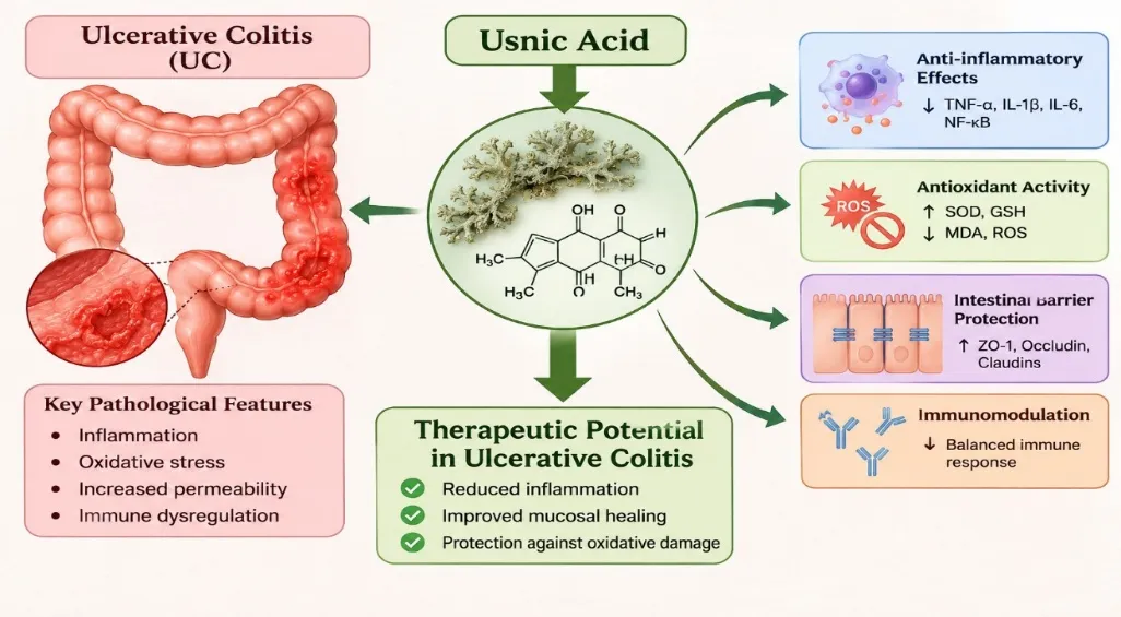
Figure 2: Proposed Therapeutic Mechanisms of Usnic Acid in Ulcerative Colitis.
6. Experimental Evidence Supporting the Therapeutic Potential of Usnic Acid
In-vitro Studies
Usnic acid has anti-inflammatory activities that have been studied through various in vitro and in vivo experiments [37-38]. Usnic acid suppressed the expression of inflammatory mediators such as TNF-α, IL-1β, IL-6, and nitric oxide (NO) in LPS-induced RAW 264.7 macrophages [39,40]. Usnic acid suppressed NF-κB activation, TNF-α expression, and iNOS, suggesting its effect on inflammatory signaling pathways [39,40]. Both (+)- and (−)-usnic acid suppressed TLR4 expression and decreased the production of cytosolic phospholipase A2 (cPLA2) in LPS-induced macrophages [23].
In-vivo Studies
Usnic acid decreased inflammatory mediators such as TNF-α, IL-1β, and IL-6 in LPS-induced acute lung injury and bleomycin-induced pulmonary fibrosis in experimental animals [41,38]. Usnic acid showed increases in SOD and GSH activities during experimental cerebral ischemia/reperfusion injury [42]. Usnic acid demonstrated decreases in lipid hydroperoxide and MPO activities in an experimental Alzheimer’s disease model [36].
Evidence from Experimental Colitis Models
Animal models of IBD are still required for the study of histopathological alterations in the gastrointestinal tract and are crucial in the development of new pharmacological strategies [54]. Currently, the collected literature provides evidence of the anti-inflammatory and antioxidant properties of usnic acid in different experimental models. However, these findings do not establish its therapeutic efficacy in TNBS-induced colitis.
7. Safety, Toxicity and Limitations of Usnic Acid
Dose-Dependent Toxicity
Earlier experiments on toxicity of usnic acid have shown toxic doses of usnic acid less than 50 mg/kg, which means that there may be some risks associated with using high doses in pharmacological experiments [46]. The effective dose of 25 mg/kg in one of the experimental studies was lower than those doses which were considered toxic in earlier experiments on toxicity [36,46]. In some studies which were taken into account in this review, the effective doses of usnic acid were as high as 100 mg/kg [38,41,47].
Hepatotoxicity Concerns
Although usnic acid is characterized by promising pharmacological activities, it shows certain toxicity risks especially hepatotoxicity [46,48]. There are very few experimental studies which show the impact of usnic acid on the liver and other organs [46,48]. More research is needed to prove the liver toxicity of usnic acid especially at high doses [46,48].
Limitations of Existing Experimental Studies
In vitro studies that have explored the anti-inflammatory action of usnic acid are few, and these studies do not provide sufficient information about the molecular mechanism of the compound [39-49]. It is necessary to conduct more elaborate studies to reveal the impact of usnic acid on the production of inflammatory mediators [39-49]. The in vivo studies are preliminary since a small number of animals were used in them [38-47]. Further investigations are necessary to assess the efficacy and toxicity of (+)- and (−)-enantiomers of usnic acid [23,36]. There is also need to explore the possibility of using usnic acid in treatment of inflammatory conditions [38,39].
Table 2: Safety Concerns, Limitations and Future Research Directions of Usnic Acid
| Aspect | Current evidence/ Limitation | Future research direction |
|---|---|---|
| Hepatotoxicity | Potential liver toxicity | Detailed liver-function and histopathological studies |
| Dose-Dependent Toxicity | Toxicity concerns at higher doses | Establish safe and effective dose ranges |
| Molecular mechanisms | Incomplete understanding of mechanisms | Investigate NF-κB, TLR4 and antioxidant pathways |
| Intestinal barrier protection | Limited direct evidence | Evaluate ZO-1, occludin and claudins |
| Pharmacokinetics | Incomplete ADME profile | Conduct detailed pharmacokinetic studies |
| Experimental validation | Poor aqueous solubility | Develop colon-targeted and nanoparticle formulations |
8. Future Perspectives
Further Investigation of Its Molecular Mechanisms
In vitro studies that have explored the anti-inflammatory action of usnic acid are few, and these studies do not provide sufficient information about the molecular mechanism of the compound [39-49]. It is necessary to conduct more elaborate studies to reveal the impact of usnic acid on the production of inflammatory mediators [39-49].
Dose Optimisation and Safety Evaluation
More research is needed to prove the liver toxicity of usnic acid especially at high doses [46,48]. Further investigations are necessary to assess the efficacy and toxicity of (+)- and (−)-enantiomers of usnic acid [23,36].
Development of Targeted Drug Delivery Systems and Nanoparticle-Based Formulations
The delivery of drugs using targeted drug delivery system can help in improving the solubility, stability and targeted delivery of usnic acid. Colon targeted drug delivery can be used to achieve better drug delivery to inflamed tissues of intestines. Further investigations need to be carried out for their effectiveness and safety in colitis [50]. The nanosystems developed through nanotechnology, such as liposomes and polymeric nanoparticles, may enhance the properties of usnic acid in terms of solubility and delivery into targeted regions. Additional research is required in order to evaluate the properties of these formulations [50].
Pharmacokinetic and Pharmacodynamic Investigations
Further studies on pharmacokinetics and pharmacodynamics should be conducted to understand the ADME characteristics, distribution, hepatic metabolism, dose-related toxicity, and therapeutically effective levels of usnic acid [51,52,60].
Further Preclinical and Clinical Studies
The in vivo studies are preliminary since a small number of animals were used in them [38-47]. There is also need to explore the possibility of using usnic acid in treatment of inflammatory conditions [38-39].
CONCLUSION
Usnic acid has shown interesting anti-inflammatory properties in different in vitro and in vivo models [37-38]. The anti-inflammatory properties of usnic acid are related to the modulation of different inflammatory mediators such as TNF-α, IL-1β, IL-6, NO and NF-κB [23-39]. Usnic acid has also been shown to exhibit antioxidant properties by altering the levels of different parameters like SOD, GSH, MDA, MPO and lipid hydroperoxides [36,38,41,42]. More research work needs to be done to elucidate its molecular mechanisms, hepatotoxicity, enantiomers and derivatization for better drug properties [23,36,48]. Usnic acid demonstrates good anti-inflammatory and antioxidant effects, making it a prospective compound to be studied in UC treatment. Still, information concerning the effects of usnic acid in the case of TNBS-induced colitis and its influence on the tight-junction proteins is rather scarce [23,36,38-42,51,53].
REFERENCES

Gade Indu Vimala Devi, Venu Talla, Syeda Sumaiya, Nomula Pavani, Dr. Madathala Sreekanth, Usnic Acid as a Potential Therapeutic Agent for Ulcerative Colitis: Anti-Inflammatory, Antioxidant and Intestinal Barrier-Protective Properties, Int. J. of Pharm. Sci., 2026, Vol 4, Issue 11, 1698-1711. https://doi.org/10.5281/zenodo.24143745
 10.5281/zenodo.24143745