
We use cookies to ensure our website works properly and to personalise your experience. Cookies policy
Department of Pharmacology, Womens College of Pharmacy, Peth-Vadgoan, Kolhapur, Maharashtra, India.
Antihelmintic drug affect the human body and its normal functions by causing helminthiasis, a serious parasitic infection. Most parasites affecting humans and livestock develop resistance to these drugs, leading to increased morbidity and mortality.[1] The impact of parasitic worm infections on vulnerable hosts is a major concern, prompting research into the infection, prevalence, host-parasite interactions, and the parasite's resistance to treatment. This review highlights the rising prevalence of parasitic worm infections, which is linked to factors such as population growth, climate change, low living standards, particularly in conflict zones, and in developing countries.[2] This review aims to classify different species of anthelmintic plants based on their in vitro effectiveness. A range of plant species known for their anthelmintic activity were selected for evaluation. In vitro assays, including the paralysis time, death time, egg hatch test, larval migration assay, larval development test, and adult motility assay were included to measure the anthelmintic potential of the plant. Also focused on the use of plants and secondary metabolites against intestinal parasites. We discuss the use of plants in traditional medicine and the use of plant secondary metabolites tried in in vitro test.
Helminth parasites infect nearly one-third of the global population, predominantly in tropical regions. Interestingly, areas where these parasites are widespread tend to have significantly lower rates of allergies and autoimmune diseases, suggesting a potential protective effect against immunopathological conditions. Many helminth infections vary in severity, with most cases being mild or asymptomatic, while a smaller group of individuals develop severe complications. The ability to maintain an asymptomatic state is now understood to result from an immunoregulatory environment influenced by the parasites. This regulation involves various host immune cells and cytokines, and when it fails, disease symptoms become more pronounced. There is growing interest in exploring whether helminth-induced immune modulation could help alleviate allergies and autoimmune disorders, with ongoing research in both experimental models and human studies. Gaining deeper insights into these parasite-host interactions could lead to innovative strategies for managing both infectious diseases and immune-related conditions. More than 25% of the global population is at risk of infection by soil-transmitted helminths, including Ascaris lumbricoides, hookworms (Ancylostoma duodenale and Necator americanus), Trichuris trichiura, and Strongyloides stercoralis. Both children and adults affected by these parasites can experience a variety of medical and surgical complications, making it essential for healthcare providers to consider helminth infections in individuals from endemic regions or those returning from such areas. Although safe and effective treatments are available and donated to endemic countries at no cost, only half of the at-risk children received medication in 2016. This review examines the epidemiology, life cycles, disease mechanisms, clinical presentation, diagnosis, treatment, and public health strategies related to soil-transmitted helminths. While the effectiveness of mass deworming programs has been debated, there is clear evidence that treatment significantly reduces the severe outcomes of these infections. We emphasize the urgent need for improved diagnostic methods and enhanced control strategies to strengthen public health efforts and enhance the clinical management of these parasitic diseases. [4,5]
B) Mode Of Transmission of Helminths:
1) Direct method:
Transmission occurs when the host comes into direct contact with the eggs, without the involvement of an intermediate host. Helminth eggs are expelled through feces, where they hatch into larvae and subsequently reinfect either the same host or a new one. A notable example of this mode of transmission is Enterobius vermicularis.
2) Modified direct method:
Helminth eggs are excreted in the feces of the host, especially in areas where open defecation occurs, contaminating the soil. Over the course of several days, the eggs undergo developmental changes. A susceptible host may ingest the eggs either through direct contact with contaminated soil or through contaminated food. Once inside the body, the eggs hatch into larvae, which penetrate the stomach wall and enter the bloodstream. The larvae then migrate to the lungs, where they are coughed up and subsequently swallowed again, allowing them to reach the intestines. There, they mature into adult worms. Ascaris lumbricoides and Trichuris trichiura are examples of helminths that follow this life cycle.
3) Skin penetration:
The eggs produced by adult helminths are expelled through the host’s feces or urine, contaminating the soil or freshwater. Depending on the type of parasite, the eggs develop into infective larvae either independently or with the assistance of an intermediate host. Once matured, the larvae penetrate the skin of a susceptible host and enter the bloodstream. For example, the larvae of Ancylostoma duodenale and Strongyloides stercoralis migrate to the lungs, where they are coughed up, swallowed, and eventually mature into adult worms in the intestine. In contrast, the larvae of parasites responsible for schistosomiasis remain within the blood vessels, where they develop into their adult form. The fully matured worms then reside in the mesenteric venules at different locations within the body.
4) Bites from infected insect vector:
In diseases such as lymphatic filariasis, loiasis, and onchocerciasis, parasitic worms are transmitted by vector insects. Mosquitoes spread lymphatic filariasis, deer flies are responsible for transmitting loiasis, and black flies facilitate the spread of onchocerciasis. These insects transmit the parasites by biting a host and depositing infectious larvae in or near the wound. Once inside the body, the larvae mature into adult worms beneath the skin (in the subcutaneous tissue). The adult worms then produce microfilariae, which migrate to the skin, where they remain until another vector takes them up during a blood meal. Inside the insect’s gut, the microfilariae develop into infective larvae, continuing the transmission cycle.
5) Foodborne helminths:
Infection occurs when a host consumes raw or undercooked fish, vegetables, crustaceans, or meat, or drinks water contaminated with parasite larvae, eggs, or cysts. Several helminthic diseases can be transmitted through contaminated food, including cysticercosis, dracunculiasis, ascariasis, and food-borne trematodiases.[2]
C) Symptoms And Diagnosis:
Common symptoms of helminth infections include:[2]
1. Stomach pain
2. Diarrhea
3. Nutritional deficiencies
4. Fatigue
5. An enlarged liver or spleen
6. Inflammation of the digestive
7. Lung inflammation
8. Vision impairment
9. An increased eosinophil count
10. Intestinal blockage
11. Anaemia
12. Nausea
13. Constipation
14. Swelling of the lymphatic system
15. Unintended weight loss
16. Itching of the skin or anal area
There are various diagnostic methods available for detecting helminth infections, including:
1. Fecal egg examination
2. Antigen test
3. Serological assay
4. Nucleic Acid-based Diagnosis
5. Urine examination
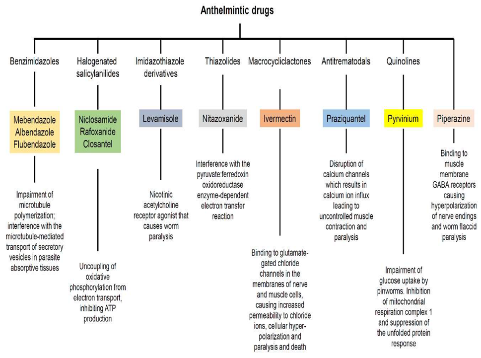
D) Classification Of Synthetic Anthelmintic Drugs:
E) Scope Of Herbal Drugs as Anthelmintics:
In recent years, herbal medicines have gained significant importance in the field of healthcare due to their minimal side effects. As a result, the demand for herbal formulations continues to rise steadily. The identification and standardization of phytochemical compounds have advanced with the development of instrumental analysis, making this area of research increasingly relevant and promising.A large portion of the global population suffers from bacterial and helminth infections, primarily due to common factors such as inadequate sanitation, poor hygiene, malnutrition, and overcrowded living conditions. The main sources of these infections include:
1. Humans – The most common source of infection, as individuals can transmit pathogens to one nother.
2. Animals – Many infectious agents affect both humans and animals, with animals often serving as reservoirs for human infections.
3. Insects – Blood-feeding insects can spread pathogens to humans. In addition to acting as disease carriers, some insects may also serve as reservoirs for these microorganisms.
4. Soil and Water – Certain pathogens can persist in the soil for extended periods, while contaminated water can transmit infections either through direct exposure to microorganisms or by harboring aquatic vectors.
5. Food – Consuming contaminated food is another significant route of infection.
Given the widespread nature of bacterial and helminth infections, there is an urgent need to develop antibacterial and anthelmintic drugs derived from herbal sources to provide safer and more effective treatment options.[1]
F) Commonly Used Worms For In Vitro Tests Anthelmintic Activity:
Table No. 01: Common worms used for in vitro tests:
|
Sr. No. |
Class |
Species |
|
01 |
Earthworms |
1) Pheretimaposthuma 2) Eisenia foetida |
|
02 |
Roundworms (Nematodes) |
1) Ascaridia galli 2)Ascaris lumbricoides 3) Caenorhabditis elegans 4) Haemonchuscontortus |
|
03 |
Tapeworms (Cestodes) |
1) Hymenolepisdiminuta 2) Taenia saginata |
|
04 |
Flukes (Trematodes) |
1) Fasciola hepatica (Liverfluke) 2) Paramphistomumcervi |
1) Pheretima Posthuma:
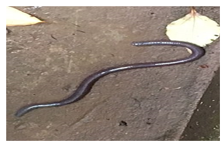
Fig: Pheretima Posthuma
2) Eisenia Foetida:
Fig: Eisenia Foetida
3) Ascaridia galli:
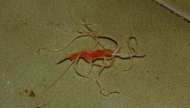
Fig: Ascaridia Gallis
4) Ascaris lumbricoides:

Fig: Ascaris Lumbricoides
5) Caenorhabditis elegans:
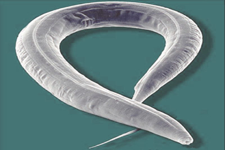
Fig: Caenorhabditis Elegans
6) Haemonchus Contortus:
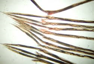
Fig: Haemonchus Contortus
7) Hymenolepis Diminuta:
Fig: H. Diminuta
8) Taenia Saginata:
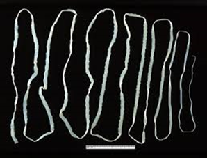
Fig: Taenia Saginata
9) Fasciola hepatica (Liver fluke):
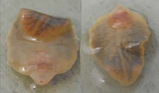
Fig: Fasciola Hepatica
10) Paramphistomum cervi:
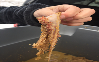
Fig: Paramphistomum Cervi
G) Common Phytochemicals Responsible for Anthelmintic Activity:
There are some phytochemicals present in plants which possess the potential anthelmintic activity.[8]
|
Sr. No. |
Plant Secondary Metabolites |
Examples |
|
01 |
Alkaloids |
1) Nicotine 2) Palmatine 3) Berberine |
|
02 |
Tannins
|
1) Condensed tannins 2) Hydrolyzable tannins |
|
03 |
Terpenoids |
1) Thymol 2) Carvacrol 3) Artemisinin |
|
04 |
Phenolic Compounds |
1) Caffeic acid 2) Gallic acid 3) Ellagic acid |
|
05 |
Flavonoids |
1) Quercetin 2) Kaempferol 3) Luteolin |
|
06 |
Saponins |
1) Diosgenin 2) Hederacoside C |
|
07 |
Coumarins |
1) Scopoletin 2)Aflatoxin |
H) In Vitro Tests for Assessment of Anthelmintic Activity:
There are different methods for assessment of anthelmintic potential. Some commonly used methods for assessment of anthelmintic activity are as follows:
1) Paralysis Time Analysis
2) Death Time Analysis
3) Egg Hatch Assay
4) Larval Migration Assay
5) Larval Development Test
6) Adult Motility Assay
1) Paralysis Time Analysis:
Generally,paralysis time is the period required for a substance to cause immobilization in parasitic worms. This measure is essential for evaluating the effectiveness of anthelmintic drugs, as paralysis disrupts the worms' ability to remain in the host, ultimately resulting in their removal or death.[9,10,11]
1. Prepare Parasite Culture: Select the appropriate parasitic species (e.g., nematodes or trematodes).Ensure the parasites are healthy and active before starting the test.
2. Prepare Anthelmintic Solutions: Prepare various concentrations of the anthelmintic agent for testing.Set up a control group without any anthelmintic for comparison.
3. Set Up the Test Environment: Place the parasites into separate wells or Petri dishes with an appropriate medium (such as saline or water).Ensure a sufficient number of active parasites in each well for accurate results.
4. Administer the Anthelmintic Treatment: Introduce the anthelmintic solution to the wells containing the parasites.Make sure the parasites come into contact with the solution.
5. Monitor for Paralysis: Continuously observe the parasites for signs of paralysis, such as lack of movement.Record the time it takes for paralysis to appear after exposure to the anthelmintic.The worms uncurls their body, relaxes and stop moving.The time taken by test organisms to paralyse completely is measured in min.
6. Control Group Observation: Keep a control group (no treatment) under identical conditions to track natural movement or any environmental effects.
7. Record and Analyze Paralysis Onset: Document the time taken for paralysis to occur at each anthelmintic concentration.Compare the results from different concentrations with the control group.
8. Results: A quicker onset of paralysis at lower concentrations suggests the anthelmintic is more effective.
2) Death Time Analysis:
Death time is the time it takes for a compound to fully eradicate a parasitic worm. This factor is important in anthelmintic research, as it helps assess a drug's strength and efficiency. A faster death time suggests a more potent anthelmintic agent. [9,10,11]
1. Prepare Parasite Culture: Select the target parasitic species (e.g., nematodes, trematodes). Ensure the parasites are healthy and active before starting the experiment.
2. Prepare Anthelmintic Solutions: Prepare different concentrations of the anthelmintic compound to be tested.Set up a control group with no anthelmintic for comparison.
3. Set Up Test Environment: Place the parasites in individual test wells or Petri dishes containing a suitable medium (such as saline or distilled water). Ensure a sufficient number of active parasites are in each test well.
4. Administer Anthelmintic Treatment: Add the prepared anthelmintic solutions to the test wells with the parasites.Ensure that the parasites are in direct contact with the solution.
5. Observe for Signs of Death: Monitor the parasites closely for signs of death, such as cessation of movement or inability to respond to stimuli, the worms after treatment until they become completely unresponsive, even when stimulated. Record the time taken by test organisms for death Record the time taken for each parasite to exhibit signs of death after exposure to the anthelmintic.
6.Control Group Monitoring: Keep a control group (without anthelmintic) under identical conditions to monitor natural behavior and check for any environmental effects.
7.Record and Analyze Death Time: Note the exact time it takes for the parasites to die at each concentration of the anthelmintic.Compare the death time across different concentrations of the anthelmintic and the control group.
3) Egg Hatch Assay:
An egg hatch test for anthelmintic activity is a lab-based method used to evaluate the effectiveness of anthelmintic (anti-parasitic) medications, especially in relation to nematode eggs. This test measures how effectively a particular anthelmintic prevents egg hatching or impacts larval development, offering insight into the drug's efficacy.[12]
1.Choose the Species: Select a species with well-known egg development and hatching patterns, such as marine organisms like Artemia (brine shrimp) or Corbicula (freshwater mussels).
2. Collect Eggs: Obtain freshly laid eggs from a suitable source, ensuring they are at a comparable developmental stage. If necessary, incubate eggs in a suitable medium (e.g., seawater, freshwater, or artificial water) until they are ready for testing.
3. Prepare Test Solutions: Prepare the test substance or chemicals to be tested in varying concentrations (e.g., toxins, pollutants, or environmental factors like temperature). Include control groups with no added test substance and solvent control if needed.
4. Set Up Experimental Containers: Place the eggs into individual test containers (e.g., Petri dishes, beakers, or incubation wells). Ensure the eggs are placed in a suitable environment for hatching, with appropriate water quality (e.g., pH, salinity, temperature).
5. Introduce Test Substances: Add the prepared test substances or chemicals to the appropriate containers, ensuring the desired concentrations are achieved. Maintain separate groups for the experimental substance, controls, and solvent controls.
6. Incubation and Monitoring: Incubate the eggs under optimal conditions (e.g., temperature, light) to allow for proper hatching. Monitor the eggs periodically for signs of hatching, mortality, and abnormal development.
7. Record Hatching Success: Record the number of eggs that hatch during the exposure period (typically 24-48 hours, depending on the species). Calculate the hatching success percentage for each group (e.g., number of hatched eggs divided by the total number of eggs).
8. Post-Exposure Observations: After the exposure period, observe and document any developmental abnormalities or delayed hatching in larvae. Assess the larvae’s survival rate, if applicable, during the early stages post-hatching.
9. Data Analysis: Analyze the data by comparing the hatching success rates between the control and experimental groups. Use statistical tests (e.g., t-tests, ANOVA) to determine any significant effects of the test substances on egg hatching success.
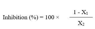
Where,
X1 = The number of eggs hatched in test extracts.
X2 = The respective number in PBS control.
4) Larval Migration Assay:
A larval migration assay is a laboratory method designed to evaluate the movement of larvae, particularly those of parasitic nematodes or insects, across various substrates. It is widely utilized in parasitology, toxicology, and developmental biology to analyze larval motility, reactions to environmental factors, and the impact of drugs or chemicals. [13,14]
1. Larvae Preparation: Collect and rinse the larvae using an appropriate solution, such as phosphate-buffered saline.
2. Selection of Migration Surface: Choose a suitable medium like agar, mesh, or filter paper, depending on the experiment.
3. Larvae Placement: Introduce a specific number of larvae onto the chosen substrate or into a migration chamber.
4. Incubation Period: Maintain the larvae under controlled environmental conditions, including temperature, humidity, and light, for a set duration.
5. Observation & Data Collection: Monitor larval movement using imaging techniques or microscopy. Count the number of larvae that have migrated and those that have not.
6. Data Analysis: Determine the percentage of larvae that migrated and evaluate factors affecting their movement.
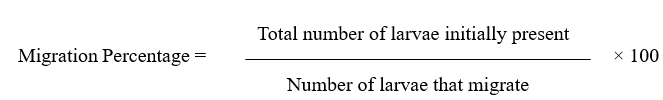
Where,
Number of migrated larvae = Larvae that successfully moved through the substrate or filter.
Total number of larvae = Sum of migrated and non-migrated larvae.
5) Larval Development Test:
The Larval Development Assay (LDA) is a method used to determine the anthelmintic potential of test compounds by examining their impact on the growth of Haemonchus contortus larvae, from the egg stage to the infective L3 stage. [15,16]
1. Egg Collection: Obtain Haemonchus contortus eggs from the feces of infected animals.Clean and count the eggs using a microscope.
2. Preparation of Test Solutions:Dilute plant extracts in PBS at different concentrations.Prepare albendazole as a positive control and PBS as a negative control.
3. Experimental Setup: Distribute about 100 eggs into each well of a 24-well plate or Petri dish.Add 2 mL of the respective test solutions.Incubate at 25–30°C for 48–72 hours.
4. Observation and Counting: Examine larvae under a microscope to check for developed (L3) and undeveloped forms.
5. Inhibition Calculation:
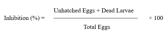
6. Data Analysis: Compare plant extract effects with control groups.Greater inhibition indicate stronger anthelmintic activity.
6) Adult Motility Assay:
The Adult Motility Assay (AMA) is a technique used to assess the anthelmintic properties of test compounds by measuring their impact on the movement of adult Haemonchus contortus worms.[17,18,19]
1. Collection of Worms:Adult H. contortus worms are collected from the abomasum of infected animals.The worms are washed with PBS to remove any debris or contaminants.
2. Preparation of Test Solutions:Prepare different concentrations of the crude plant extracts in PBS (e.g., 1.25, 2.5, 5, and 10 mg/mL).Prepare a solution of standard drug at 0.25 mg/mL as a positive control.Use PBS as a negative control.
3. Exposure of Worms to Test Solutions:Transfer ten worms into each Petri dish.Add 5 mL of the respective test solution to each dish.Set up the experiment in triplicate for each concentration.Maintain the Petri dishes at room temperature (25–30°C).
4. Observation and Data Collection: Monitor worm motility at different time intervals (e.g., 1, 3, 6, and 12 hours). Consider worms immobile if they do not move even after gentle probing with a needle. Compare motility inhibition between test groups and controls.
5. Analysis of Results: Calculate the percentage of motility inhibition for each treatment. Compare the effectiveness of plant extracts against the positive and negative controls.
CONCLUSION:
This review highlights the growing importance of exploring plant secondary metabolites as potential anthelmintic agents in light of rising concerns over synthetic drug resistance. A thorough understanding of the mode of transmission and classification of helminths is crucial for the development of targeted interventions. The scope of plant-based anthelmintics is expanding, with a variety of secondary metabolites including alkaloids, flavonoids, saponins, tannins ,terpenoids and coumarins showing promising bioactivity against different helminth species. In vitro anthelmintic tests have proven to be an effective and reliable approach for screening and evaluating the efficacy of these plant-derived compounds, providing valuable insights into their effect. Despite the promising results, further research is needed to assess the in vivo effectiveness and safety of these natural products, as well as to address challenges related to standardization and toxicity. The integration of traditional knowledge with modern pharmacological techniques offers a promising avenue for discovering novel, sustainable anthelmintic solutions to combat parasitic infections.
REFERENCES

Repe Sakshi, Gurav Vasudha, Parit Pratiksha, Kamble Ravina*, Dr. Jadge Dhanraj, A Comprehensive Review of Methods for Testing in-vitro Anthelmintic Activity, Int. J. of Pharm. Sci., 2025, Vol 3, Issue 4, 3396-3408 https://doi.org/10.5281/zenodo.15309277
 10.5281/zenodo.15309277