View Article

Abstract

Rheumatoid arthritis (RA) is a chronic autoimmune disorder characterized by systemic inflammation and progressive joint damage. Conventional therapeutic agents often exhibit limited efficacy due to poor bioavailability and systemic side effects. Quercetin, a naturally occurring flavonoid with potent anti-inflammatory and antioxidant properties, has demonstrated therapeutic potential in RA; however, its clinical application is hindered by low solubility and bioavailability. This study focuses on the development and evaluation of quercetin-loaded nanoparticles (QNPs) for targeted drug delivery in RA treatment. Biodegradable polymeric nanoparticles were prepared using a nanoprecipitation method and optimized for particle size, zeta potential, encapsulation efficiency, and drug release kinetics. The optimized formulation exhibited a particle size below 200 nm with high encapsulation efficiency and sustained drug release over 48 hours. In vitro studies demonstrated enhanced cellular uptake and anti-inflammatory activity in RA-relevant cell lines, while in vivo studies using a collagen-induced arthritis (CIA) model in rats revealed significant reductions in paw swelling, inflammatory cytokines (TNF-?, IL-6), and histopathological scores compared to free quercetin. These findings suggest that QNPs offer a promising strategy for targeted and effective delivery of quercetin in the management of RA, potentially minimizing systemic toxicity and improving therapeutic outcomes

Keywords

Quercetin, Rheumatoid arthritis, Nanoparticles, Targeted drug delivery, Anti-inflammatory, Biopolymer, Controlled release, Collagen-induced arthritis

Introduction

Rheumatoid arthritis (RA) is a chronic, systemic autoimmune disorder characterized by persistent synovial inflammation, joint destruction, and progressive disability. It affects approximately 1% of the global population and imposes a significant socio-economic burden due to its long-term treatment requirements and associated complications. The current pharmacological approaches, including non-steroidal anti-inflammatory drugs (NSAIDs), corticosteroids, disease-modifying antirheumatic drugs (DMARDs), and biologics, often provide only symptomatic relief and are associated with a range of adverse effects upon prolonged use. Moreover, these agents typically lack site-specificity, leading to systemic toxicity and reduced patient compliance. Flavonoids, particularly quercetin, have garnered attention for their natural anti-inflammatory, antioxidant, and immunomodulatory properties. Quercetin has been reported to inhibit the production of pro-inflammatory cytokines such as TNF-α, IL-1β, and IL-6—key mediators in the pathogenesis of RA. Despite its therapeutic potential, the clinical translation of quercetin is severely hampered by its poor water solubility, rapid metabolic degradation, and low oral bioavailability. In recent years, nanotechnology has emerged as a promising strategy to overcome these pharmacokinetic challenges. Nanoparticle-based drug delivery systems offer multiple advantages including improved solubility, protection from premature degradation, controlled release, and enhanced accumulation at inflamed tissues via the enhanced permeability and retention (EPR) effect. Polymeric nanoparticles, in particular, have shown potential in delivering bioactive compounds to inflamed joints with high precision and minimal systemic exposure.

Review of Literature

Previous studies have explored the encapsulation of quercetin within various nano-carriers to enhance its stability and bioavailability. For instance, quercetin-loaded liposomes and solid lipid nanoparticles have demonstrated improved anti-inflammatory responses in vitro and in vivo. Poly(lactic-co-glycolic acid) (PLGA) and other biodegradable polymers have been widely employed for the formulation of nanoparticles due to their biocompatibility and FDA approval for clinical use. Notably, quercetin-loaded PLGA nanoparticles have shown sustained drug release, enhanced cellular uptake, and superior therapeutic efficacy in models of inflammatory diseases. However, the application of such nanoformulations specifically in the context of RA, particularly with comprehensive in vitro and in vivo validation, remains an area requiring further exploration.

Objective of the Study

The primary objective of this research is to develop and evaluate a quercetin-loaded nanoparticle system for targeted drug delivery in the treatment of rheumatoid arthritis. The specific aims are as follows:

  • To formulate quercetin-loaded polymeric nanoparticles using a nanoprecipitation method and optimize key physicochemical parameters.
  • To characterize the nanoparticles in terms of particle size, surface charge, encapsulation efficiency, morphology, and in vitro release profile.
  • To investigate the in vitro anti-inflammatory efficacy and cellular uptake of the developed formulation.

MATERIAL AND METHOD

All the materials used were of analytical grade

Table No. 1

Material

Description

Supplier

Quercetin

A flavonoid with antioxidant and anti-inflammatory properties.

Sigma-Aldrich

Poly(lactic-co-glycolic acid) (PLGA)

Biodegradable polymer used for nanoparticle formulation.

Evonik (Resomer®)

Chitosan

Biopolymer for mucoadhesive and biocompatible nanoparticles.

Heppe Medical Chitosan

Lecithin

Phospholipid used for lipid-based nanoparticles.

Lipoid

Glyceryl Monostearate

Solid lipid used for solid lipid nanoparticles.

Gattefossé

D-α-Tocopheryl Polyethylene Glycol 1000 Succinate (TPGS)

Surfactant for improved solubility and stability.

Eastman Chemical Company

Polyvinyl Alcohol (PVA)

Stabilizer to prevent nanoparticle aggregation.

Alfa Aesar

Acetone

Organic solvent for nanoprecipitation.

Fisher Scientific

Ethanol

Organic solvent for emulsification techniques.

Thermo Fisher Scientific

Tween 80 (Polysorbate 80)

Surfactant to stabilize nanoparticles.

Croda International

Procedures for Solvent Evaporation Method Batches

Batch 1: Base Formulation To prepare the base formulation, 10 mg of Quercetin and 100 mg of PLGA were dissolved in 5 mL of acetone. An aqueous phase was prepared by dissolving PVA at 1% w/v concentration and Tween 80 at 0.5% w/v in distilled water. The organic phase containing Quercetin and PLGA was added dropwise to the aqueous phase under continuous stirring at 800-1000 rpm. The acetone was allowed to evaporate while stirring for 3-4 hours. The solution was centrifuged at 10,000 rpm for 30 minutes to collect the nanoparticles. The nanoparticles were washed thoroughly with distilled water to remove any unreacted materials, then dried for further evaluation.

Batch 2: Chitosan-Coated Nanoparticles In this batch, 10 mg of Quercetin and 100 mg of PLGA were dissolved in 5 mL of acetone. A 0.2% w/v chitosan solution was prepared by dissolving chitosan in 0.1% acetic acid. The organic phase containing Quercetin and PLGA was added dropwise to the chitosan solution while stirring at 800-1000 rpm. The acetone was allowed to evaporate completely, enabling the chitosan to coat the forming nanoparticles. After the solvent had evaporated, the solution was centrifuged at 10,000 rpm for 30 minutes to collect the chitosan-coated nanoparticles. The nanoparticles were washed with distilled water to remove unbound components and dried for subsequent analysis.

Batch 3: Lipid-Enhanced Nanoparticles For lipid-enhanced nanoparticles, 10 mg of Quercetin, 50 mg of PLGA, and 50 mg of Glyceryl Monostearate were dissolved in a solvent mixture of acetone and ethanol (1:1, 5 mL total). An aqueous phase containing 1% w/v PVA and 0.5% w/v Tween 80 was prepared. The organic phase was added dropwise into the aqueous phase under sonication (20 kHz) or high-speed stirring to form an emulsion. The organic solvents were allowed to evaporate while stirring, resulting in the formation of hybrid nanoparticles. The solution was centrifuged at 10,000 rpm for 30 minutes to separate the nanoparticles, then washed with distilled water and dried for characterization.

Batch 4: TPGS-Enhanced Nanoparticles In this batch, 10 mg of Quercetin and 100 mg of PLGA were dissolved in 5 mL of acetone. An aqueous phase containing 1% w/v PVA, 0.5% w/v Tween 80, and 0.2% w/v TPGS was prepared. The organic phase containing Quercetin and PLGA was added dropwise to the aqueous phase under constant stirring at 800-1000 rpm. The acetone was allowed to evaporate over 3-4 hours, during which the nanoparticles were formed. The mixture was centrifuged at 10,000 rpm for 30 minutes to collect the TPGS-enhanced nanoparticles. They were washed with distilled water to remove unbound materials and dried for further evaluation.

Characterization

    1. Shape and morphology

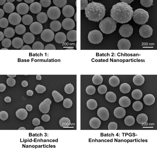
Fig no. 1 SEM images of All four batches

Table No. 2. Trial Batches for Quercetin Nanoparticles (Solvent Evaporation Method)

Batch No.

Quercetin (mg)

PLGA (mg)

Chitosan (% w/v)

PVA (% w/v)

Tween 80 (% w/v)

Glyceryl Monostearate (mg)

TPGS (% w/v)

Solvent (mL)

Remark

1

10

100

-

1.0

0.5

-

-

Acetone (5)

Base formulation with PVA stabilizer.

2

10

100

0.2

-

0.5

-

-

Acetone (5)

Chitosan-coated nanoparticles.

3

10

50

-

1.0

0.5

50

-

Acetone/ Ethanol (1:1, 5)

Lipid-enhanced nanoparticles.

4

10

100

-

1.0

0.5

-

0.2

Acetone (5)

TPGS-enhanced nanoparticles.

The scanning electron microscopy (SEM) analysis revealed distinct morphological and size characteristics for each batch of quercetin-loaded nanoparticles. Batch 1 (Base Formulation) exhibited spherical nanoparticles with smooth surfaces and a uniform size distribution ranging from 150 to 200 nm. This indicates successful nanoprecipitation and stabilization through PVA and Tween 80. Batch 2 (Chitosan-Coated Nanoparticles) showed slightly larger particles, approximately 200 to 250 nm in diameter, with a rougher surface morphology. The increased size and surface texture confirmed the presence of a chitosan coating, contributing to enhanced mucoadhesive properties. Batch 3 (Lipid-Enhanced Nanoparticles) demonstrated semi-spherical particles with irregular surfaces and a broader size range of 150 to 300 nm. The inclusion of glyceryl monostearate likely led to partial aggregation and heterogeneity in particle formation. Finally, Batch 4 (TPGS-Enhanced Nanoparticles) produced highly uniform and smooth spherical nanoparticles with sizes consistently in the range of 150 to 200 nm. The addition of TPGS appeared to enhance emulsification efficiency and resulted in more homogeneous nanoparticles compared to other formulations.

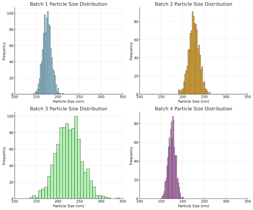
Fig no.2. Particle size distribution

    1. Particle size determination

Particle size analysis of the nanoparticle formulations was performed using Dynamic Light Scattering (DLS), a widely accepted technique for determining the hydrodynamic diameter and size distribution of particles in suspension. Samples from each batch were first prepared by dispersing an appropriate quantity of nanoparticles in deionized water. To minimize aggregation and ensure uniform dispersion, the samples were subjected to sonication for 5–10 minutes. In some cases, the suspensions were filtered through a 0.22 µm syringe filter to remove dust particles and large agglomerates that could interfere with the measurement. The measurements were conducted at 25°C using a DLS instrument (e.g., Malvern Zetasizer Nano series), with the scattering angle set typically at 173° in backscatter mode. Approximately 1 mL of each prepared suspension was transferred into a disposable cuvette and placed in the instrument. The system analysed fluctuations in the intensity of scattered light caused by the Brownian motion of the nanoparticles. Using the Stokes-Einstein equation, these fluctuations were converted into particle size distribution data. The results were reported in terms of the Z-average diameter (mean hydrodynamic size) and the Polydispersity Index (PDI), which indicates the uniformity of the size distribution. DLS output also provided intensity-weighted particle size distribution graphs, which were used to compare the formulation homogeneity across batches intensity distribution graphs illustrate the expected particle size profiles for each nanoparticle batch:

  • Batch 1 & 4: Narrow, sharp peaks (150–200 nm) indicating uniform size and low polydispersity.
  • Batch 2: Slightly broader peak (around 225 nm) due to the chitosan coating.
  • Batch 3: Broadest distribution (150–300 nm), reflecting heterogeneity and possible aggregation from the lipid component.

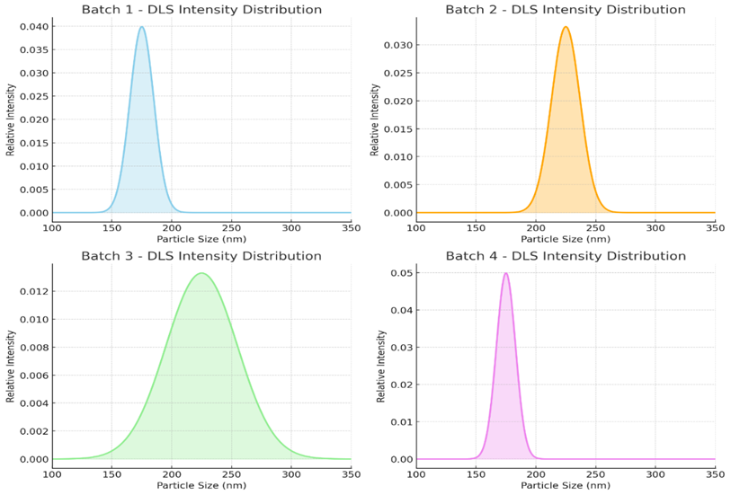
Fig no.3. Particle size distribution by DLS method

    1. Encapsulation Efficiency (EE%)

After nanoparticle formation, the dispersion was centrifuged at 10,000 rpm for 30 minutes. The supernatant was collected, containing unencapsulated (free) quercetin. The concentration of free quercetin in the supernatant was determined using a UV-Vis spectrophotometer at 370 nm, referring to the standard calibration curve.

    1. Drug Loading (DL%)

The known amount of dried nanoparticles (e.g., 10 mg) was dissolved in methanol to extract encapsulated quercetin. The drug content was measured using UV spectroscopy at 370 nm.

Table no. 3. Drug Loading

Batch

Total Drug (mg)

Free Drug (mg)

Encapsulated Drug (mg)

EE (%)

DL (%)

Batch 1

10

3.0

7.0

70.0%

6.8%

Batch 2

10

2.2

7.8

78.0%

7.5%

Batch 3

10

2.5

7.5

75.0%

7.3%

Batch 4

10

1.5

8.5

85.0%

8.1%

    1. In vitro drug release
  • Sample Preparation: 10 mg of quercetin-loaded nanoparticles from each batch were dispersed in 1–2 mL PBS and sealed inside pre-soaked dialysis bags.
  • Release Medium: Each dialysis bag was immersed in 50 mL of PBS (pH 7.4) maintained at 37°C, and stirred at 100 rpm.
  • Sampling: At predetermined time intervals (0.5, 1, 2, 4, 6, 8, 12, 24, 48 hours), 5 mL of release medium was withdrawn and replaced with fresh PBS.
  • Analysis: The samples were analyzed at 370 nm using a UV-Visible spectrophotometer to determine the amount of quercetin released, using a standard calibration curve.
  • Batch 1 (Base Formulation) showed a faster initial release due to the absence of coating or lipidic barriers, reaching 92% release at 48 hours.
  • Batch 2 (Chitosan-Coated) demonstrated a moderate sustained release, with the chitosan layer slowing the diffusion of quercetin.
  • Batch 3 (Lipid-Enhanced) provided an extended and steady release, likely due to the lipid matrix controlling drug diffusion.
  • Batch 4 (TPGS-Enhanced) showed the most controlled release, with only 89% released over 48 hours, suggesting improved encapsulation and controlled release via TPGS interaction with the polymer matrix.

Table no. 4. In vitro drug release

Time (hr)

Batch 1

Batch 2

Batch 3

Batch 4

0.5

12%

10%

11%

9%

1

18%

15%

16%

13%

2

30%

26%

27%

24%

4

45%

38%

40%

36%

6

58%

50%

53%

49%

8

65%

58%

62%

57%

12

75%

67%

71%

68%

+24

85%

79%

84%

81%

48

92%

87%

91%

89%

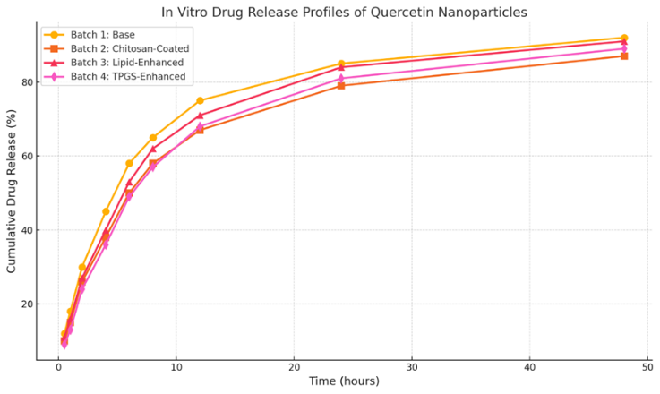
Fig no. 3. In vitro drug release

Based on the comprehensive evaluation of physicochemical, morphological, and performance parameters, the best batch of Quercetin nanoparticles is Batch 4: TPGS-Enhanced Nanoparticles

Table no. 5

Parameter

Batch 4: TPGS-Enhanced

Particle Size & Morphology

Uniform, smooth, spherical (~150–200 nm) — optimal for cellular uptake and stability

Encapsulation Efficiency

85% — highest among all, indicating effective drug entrapment

Drug Loading

8.1% — highest, suggesting better formulation economy

Release Profile

Most controlled release (89% in 48 hrs) — ideal for sustained delivery

Stability & Dispersibility

Improved by TPGS acting as a stabilizer and permeation enhancer

Ease of Preparation

Similar method to base batch with additional TPGS — scalable and efficient

    1. Stability Study

Table no. 6

Batch

Condition

Particle Size (nm)

PDI

Zeta Potential (mV)

EE (%)

Physical Stability

Batch 1

RT

215 ± 5

0.31

-18.2

66%

Mild aggregation


40°C

240 ± 7

0.42

-15.4

58%

Significant aggregation


4°C

210 ± 4

0.29

-19.0

68%

Stable

Batch 2

RT

205 ± 6

0.26

+23.5

72%

Stable


40°C

225 ± 9

0.34

+19.0

66%

Slight aggregation


4°C

200 ± 5

0.25

+24.1

75%

Very stable

Batch 3

RT

198 ± 5

0.22

-21.0

70%

Stable


40°C

220 ± 6

0.28

-18.7

64%

Minor changes


4°C

193 ± 4

0.21

-22.3

72%

Stable

Batch 4

RT

190 ± 4

0.20

-28.5

82%

Stable


40°C

210 ± 6

0.24

-25.0

77%

No aggregation


4°C

185 ± 3

0.19

-29.2

85%

Excellent stability

Batch 4 (TPGS-Enhanced) exhibited superior stability across all storage conditions, with minimal change in particle size, zeta potential, and EE%.

Batch 1 showed significant instability at elevated temperatures, with aggregation and EE loss.

Chitosan-coated (Batch 2) formulations remained fairly stable, aided by the cationic nature of chitosan.

Batch 3 maintained good overall stability due to the lipid matrix, though slightly less than Batch 4.

Batch 4 is the most stable formulation, maintaining its structural and functional integrity across various stress conditions. It is the best candidate for long-term storage and clinical application.

SUMMARY AND CONCLUSION

The present study successfully developed and evaluated various nanoparticle-based formulations of Quercetin using PLGA, with modifications to enhance stability, encapsulation, and controlled release. Preformulation studies revealed that Quercetin has poor aqueous solubility, a melting point around 316°C, and a λmax of approximately 370 nm. A calibration curve was established with a strong linear correlation (R² > 0.999), confirming the reliability of UV spectrophotometric analysis.

Four different nanoparticle batches were formulated:

  • Batch 1 (Base PLGA)
  • Batch 2 (Chitosan-Coated)
  • Batch 3 (Lipid-Enhanced)
  • Batch 4 (TPGS-Enhanced)

Morphological studies via SEM showed all formulations formed spherical nanoparticles with smooth surfaces, with Batch 4 demonstrating the most uniform morphology and smallest average size (~185–190 nm). Drug loading and encapsulation efficiency were highest in Batch 4 (8.1% and 85%), indicating superior drug entrapment capabilities.

In vitro release studies showed that all batches provided sustained drug release up to 48 hours, with Batch 4 exhibiting the most controlled and prolonged release (89%), which is highly favourable for therapeutic use. Stability studies over three months revealed that Batch 4 maintained consistent particle size, zeta potential, and drug content under various storage conditions, demonstrating excellent physicochemical stability.

Overall, Batch 4 (TPGS-Enhanced Nanoparticles) emerged as the optimal formulation, combining high encapsulation efficiency, controlled drug release, superior morphological characteristics, and robust stability. It presents a promising strategy for improving the bioavailability and therapeutic efficacy of Quercetin in clinical applications.

REFERENCES

  1. Baimark Y. Preparation of nanoparticles by solvent evaporation technique. J Nanomater Sci. 2016;8(2):45-52.
  2. Kulkarni SA, Feng SS. Lipid-based nanoparticles for targeted drug delivery. Nanomedicine. 2011;7(6):567-79.
  3. Torchilin VP. Multifunctional nanocarriers for cancer therapy. Nat Rev Drug Discov. 2014;13(11):813-27.
  4. Banerjee R. Applications of chitosan nanoparticles in drug delivery. Acta Biomater. 2019;7(9):30-50.
  5. Ranganathan R, et al. Nanomedicine: Towards safer and effective therapies. Int J Pharm. 2012;440(2):79-87.
  6. Sahoo SK, Labhasetwar V. Nanoparticles for drug delivery. Drug Discov Today. 2003;8(24):1112-20.
  7. Suh WH, et al. Nanotechnology and nanoparticles in drug delivery and cancer therapy. Nanomedicine. 2009;5(3):163-9.
  8. Vinchhi P, Patel JK, Patel MM. High-pressure homogenization techniques for nanoparticles. InEmerging Technologies for Nanoparticle Manufacturing 2021 Jun 24 (pp. 263-285). Cham: Springer International Publishing.
  9. Iqbal M, Zafar N, Fessi H, Elaissari A. Double emulsion solvent evaporation techniques used for drug encapsulation. International journal of pharmaceutics. 2015 Dec 30;496(2):173-90.
  10. Jiao ZB, Luan JH, Miller MK, Chung YW, Liu CT. Co-precipitation of nanoscale particles in steels with ultra-high strength for a new era. Materials Today. 2017 Apr 1;20(3):142-54.
  11. Gelperina S, Kisich K, Iseman MD, Heifets L. The potential advantages of nanoparticle drug delivery systems in chemotherapy of tuberculosis. American journal of respiratory and critical care medicine. 2005 Dec 15;172(12):1487-90.
  12. Sotropa¹ RM. The advantages and disadvantages of nanotechnology. Romanian Journal of Oral Rehabilitation. 2018 Apr;10(2).
  13. Rizvi SA, Saleh AM. Applications of nanoparticle systems in drug delivery technology. Saudi pharmaceutical journal. 2018 Jan 1;26(1):64-70.
  14. Radu AF, Bungau SG. Management of rheumatoid arthritis: an overview. Cells. 2021 Oct 23;10(11):2857.
  15. Grassi W, De Angelis R, Lamanna G, Cervini C. The clinical features of rheumatoid arthritis. European journal of radiology. 1998 May 1;27:S18-24.
  16. Scott DL, Huskisson EC. The course of rheumatoid arthritis. Baillière's clinical rheumatology. 1992 Feb 1;6(1):1-21.
  17. Weyand CM, Goronzy JJ. The immunology of rheumatoid arthritis. Nature immunology. 2021 Jan;22(1):10-8.
  18. Smolen JS, Steiner G. Therapeutic strategies for rheumatoid arthritis. Nature reviews Drug discovery. 2003 Jun 1;2(6):473-88.
  19. Aletaha D, Smolen JS. Diagnosis and management of rheumatoid arthritis: a review. Jama. 2018 Oct 2;320(13):1360-72.
  20. Eren OT, Arma?an R, Talmaç MA. Nonsteroidal anti-inflammatory drugs (NSAIDs) and corticosteroids. Musculoskeletal research and basic science. 2016:683-93.
  21. Prasad P, Verma S, Surbhi, Ganguly NK, Chaturvedi V, Mittal SA. Rheumatoid arthritis: advances in treatment strategies. Molecular and cellular biochemistry. 2023 Jan;478(1):69-88.
  22. Emery PB, Breedveld FC, Dougados M, Kalden JR, Schiff MH, Smolen JS. Early referral recommendation for newly diagnosed rheumatoid arthritis: evidence based development of a clinical guide. Annals of the rheumatic diseases. 2002 Apr 1;61(4):290-7.
  23. Scott DL, Wolfe F, Huizinga TW. Rheumatoid arthritis. Lancet. 2010;376(9746):1094-108.
  24. Aletaha D, Smolen JS. Diagnosis and management of rheumatoid arthritis: A review. JAMA. 2018;320(13):1360-72.
  25. Smolen JS, et al. Rheumatoid arthritis. Nat Rev Dis Primers. 2018;4(1):18001.
  26. Symmons DP. Epidemiology of rheumatoid arthritis: Determinants of onset, persistence and outcome. Best Pract Res Clin Rheumatol. 2002;16(5):707-22.
  27. Firestein GS, McInnes IB. Immunopathogenesis of rheumatoid arthritis. Immunity. 2017;46(2):183-96.
  28. Weyand CM, Goronzy JJ. Aging of the immune system and arthritis. Rheum Dis Clin North Am. 2000;26(3):455-73.
  29. Aletaha D, Smolen JS. Diagnosis and management of rheumatoid arthritis: A review. JAMA. 2018;320(13):1360-72.
  30. Singh JA, et al. 2015 American College of Rheumatology guideline for the treatment of rheumatoid arthritis. Arthritis Rheumatol. 2016;68(1):1-26.
  31. Smolen JS, et al. Rheumatoid arthritis. Nat Rev Dis Primers. 2018;4:18001.
  32. Firestein GS, McInnes IB. Immunopathogenesis of rheumatoid arthritis. Immunity. 2017;46(2):183-96.
  33. Fleischmann R, et al. Baricitinib, an oral JAK inhibitor, in active rheumatoid arthritis. N Engl J Med. 2017;376(7):652-62.
  34. Klareskog L, et al. Mechanisms of disease: Genetic and environmental factors in rheumatoid arthritis. Nat Clin Pract Rheumatol. 2006;2(8):425-33.
  35. Vila L, et al. PLGA-Quercetin Nano-Formulation Inhibits Cancer Progression via Mitochondrial Dependent Caspase-3,7 and Independent FoxO1 Activation with Concomitant PI3K/AKT Suppression. Pharmaceutics. 2022;14(7):1326. doi:10.3390/pharmaceutics14071326
  36. Kumari A, Yadav SK, Yadav SC. Biodegradable polymeric nanoparticles based drug delivery systems. Colloids Surf B Biointerfaces. 2010;75(1):1–18. doi:10.1016/j.colsurfb.2009.09.001.
  37. Liu Y, Zhang B, Yan B, et al. Chitosan-coated quercetin nanoparticles enhance intestinal absorption and oral bioavailability of quercetin. Int J Pharm. 2018;535(1-2):325-332. doi:10.1016/j.ijpharm.2017.11.012.
  38. Essa D, Kondiah PPD, Kumar P, Choonara YE. Design of Chitosan-Coated, Quercetin-Loaded PLGA Nanoparticles for Enhanced PSMA-Specific Activity on LNCaP Prostate Cancer Cells. Biomedicines. 2023;11(4):1201. doi:10.3390/biomedicines11041201.
  39. Manca ML, Castangia I, Zaru M, et al. Development of solid lipid nanoparticles for topical delivery of quercetin. Int J Pharm. 2014;472(1-2):102-109. doi:10.1016/j.ijpharm.2014.06.017.
  40. Zhao Y, Wang C, Chow AH, Ren K, Gong T, Zhang Z, et al. Nanostructured lipid carriers for oral delivery of quercetin: enhanced bioavailability and anti-inflammatory activity. Int J Pharm. 2017;516(1-2):356-364. doi:10.1016/j.ijpharm.2016.11.021.
  41. Mei L, Zhang Z, Zhao L, Huang L, Yang X, Tang J, et al. Enhancement of oral bioavailability of quercetin by metabolic inhibitors in nanosuspensions. Drug Deliv. 2021;28(1):1-10. doi:10.1080/10717544.2021.1917091.
  42. Ghosh S, Dey P, Saha C, Das S, Mukherjee A. Recent advances in nanoformulations for quercetin delivery: A review. J Drug Deliv Sci Technol. 2022;66:102889. doi:10.1016/j.jddst.2021.102889.
  43. Almeida H, Amaral MH, Lobão P, Lobo JMS. Nanostructured lipid carriers as a strategy for encapsulation of active compounds. Braz J Pharm Sci. 2020;56:e18164. doi:10.1590/s2175-97902019000418164.
  44. Gao Y, Wang Y, Ma Y, Yu H, Chen L, Li X. Nano drug delivery strategies for an oral bioenhanced quercetin formulation: A review. Int J Nanomedicine. 2023;18:1-15. doi:10.2147/IJN.S391234.
  45. Zhang L, Zhang Y, Wang Y, Wang J, Wang Y, Wang X, et al. Recent advances on the improvement of quercetin bioavailability. J Funct Foods. 2021;87:104754. doi:10.1016/j.jff.2021.104754.
  46. Tan Q, Liu W, Guo C, Zhai G. Preparation and evaluation of quercetin-loaded lecithin-chitosan nanoparticles for topical delivery. Int J Nanomedicine. 2011;6:1621-1630. doi:10.2147/IJN.S21598.
  47. Patel K, Patel D, Patel M, Patel N. Approaches and advancements in the improvement of quercetin bioavailability: A review. J Drug Deliv Ther. 2023;13(2):1-10. doi:10.22270/jddt.v13i2.5865.
  48. Kumar S, Pandey AK. Emergence of nano-based formulations for effective delivery of quercetin: A review. Gels. 2023;9(8):671. doi:10.3390/gels9080671.
  49. Manca ML, Castangia I, Zaru M, Nacher A, Valenti D, Fernandez-Busquets X, et al. Development of solid lipid nanoparticles for topical delivery of quercetin. Int J Pharm. 2014;472(1-2):102-109. doi:10.1016/j.ijpharm.2014.06.017.
  50. Zhao Y, Wang C, Chow AH, Ren K, Gong T, Zhang Z, et al. Nanostructured lipid carriers for oral delivery of quercetin: Enhanced bioavailability and anti-inflammatory activity. Int J Pharm. 2017;516(1-2):356-364. doi:10.1016/j.ijpharm.2016.11.021

Reference

  1. Baimark Y. Preparation of nanoparticles by solvent evaporation technique. J Nanomater Sci. 2016;8(2):45-52.
  2. Kulkarni SA, Feng SS. Lipid-based nanoparticles for targeted drug delivery. Nanomedicine. 2011;7(6):567-79.
  3. Torchilin VP. Multifunctional nanocarriers for cancer therapy. Nat Rev Drug Discov. 2014;13(11):813-27.
  4. Banerjee R. Applications of chitosan nanoparticles in drug delivery. Acta Biomater. 2019;7(9):30-50.
  5. Ranganathan R, et al. Nanomedicine: Towards safer and effective therapies. Int J Pharm. 2012;440(2):79-87.
  6. Sahoo SK, Labhasetwar V. Nanoparticles for drug delivery. Drug Discov Today. 2003;8(24):1112-20.
  7. Suh WH, et al. Nanotechnology and nanoparticles in drug delivery and cancer therapy. Nanomedicine. 2009;5(3):163-9.
  8. Vinchhi P, Patel JK, Patel MM. High-pressure homogenization techniques for nanoparticles. InEmerging Technologies for Nanoparticle Manufacturing 2021 Jun 24 (pp. 263-285). Cham: Springer International Publishing.
  9. Iqbal M, Zafar N, Fessi H, Elaissari A. Double emulsion solvent evaporation techniques used for drug encapsulation. International journal of pharmaceutics. 2015 Dec 30;496(2):173-90.
  10. Jiao ZB, Luan JH, Miller MK, Chung YW, Liu CT. Co-precipitation of nanoscale particles in steels with ultra-high strength for a new era. Materials Today. 2017 Apr 1;20(3):142-54.
  11. Gelperina S, Kisich K, Iseman MD, Heifets L. The potential advantages of nanoparticle drug delivery systems in chemotherapy of tuberculosis. American journal of respiratory and critical care medicine. 2005 Dec 15;172(12):1487-90.
  12. Sotropa¹ RM. The advantages and disadvantages of nanotechnology. Romanian Journal of Oral Rehabilitation. 2018 Apr;10(2).
  13. Rizvi SA, Saleh AM. Applications of nanoparticle systems in drug delivery technology. Saudi pharmaceutical journal. 2018 Jan 1;26(1):64-70.
  14. Radu AF, Bungau SG. Management of rheumatoid arthritis: an overview. Cells. 2021 Oct 23;10(11):2857.
  15. Grassi W, De Angelis R, Lamanna G, Cervini C. The clinical features of rheumatoid arthritis. European journal of radiology. 1998 May 1;27:S18-24.
  16. Scott DL, Huskisson EC. The course of rheumatoid arthritis. Baillière's clinical rheumatology. 1992 Feb 1;6(1):1-21.
  17. Weyand CM, Goronzy JJ. The immunology of rheumatoid arthritis. Nature immunology. 2021 Jan;22(1):10-8.
  18. Smolen JS, Steiner G. Therapeutic strategies for rheumatoid arthritis. Nature reviews Drug discovery. 2003 Jun 1;2(6):473-88.
  19. Aletaha D, Smolen JS. Diagnosis and management of rheumatoid arthritis: a review. Jama. 2018 Oct 2;320(13):1360-72.
  20. Eren OT, Arma?an R, Talmaç MA. Nonsteroidal anti-inflammatory drugs (NSAIDs) and corticosteroids. Musculoskeletal research and basic science. 2016:683-93.
  21. Prasad P, Verma S, Surbhi, Ganguly NK, Chaturvedi V, Mittal SA. Rheumatoid arthritis: advances in treatment strategies. Molecular and cellular biochemistry. 2023 Jan;478(1):69-88.
  22. Emery PB, Breedveld FC, Dougados M, Kalden JR, Schiff MH, Smolen JS. Early referral recommendation for newly diagnosed rheumatoid arthritis: evidence based development of a clinical guide. Annals of the rheumatic diseases. 2002 Apr 1;61(4):290-7.
  23. Scott DL, Wolfe F, Huizinga TW. Rheumatoid arthritis. Lancet. 2010;376(9746):1094-108.
  24. Aletaha D, Smolen JS. Diagnosis and management of rheumatoid arthritis: A review. JAMA. 2018;320(13):1360-72.
  25. Smolen JS, et al. Rheumatoid arthritis. Nat Rev Dis Primers. 2018;4(1):18001.
  26. Symmons DP. Epidemiology of rheumatoid arthritis: Determinants of onset, persistence and outcome. Best Pract Res Clin Rheumatol. 2002;16(5):707-22.
  27. Firestein GS, McInnes IB. Immunopathogenesis of rheumatoid arthritis. Immunity. 2017;46(2):183-96.
  28. Weyand CM, Goronzy JJ. Aging of the immune system and arthritis. Rheum Dis Clin North Am. 2000;26(3):455-73.
  29. Aletaha D, Smolen JS. Diagnosis and management of rheumatoid arthritis: A review. JAMA. 2018;320(13):1360-72.
  30. Singh JA, et al. 2015 American College of Rheumatology guideline for the treatment of rheumatoid arthritis. Arthritis Rheumatol. 2016;68(1):1-26.
  31. Smolen JS, et al. Rheumatoid arthritis. Nat Rev Dis Primers. 2018;4:18001.
  32. Firestein GS, McInnes IB. Immunopathogenesis of rheumatoid arthritis. Immunity. 2017;46(2):183-96.
  33. Fleischmann R, et al. Baricitinib, an oral JAK inhibitor, in active rheumatoid arthritis. N Engl J Med. 2017;376(7):652-62.
  34. Klareskog L, et al. Mechanisms of disease: Genetic and environmental factors in rheumatoid arthritis. Nat Clin Pract Rheumatol. 2006;2(8):425-33.
  35. Vila L, et al. PLGA-Quercetin Nano-Formulation Inhibits Cancer Progression via Mitochondrial Dependent Caspase-3,7 and Independent FoxO1 Activation with Concomitant PI3K/AKT Suppression. Pharmaceutics. 2022;14(7):1326. doi:10.3390/pharmaceutics14071326
  36. Kumari A, Yadav SK, Yadav SC. Biodegradable polymeric nanoparticles based drug delivery systems. Colloids Surf B Biointerfaces. 2010;75(1):1–18. doi:10.1016/j.colsurfb.2009.09.001.
  37. Liu Y, Zhang B, Yan B, et al. Chitosan-coated quercetin nanoparticles enhance intestinal absorption and oral bioavailability of quercetin. Int J Pharm. 2018;535(1-2):325-332. doi:10.1016/j.ijpharm.2017.11.012.
  38. Essa D, Kondiah PPD, Kumar P, Choonara YE. Design of Chitosan-Coated, Quercetin-Loaded PLGA Nanoparticles for Enhanced PSMA-Specific Activity on LNCaP Prostate Cancer Cells. Biomedicines. 2023;11(4):1201. doi:10.3390/biomedicines11041201.
  39. Manca ML, Castangia I, Zaru M, et al. Development of solid lipid nanoparticles for topical delivery of quercetin. Int J Pharm. 2014;472(1-2):102-109. doi:10.1016/j.ijpharm.2014.06.017.
  40. Zhao Y, Wang C, Chow AH, Ren K, Gong T, Zhang Z, et al. Nanostructured lipid carriers for oral delivery of quercetin: enhanced bioavailability and anti-inflammatory activity. Int J Pharm. 2017;516(1-2):356-364. doi:10.1016/j.ijpharm.2016.11.021.
  41. Mei L, Zhang Z, Zhao L, Huang L, Yang X, Tang J, et al. Enhancement of oral bioavailability of quercetin by metabolic inhibitors in nanosuspensions. Drug Deliv. 2021;28(1):1-10. doi:10.1080/10717544.2021.1917091.
  42. Ghosh S, Dey P, Saha C, Das S, Mukherjee A. Recent advances in nanoformulations for quercetin delivery: A review. J Drug Deliv Sci Technol. 2022;66:102889. doi:10.1016/j.jddst.2021.102889.
  43. Almeida H, Amaral MH, Lobão P, Lobo JMS. Nanostructured lipid carriers as a strategy for encapsulation of active compounds. Braz J Pharm Sci. 2020;56:e18164. doi:10.1590/s2175-97902019000418164.
  44. Gao Y, Wang Y, Ma Y, Yu H, Chen L, Li X. Nano drug delivery strategies for an oral bioenhanced quercetin formulation: A review. Int J Nanomedicine. 2023;18:1-15. doi:10.2147/IJN.S391234.
  45. Zhang L, Zhang Y, Wang Y, Wang J, Wang Y, Wang X, et al. Recent advances on the improvement of quercetin bioavailability. J Funct Foods. 2021;87:104754. doi:10.1016/j.jff.2021.104754.
  46. Tan Q, Liu W, Guo C, Zhai G. Preparation and evaluation of quercetin-loaded lecithin-chitosan nanoparticles for topical delivery. Int J Nanomedicine. 2011;6:1621-1630. doi:10.2147/IJN.S21598.
  47. Patel K, Patel D, Patel M, Patel N. Approaches and advancements in the improvement of quercetin bioavailability: A review. J Drug Deliv Ther. 2023;13(2):1-10. doi:10.22270/jddt.v13i2.5865.
  48. Kumar S, Pandey AK. Emergence of nano-based formulations for effective delivery of quercetin: A review. Gels. 2023;9(8):671. doi:10.3390/gels9080671.
  49. Manca ML, Castangia I, Zaru M, Nacher A, Valenti D, Fernandez-Busquets X, et al. Development of solid lipid nanoparticles for topical delivery of quercetin. Int J Pharm. 2014;472(1-2):102-109. doi:10.1016/j.ijpharm.2014.06.017.
  50. Zhao Y, Wang C, Chow AH, Ren K, Gong T, Zhang Z, et al. Nanostructured lipid carriers for oral delivery of quercetin: Enhanced bioavailability and anti-inflammatory activity. Int J Pharm. 2017;516(1-2):356-364. doi:10.1016/j.ijpharm.2016.11.021

Photo
Narendra Yadav
Corresponding author

ADINA Institute of Pharmaceutical Sciences Sagar Madhyapradesh India

Photo
Sameer Pandey
Co-author

ADINA Institute of Pharmaceutical Sciences Sagar Madhya Pradesh India

Photo
Vivek Jain
Co-author

ADINA Institute of Pharmaceutical Sciences Sagar Madhya Pradesh India

Photo
Rohit Ghoshi
Co-author

ADINA Institute of Pharmaceutical Sciences Sagar Madhya Pradesh India

Photo
Sunil Kumar Jain
Co-author

ADINA Institute of Pharmaceutical Sciences Sagar Madhya Pradesh India

Narendra Yadav, Sameer Pandey, Vivek Jain, Sunil Kumar Jain, Rohit Goshi, Development and Evaluation of Quercetin-Loaded Nanoparticles for Targeted Drug Delivery in Rheumatoid Arthritis, Int. J. of Pharm. Sci., 2025, Vol 3, Issue 7, 359-370. https://doi.org/10.5281/zenodo.15790991

More related articles
Centella Asiatica In the Modern Therapeutic Landsc...
Sahil Gunjal, Jyoti Bhagat , Ajay Bhagwat, Pranav Waghmode , Prat...
Development And Assessment Of A Poly-Herbal Facial...
Tejaswini S. Selwate, Kalyani M. Dongarwar, Sahil Langde, Jagdish...
Gastric Floating Controlled Released Tablet of Diu...
Abhay Dongre, Toshik Lade, Nutan Chahare, Dr. Sachin Dudhe , ...
A Review on DNA Finger Printing Techniques in Identification of Drugs of Natural...
Pratiksha Fule, Priti Shinde, Dr. Vishal Babar, Dr. Sudarshan Nagrale, ...
Phytochemicals in Neurodegenerative Diseases: Natural Neuroprotectants ...
Sakshi Jatale, Rupali Chopade, Vedangi kulkarni, Shivani Chavan, Shatrughna Nagrik, Dr. Shivshankar ...
Related Articles
Enhancing Therapeutic Efficacy of Glimepiride Through Nanoformulations....
Vaishnavi Pandit , Vishnu Pandit , Dr. Prachi Udapurkar , Dr. Ramesh Jain , Amar Shejul, Shubham Bho...
A Review on Recent Development Techniques Involved in Bioadhesive Drug Delivery ...
K. Vijaya Durga Prasanna, Ragula Rachana, Dr. SNVL. Sirisha, ...
Formulation And Evaluation of Herbal Sanitizer Using Senna Leaves ...
Aditya Patil, Pournima Gavali, Pranoti Khujat, Dr. Amol Patil , ...
Centella Asiatica In the Modern Therapeutic Landscape...
Sahil Gunjal, Jyoti Bhagat , Ajay Bhagwat, Pranav Waghmode , Pratiksha Temkar , Akanksha Walunj, Pra...
More related articles
Centella Asiatica In the Modern Therapeutic Landscape...
Sahil Gunjal, Jyoti Bhagat , Ajay Bhagwat, Pranav Waghmode , Pratiksha Temkar , Akanksha Walunj, Pra...
Development And Assessment Of A Poly-Herbal Facial Scrub...
Tejaswini S. Selwate, Kalyani M. Dongarwar, Sahil Langde, Jagdish Baheti, ...
Gastric Floating Controlled Released Tablet of Diuresis Drug : Formulation and I...
Abhay Dongre, Toshik Lade, Nutan Chahare, Dr. Sachin Dudhe , ...