
We use cookies to ensure our website works properly and to personalise your experience. Cookies policy
1-5, Department of Pharmaceutics- Jamia Salafiya Pharmacy College, Pulikkal, Malappuram, Kerala.
Nanoparticles are ultrafine particles size range from 1-1000nm which known to produce distinctive physical, chemical and biological properties that render them suitable candidates in drug delivery, cancer therapy and imaging. In this article involved the review about green synthesis of copper oxide nanoparticle due to the enhanced bioactivity of copper oxide nanoparticle. Curcumin is a polyphenolic compound with enhanced antimicrobial and anti-inflammatory activity which is entrapped as a herbal constituent in the green synthesis of copper oxide nanoparticle. Curcumin-based copper oxide nanoparticle can prepare by co-precipitation method using Curcumin and copper sulphate. The physicochemical properties of the Nanoparticles are characterized by FTIR, XRD, SEM, Zeta Potential and UV Visible Spectroscopy. The biological properties like antioxidant activity can evaluate by DPPH assay and the antimicrobial efficacy can determine by agar well diffusion method. A wound healing assay (scratch test) is for evaluate the cell migration in a fibroblast cell. The Nanoparticles are incorporate into a topical cream and its evaluation studies like organoleptic characters, pH determination, viscosity determination, UV Visible spectroscopy and diffusion studies. Pharmacokinetic studies involve zero-order, first-order, Higuchi and Korsmeyer-Peppas models.
Nano drug delivery system is the method of transferring the drug into the targeted site at specific time and dose, to improves the absorption and bioavailability of the drug and reduce the adverse effect. In this system the drug molecule either in nano-form or they dispersed in a polymeric solution.
Nanoparticles are ultrafine particles, having a size range of 10-1000nm. It exhibits unique physical, chemical and biological properties that differ from their bulk drugs. Because of these properties, they have various applications in drug delivery, diagnostic imaging, and cancer therapy. Nanospheres and Nanocapsules are the two types of Nanoparticles. In nanospheres the drug is uniformly distributed in polymeric matrix, while in nanocapsules the drug core is surrounded by polymeric membrane.1 Nanoparticles can deliver both hydrophilic and hydrophobic drugs to the targeted site in the body. After parenteral administration Nanoparticles can change their surface characteristics and improve the bioavailability of drug.2
Nanoparticles are mainly three types. Organic Nanoparticles include carbohydrates, proteins, lipids and polymers. They are generally non-toxic and biodegradable, so it is suitable in biomedical applications. Common examples are liposomes, dendrimers, and micelles.3 Carbon-based Nanoparticles mainly found in crystalline and amorphous form. It classified into fullerenes, nanotubes, graphene, carbon dots and nanodiamonds. It exhibits electrical and thermal conductivity, high stability, biocompatibility and mechanical strength.4 The Nanoparticles which are not included in previous two types are called inorganic Nanoparticles. It includes metallic, ceramic, semiconductor and lipid-based Nanoparticles.5
The actual mechanism of Nanoparticles is unknown. But Nanoparticles can bind to the bacterial cell wall and produce reactive oxygen species, which causes the inhibition of bacterial growth by oxidation of bacterial cell components.6
Green synthesis of Nanoparticles is the method of production of Nanoparticles from the herbal extract. They are produced by either top-down or bottom-up synthesis.
Metallic Nanoparticles mainly contain metal elements. They can be uni-metallic, bimetallic and polymetallic depends on number of metals contained in it.3 Localized Surface Plasmon Resonance (LSPR) in metallic Nanoparticles result in unique optical and electrical properties. They are made from copper, silver, zinc or gold metals.5
Curcumin is a natural polyphenolic compound having anti-inflammatory, anti-microbial, anti-tumor and immunomodulatory activities. Chemically Curcumin is diferuloylmethane, having a molecular formula of C21H20O6.7 Curcumin is usually found in powder form as yellow colored crystalline substance with slightly bitter taste and earthy mustard like aroma. It has poor water solubility and poor chemical stability in alkaline condition.8
Curcumin Nanoparticles are prepared from turmeric extract or Curcumin active constituent itself. The formed Nanoparticles undergo characterization studies such as FTIR, SEM, XRD, Zeta Potential, UV-Visible Spectrophotometry and Scratch test.
Creams are viscous or semisolid dosage form applied topically on the skin. They may be either W/O type or O/W type. They used for various cosmetics purpose such as cleansing, beautifying, enhancing appearance provide protection or therapeutic use.9 Pharmaceutically creams are prepared through specific method like trituration, levigation and fusion method. They can be evaluated by physical evaluation, determination of pH, viscosity, spreadability and pharmacokinetic studies.
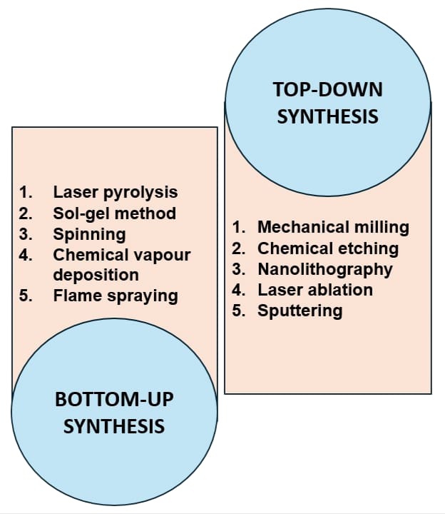
Figure 1: Method of preparation of Nanoparticles 34
METAL CONJUGATED NANOPARTICLES
Nanoparticle obtained from metal including compounds like salts; oxides represent a wide range of Nanoparticles which carry essential metal ions combined with other organic or inorganic molecules. Iron, copper, zinc and other noble metals like gold and silver work as major metal constituents of biomedical Nanoparticles. These metal conjugated Nanoparticles are suitable as drug delivery and diagnostic tool in antibacterial treatment. Metal conjugated Nanoparticles produce reactive oxygen species and produce reactive nitrogen species which leads to damage of protein and DNA which later leads to cell death. Even so Nanoparticles may produce unacceptable actions in normal cells; the unwanted inflammatory response and toxicity may inhibit the clinical benefits. These disadvantages can be avoided when metal- based Nanoparticles are used in conjugation with organic compounds, inorganic compounds, nucleic acids or proteins. It leads to increased efficiency, safety and specificity.10
METHODOLOGY
Green Synthesis of Curcumin Loaded Copper Nanoparticles
Co-precipitation method is used for the preparation. Here, 100ml of copper sulphate solution is added into 100ml Curcumin solution until green color obtained. Stir the mixture in 500rpm at ±70°C. Then add 1M NaOH and keep it for change into dark brown. This indicates the formation of Nanoparticles. Keep it for 24h and separate out the precipitate, and wash with pure water followed by ethanol for 3 times by centrifuging about 10 min at 4100rpm to separate the impurities. The final dark brown colored residue subjected to dry in an oven at 60? for 24h and then size reduced to fine powder for storage.11
CHARACTERIZATION STUDIES
FTIR spectroscopy is used for the characterization of molecular structure, and the impurities present in the Nanoparticles. KBr pellet method is used for the sample preparation. Here, 2-3mg sample Nanoparticles and 2g of potassium bromide is dehydrated at 200°C. The dried sample was pressed into pellet with 100mg of anhydrous KBr to obtain a homogeneous mixture and scanned on wavenumber ranging from 4000-400cm-1 with KBr pellet as control.12
Microstructural characterization and particle size distribution of Nanoparticles were studied using SEM-EDX (Energy Dispersive X-ray). About 1mg of sample was placed on a smooth carbon coated tape to produce a thin layer and then the film is coated with carbon. Various magnifications were used to capture the images at 15-20 kV.12
XRD was used to find the crystalline nature of the Nanoparticles by using a Rigaku diffractometer, with Cu Kα radiation at 25?. The scans were taken over a range of 2θ from 10º to 70º, with a step interval of 0.02º and scanning speed of 6º/min. 13
Zeta potential characterization was conducted in a Zetasizer Nano ZS90 at 25? to analyze the surface charge and stability of the Nanoparticles. Copper oxide Nanoparticles were diluted to 1mg/ml in ultra-pure water and filled in a disposal cell. Measurement was conducted by electrophoretic light with 60 runs per scan.12
UV-Visible spectroscopy was used to analyze the properties of the Nanoparticles. Stock solution is prepared into 100µg/ml with methanol and 1-7 µg/ml were prepared from stock solution by proper dilution with methanol. The maximum absorption wavelength (λmax) was determined by taking a 5µg/ml solution of Curcumin and scanning it from 200 to 700 nm using methanol as the reference.14
The wound healing assay is conducted in the fibroblast cell lines and is cultured in the Dulbecco’s phosphate buffer medium. It is incubated at a temperature of 37°C with air and carbon (19:1) for an overnight. After that the confluent monolayer is formed, and the wound is created with a sterile micropipette tip. Then wash the wound with phosphate buffer and add the 100µg/ml of nanoparticle. Cipladine is used as the standard drug. Then it is monitored using inverted microscope at different time interval.15
Curcumin-loaded copper oxide Nanoparticles exhibit strong antioxidant activity through effective free radical scavenging, which leads to enhanced wound healing effect.
Here, 100 µL of the Nanoparticles was mixed with 0.5 mL of 0.1 mM DPPH in 95% ethanol. Ten different concentrations were used. Methanol and butylated hydroxytoluene is used as positive and negative control. Absorbance was measured at 518 nm to calculate the IC50 value, and the percentage of scavenging activity was determined using a standard formula.16
The antimicrobial activity of nanoparticle is performed by agar well diffusion method. Here wells are made after the nutrient agar media get solidified. E. coli and Bacillus subtilis is swabbed on the agar plate. Then the samples are loaded at concentration of 20,40,60, and 80µl. Chloramphenicol is used as the positive control.17
FORMULATION OF NANOPARTICLES
Table 1: Formulation of Cream 18
|
Sl. No |
Phase |
Ingredient |
Quantity (%W/W) |
|
1 |
API |
Curcumin-loaded copper Nanoparticles (95%) |
1 |
|
2 |
Aqueous Phase |
Triethanolamine |
3 |
|
Glycerin |
20 |
||
|
Water |
q.s |
||
|
3 |
Oil Phase |
Stearic acid |
20 |
|
Liquid paraffin |
7 |
||
|
Cetyl alcohol |
4 |
||
|
4 |
Preservatives |
Propyl paraben |
0.04 |
|
Sodium metabisulphite |
0.2 |
||
|
EDTA |
0.2 |
EVALUATION OF CREAM
The appearance of the cream is evaluated by using vision, where we can identify the color, freshness, glossy, etc. By smelling the cream, we can identify the aroma and odour. Touching of the cream will help to identify the texture and the temperature of the cream.
Standard buffer (pH 7, 4.01 and 10) is used for the determination of pH. After calibration, about 0.5g of cream is dissolved in 50ml of water. Then it is measured by using pH meter at room temperature.19
Viscosity of cream is determined by using Brook field viscometer at room temperature. The results were measured by centipoise. Take a little quantity of cream and keep it in room temperature for some time and mix it thoroughly until sample attain homogeneity. Transfer the cream into the brook field viscometer and immerse the spindle until it completely dip. Then record the viscosity after 30seconds. Repeat the procedure three times to get the correct value.20
Spreadability test is used to check how easily the cream is spreading on the skin. An excess amount of cream was placed between two glass slides, and a 100 g weight was applied for 5 minutes to get a uniform thickness. Then a 250 g weight was added to the pan. The time taken for the slides to separate is measured for spreadability.21
Both Staphylococcus aureus and Escherichia coli were used to evaluate the antibacterial activity. Among the two bacterial strain, Staphylococcus aureus was found to be more effective. Then the minimum inhibitory concentration of Staphylococcus aureus is around 50g/ml and for Escherichia coli it is around 70g/ml. Which means lower amount of cream is needed to inhibit the bacterial growth.22
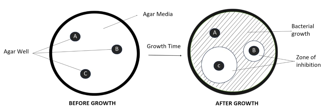
Figure 2: Demonstration of Agar well diffusion method.
The diffusion study of nanoparticle is done by using dialysis bag diffusion method. The accurately weighed sample is placed on the previously hydrated diffusion bag, and which is immersed in already prepared phosphate buffer (50ml) at 37°C and 120rpm. 1ml of release media is withdrawn in a predetermined interval, and 1ml of freshly prepared medium is added to the flask. It is continued for several time and finally the sample is filtered using 0.22-µm Cameo Acetate membrane filter. Filtered sample is analyzed by UV-Visible spectrophotometer at 420nm.15,23
PHARMACOKINETIC STUDY
It is the reaction in which the rate is independent of the concentration of the drug in the reaction. If the absorbance is obtained, then the concentration will get through the beer lambert’s law. Which is then applied to the below equation, then plot the graph using time vs concentration. If it is straight line with a negative slope, then it is confirmed as zero order kinetics.
c=a-kt?

Where ‘c’ is the concentration at time ‘t’ and ‘a’ is the initial concentration.24
The reaction in which the rate is dependent upon the concentrations of the drug. The method is same as that of the zero-order reaction, but the graph is plot between time vs log C and get a straight line of negative slope, which means it follow first-order kinetics.
logC=logCo-kt2.303
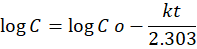
Here ‘C’ is the concentration at time ‘t’ and ‘Co’ is the initial concentration.24
It is the mathematical order in which describes the drug release from matrix-based system. After getting the concentration from the absorbance, it is easy to calculate the percentage drug release. Then plot a graph between percentage drug release and t, if it is straight line then it follows Higuchi kinetics.
MA=2Ci-Cst

Here, ‘M’ is the cumulative amount of drug released at time ‘t’, whereas ‘A’ is the surface area, ‘Ci’ is the initial concentration and ‘Cs’ is the drug solubility. Higuchi equation describes as the square root of time.25
This model explains the drug release mechanism of polymeric nanoparticle. Here the percentage drug release is calculated from the concentration. Then convert the equation into logarithm and plot a graph between log Qt vs log t

Qt = Atn
Here ‘Qt’ is the rate of drug release at time ‘t’, ‘A’ is the kinetic constant and ‘n’ is the release exponent.26
DISCUSSION
Characterization study
Table 2: FTIR stretches and its region.27
|
Stretch |
Region (Cm?¹) |
|
Single bond stretches |
4000–2500 |
|
Triple bond regions |
2500–2000 |
|
Double bond vibrations |
2000–1500 |
|
Hydroxyl stretching |
3700–3200 |
|
Fingerprint region |
1500–500 |
|
Curcumin conjugation with Nanoparticles |
1700–600 |
Using scanning electron microscopy together with Energy dispersive X-ray (EDX) analysis found that Curcumin loaded copper oxide Nanoparticles can be found in both rod shaped and spherical structure. The diameter of these Nanoparticles is showed in a range of 140-190nm.12
XRD analysis shows the strong diffraction peaks between 10° and 30° (2θ), confirming the crystalline structure of Curcumin. CuO Nanoparticles displayed distinct peaks at 2θ values of 30-75°, consistent with a monoclinic crystal structure. Curcumin exhibited characteristic peaks at 7-18°. The XRD pattern of Nanoparticles shows the combined features of both CuO and Curcumin, indicating successful integration.28
Zeta potential analyzes the stability of the nanoparticle. High zeta potential means the nanoparticle is in stable, while the low zeta potential indicates that the nanoparticle may aggregate or undergo coagulation. Using the zetasizer, the hydrodynamic diameter of the Curcumin loaded copper Nanoparticles was found to be around 13nm and the zeta potential value of the Nanoparticles should be in range of above ±40mV. Which shows the greater stability of Nanoparticles.29
Table 3: stability of Nanofluids for different zeta potential values 35
|
ZETA POTENTIAL [mV] |
STABILITY BEHAVIOUR OF THE COLLOID |
|
0 to ±5 |
Rapid coagulation or flocculation |
|
±10 to ±30 |
Incipient instability |
|
±30 to ±40 |
Moderate stability |
|
±40 to ±60 |
Good stability |
|
more than ±61 |
Excellent stability |
The Curcumin-loaded copper Nanoparticles exhibited a distinct absorption peak at a range of 415-430nm, confirming their formation and no other prominent or measurable peaks were detected. 30
The Curcumin loaded copper Nanoparticles show the width of wound healing is about 45nm in first 24h and in the next 48h it is about 80nm. Which is greater than the Curcumin itself. That means the cell migration is more into wound that applied Curcumin Nanoparticles than Curcumin alone.31
Curcumin loaded copper Nanoparticles have greater free radical scavenging activity than the Curcumin itself. It produces an oxidative stress against the microbes. The scavenging activity are directly proportional to the concentration of Curcumin loaded copper Nanoparticles.32
The antimicrobial activity is measured by the zone of inhibition. It is calculated by measuring the radius from the center of well to the site where bacteria growth is inhibited. The Curcumin loaded copper Nanoparticles shows greater zone of inhibition in both E. coli and B. subtilis.33
CONCLUSION
The green synthesis of Curcumin-loaded copper Nanoparticles offers a sustainable approach for developing an improved topical drug delivery system. These Nanoparticles combine the therapeutic effects of Curcumin and copper, including antimicrobial, antioxidant, and wound healing activities, while reducing the toxicity and increasing the bioavailability. Characterization techniques such as FTIR, SEM, XRD, zeta potential, and UV-Visible spectroscopy helps to confirm its formation, stability, and integration of Curcumin with copper Nanoparticles. The cream will be exhibited favorable physicochemical properties including appropriate pH, viscosity, spreadability, and significant antimicrobial activity against Staphylococcus aureus and E. coli. Kinetic studies help to know about the controlled and sustained drug release behavior of the cream. Overall, Curcumin-loaded copper nanoparticle cream shows great potential as a novel drug delivery and a better topical therapeutic formulation, with antioxidant and antimicrobial applications.
ACKNOWLEDGEMENT
We would like to express our sincere gratitude to all those who supported us in the completion of this review. We are especially thankful to our guide and the institute, for their invaluable guidance, encouragement, and continuous support throughout the work.
REFERENCES

Hani Jaleel V., Nidha V. P.*, Shahala Sharin, Shahana Shirin KM., Suhaila N., Nishad KM., Sirajudheen MK., A Review on Curcumin-Based Copper Nanoparticle Loaded Cream, Int. J. of Pharm. Sci., 2025, Vol 3, Issue 12, 1538-1548 https://doi.org/10.5281/zenodo.17862574
 10.5281/zenodo.17862574