
We use cookies to ensure our website works properly and to personalise your experience. Cookies policy
Shree Venkateshwara College of Paramedical Sciences, College of Pharmacy.
Arne Tiselius, made a special machine called Tiselius apparatus to separate chemicals using electric current. Electrophoresis is defined as the migration of the charged particles through solution under the influence of an external electrical field. It has a wide application in separating and in analyzing the biomolecules such as protein, plasmids, DNA and RNA. Electrophoresis in 1930s types include capillary electrophoresis, immunoelectrophoresis, isoelectric focusing and isotachophoresis. Moving boundary electrophoresis is the method that allows the charged species to migrate within a free moving solution in the absence of supporting medium. Capillary electrophoresis works by separating molecules based on how fast they move in an electric field. Immunoelectrophoresis is a technique in which an antigen mixture is separated into its component parts. Isotachophoresis technique is used for the separation of ionic species based on their effective mobilities. In isoelectric focusing the charged molecules are separated based on their isoelectric point.
Electrophoresis is defined as the migration of the charged particles through solution under the influence of an external electric field. Ions between two electrodes moves the direction of the electrodes with the opposing charges. Movement of cations and anions are based on the strength of the buffer solution. Electrophoresis is an analytical technique used in biochemistry for separating particles, molecules or ions by size or by binding affinity either freely or through a supportive medium using one directional flow of electrical charge. Electrophoresis has a wide application in separating and analyzing biomolecules such as protein, plasmids, DNA, RNA and nucleic acids.
Classification of electrophoresis:
They are two types:
Moving Boundary Electrophoresis:
The moving boundary electrophoresis allows the charged particles to migrate free solution in the absence of supporting media.
In capillary electrophoresis ions are separated using high voltage. It is electrokinetic method.
Immunoelectrophoresis involves an additional step of antigen-antibody complex formation in the equivalence zone.
The separation of ionic particles is based on their effective mobilities in isotachophoresis.
The charged molecules are separated on the basis of their isoelectric point (i.e.) the pH at which the molecules as no charge.
2. Moving Boundary Electrophoresis
The migration of charged species with the free solution without the help of supporting media is called as moving boundary electrophoresis. It is one of the reference methods for determining the electrophoretic mobility. Samples having minute concentration can also be easily detected.
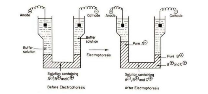
Fig.1 Schematic diagram of moving boundary electrophoresis
Its component includes U shaped cell filled with buffer solution in which the ends of the electrode are immersed. Any mixture of charged species such as protein can be used as the sample. when voltage is applied, depending on the charges the compounds migrate to the anode are cathode.
Types of moving boundary electrophoresis
There are four types of moving boundary electrophoresis. They are:
3. Capillary Electrophoresis
In 1930, Arne Tiselius showed that electrophoresis could separate proteins, in a liquid. But people didn’t pay much attention to his work until 1960s, when Hjerten started using capillaries, for this method. Capillary electrophoresis is an electro kinetic method used to separate ions using high voltage and how fast an ion moves depends on its charge, size and how thick the liquid is. Bigger electric fields make ions moves faster. Neutral particles don’t move, only charged ones do. If two ions are the size, the one with more charge moves faster. If they have the same charge, the smaller one moves faster then, this method is popular because it gives quick and clear results, and there are many ways to detect the ions.
Principle
Capillary electrophoresis works by separating molecules based on how fast they move in an electric field. This speed depends on their charge, size and shape as per the equation given below. The movement of a molecule is called Electrophoretic mobility (µe).
µ=q/6πηr
Whereas,
q is the charge of the molecule
? is the viscosity of the liquid
r is the radius of the molecule.
In capillary electrophoresis, a very thin tube made up of fused silica (usually 25-100micrometer wide) is filled with a liquid called buffer. When a high voltage (between 10,000 to 30,000 volts) is applied, molecules move through the tube at different speeds depending on their charge and size. This is also called Electro-osmotic flow (EOF), which means the whole liquid inside the tube moves and helps carry molecules along. To detect and measure the molecular technique like UV- Visible light absorption, fluorescence or mass spectrometry is used, depending on what kind of molecule are being analyzed.
Types
Five types of commonly known capillary electrophoresis:
Apparatus
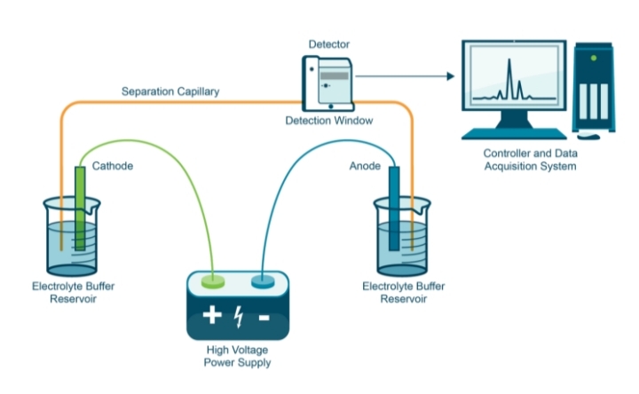
Fig.2 Schematic diagram of Capillary Electrophoresis
Instrumentation
The main parts of capillary electrophoresis include, a high voltage power supply that provides more than 30,000 volts. The capillaries are about 25-100 micrometers wide. The electric circuit is completed with buffers like citrate, phosphate or acetate. A detector, usually one that uses UV light to detect the separated substances.
Working
During the process, a small amount sample is placed into one end of the capillary. This is usually an electric field (electro migration). A high voltage power supply creates an electric current that pushes the sample through the capillary. As different part of the sample moves through, they were separated and eventually detected by a sensor at the other end. The data is then recorded. Capillary electrophoresis is easy to develop methods for reliable, fast and flexible. Its especially useful because it can separate substance that are hard to separate using HPLC. Also, Capillary electrophoresis can be automated to measure the amounts of substance quickly and accurately. Capillary electrophoresis is important in industries where it can be used as part of process analytical technology (PAT).
Advantages
Disadvantages
Applications
4. Immunoelectrophoresis
In 1953 Grabar and Williams coined the term immunoelectrophoresis. It is a technique in which an antigen mixture is firstly separated into its component’s parts. The presence of antigen is determined by formation of precipitin arc or line by immune – diffusion in agar. Immunoelectrophoresis plays a major role in diagnostic purpose and for separation, identification and /or quantification of target proteins in complex biological samples (plasma, serum). It involves an additional step of antigen – antibody complex formation.
Principle
A slide is layered with the gel and electric current is applied to it. According to their charge and size the antigen mixture placed in the wells is separated into individual antigen components. Following the electrophoresis specific antisera and antibody is reacted with the separated antigens which is placed parallel to the electrophoretic migration where diffusion is allowed to occur. Separation precipitin lines are formed as the result of movement of antiserum towards antigen components which indicates the reaction between individual proteins with its antibody.
Types
Classical immunoelectrophoresis is also known as gamma globulin electrophoresis and it represent an important advance in the analysis of biological samples separation and detection of antigens is based on their size and net charge. The proteins are first separated by electrophoresis. The antigens are allowed to diffuse towards a reservoir punched into the gel that contain specific antibodies. An antigen antibody complex precipitate in the form of arc if target antigens are present in the sample.
Rocket immunoelectrophoresis is an adaptation of radial immunoassay or electro immune-diffusion. It is a quantitative one-dimensional single electro immunodiffusion technique. In this method antibody is incorporated in the gel at pH value in which the antibodies remain essentially immobile.
Crossed immunoelectrophoresis is performed in two steps. In the first step the antigens are separated, and in the second the separated antigens are forced into an antibody containing gels slab by application of electric field at right angles to the direction of separation of the antigen in the first step.
Immunofixation electrophoresis is also known as serum protein electrophoresis. It is used for detecting monoclonal antibodies or immunoglobulins (Ig) in serum or urine. It plays a major role in diagnosis and monitoring blood related disease like myeloma. It takes place in two steps. In the first step the Ig present in the serum or urine are deposited on agarose gel allowed by separation of electrophoretic mobility under the effect of electric field. Once Ig is separated second step is migration of serum proteins. Appearance of a narrow band will be observed as precipitates if there is presence of monoclonal Ig.
Apparatus
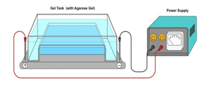
Fig.3 Schematic diagram of immunoelectrophoresis
Instrumentation
Immunoelectrophoresis includes power supply, electrophoresis tank, gel support system and detector. Power supply provides electrical current (typically 200-300V) necessary for electrophoresis. Electrophoresis tank holds the gel and buffer solutions with electrodes to create the electric field. Gel support system cast the agarose gel on a glass or plastic plate providing the medium for protein separation. The detection method relies on the formation of precipitin arcs where antibodies bind to antigens in the gel medium.
Working
Using the sample template, agarose gel is prepared on a glass slide. Across each corresponding slit 5µl of sample and control is applied. On the cathodic side gel is placed into the electrophoresis chamber with the samples and electrophoresis runs for 20mins /100 volts.
Advantages
Disadvantages
Limitation of immunoelectrophoresis
Application
5. Isotachophoresis
Isotachophoresis is an electrophoresis technique used for the separation of ionic species based on their effective mobility’s under an electrical field. Isotachophoresis (Greek: iso = equal, tachos = speed, phoresies = migration) is an advanced electrophoresis method used for the qualitative and quantitative analysis of ions. The method characterized by the equal migration velocities of the separated ions once a steady state is reached. Techniques like capillary electrophoresis and isotachophoresis are also effective for separating ionic substances.
Principle
Isotachophoresis requires a system of three types of ions with the same charge polarity.
Contain fast moving leading ions (L ?) with higher mobility than any sample ion.
Contains a slow-moving terminating ion (T ?) with slow mobility than any sample ion.
Contains ions with intermediate motilities.
When an electric field is applied
Apparatus
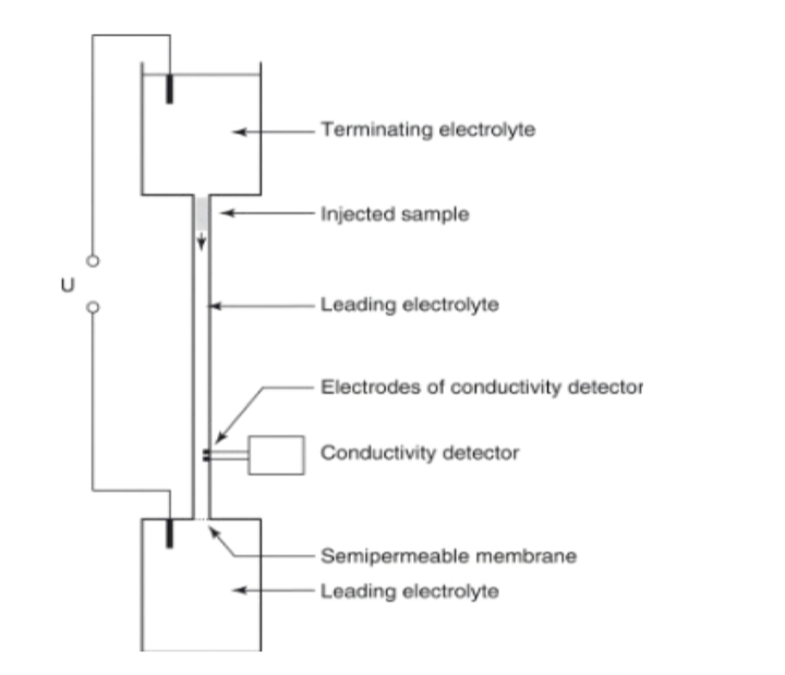
Fig .4 Schematic diagram of isotachophoresis
Instrumentation
Isotachophoresis consist of a separation column placed between two electrodes. The sample is introduced between two electrolytes: a leading electrolyte (with higher ion mobility than the sample) and trailing electrolyte (with lower ion mobility). A detector is integrated with the column to monitor the separated zone. The separation occurs based on the differential mobility of ions under an electric field, forming sharp and distinct zones. Detection is typically performed using conductivity or UV/Visible detectors, while thermal optical detectors may be used to enhance resolution.
A standard isotachophoresis system includes:
Working:
The sample analytical mixture is placed between the trailing electrolytes in an aqueous medium. When an electric field migrate between leading electrolyte and terminating electrolyte. Ions separate into discrete zones based on their individuals mobility ? s. The analytics form sharp, stacked bands in decreasing order of mobility. The differential in ionic mobility across leading electrolyte and terminating electrolytes creates a self- After the gradient is established, all zones migrate at the same velocity–hence the name isotachophoresis (equal speed migration).
Advantages
Disadvantages
Applications
6. Isoelectric Focusing Electrophoresis
Isoelectric focusing is also known as electro focusing. It is a technique is used for separated charged molecules like protein are peptide depending on their isoelectric point (i.e) the pH at which the molecules as no charge. It has better resolution and quantification than the gel electrophoresis technique so it is widely used in molecular biology labs and biotech labs. Further it is easier to perform as the samples do not have stress of the placement.
Principle
Slab of polyacrylamide or agarose gel that contains a mixture of amphoteric electrolytes (ampholytes) is used to carry out separation. The ampholyte starts to migrate in the gel which creats the pH gradient when subjected to an electrical field. When the applied protein reaches the gel fraction that has the pH that is same as their isoelectric point their charge is neutralized migration access. The separation is estimated by determining the minimum pH difference.
ΔpI=3D(dpHdx)/E(-dμdpH)

Where,
D is the diffusion coefficient of the protein
D pH /dx is the pH gradient
E is the intensity of the electrical field in volts per centimeter -du /d pH is the variation of the solute mobility with the pH in the region close to the pI
Types if isoelectric focusing
Apparatus

Fig.5 Schematic diagram of isoelectric focusing
Instrumentation
Isoelectric focusing electrophoresis consist of IEF gel, ampholyte and Buffer systems. The electrical field apparatus capable of providing a stable and adjustable electrical field, along with the electrodes and the gel casting apparatus. The gel is the through which the protein will migrate. These ampholytes are small, multi charged molecules that have a range of isoelectric point and distributed themselves throughout the gel to create a stable pH gradient. The pH and ionic strength required for IEF are maintained using buffers.
Working
The gel is prepared by mixing the acrylamide or agarose with the ampholytes and polymerizing reagents. The casting tray is filled with the gel and allowed to polymerize, forming a solid matrix. Ampholytes are added to the gel mixture before polymerization. Their concentration and range should be selected based on the expected pH range of the protein to be separated.
Sample application After polymerization, the gel is carefully mounted in the electrophoresis chamber. Protein samples are applied to the wells or spots on the gel, ensuring even distribution and avoiding cross contamination.
The gel is subjected to an electric field generated by the power supply. Based on the gel type and some characteristics current and voltage are adjusted. The electric field is applied for a period of time sufficient to allow protein to migrate and focus at their isoelectric point.
Once focusing is completed, gel is removed from the chamber. Staining methods like Coomassie brilliant blue, silver staining or other techniques are used for protein visualization based on their protein types. In image analysis, the stained gel is analyzed to interpret the separation patterns. Proteins are identified based on their position in pH gradient, corresponding to their isoelectric points.
Advantages
Disadvantages
Applications
7. DISCUSSION
Moving boundary electrophoresis is a classical electrophoretic technique used to study the migration of charged particles or molecules in a free solution under the influence of an electric field. Unlike zone electrophoresis, MBE doesn’t require a supporting medium like gel or paper. This approach provided early and critical insights into the physicochemical properties of biomolecules, particularly proteins and nucleic acid.
8. CONCLUSION
Moving boundary electrophoresis played a foundational role in the development of electrophoretic separation methods. It provided essential into the behavior of charged molecules in electric field and laid the groundwork for modern electrophoretic techniques. Moving boundary electrophoresis continues to hold educational and theoretical value, offering a fundamental understanding of electrophoretic principles and molecular mobility in solution.
REFERENCES

G. M. Srimyvizhiy*, K. B Ilango, S. Abinaya, X. Marcelin Brigith, S. Thanikadevi, C. Thirupathi, V. Rajarajan, A Review on Moving Boundary Electrophoresis, Int. J. of Pharm. Sci., 2025, Vol 3, Issue 9, 2119-2131 https://doi.org/10.5281/zenodo.17157473
 10.5281/zenodo.17157473