1Sir Dr. M.S. Gosavi College of Pharmaceutical Education and Research, College Rd, Vidya Nagar, Krishi Nagar, Nashik, 422005, Maharashtra, India.
2AISSMS College of Pharmacy, Raja Bahadur Motilal Rd, near RTO Pune, Sangamvadi, Pune, 411001, Maharashtra, India.
3Datta Meghe Institute of Higher Education & Research, Sawangi, Wardha, 442107, Maharashtra, India.
4MCE Society’s. Allana College Of Pharmacy, Hidayatullah Rd, New Modikhana, Azam Campus, Camp, Pune, 411001, Maharashtra, India.
5R.G Sapkal College of Pharmacy, Sapkal Knowledge Hub, Kalyani Hills, Anjaneri, Trimbakeshwar Rd, Nashik, 422213, Maharashtra, India.
6Dr. Vithalrao Vikhe Patil Foundation’s College of Pharmacy, Vadgaon Gupta Rd, Vilad Ghat, Ahilyanagar, 414111, Maharashtra, India.
Cancer continues to be one of the primary causes of illness and death across the globe, with standard treatment methods often constrained by limited specificity and systemic toxicity. Recent developments in nanotechnology have transformed the field of oncology by creating nanoparticle-based drug delivery systems aimed at enhancing therapeutic accuracy and results. Due to their adjustable size, surface properties, and material composition, nanoparticles facilitate both passive and active targeting of tumours, promoting drug concentration in cancerous tissues while reducing harm to healthy cells. A range of nanoparticle platforms including polymeric nanoparticles, liposomes, micelles, nanogels, and inorganic carriers has been investigated for controlled release, enhanced bioavailability, and targeted therapeutic effects. Nonetheless, challenges such as immune clearance, stability, safety concerns, andlarge-scale manufacturing pose significant barriers to clinical application. This review emphasizes recent progress in nanoparticle design, targeting methods, and clinical uses, while also discussing the challenges that need to be addressed to fully harness the promise of nanomedicine in cancer treatment.
Cancer encompasses various diseases marked by abnormal cell growth, frequently resulting in the spread of malignant cells to multiple body parts. On a worldwide scale, mortality rates from cancer rank second only to deaths caused by heart and vascular diseases, making it one of the most serious global health challenges. As a result, discussions about cancer therapy have evolved into a joint initiative involving medical practitioners and scientific researchers. At present, limited therapeutic options exist for cancer patients, with treatments mainly restricted to approaches such as chemotherapy1,2. A major concern with one of the most commonly employed cancer treatments is its inability to target tumour cells specifically, creating significant obstacles and limitations. Furthermore, the complex characteristics of tumour environments and formations can make it difficult to create new and more effective therapeutic approaches. The delivery of pharmaceutical compounds to target sites represents a major obstacle in numerous cancer treatment approaches. In response to this challenge, substantial progress has been made in research and practical applications of nanoscience and nanotechnology. These scientific disciplines are employed for both diagnosing and treating medical conditions. Nanoparticles are characterized as particles measuring less than 0.1 µm or 100 nm in size2. Within drug delivery applications, nanoparticles exceeding 100 nm may be necessary to accommodate sufficient drug quantities. Additionally, in therapeutic delivery systems, engineered particles can function as transport vehicles, while medications themselves may be formulated at the nanoscale, essentially becoming their own delivery mechanism3. The composition of these manufactured nanoparticles varies considerably. Raw materials may come from biological origins including phospholipids, lipids, lactic acid, dextran, or chitosan. They may also exhibit more synthetic properties, incorporating different polymers, carbon, silica, and metallic elements. Nanoparticles function as potent radiosensitizers in healthcare applications3,4, especially for drug transport and cancer therapy. Based on their geometric configuration, these particles are categorized as 0D, 1D, 2D, or 3D. As an example, gold nanoparticles (Au NPs) are recognized for their ability to improve radiation treatment outcomes in clinical settings. This article examines how nanoparticles can be utilized as a drug delivery approach for cancer treatment5. The primary objectives of employing nanoparticles for drug encapsulation involve enhancing drug transport to target cells and improving cellular uptake, while simultaneously reducing harmful effects of free drugs on non-target tissues. Meeting these aims will improve the therapeutic index, which represents the margin between doses that deliver clinical benefits, like destroying cancer cells, and doses that harm other body systems. To reach these targets, researchers must create nanoparticles with extended circulation time and precise targeting capabilities6.
Methods For Producing Nanoparticles:
Nanoparticles vary in their structure, size, and material composition. Their fabrication encompasses various synthesis techniques, which are primarily classified into two main approaches: 1) the bottom-up strategy and 2) the top-down strategy. These methods can be further divided into specific subcategories depending on the particular reaction conditions and processes utilized7,8, as shown in Figure 1. The top-down strategy involves altering bulk materials through subtraction or addition processes to create nano-scale formations. Several techniques exist for producing nanostructures using this method. Conversely, the bottom-up approach constructs nanostructures by assembling single atoms or molecules. This method allows for exact control over atom or molecule placement during the creation of desired nanostructures, typically within a 2 to 10 nanometre dimensional range. The engineering and dimensioning of materials represents the foundation of nanotechnology, with the objective of developing drug delivery systems especially for cancer treatment. We have methodically classified the materials currently being developed into different categories: polymeric nanoparticles (covering both natural and synthetic varieties), liposomes, micelles, hydrogels, exosomes, and other extracellular vesicles. This includes natural membrane-encased nanoparticles, such as blood cell nanoparticles, white blood cell-derived delivery mechanisms, and platelet-based materials, together with viruses and inorganic nanoparticles, including mesoporous silica, gold nanoparticles, and carbon-derived nanomaterials7,9.
Polymeric Nanoparticles:
Polymeric nanoparticles are a predominant type of nanoparticle due to their small dimensions, which range from 10 to 100 nm. These nanoparticles provide several benefits as drug delivery systems, such as enabling controlled release, protecting therapeutic agents and biologically active substances from environmental degradation, improving bioavailability, and enhancing the therapeutic index. Generally, polymeric nanoparticles can be classified into three primary categories: synthetic polymers, natural materials, and hybrid combinations that merge both synthetic and natural elements to achieve various functionalities10.
Liposomes:
Liposomes are spherical entities that form spontaneously and consist of membranes built from phospholipid bilayers. Their sizes can range from 25 nm to 10 μm, depending on the preparation method used. Having been examined as potential drug delivery systems for over fifty years since their discovery by Bangham, liposomes have demonstrated potential. Nonetheless, traditional liposome-based drug delivery systems encounter challenges due to their short circulation duration in the bloodstream, primarily attributed to rapid removal by macrophages in the reticuloendothelial system (RES). Advances such as second-generation polymer-coated liposomes have notably improved blood circulation times, increasing them from merely a few minutes to as long as three days11,12.
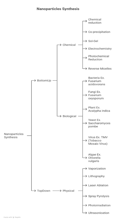
Figure 1. Classification chart of nanoparticle synthesis methods highlighting chemical reduction, biological sources, and physical techniques13.
Polymeric Micelles:
Polymeric micelles are formed through the self-assembly of amphiphilic di- or tri-block copolymers, resulting in nanosized core/shell structures in water. Recent studies emphasize the promise of specific micelle-based anticancer therapies as efficient drug delivery systems in oncology14. Genexol-PM (PEG-poly(D,L-lactide) paclitaxel) is the first polymeric micelle formulation of paclitaxel, notably lacking Cremophor, a component present in other paclitaxel formulations that utilize polyethoxylated castor oil. Furthermore, there is ongoing progress in creating multifunctional polymeric micelles that integrate targeting ligands along with imaging and therapeutic agents15.
Nanogels (Hydrogels):
Nanogels are three-dimensional hydrogel materials at the nanoscale formed via cross-linked, swellable polymer networks. They can effectively retain water while remaining insoluble in the surrounding solution. These nanogels combine the characteristics of nanoparticles and hydrogels, with dimensions varying between 20 and 200 nm. Hydrogels are well-known for their superior biocompatibility, biodegradability, and their abilities to load and release drugs in a controlled manner. As a result, they are widely utilized in a variety of treatments, including radiotherapy, chemotherapy, immunotherapy, hyperthermia, photodynamic therapy, and photothermal therapy16,17.
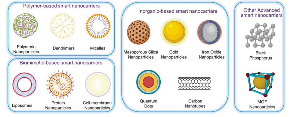
Figure 2: Classification of Nanocarriers for Drug Delivery18
Delivery of Therapeutic Nanoparticles to Targeted Areas:
Passive Targeting:
The concept of "passive targeting" is commonly used in nanomedicine to describe the phenomenon of nanoparticle accumulation in solid tumours. Unlike passive targeting nanoparticles, those used for active targeting are specifically designed with particular ligands on their surfaces. As tumours grow and begin to outstrip the available oxygen and nutrients, they release cytokines and other signalling molecules that trigger the creation of new blood vessels through a process called angiogenesis. The blood vessels generated during angiogenesis within tumour tissue display gaps of approximately 600 to 800 nm between adjacent endothelial cells, unlike the tightly connected vessels found in healthy tissues. This abnormal vascular structure, along with insufficient lymphatic drainage, leads to a phenomenon known as enhanced permeability and retention (EPR). The EPR effect arises because tumours require increased blood flow to supply the essential nutrients and oxygen needed for their unregulated cell proliferation19. As a result, nanoparticles tend to accumulate more in the tumour interstitium. Generally, several factors influence the extent of nanoparticle accumulation in tumour tissues, including the size and surface characteristics of the nanoparticles, their circulation half-life, and the level of angiogenesis present in the tumour. It is thought that nanoparticles in the size range of 10 to 100 nm are most effective for tumour accumulation. Appropriate surface characteristics and prolonged circulation times for nanoparticles can improve their uptake by tumours. The unmodified phospholipid surface of liposomes tends to bind plasma proteins, causing them to be recognized by the mononuclear phagocytic system (MPS) and leading to their rapid removal from the bloodstream. This aspect can impede the effective delivery of drugs encapsulated in liposomes to solid tumours. Stealth liposomes, which have modified surfaces, address the challenge of rapid clearance, resulting in liposomes with significantly longer circulation half-lives and reduced clearance rates. Passive targeting capitalizes on the inherent size of nanoparticles along with the unique properties of tumour blood vessels20.
Table 1. Types of nanoparticles in cancer therapy with corresponding formulations and therapeutic agents21
|
Type of Nanoparticle |
Name and References |
Therapeutic Agent |
|
Liposomes |
DaunoXome |
Dox |
|
Polymeric micelles |
Genexol-PM |
Paclitaxel |
|
Polymer-drug conjugate-based nanoparticles |
Xyotax |
Paclitaxel |
|
Albumin-based nanoparticles |
Abraxane |
Paclitaxel |
Active Targeting:
Active targeting is vital for the efficient delivery of drugs. This method employs affinity ligands to improve the attachment of nanoparticles (NPs) to antigens that are overexpressed on the surfaces of diseased cells or to extracellular matrix proteins found in diseased tissue. Actively targeted NPs can be utilized in situations where drug release occurs either externally or internally within the cells. This strategy significantly boosts the quantity of drug delivered to the target cells when compared to free drugs or passively targeted nano systems. The initial examples of targeted NPs emerged in the 1980s, focusing on modifying liposome surfaces with monoclonal antibodies (mAbs) that specifically bind to antigens on target cells. To date, 30 mAbs have received approval for clinical use, with Muromonab-CD3 (OKT3), an immunosuppressive agent, being the first to be authorized in 198622,23. While targeted nanoparticles may not always enhance drug accumulation in tumours compared to non-targeted nanoparticles, they do facilitate improved delivery of drugs to cancer cells, leading to a considerable increase in antitumor effectiveness. The nanoparticles listed in Table 1, which have been used in clinical contexts, primarily take advantage of the enhanced permeability and retention (EPR) effect of tumours and their surrounding environments for selective delivery. A range of targeting elements have been incorporated into drug delivery systems, such as antibody fragments, peptides, phage display-identified sequences, small molecules, or aptamers. Below are detailed descriptions of several actively targeted NPs to illustrate the ligands involved24.
Antibodies:
Monoclonal antibodies (mAbs) and their fragments, such as Fab’ and single-chain Fv (scFv), have been widely utilized as ligands for targeting cancerous cells with specific receptors. Poly(lactic-co-glycolic acid) (PLGA) nanoparticles (NPs) bound to mAbs specifically targeted MCF10A neo T cells, whereas uncoated NPs were randomly dispersed. In a different study, PEGylated liposomes modified with mAb 2C5 showed a three- to eight-fold increase in binding and internalization across various cancer cell lines from diverse origins, indicating greater cytotoxicity against numerous cancer cells and notable therapeutic benefits compared to control liposomes that were modified with a nonspecific IgG. However, to minimize immunogenicity and prevent clearance via Fc receptor-mediated mechanisms, fragments such as Fab’ and scFv are frequently preferred over full mAbs. Kou et al. developed PLGA NPs coated with the SM5-1 monoclonal antibody (scFv), which enhanced in vitro cytotoxicity against human hepatocellular carcinoma cell lines and resulted in significant tumour growth inhibition and regression25. Herceptin® is a therapeutic antibody specifically designed to target the human EGF receptor-2 (HER2), which is often overexpressed on breast cancer cells. Investigations involving anti-HER2 immunoliposomes, formed by attaching anti-HER2 antibody fragments to PEGylated liposomes, have shown that these immunoliposomes are capable of effectively delivering drugs into cells via mAb-mediated endocytosis. Conversely, nontargeted liposomes primarily reside in extracellular stroma or within macrophages. Additionally, DOX-loaded anti-HER2 immunoliposomes exhibited a significant antitumor effect compared to nontargeted liposomes. Nonetheless, despite the documented high uptake of these immunoliposomes, the relevance of active targeting is still debatable. In fact, the same research presented findings of comparable high accumulation levels in tumour tissue between anti-HER2 immunoliposomes and nontargeted liposomes in HER2-overexpressing breast cancer xenografts (BT-474). As a result, targeted nanocarriers did not improve tumour accumulation in comparison to their nontargeted equivalents. One factor that may account for the minimal accumulation observed is the study model, which concentrated on tumour cells rather than tumour endothelial cells, the intended target. Another potential explanation is the high density of ligands present on the nanoparticle (NP) surface. An overabundance of active ligands can hinder the long-circulation abilities of PEG, leading to a faster clearance of NPs from the bloodstream. Furthermore, several factors need to be considered when employing antibodies as targeting agents, including:
• The conjugation method for attaching antibodies to nanocarriers
• The effect of freely circulating antibodies25,26,27
Advantages of Nanoparticles in Cancer Therapy:
The integration of nanotechnology in cancer diagnosis, treatment, and management has marked the beginning of a transformative period. Nanoparticles (NPs), whether through active or passive targeting strategies, increase the concentration of drugs within cells while reducing toxicity to healthy tissues. These targeted NPs can be designed to be pH-sensitive or temperature-sensitive, enabling controlled drug release. The pH-sensitive delivery system is especially effective in administering drugs within the acidic tumour microenvironment (TME). Similarly, temperature-sensitive NPs release drugs at the target site due to temperature fluctuations caused by techniques like magnetic fields and ultrasound28. Moreover, the “physicochemical properties” of NPs—such as shape, size, molecular weight, and surface chemistry—significantly impact drug delivery targeting. NPs can be tailored to match specific targets and guide themselves toward particular molecules. Traditional chemotherapy and radiation treatments present various challenges, especially regarding their efficacy and side effects that arise from uneven distribution and cytotoxicity. As a result, meticulous dosing is essential to eliminate cancer cells while minimizing toxicity29. To achieve their goal, drugs must overcome several obstacles. The process of drug metabolism is intricate; under normal physiological conditions, a drug must pass through the tumour microenvironment (TME), the reticuloendothelial system (RES), and the blood-brain barrier (BBB), while also undergoing kidney filtration. The RES, consisting of blood monocytes, macrophages, and other immune cells, interacts with drugs in organs such as the liver, spleen, or lungs, activating macrophages and leukocytes that quickly eliminate the drug, leading to a reduced half-life. To counter this, nanoparticles (NPs) with surface modifications, like polyethylene glycol (PEG), can bypass this mechanism, prolonging the drug's half-life. Furthermore, kidney filtration is crucial in minimizing NP-related toxicity30. The blood-brain barrier (BBB) acts as a specialized defines mechanism designed to protect the central nervous system (CNS) from harmful substances. It is composed of brain capillary endothelial cells that create a barrier to ensure vital nutrients reach the brain while restricting access to harmful agents. Consequently, current chemotherapy options for brain cancer are mainly restricted to intraventricular or intracerebral infusions. In contrast, NPs possess the capability to penetrate the BBB. Various methods, such as the enhanced permeability and retention (EPR) effect, focused ultrasound, peptide-modified endocytosis, and transcytosis are employed for NP delivery. For example, glutathione-PEGylated liposomes loaded with methotrexate showed improved uptake of methotrexate in rat studies. Gold nanoparticles (Au-NPs) are frequently used due to their efficiency in drug delivery and the ability to induce apoptosis31. Moreover, NPs functioning as carriers improve drug stability by preventing the degradation of encapsulated substances, which permits a greater volume of drugs to be contained without undergoing chemical reactions. Dry solid dosage forms typically exhibit greater stability when compared to nano-liquid formulations. The stability can be further enhanced with the use of stabilizers, and employing porous NPs is another method to improve stability. Tumours display unique pathophysiological characteristics, including pronounced angiogenesis, irregular vascular structures, and inadequate lymphatic drainage. Nanoparticles (NPs) exploit these features to effectively target tumour tissues. Due to decreased venous return and poor lymphatic drainage in tumour areas, NPs tend to be retained more easily, a phenomenon referred to as the EPR effect. Additionally, focusing on adjacent tissues can further enhance localization to the tumour. NPs can be administered through various methods, such as oral, nasal, parenteral, and intraocular routes. With their high surface-to-volume ratios and ability for cellular uptake, NPs have demonstrated greater effectiveness compared to microparticles when used as drug carriers32.
Significant Challenges in the Clinical Application of Nanoparticles:
It is widely acknowledged that the tumour microenvironment (TME) plays a crucial role in the poor outcomes seen in nanomedicine treatments. The TME, which contains malignant cells, tumour-associated fibroblasts (CAFs or TAFs), a variety of immune cells, and the stroma (comprising blood vessels and the extravascular matrix), significantly contributes to cancer’s resistance to therapy33. Presently, with the swift progress of nanotechnology, there has been a substantial increase in the understanding and investigation of nanoparticles. Nevertheless, only a small fraction of these nanoparticles advances to clinical trials, with the majority remaining in the in vivo and in vitro testing phases. The approval rate for novel nano-drugs is less than 10%, and concerns regarding biosafety are increasing. Each specific nano formulation faces unique challenges in clinical translation, yet many nanoparticles encounter shared obstacles that can be categorized into biological, technological, and study-design-related issues34. Moreover, one complex challenge is eluding the “mononuclear phagocytic system (MPS).” In biological fluids, nanoparticles attract proteins, creating a protein corona that enhances MPS uptake. To counteract this, nanoparticles have been coated with materials aimed at preventing protein corona formation, but these strategies have not produced significant results. Developing nanoparticles that directly target macrophages and using them as innovative drug carriers can help mitigate this problem. Currently, tactics such as preventing macrophage recruitment, depleting and reprogramming tumour-associated macrophages (TAMs), and inhibiting the “CD47-SIRPα pathways” are frequently implemented. Another concern involves the manufacturing of nano drugs, as large-scale production of nanomedicines continues to be a significant challenge. Although these issues may appear overwhelming, targeted efforts can lead to progress35.
Toxicity And Safety Concerns of Nanoparticles in Cancer Therapy:
Nanoparticles (NPs) are being extensively studied for targeted cancer treatment due to their capacity to enhance drug delivery and minimize systemic toxicity, yet they also bring forth distinct safety issues that require careful consideration. The same nanoscale characteristics that enable NPs to infiltrate tumors and accumulate through the enhanced permeability and retention (EPR) effect can also result in unintended accumulation in healthy organs such as the liver, spleen, lungs, and kidneys, which raises concerns about long-term systemic toxicity. Numerous studies indicate that metallic and metal oxide NPs, including gold, silver, and titanium dioxide, can trigger the generation of reactive oxygen species (ROS), cause oxidative DNA damage, and lead to mitochondrial dysfunction, all of which are particularly concerning in cancer patients who already face genomic instability36. Furthermore, carbon-based nanomaterials like nanotubes can exhibit asbestos-like characteristics, resulting in chronic inflammation and fibrotic changes that may increase the risk of secondary tumor development. Another critical issue in oncology is the unintended interaction of NPs with the immune system: upon entering the bloodstream, proteins quickly adhere to NP surfaces, forming a “protein corona” that modifies biodistribution, promotes clearance by macrophages, and may even induce an immune response, thereby diminishing therapeutic effectiveness. Inhalation or skin exposure to engineered NPs during production and clinical handling poses additional occupational cancer risks, as airborne particles can migrate into systemic circulation and accumulate in various organs. Notably, some chemotherapeutic nano-formulations exhibit toxicity on their own; for instance, doxorubicin-loaded liposomes and paclitaxel-based NP carriers, while reducing cardiotoxicity in comparison to free drugs, still demonstrate hepatic and hematological side effects due to dose accumulation. These findings emphasize that although nanocarriers have significant potential for targeted cancer treatment, their toxicological assessment must be equally thorough. Comprehensive studies in nanotoxicology focusing on parameters such as size, shape, surface charge, and degradation profiles are crucial to achieving a balance between therapeutic efficacy and biosafety, ensuring that NPs developed for cancer treatment do not unintentionally result in secondary malignancies or systemic toxicity37,38.
Innovative Outlooks and Tailored Nanomedicine:
The advancement of nanoparticle-based treatments for cancer is expected to merge precision medicine with sophisticated nanotechnology, leading to exceptionally targeted, individualized therapies. Current obstacles including differences within the tumour microenvironment, elimination by the immune system, and overall toxicity underscore the necessity for next-generation nanocarriers that can adjust according to each patient's characteristics39. New approaches are concentrating on biodegradable and responsive nanoparticles that can release drugs based on specific tumour signals such as pH levels, enzyme activity, redox potential, or temperature, which would reduce unintended side effects while enhancing treatment effectiveness. Concurrently, personalized nanomedicine aims to utilize genomic, proteomic, and metabolomic information to create nanocarriers specifically designed for the unique biology of a patient's tumour, allowing for more precise targeting of oncogenic pathways. Recent advancements in liquid biopsies and the detection of circulating tumour DNA (ctDNA) have further paved the way for theranostics that employ nanoparticles, allowing for the simultaneous diagnosis, monitoring, and treatment of cancer in real-time. Additionally, artificial intelligence (AI) and machine learning are being utilized to anticipate interactions between nanoparticles and tumours, enhance formulation designs, and determine the most suitable nano-therapies for different patients40. Moreover, an increasing number of clinical trials are examining combination therapies, wherein nanoparticles convey multiple agents such as chemotherapy drugs, siRNA, or immune checkpoint inhibitors to address drug resistance and promote synergistic effects. Although challenges related to regulation, ethics, and large-scale production persist, the overarching goal is to establish a tailored nanomedicine framework in which cancer patients receive personalized nanoparticle treatments that not only enhance survival rates but also reduce side effects, thereby advancing toward safer and more effective cancer care41.
CONCLUSION:
Nanoparticle-based drug delivery systems represent an exciting new approach in cancer treatment, providing opportunities to increase drug specificity, minimize overall toxicity, and address the limitations of traditional therapies. By utilizing innovative passive and active targeting methods, nanoparticles enhance therapeutic effectiveness by taking advantage of the distinct characteristics of the tumour microenvironment and facilitating the controlled release of medications. Various platforms, such as polymeric carriers, liposomes, micelles, nanogels, and inorganic nanoparticles, have shown considerable promise in both preclinical and clinical trials. Nevertheless, the broader application of these technologies in clinical settings is hindered by challenges including immune system recognition, stability, large-scale manufacturing, and safety concerns. Overcoming these hurdles demands interdisciplinary collaboration among material scientists, biologists, and healthcare professionals to refine designs, create standardized production processes, and guarantee long-term safety. With ongoing advancements, nanomedicine has the potential to transform oncology by enabling targeted, personalized, and effective cancer treatment approaches.
REFERENCES


Aaditi Patil*, Sakshi Bhalerao, Fardeen Ansari, Heba Khan, Rutuja Baburao Shinde, Anuradha Pokale, Advancements in Targeted Nanoparticle-Based Drug Delivery Systems: Improving Therapeutic Outcomes in Oncology, Int. J. of Pharm. Sci., 2025, Vol 3, Issue 8, 2987-2999 https://doi.org/10.5281/zenodo.16992503
 10.5281/zenodo.16992503
