View Article

Abstract

Liposomes are spherical vesicles composed of one or more phospholipid bilayers encapsulating an aqueous core, widely used as drug delivery systems due to their biocompatibility, biodegradability, and capacity to encapsulate both hydrophilic and hydrophobic molecules. Their preparation typically involves lipids (such as synthetic or natural phosphatidylcholines, cholesterol) and aqueous media, employing methods like thin-film hydration, ethanol injection, reverse-phase evaporation, and extrusion. Critical formulation parameters (e.g., lipid composition, lipid-to-drug ratio, chain length, unsaturation, presence of cholesterol) strongly influence properties such as particle size, lamellarity, polydispersity, and encapsulation efficiency. Characterization and evaluation of liposomes involve a set of physicochemical and performance parameters: particle size and size distribution (often via Dynamic Light Scattering), zeta potential (for surface charge and colloidal stability), morphology (by electron microscopy), encapsulation efficiency (percentage of drug loaded), in vitro release profiles, membrane fluidity, and sometimes in vitro/in vivo stability or biodistribution. Optimization of these features is essential for ensuring effective delivery, minimizing toxicity, and achieving controlled release. liposomal properties are highly dependent on both formulation and process variables, a systematic design and thorough analytical characterization are critical in developing clinically viable liposome-based therapeutics.

Keywords

Liposome, Drug Delivery System, Microfluidic, Phospholipid

Introduction

The name liposome is derived from two Greek words: Lipo = "fat" and Soma = "body". A liposome is the drug delivery system which is structurally seeing like a colloidal, vesicular and made up one or more than one lipid bilayer (outer layer) in which the equal number of aqueous layer (inner layer) is inclosed into it shown in Figure 1 which contains a substance like peptides and protein, hormones, enzymes, antibiotics, antifungal and anticancer agent in this delivery system drug achieve the long therapeutic effect for the treatment of particular disease without affected to another part of the body.1,2 Liposomes are composed of small vesicles of phospholipids encapsulating an aqueous space ranging from about 0.03-to 10 µm in diameter.3 Liposomes can be produced with various lipid compositions or by different methods, leading to differences in parameters such as size, size distribution, surface electrical potential, number of lamellae, and encapsulation efficiency. Surface modification offers significant advantages in generating liposomes with distinct mechanisms, kinetic properties, and biodistribution profiles. Available products include Doxorubicin (Doxil, Myocet) for Kaposi's sarcoma, Daunorubicin (Daunoxome), and Cytarabine. These artificial microscopic vesicles consist of an aqueous core surrounded by one or more layers of phospholipids and are utilized to deliver vaccines, drugs, enzymes, or other agents to specific cells or organs.4 Liposomes are nano-sized artificial vesicles characterized by a spherical shape. They can be created from natural phospholipids and cholesterol. When phospholipids interact with water, they immediately form a double-layered sphere.5 Consequently, Dr. Bauman Cosmetic exclusively creates liposome products that are free from fragrances and artificial preservatives. The same phospholipids that make up the liposome membrane also constitute the walls of skin cells. Likewise, the substance found between skin cells is made up of phospholipids, ceramides, triglycerides, free fatty acids, cholesterol, and water. If skin cells are slightly compromised or if the intercellular substance is diminished due to harsh cleansing practices, liposomes can effectively restore the missing lipids. Thus, the combination of phospholipids, ceramides, and other lipids naturally found in the skin is beneficial. A liposome is a small bubble (vesicle) composed of materials similar to those of the cell membrane. Liposomes can be loaded with medications and utilized to deliver drugs for cancer treatment and other illnesses.6,7,8

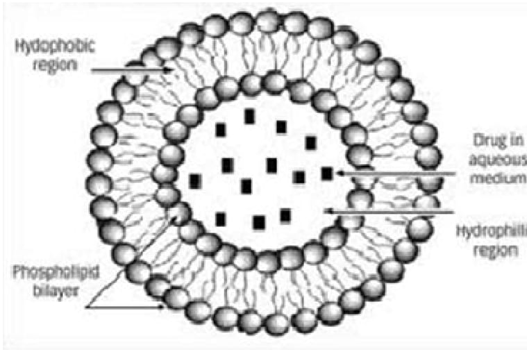
Fig No1: Structure of liposome

Structure of liposomes:9,10

Phospholipids

  • Naturally occurring phospholipids used in liposome:
  • Phosphatidylethanolamine
  • Phosphatidylcholine
  • Phsphatidylserine
  • Synthetic phospholipids used in the liposomes are:
  • Dioleoyl phosphatidylcholine
  • Disteroyl phosphatidylcholine
  • Dioleoyl phosphatidylethanolamine

Cholesterol

Cholesterol can be incorporated into phospholipid membranes at very high ratios, reaching up to 1:1 or 2:1 molar ratio of cholesterol to phosphatidylcholine. As an amphipathic molecule, cholesterol embeds itself within the membrane with its hydroxyl group directed towards the aqueous environment and its aliphatic chain aligned parallel to the acyl chains in the interior of the bilayer. Additionally, it increases the spacing between choline head groups and disrupts the usual electrostatic and hydrogen bonding interactions.

Fig No2: An illustration of liposome and its structural component

History Of Liposome:

Liposomes were initially identified in the mid-1960s by British hematologist Dr. Alec D. Bangham. His research focused on the structure of cell membranes, and he inadvertently discovered liposomes while using an electron microscope. Liposomes are minuscule spherical entities composed of lipid bilayers, resembling the architecture of cell membranes. Dr. Bangham's pioneering research established the groundwork for comprehending and developing liposomes for a variety of applications.11

In the subsequent decades, liposomes were acknowledged for their promise in drug delivery. Their capability to encapsulate pharmaceuticals and transport them to targeted areas within the body transformed the landscape of pharmacology. Liposomal drug delivery systems enabled controlled release and minimized the side effects associated with numerous medications.12

Over time, liposomes have been utilized not only in drug delivery but also in cosmetics, food technology, and gene therapy. Researchers have created various liposome types featuring different sizes, compositions, and surface modifications to enhance their performance for particular uses.13,14

ADVANTAGES:15,16

  • Some of the advantages of liposome are as follows
  • Provides selective passive targeting to tumour tissues (Liposomal doxorubicin).
  • Reduction in toxicity of the encapsulated agents.
  • Increased efficacy and therapeutic index.
  • Increased stability via encapsulation.
  • Improved pharmacokinetic effects (reduced elimination, increased circulation life times).
  • Flexibility to couple with site specific ligands to achieve active targeting.

DISADVANTAGES:15,16

  • Short half-life.
  • Low solubility.
  • Leakage and fusion of encapsulated drug/ molecules.
  • Production cost is high.
  • Fewer stables.
  • Sometimes phospholipids undergo oxidation and hydrolysis-like reaction.

Classification of Liposomes:17

A] Based on structure parameter

  1. Multilamellar vesicles (MLV) – (> 0.5 mm)
  2. Oligolamellar vesicles (OLV) – (0.1-1.0 mm)
  3. Unilamellar vesicles (ULV) – (all size ranges)
  • Medium Unilamellar vesicle (MUV)
  • Small Unilamellar vesicle (SUV) – (20-100 nm)
  • Glant Unilamellar vesicle (GUV) – (>1.0 mm)
  • Large Unilamellar vesicle (LUV) – (>100 nm)
  1. Multivesicular vesicles (MVV) – (>1.0 mm)

B] Based on Method of Preparation

  1. Dehydration rehydration method (DRV)
  2. SUVs/OLVs made by reverse phase evaporation method (REV)
  3. MLVs made by reverse phase evaporation (MLV-REV)
  4. Stable plurilamellar. Vesicles (SPLV)
  5. Frozen and thawed MLV (FATMLV)
  6. Vesicles prepared by Extrusion Technique (VET)

C] Based on Composition and Application

  1. CL (conventional Liposomes) - neutral/negatively charged phospholipids and cholesterol
  2. Fusogenic Liposomes - RSVE Reconstituted Sendai Virus
  3. pH sensitive Liposomes - Using phospholipids such as PE or DOPE with OA
  4. Cationic Liposomes - cationic lipids with DOPE
  5. Long Circulatory (Stealth) liposomes -  Made using cholesterol and 5-10% PEG-DSPE or GM1
  6. Immuno-liposomes - With attached monoclonal antibody

Mechanism of formation of Liposome

  • As liposomes are made up of phospholipids, they are amphipathic in nature and have ability to binds both aqueous and polar moiety. They have polar head and non-polar tail.
  • The polar end is mainly phosphoric acid and it will bound to water soluble molecule.
  • In aqueous medium the molecules in self-assembled structure are oriented in such way that the polar portion of the molecule remain in contact with in polar environment and at same time shields the non polar part. Liposomes are formed when the thin films are hydrated and stacks of liquid crystalline bilayers become fluid and swells.
  • Once these vesicles get formed, a change in vesicle shape and morphology required energy input in the form of …. Sonic energy to get SUVs and mechanical energy to get LUVs.
  • However, in aqueous mixtures these molecules are able to form various phases, some of them are stable and other remains in metastable form.18,19

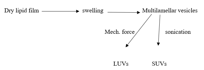
Fig No3: Mechanism of liposome preparation

Method Of Preparation:

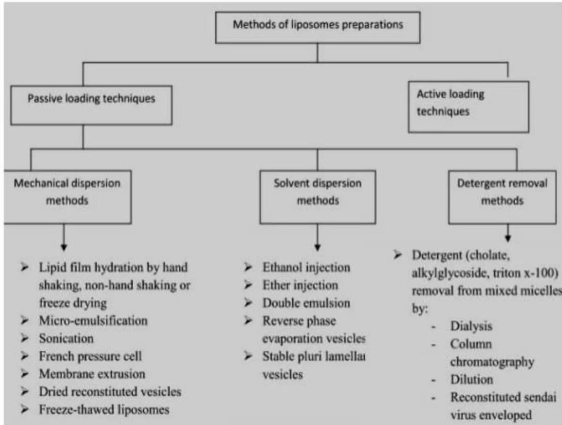
Fig No4: Different Methods of Liposome Preparations

A] Mechanical Dispersion Method

1) Lipid Film Hydration by hand shaking method:20

Liposomes were created using a physical dispersion technique with varying ratios of lipids. In this process, the lipids were dissolved in chloroform. This chloroform solution of lipids was spread over a flat-bottomed conical flask. The solution was then evaporated at room temperature without disturbing it. The hydration of the lipid film was performed with phosphate buffer (pH 7.4) while tilting the flask to one side, and the aqueous medium containing the drug to be encapsulated was introduced into the side of the flask as it was slowly returned to an upright position. The fluid was gently allowed to flow over the lipid layer, and the flask was left standing for 2 hours at 37°C for complete swelling; after swelling, the vesicles were collected by swirling the contents of the flask to produce a milky white suspension. The formulations were then subjected to centrifugation. Various batches of liposomes were prepared to identify the optimum formulation.

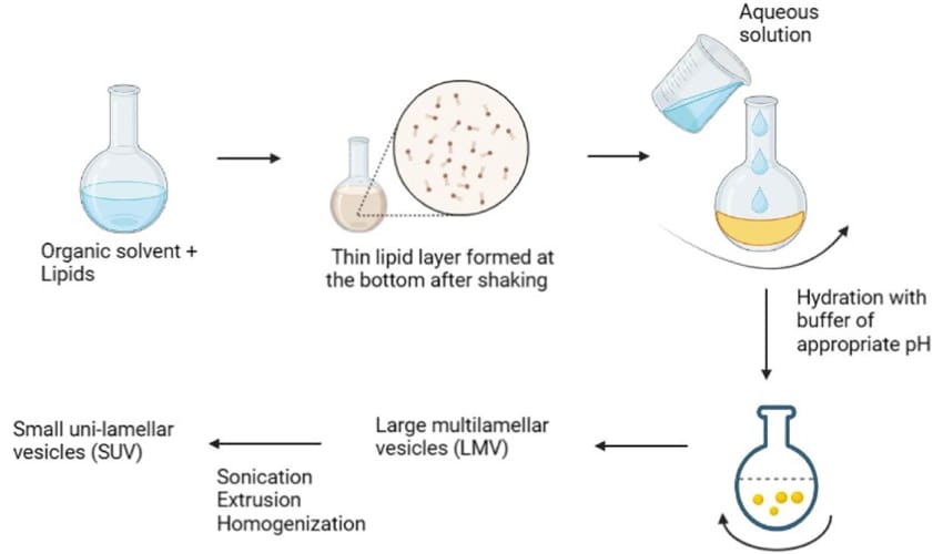
Fig No5 : Lipid Film Hydration method

2) Micro-emulsification:21

“Micro fluidizer” is used to prepare small MLVs from concentrated Lipid dispersion. Micro fluidizer pumps the fluid at very at very high pressure (10,000 psi), through a 5 micrometer orifice. Then, it is forced along defined micro channels which direct two streams of fluid to collide together at the right angles at a very high velocity, there by affecting an efficient transfer of energy. The lipids can be introduced into the fluidizer, either as large MLVs or as the slurry of un hydrated lipid in organic medium. The fluid collected can be recycled through the pump and interaction chamber until vesicles of spherical dimensions are obtained. diameter After a single pass, the size of vesicles is reduced to a size 0.1 and 0.2um .

3) Sonication:

This is the procedure by which Multi Lamellar Vesicles (MLVs) are converted into small Uni Lamellar Vesicles (SUVs). Ultrasonic irradiation is applied to the MLVs to produce the SUVs. Two techniques are utilized: a) Probe sonication method, and b) Bath sonication method. The probe method is used for dispersion and requires high energy for small volumes (for instance, high concentrations of lipids or a viscous aqueous phase), while bath sonication is better suited for larger volumes of diluted liquids. The probe tip sonicator delivers a substantial amount of energy to the liquid dispersion but can lead to overheating of the liposomal dispersion, resulting in lipid degradation. Additionally, the sonication tip may introduce titanium into the liposome dispersion, which can be removed through centrifugation before use. For these reasons, bath sonicator  tend to be more commonly employed. The sonication of MLVs is performed by either placing the dispersion in a bath sonicator or immersing the probe tip into the test tube containing the dispersion (for a duration of 5-10 minutes). Following sonication, the resulting dispersion is centrifuged, and as illustrated in the diagram, the SUVs will remain at the top while the smaller MLVs and aggregated lipids settle at the bottom. The upper layer consists of  a pure dispersion of SUVs with varying diameters, as size is influenced by factors such as composition, concentration, temperature, duration of sonication, volume, and sonication tuning.

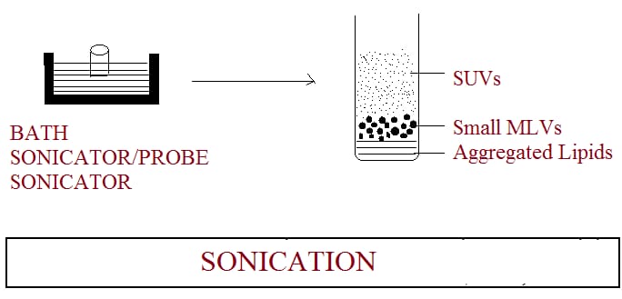
Fig No6 : Method of preparation of liposomes by sonication

4) French-Pressure Cell :22,23

The French pressure cell technique consists of extruding MLV through a small orifice. A key characteristic of the French pressure vesicle method is that proteins do not seem to be significantly altered during the process, unlike in sonication. This technique involves gentle handling of sensitive substances. It offers several benefits compared to sonication. The resulting liposomes are generally larger than the small unilamellar vesicles produced by sonication. However, the method has some limitations, such as the difficulty in achieving high temperatures and the relatively small working volumes, which are capped at about 50 ml.

5) Membrane Extrusion :

This technique can effectively process both LUVs and MLVs. The liposome size is minimized by gently forcing them through a membrane filter with a specific pore size, achieved at much lower pressures (<100 psi). During this process, the contents of the vesicles are exchanged with the dispersion medium as the phospholipid bilayers break and reseal while passing through the polycarbonate membrane. Liposomes generated through this method are referred to as LUVETs. This technique is the most commonly utilized method for producing SUVs and LUVs for both in vitro and in vivo research.

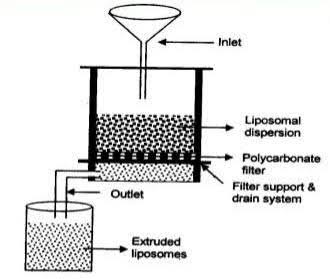
Fig No7 : Liposome Prepared by Membrane Extrusion method

6) Dried Reconstituted Vesicles:

This method starts with freeze drying of a dispersion of empty SUVs. After freeze drying the freeze dried membrane is obtained. Then these freeze dried SUVs are rehydrated with the use aqueous fluid containing the material to be entrapped. This leads to formation of the solutes in oligolamellar vesicles.

7) Freeze Thaw Sonication:

This method is based upon freezing of a unilamellar dispersion(SUV). Then thawing by standing at room temperature for 15min.Finally subjecting to a brief Sonication cycle which considerably reduces the permeability of the liposomes membrane. In order to prepare GIANT VESICLES of diameter between 10 and 50um, the freeze thaw technique has been modified to incorporate a dialysis step against hypo- osmolar buffer in the place of sonication. The method is simple, rapid and mild for entrapped solutes, and results in a high proportion of large unilamellar vesicles formation which are useful for study of membrane transport phenomenon. This method is based upon freezing of a unilamellar dispersion(SUV). Then thawing by standing at room temperature for 15min. Finally subjecting to a brief Sonication cycle which considerably reduces the permeability of the liposomes membrane. In order to prepare GIANT VESICLES of diameter between 10 and 50um, the freeze thaw technique has been modified to incorporate a dialysis step against hypo- osmolar buffer in the place of sonication. The method is simple, rapid and mild for entrapped solutes, and results in a high proportion of large unilamellar vesicles formation which are useful for study of membrane transport phenomenon.3,24,25

Fig No8: Liposome Prepared by Dried Reconstitute Vesicles/Freeze Thaw Sonication Method

B] Solvent Dispersion Method

1) Ethanol Injection Method :26

A lipid solution containing ethanol is quickly injected into a large amount of buffer, resulting in the immediate formation of MLVs. The challenges associated with this method include a heterogeneous population size (ranging from 30 to 110 nm), a very low concentration of liposomes, difficulties in completely removing all ethanol due to its formation of an azeotrope with water, and the risk of inactivating various biologically active macromolecules even with minimal ethanol exposure.

2) Ether Injection Method :

A lipid solution in diethyl ether or an ether/methanol combination is gradually added to an aqueous solution containing the material intended for encapsulation at a temperature of 55-65°C or under reduced pressure. Removing the ether under vacuum afterwards results in the creation of liposomes. The key disadvantages of this method include a heterogeneous particle

size distribution (ranging from 70 to 190 nm) and the exposure of the encapsulated compounds to organic solvents or elevated temperatures.

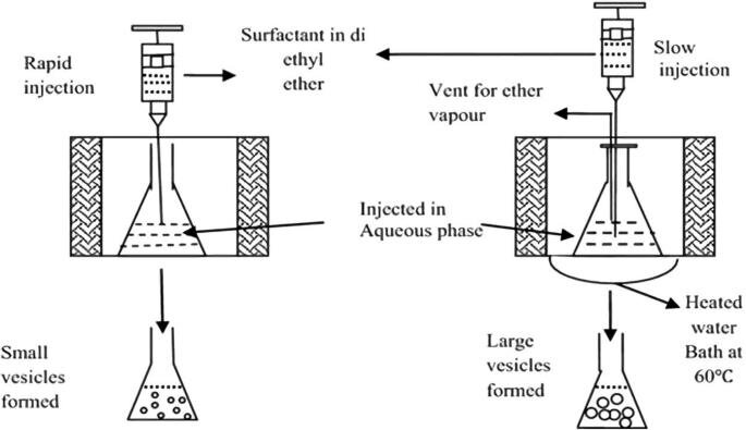
Fig No9 : Liposomes prepared by (1) Ethanol injection and (2) Ether injection method

3) Reverse Phase Evaporation Method :27

Initially, a water-in-oil emulsion is created by briefly sonicating a biphasic system that includes phospholipids dissolved in an organic solvent (such as diethyl ether, isopropyl ether, or a combination of isopropyl ether and chloroform) along with an aqueous buffer. The organic solvents are subsequently eliminated under low pressure, leading to the development of a thick gel. Liposomes are produced when the remaining solvent is effectively removed through ongoing rotary evaporation under reduced pressure. This technique can achieve a high encapsulation efficiency of up to 65% in a medium with low ionic strength, for instance, 0.01M NaCl. This method has been utilized for encapsulating both small molecules and large macromolecules. A significant drawback of this technique is that it exposes the encapsulated materials to organic solvents and to short intervals of sonication.

C] Detergent Removal Method :28

Detergents at their critical micelle concentrations have been utilized to solubilize lipids. As the detergent is eliminated, the micelles gradually become more enriched in phospholipids and ultimately merge to create IUVs. The removal of detergents can be achieved through dialysis. The benefits of the detergent dialysis method include high reproducibility and the generation of liposome populations that are uniform in size. The primary disadvantage of this technique is the potential for residual detergent(s) remaining within the liposomes. A commercial device known as LIPOPREP (Diachema AG, Switzerland), which is a type of dialysis system, is available for detergent removal. Other methods that have been employed for detergent removal include (a) Gel Chromatography using a Sephadex G-25 column, (b) the adsorption or binding of Triton X-100 (a detergent) to Bio-Beads SM-2, and (c) the binding of octyl glucoside (a detergent) to Amberlite XAD-2 beads.

Evaluation Of Liposome29

The liposomal formulation and processing for a designated purpose are defined to guarantee their consistent performance both in vitro and in vivo. The characterization parameters for evaluation can be categorized into three stages: physical, chemical, and biological parameters. Physical characterization focuses on evaluating aspects such as size, shape, surface characteristics, and drug release profiles. Chemical characterization involves studies that determine the purity and potency of various lipophilic components. Biological characterization parameters aid in establishing the safety and appropriateness of the formulation for therapeutic use. Some of the parameters include:28

Characterization of Liposome Structure

Morphology:

  • Transmission Electron Microscopy (TEM): Provides high-resolution images of liposome size, shape, and lamellarity.
  • Scanning Electron Microscopy (SEM): Offers surface information and morphology details.

Size and Size Distribution:

  • Dynamic Light Scattering (DLS): Measures particle size, size distribution, and polydispersity.
  • Nanoparticle Tracking Analysis (NTA): Tracks and sizes individual liposomes in a liquid suspension.

Zeta Potential:30

  • Electrophoretic Light Scattering: Determines the surface charge of liposomes, which affects stability and colloidal behavior

Lipid Composition Analysis:

  • High-Performance Liquid Chromatography (HPLC): Identifies and quantifies lipids in liposomal formulations.

Liposome Properties:

Encapsulation Efficiency :

  • UV-Visible Spectroscopy or Fluorescence Spectroscopy: Measures the concentration of encapsulated drugs or molecules.

Stability:

  • Assessing changes in size, poly dispersity, and zeta potential over time under various storage conditions (e.g., temperature, pH).

Drug Release Kinetics:

  • In vitro release studies to determine the rate and extent of drug release from liposomes.

Biological Evaluation:31

In vitro Cell Studies:

  • Cell viability assays (MTT, Alamar Blue) to assess liposome cytotoxicity.
  • Cellular uptake studies to evaluate liposome internalization and drug delivery efficiency.

In vivo Studies:

  • Animal models to evaluate the pharmacokinetics, biodistribution, and therapeutic efficacy of liposomal drug formulations.

Biocompatibility and Toxicity Assessment:

  • Hemolysis Assay: Measures the potential for liposomes to cause red blood cell damage.
  • Immunogenicity Assessment: Investigates the immune response to liposomes.

Drug Release Studies

  • Dialysis Method: Evaluates drug release kinetics under sink conditions by dialyzing liposomal suspensions against a release medium.
  • Franz Diffusion Cell: Measures drug permeation through a membrane to mimic transdermal drug delivery.

Surface Modification Analysis

  • Surface Characterization: Techniques like X-ray Photoelectron Spectroscopy (XPS) and Fourier-Transform Infrared (FTIR) spectroscopy to analyze modifications made to the liposome surface.

Marketed Formulations of Liposomes:18

Product

Drug

Company

AmbisomeTM

Amphotericin B

Nexstar pharmaceuticals Inc., CO

AbelcetTM

Amphotericin B

The Liposome Company, NJ

AmphocilTM

Amphotericin B

Sequus pharmaceuticals, Inc., C.A

DoxilTM

Doxorubicin

Sequus pharmaceuticals, Inc., C.A

DaunoxomeTM

Daunorubicin

Nexstar pharmaceuticals, Inc., CO

MikasomeTM

Amikacin

Nexstar pharmaceuticals, Inc., CO

DC99TM

Doxorubicin

Liposome CO., NJ, USA

EpaxelTM

Hepatitis A Vaccine

Swiss Serum Institute, Switzerland

ELA-MaxTM

Lidocaine

Biozone Labs, CA, USA

Applications Of Liposome:

Drug Delivery:32

  • Liposomes are commonly used as drug delivery vehicles to encapsulate and deliver both hydrophobic and hydrophilic drugs.
  • They can improve drug solubility, stability, and bioavailability.
  • Liposomal drug formulations can target specific tissues or cells, reducing systemic side effects.

Vaccines:33

  • Liposomes are used as adjuvants or carriers for vaccines to enhance immunogenicity.
  • They can improve antigen delivery to immune cells, leading to a stronger immune response.

Cosmetics and Skincare:

  • Liposomes are utilized in cosmetics and skincare products for controlled release of active ingredients, such as vitamins and antioxidants.
  • They can enhance the penetration of ingredients into the skin, improving their efficacy.

Gene Delivery:34

  • Liposomes can be used to deliver genetic material, including DNA and RNA, for gene therapy applications.
  • They protect and facilitate the transport of genetic cargo into target cells.

Diagnostics:

  • Liposomes can serve as carriers for contrast agents in medical imaging, such as magnetic resonance imaging (MRI) and ultrasound.
  • They enable targeted imaging of specific tissues or cells.

 Cancer Therapy:35

  • Liposomal formulations of chemotherapy drugs, like Doxil (liposomal doxorubicin), are used to treat cancer.
  • They can improve drug circulation time and reduce damage to healthy tissues.

Food Technology

  • Liposomes are applied in the food industry for encapsulating and protecting sensitive ingredients, such as vitamins, flavors, and antioxidants.
  • They can improve the stability and bioavailability of these additives in food products.

Biotechnology

  • Liposomes are used in research and biotechnology applications for drug screening and delivery to cells in vitro.
  • They are valuable tools for studying cell membrane interactions and drug transport mechanisms

 Transdermal Drug Delivery:36

  • Liposomal formulations can be applied topically to deliver drugs through the skin.
  • They offer controlled release and can avoid the first-pass metabolism in the liver.

 Personal Care Products

  • Liposomes are employed in personal care products such as sunscreens and moisturizers to enhance the delivery of active ingredients.

 Veterinary Medicine

  • Liposomes are used in veterinary medicine for drug delivery to animals, similar to their applications in human medicine.

 Environmental Remediation:

  • Liposomes can be utilized for the controlled release of remediation agents in environmental cleanup efforts.

 Intracellular Delivery:37

  • Liposomes are valuable tools in research for delivering molecules into specific organelles within cells.

 Nutraceuticals:33

  • Liposomes are used to enhance the bioavailability of nutraceutical compounds in dietary supplements.

Wound Healing

  • Liposomal formulations can be applied to wound dressings to promote the controlled release of wound-healing agents.

CONCLUSION

Liposomes are a promising and groundbreaking system for delivering drugs, with numerous applications in the pharmaceutical sector. Extensive studies over the years have shown their ability to address many challenges linked to conventional drug delivery methods. Liposomes have become an encouraging category of drug delivery systems that provide considerable benefits for improving the effectiveness and safety of various medications. Although challenges persist, ongoing innovations and improvements in liposomal technologies offer great potential for the future of drug delivery within the pharmaceutical domain. Liposomes offer an exciting and adaptable strategy for drug delivery, with the capacity to transform the pharmaceutical landscape by enhancing drug effectiveness, minimizing side effects, and allowing targeted therapies. Further progress in liposomal technology is expected to broaden their application across a diverse array of medical uses.

Conflict Of Interest

The authors declare that there is no conflict of interest.

ACKNOWLEDGMENT

The authors would like to acknowledge the library facility of the college for this work.

REFERENCES

  1. Kant S, Kumar S, Prashar B,  a complete review on: liposomes. From international research journal of pharmacy. ISSN 2230. Available from: https://www.researchgate.net/publication/285487882_A_complete_review_on_Liposomes.
  2. Yadav D, Sandeep k, Pandey D, and Dutta R K. Liposomes for Drug Delivery from Journal of Biotechnology & Biomaterials .Available from: https://www.omicsonline.org/peer-reviewed/liposomes-for-drug-deliveryp-97370.html
  3. Riaz Mohammad ,Liposomes Preparation Methods” Pakistan Journal of Pharmaceutical Sciences Vol.19(1), January 1996, 65-77.
  4. Barani, H. & Montazer, M. “A Review on Applications of Liposomes in Textile Processing. Journal of Liposome Research, 2008, 18, 249-262.
  5. http.www nanolifeneutra .com liposomes.
  6. Www.News-Medical.Net/Health/What-Is-A-Liposome.Aspx, What Is A Liposome? - From News-Medical.Net.
  7. Dosi Abha Created By Mehta Akul Www Pharmaxexchange Info…Author Acer Flexible Liposomes For Topical Applications In Cosmetics Blume Gabriele.
  8. Satynarayna U, Biochemistry, 3rd Edition Published By Arunabha Sen Book And Allid (P) Ltd First Published 2008: 35.
  9. Dr. Tiwari,Talreja S & Ms Swarnima Pandey. A review on use of novel drug delivery systems in herbal medicines, Science and Engineering Journal. 2020; 24(8):190-197.
  10. Dwivedi C, Yadav R, Tiwari S, T. Satapathy. Roy A, Role of Liposome in novel drug delivery systems. Journal of Drug Delivery & Therapeutics; 2014; 4(2):116-129.
  11. A. D. Bangham. Liposome: The Babraham Connection. Chemistry and Physics of Lipids. 1993; 64:1-3.
  12. Touitou, E. Drug delivery across the skin. Expert Opin. Biol. Ther. 2002; 2: 723–733.
  13. Abd El-Alim, S. H.; Kassem, A. A. Basha, M.; Salama, A. Comparative study of liposomes, ethosomes and transfersomes as carriers for enhancing the transdermal delivery of diflunisal. In vitro and in vivo evaluation. Int. J. Pharm. 2019; 563, 293–303.
  14. Ramadon, D. Mc Crudden, M. T. C. Courtenay, A. J. Donnelly, R. F. Enhancement strategies for transdermal drug delivery systems. Current trends and applications. Drug Deliv. Transl. Res.2022 12(4):758-791.
  15. Thulasiramaraju TV, Babu AS, Arunachalam A, Prathap M, Srikanth S, Sivaiah P. Liposome: A novel drug delivery system. International Journal of Biopharmacuties 2012; 2229:7499.
  16. Shashi K, Satinder K, Bharat P. A complete review on: Liposome. International Research Journal of Pharmacy. 2012; 3(7):10-6.
  17. Dua JS, Rana AC, Bhandari AK. Liposome: methods of preparation and applications. International Journal Pharmaceutical Studies Research. 2012; 3:14-20
  18. Cabral E. C. M., Zollner R. L. And Santana M. H. A. Brazilian Journal of Chemical Engineering Preparation And Characterization Of Liposmes Entrapping Allergenic Protein Received: February 13, 2003003.
  19.  Bangham, A.D. and R.W. Horne, 1964, Negative staining of phospholipids and their structured by surface active agents as observed in the electron microscope, J. Mol. Biol. 8,660–668.
  20. Shivhare UD, Ambulkar DU, Mathur VB, Bhusari KP, Godbole MD. Formulation and evaluation of pentoxifylline liposome formulation. Digest Journal Nanomaterials and Bio structures. 2009 4(4):857-62.
  21. Akbarzadeh A, Rezaei-Sadabady R, Davaran S, Joo SW, Zarghami N, Hanifehpour Y, Samiei M, Kouhi M, Nejati-Koshki K. Liposome: Classification, Preparation, and Applications. Nanoscale Research Letters. 2013; 8(1):102.
  22. Jain N.K. “Controller and Novel Drug Delivery” CBS Publisher and Distributors, New Delhi. 2009; 1; 278-283.
  23.  Saraswathi Marripati, K. Umasankar.” A Review on Liposome” International Journal of Research in Pharmaceutical and Nano Sciences. 2014; 3(3), 159 - 169.
  24. Deepak, By Preparation of Liposomes Www.Google .Com.
  25. Xiaoling Li, Bhaskara R. Jasti, Mcgram Hill, Chemical Enginerring, Design Of Controlled Release Drug Delivery System., Page No 65,367.
  26. Deamer D and Rangham ATD Blockon. Biophys, Act, 1976; 443: 629.
  27. Bateri S and Kom RD Nichum. Biopic. Acta 1973: 298 1015.
  28. Enoch IHG. and Strittmatter P. (1979). Proc. Natl. Acad. Sci. USA. 76: 145.
  29. Sawant GS, Sutar KV, Kanekar AS. Liposome: A Novel Drug Delivery System. International Journal of Research and Review. 2021; 8(4): 252-268.
  30. Zili Gu, Candido G. Da Silva, Koen van der Maaden, Ferry Ossendorp and Luis J. Cruz. Liposome-Based Drug Delivery Systems in Cancer Immunotherapy. Pharmaceutics, 2020; 12.1-25.
  31. Ramkrishna S, Sunit K. S, Soumyaranjan S. Liposome as drug delivery system: A brief review. International Journal of Research in Pharmaceutical Science. 2014; 5(4): 309-321.
  32. S. Marripati, K. Umashankar, P. Jayachandra Reddy. A Review on Liposomes. International Journal of Research in Pharmaceutical and Nanoscience. 2014; 3(3): 159-169.
  33. Shashi K. Satinder K.Bharat P. A Complete Review on Liposome. International Research Journal of Pharmacy 2012; 3(7): 10-16.
  34. Gabrijelcic V and Sentjure M. Influence of hydrogel on liposome stability and on liposome stability and on the transport of liposome entrapped substance in to the skin. Int J Pharm .1995; 118: 209-212.
  35. Hatwar P. R, Dr. Bakal R. L, Dumare A. A, Dere M. D, Hage. P. D, Targeted drug delivery system for the treatment of Cancer, WJPR. 2023; 12(12): 808-816.
  36. Deulkar D. A, kubade J. A, Hatwar P. R, Dr. Bakal R. L. A review on transdermal drug delivery system, GSC Advances Research and Reviews. 2024; 18(02): 347-361.

Reference

  1. Kant S, Kumar S, Prashar B,  a complete review on: liposomes. From international research journal of pharmacy. ISSN 2230. Available from: https://www.researchgate.net/publication/285487882_A_complete_review_on_Liposomes.
  2. Yadav D, Sandeep k, Pandey D, and Dutta R K. Liposomes for Drug Delivery from Journal of Biotechnology & Biomaterials .Available from: https://www.omicsonline.org/peer-reviewed/liposomes-for-drug-deliveryp-97370.html
  3. Riaz Mohammad ,Liposomes Preparation Methods” Pakistan Journal of Pharmaceutical Sciences Vol.19(1), January 1996, 65-77.
  4. Barani, H. & Montazer, M. “A Review on Applications of Liposomes in Textile Processing. Journal of Liposome Research, 2008, 18, 249-262.
  5. http.www nanolifeneutra .com liposomes.
  6. Www.News-Medical.Net/Health/What-Is-A-Liposome.Aspx, What Is A Liposome? - From News-Medical.Net.
  7. Dosi Abha Created By Mehta Akul Www Pharmaxexchange Info…Author Acer Flexible Liposomes For Topical Applications In Cosmetics Blume Gabriele.
  8. Satynarayna U, Biochemistry, 3rd Edition Published By Arunabha Sen Book And Allid (P) Ltd First Published 2008: 35.
  9. Dr. Tiwari,Talreja S & Ms Swarnima Pandey. A review on use of novel drug delivery systems in herbal medicines, Science and Engineering Journal. 2020; 24(8):190-197.
  10. Dwivedi C, Yadav R, Tiwari S, T. Satapathy. Roy A, Role of Liposome in novel drug delivery systems. Journal of Drug Delivery & Therapeutics; 2014; 4(2):116-129.
  11. A. D. Bangham. Liposome: The Babraham Connection. Chemistry and Physics of Lipids. 1993; 64:1-3.
  12. Touitou, E. Drug delivery across the skin. Expert Opin. Biol. Ther. 2002; 2: 723–733.
  13. Abd El-Alim, S. H.; Kassem, A. A. Basha, M.; Salama, A. Comparative study of liposomes, ethosomes and transfersomes as carriers for enhancing the transdermal delivery of diflunisal. In vitro and in vivo evaluation. Int. J. Pharm. 2019; 563, 293–303.
  14. Ramadon, D. Mc Crudden, M. T. C. Courtenay, A. J. Donnelly, R. F. Enhancement strategies for transdermal drug delivery systems. Current trends and applications. Drug Deliv. Transl. Res.2022 12(4):758-791.
  15. Thulasiramaraju TV, Babu AS, Arunachalam A, Prathap M, Srikanth S, Sivaiah P. Liposome: A novel drug delivery system. International Journal of Biopharmacuties 2012; 2229:7499.
  16. Shashi K, Satinder K, Bharat P. A complete review on: Liposome. International Research Journal of Pharmacy. 2012; 3(7):10-6.
  17. Dua JS, Rana AC, Bhandari AK. Liposome: methods of preparation and applications. International Journal Pharmaceutical Studies Research. 2012; 3:14-20
  18. Cabral E. C. M., Zollner R. L. And Santana M. H. A. Brazilian Journal of Chemical Engineering Preparation And Characterization Of Liposmes Entrapping Allergenic Protein Received: February 13, 2003003.
  19.  Bangham, A.D. and R.W. Horne, 1964, Negative staining of phospholipids and their structured by surface active agents as observed in the electron microscope, J. Mol. Biol. 8,660–668.
  20. Shivhare UD, Ambulkar DU, Mathur VB, Bhusari KP, Godbole MD. Formulation and evaluation of pentoxifylline liposome formulation. Digest Journal Nanomaterials and Bio structures. 2009 4(4):857-62.
  21. Akbarzadeh A, Rezaei-Sadabady R, Davaran S, Joo SW, Zarghami N, Hanifehpour Y, Samiei M, Kouhi M, Nejati-Koshki K. Liposome: Classification, Preparation, and Applications. Nanoscale Research Letters. 2013; 8(1):102.
  22. Jain N.K. “Controller and Novel Drug Delivery” CBS Publisher and Distributors, New Delhi. 2009; 1; 278-283.
  23.  Saraswathi Marripati, K. Umasankar.” A Review on Liposome” International Journal of Research in Pharmaceutical and Nano Sciences. 2014; 3(3), 159 - 169.
  24. Deepak, By Preparation of Liposomes Www.Google .Com.
  25. Xiaoling Li, Bhaskara R. Jasti, Mcgram Hill, Chemical Enginerring, Design Of Controlled Release Drug Delivery System., Page No 65,367.
  26. Deamer D and Rangham ATD Blockon. Biophys, Act, 1976; 443: 629.
  27. Bateri S and Kom RD Nichum. Biopic. Acta 1973: 298 1015.
  28. Enoch IHG. and Strittmatter P. (1979). Proc. Natl. Acad. Sci. USA. 76: 145.
  29. Sawant GS, Sutar KV, Kanekar AS. Liposome: A Novel Drug Delivery System. International Journal of Research and Review. 2021; 8(4): 252-268.
  30. Zili Gu, Candido G. Da Silva, Koen van der Maaden, Ferry Ossendorp and Luis J. Cruz. Liposome-Based Drug Delivery Systems in Cancer Immunotherapy. Pharmaceutics, 2020; 12.1-25.
  31. Ramkrishna S, Sunit K. S, Soumyaranjan S. Liposome as drug delivery system: A brief review. International Journal of Research in Pharmaceutical Science. 2014; 5(4): 309-321.
  32. S. Marripati, K. Umashankar, P. Jayachandra Reddy. A Review on Liposomes. International Journal of Research in Pharmaceutical and Nanoscience. 2014; 3(3): 159-169.
  33. Shashi K. Satinder K.Bharat P. A Complete Review on Liposome. International Research Journal of Pharmacy 2012; 3(7): 10-16.
  34. Gabrijelcic V and Sentjure M. Influence of hydrogel on liposome stability and on liposome stability and on the transport of liposome entrapped substance in to the skin. Int J Pharm .1995; 118: 209-212.
  35. Hatwar P. R, Dr. Bakal R. L, Dumare A. A, Dere M. D, Hage. P. D, Targeted drug delivery system for the treatment of Cancer, WJPR. 2023; 12(12): 808-816.
  36. Deulkar D. A, kubade J. A, Hatwar P. R, Dr. Bakal R. L. A review on transdermal drug delivery system, GSC Advances Research and Reviews. 2024; 18(02): 347-361.

Photo
Snehal Salve
Corresponding author

P. R. Patil Institute of Pharmacy, Talegaon, Ashti, Wardha, 442202, Maharashtra, India.

Photo
Suyog Ghatol
Co-author

P. R. Patil Institute of Pharmacy, Talegaon, Ashti, Wardha, 442202, Maharashtra, India.

Photo
Tanuja Agole
Co-author

P. R. Patil Institute of Pharmacy, Talegaon, Ashti, Wardha, 442202, Maharashtra, India.

Photo
Vaishnavi Kadu
Co-author

P. R. Patil Institute of Pharmacy, Talegaon, Ashti, Wardha, 442202, Maharashtra, India.

Photo
Yogita Talmale
Co-author

P. R. Patil Institute of Pharmacy, Talegaon, Ashti, Wardha, 442202, Maharashtra, India.

Photo
Vaishnavi Bahe
Co-author

P. R. Patil Institute of Pharmacy, Talegaon, Ashti, Wardha, 442202, Maharashtra, India.

Snehal Salve*, Suyog Ghatol, Tanuja Agole, Vaishnavi Kadu, Yogita Talmale, Vaishnavi Bahe, An Overview of Liposome, Int. J. of Pharm. Sci., 2025, Vol 3, Issue 11, 4191-4206 https://doi.org/10.5281/zenodo.17724000

More related articles
An Overview on Mucosal Drug Delivery System...
Sneha Bhalerao, Diya Gawade, G. K. Brahma, Aamena Simeem, Payal ...
Polymeric Nanoparticles: An Overview...
Prabhu K. Halakatti, Santosh Shikarmath, Jayadev N. Hiremath, Ani...
Overview Blackberry (Rubus Fruticosus) in Treatmen...
Ravina Khandekar, Unnati Patil, Monali Gangurde, Darshan Shejwal,...
An Overview of PCOD & PCOS Treatment Strategies ...
Pratiksha Rathod, Snehal Rathod, Shital Rathod, Prachi Kadam, Renuka Sagane, Pragati Nade, ...
Nanocarrier-Based Drug Delivery in Cholangiocarcinoma: The Role of Liposomes...
Pooja Paliwal , Vidhya Thorat , Madhuri Sonawane , Rohan Sawant , Yashashri Deore , Dr. P. N. Sable,...
Novel Herbal Drug Delivery Systems: An Overview...
Durvesh Agiwale, Shruti Bhosale, Divyanjali Bhise, ...
Related Articles
Overview Of Hepatoprotective Activity of Polyherb...
Shinde Karan, Shinde Samruddhi , Vikhe Atharva , Dr. Sanjay Bhawar, ...
Overview of Bioactive Phytochemicals in Withania somnifera Relieving Stress and ...
Praveen Raja S, Sri Dharshan K, Kalaivani D, Keerthana M, ...
An Overview HPLC Method Development and Validation...
Gayatri Jadhav, Monali Khatke, Aarti Nanhe, ...
An Overview on UV Spectroscopy with Applications...
Lishika Ingole, Lokesh Aglawe, Mohit Rithe, Mrunali Niwal, Mahesh Gadge, ...
An Overview on Mucosal Drug Delivery System...
Sneha Bhalerao, Diya Gawade, G. K. Brahma, Aamena Simeem, Payal Shelkhe, ...
More related articles
An Overview on Mucosal Drug Delivery System...
Sneha Bhalerao, Diya Gawade, G. K. Brahma, Aamena Simeem, Payal Shelkhe, ...
Polymeric Nanoparticles: An Overview...
Prabhu K. Halakatti, Santosh Shikarmath, Jayadev N. Hiremath, Anita R. Desai, Avinash S. G., B. Srik...
Overview Blackberry (Rubus Fruticosus) in Treatment of Diabetes Mellitus...
Ravina Khandekar, Unnati Patil, Monali Gangurde, Darshan Shejwal, Aarya Gawali, Snehal Ukhade, ...