View Article

Abstract

Natural polymers, gums, and mucilages have emerged as crucial bioadhesive components in various drug delivery routes, including oral, nasal, ocular, transdermal and vaginal applications. This study highlights the unique properties of these biopolymers, which enhance drug retention and absorption through mucoadhesion mechanisms. This comprehensive overview examines the advantages of these bioadhesives, focusing on their ability to enhance drug solubility, stability, and localized delivery. The mechanisms of adhesion, such as hydrogen bonding and van der Waals forces, are discussed in relation to different drug delivery systems. This overview explores the diverse functionalities of these biopolymers, focusing on their mucoadhesive properties that facilitate prolonged drug residence time and enhanced absorption. Case studies are presented, illustrating the various natural polymers in the pharmaceutical application like a suspending agent, emulsifying agent, mucoadhesive agent, binder, disintegrant, Drug release Biodegradable carrier for colon specific release, enteric resistant and sustained release substantial and many more. Additionally, the review addresses the physicochemical characteristics that contribute to bioadhesion and the implications for patient compliance and therapeutic efficacy. It concludes with insights into future research directions and the potential for these natural materials to improve drug delivery.

Keywords

Bioadhesion, Natural Polymers, Mucilage and Gum, various drug delivery routes

Introduction

Bioadhesion is stated as the ability of a natural or synthetic polymer to stick to a biological article. The term is referred to as mucoadhesion when the biological substrate is a mucosal layer [1].

The adherence of two materials where at least one of the components is biological is known as bioadhesion. The delivery of drugs with improved bioadhesion has drawn a lot of interest in the last few decades. Bioadhesive dosage forms can be made to stay at the application site for extended periods of time, allowing for regulated drug release and better therapeutic results. Dosage forms that are not soluble in acid or undergo severe first pass digestion can be applied to mucosal surfaces to potentially benefit medication molecules that are not suited for oral administration. A dosage form's capacity for bioadhesion is contingent upon several elements, such as the characteristics of the mucosal tissue and the physical and chemical properties of the polymeric composition. Bioadhesive materials and their synthesis, variables influencing bioadhesion, assessment techniques, and, lastly, several features of bioadhesion include various bioadhesive pharmaceutical delivery methods (buccal, nasal, ophthalmic, gastro, vaginal, and rectal) [2].

Adhesion is the bond created when a surface and pressure-sensitive adhesive (PSA) come into contact [3]. Bioadhesion is the long-term attachment of two materials—at least one of which is biological. due to interfacial forces. It can also refer to a substance's ability, whether synthetic or biological, to adhere to biological tissue for an extended period of time [4].

There are three primary categories of bioadhesion in biological systems:

Type 1: biological stages adhering to one another, as in the case of wound healing, platelet aggregation, cell fusion, and normal cells adhering to diseased or foreign materials

Type 2: refers to the attachment of a biological phase to an artificial substrate, such as the development of biofilms on prosthetic devices and inserts, microbial fouling and barnacle adherence to ships, adherence of platelets to biomaterials, and cell adhesion to culture plates.

Type 3: adhesion of an artificial material to a biological substrate, such as the adhesion of synthetic hydrogels to soft tissues or sealants to dental enamel [5,6]

A water-soluble polymer's ability to bioadhere, which turns adhesive upon hydration [7],makes it possible to target a drug to a specific area of the body for prolonged periods of time [8]

Theories Of Bioadhesion

1.1.1 The Wetting Theory:


The interaction between the thermodynamic work of adhesion, the bioadhesive, and its angle of contact are best described by the wetting theory when it comes to liquid bioadhesives. The Dupré equation describes how the work is connected to the surface tension of the adhesive and substrate [9]

WAB = γA + γB – γAB.

interfacial energy, γAB.

interface between phase A and air (γA)

the interface between phase B and air (γB).

      1. Electrostatic Theory of Bioadhesion:

When an electrical double layer arises between an adhesive and a substrate. electrostatic forces resulting from this formation must be overcome in order to separate two distinct substances. According to the electrostatic theory of repulsion, an electrical double layer is created when electrons are transferred across the adhesive interface, and a number of attractive forces keep the two layers in touch. The adhesive interface works similarly to a parallel-plate condenser, requiring labor against electrical charges to achieve separation. If no work is done to overcome van der Waal's forces, the work of adhesion can be associated to the energy of the condenser [10].

Mechanical Theory: According to this theory, bioadhesion occurs when a liquid or semi-solid bioadhesive polymer penetrates into the irregularities, pores, or crevices of a biological tissue (such as mucosa). After penetration, the polymer solidifies or swells, leading to mechanical anchoring that holds the dosage form in place.

Mechanism

  1. The bioadhesive comes in close contact with the biological surface.
  2. The polymer flows into surface irregularities of the tissue.
  3. Upon hydration, swelling, or hardening, the polymer gets locked in place.
  4. This results in adhesion due to mechanical interlocking, not chemical bonding.

Important Factors

  • Surface roughness of the biological tissue
  • Viscosity and flow properties of the polymer
  • Degree of swelling or solidification
  • Contact time and applied pressure

Significance

  • Particularly important for rough or porous surfaces
  • Commonly applies to mucoadhesive drug delivery systems
  • Does not rely on
  • chemical or molecular interactions

Limitation

  • Less effective on smooth mucosal surfaces
  • Adhesion strength is generally weaker compared to chemical or diffusion-based mechanisms

In summary, the mechanical theory states that bioadhesion occurs due to physical entrapment of the adhesive polymer within surface irregularities of the biological tissue.

Top of Form

Bottom of Form

Cohesive Theory of Bioadhesion

The cohesive theory of bioadhesion states that adhesion occurs due to the internal strength (cohesive forces) of the bioadhesive material rather than interactions with the biological surface. Once the bioadhesive polymer comes into contact with the tissue, it adheres and remains attached because of strong intermolecular forces within the polymer itself.

Mechanism

  • The bioadhesive is placed in contact with the biological surface.
  • Adhesion is maintained due to cohesive forces such as hydrogen bonding, van der Waals forces, and polymer chain entanglement within the adhesive.
  • Failure of adhesion occurs when the cohesive bonds within the polymer break, not at the tissue–polymer interface.

Important Factors

  • Molecular weight of the polymer
  • Degree of cross-linking
  • Polymer chain flexibility
  • Presence of functional groups capable of intermolecular interactions

Significance

  • Explains adhesion where polymer internal strength dominates
  • Useful for understanding bioadhesive films, patches, and gels
  • Emphasizes the role of polymer formulation and structure

Limitation

  • Does not consider specific interactions with the biological surface
  • Alone, it cannot fully explain mucoadhesion

In summary, the cohesive theory explains bioadhesion as a result of strong internal cohesive forces within the bioadhesive polymer that hold it in place on the biological tissue.

1.1.3 Theory of Diffusion:

Diffusion theory states that a semipermanent adhesive bond is formed when the polymer chains and mucus mix thoroughly enough. The diffusion coefficient and contact time determine the exact depth to which the polymer chains enter the mucus. As the cross-linking density rises, The diffusion coefficient, which is reliant on the molecular weight between cross-links, significantly decreases as shown in figure 1 [11].

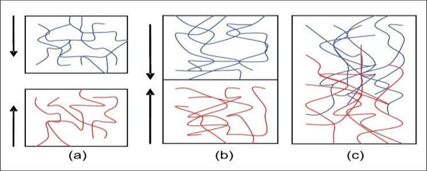
Fig.1: (a) Theory of diffusion -blue polymer layer and red mucus layer before interaction; (b) after interaction (c) The interface becomes diffuse after interaction for a period of time[11]

1.1.4 Adsorption Theory of Bioadhesion:

According to the adsorption theory, the adhesive interaction between the substrate surfaces is caused by intermolecular forces such as hydrogen bonding and VanderWaal's forces.

According to the theory, adherence to tissue is sustained by the combined effect of single or several secondary forces. The kinds of secondary forces are;

  1. Van der Waal’s forces
  2. Hydrogen bonding
  3. Hydrophobic interaction

In bioadhesion, it is not desirable for primary chemical bonds to form between tissue and adhesives used in dentist and surgery. This is due to the possibility of permanent adhesion caused by the strong ionic or covalent bonds [12].

    1. Factors Important to Mucoadhesion
      1. Factors Related to the Polymers:
  • Weight of molecule
  • Active Polymer concentration
  • Polymer Chains flexibility
  • Spatial Conformation
      1. Factors Related to the Environment:
  1. PH
  2. Strength applied
  3. Preliminary contact time
  4. Assortment of the Model Substrate Surface
  5. Swelling

1.2.3. Physiological Variables:

I] Mucin Turnover

II] Sickness States

Vaginal bioadhesive preparations have emerged as a novel controlled-release form of medication for the management of systemic and topical conditions in recent years. The biggest benefit of these dosage

forms is that they can be kept in the vagina for long stretches of time, both during the day and at night, which allows for fewer dosing intervals. [13-17].

    1. VAGINAL MUCOSA

In recent years, there has been a growing investigation into the vagina as a potential site for drug delivery due to its anatomical position and physiological attributes. This approach, when combined with the bioadhesion phenomenon, has produced successful local and systemic drug delivery outcomes.

Treatment for vaginal disorders such as candidiasis, STDs, vaginal dryness, and others has been provided by bioadhesive vaginal drug delivery systems. Also, Studies have indicated that medications can be effectively absorbed into the bloodstream through the vaginal mucosa. Furthermore, this vaginal route has been employed for medication targeting in the uterus (18).

This is especially true for API that have low absorption rates following oral administration, which makes it necessary to take use of alternate drug delivery methods. Currently under investigation are the mucosal pathways of the nasal, ophthalmic, buccal, vaginal, and rectal regions. Among With these routes, vagina appears to be a viable site for medication delivery. The vagina is a tubular, fibromuscular organ that stretches from the uterine cervix to the vaginal vestibule and measures approximately 9 cm in length. [19].

The vagina is made up of four separate layers: stratified squamous epithelium, lamina propria, and muscle layer, and adventitia as shown in the below figure-2. The mucosal layer generates a series of transverse folds known as rugae, which drastically expand its surface. Even though considered a mucosal tissue, the normal vagina does not have glands, and vaginal discharge is a mixture of fluids from a variety of sources. This mucus covering has various critical physiological functions, including drug absorption or activity. It is also worth noting that vaginal characteristics alter with the menstrual cycle, particularly pH and vaginal fluid: the usual pH ranges from 4.5 to 5.5, while vaginal fluid changes greatly in volume, content, and rheological qualities. Lactobacilli are abundant in the healthy vagina and play a crucial function in maintaining vaginal pH and controlling infection by common pathogens [20,21].

Along with age as well as menstrual cycle Vaginal histology and physiology may differ. The thickness of the vaginal epithelium fluctuates with age because the levels of ovarian steroids change. From birth until adolescence, the vaginal epithelium is very thin, but after puberty, the usual thickness of the vaginal epithelium is roughly 200 pm [22].

The vaginal BDD (body dysmorphic disorder) system has been used for both local and systemic drug delivery, particularly in female-related disorders. Traditionally, the vaginal cavity has been utilized to administer locally acting medications such as antibacterial, antifungal, antiprotozoal, antiviral, labor- inducing, spermicidal, prostaglandin, and steroid. Formulations that can prevent the transmission of sexually transmitted diseases (STDs), including AIDS, have made significant progress during the last decade [21].

Fig. 2: Illustration of the vaginal wall structure [20]

    1. ORAL MUCOSA

The oral mucosa is composed of an outermost layer of stratified squamous epithelium (about 40-50 layers thick), a lamina propria followed by the sub mucosa as the innermost layer. The composition of the epithelium varies depending on the site in the oral cavity. The mucosa of the gingival and hard palate are keratinized similar to the epidermis contain neutral lipids like ceramides and acylceramides which are relatively impermeable to water. The mucosa of the soft palate, the sublingual, and the buccal regions, however, are not keratinized contain only small amounts of ceramides.

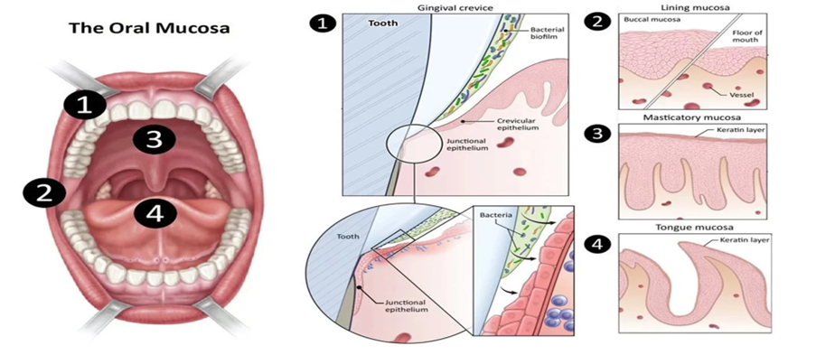
Fig 3: Illustration of the Oral Mucosa [23]

Novel buccal dosage forms: The novel type buccal dosage forms include buccal adhesive tablets, patches, films, semisolids (ointments and gels) and powders.

The oral cavity has a relatively small surface area (approximately 50 cm2). The oral mucosa is composed of stratified squamous epithelium, an outermost layer similar to that found in the rest of the body. It has a mitotically active basal cell layer, advancing through a number of differentiating. intermediate layers to the superficial layers, where cells are shed from the surface of the epithelium The turnover time for the buccal epithelium has been estimated at 5–6 days, a time probably representative of the oral mucosa as a whole. The oral mucosal thickness varies depending on the site: the buccal mucosa measures at 500–800 mm, while the mucosal thickness of the hard and soft palates, the floor of the mouth, the ventral tongue, and the gingivae measure at about 100-200 mm [23-25] .

An ideal polymer for an oral bioadhesive drug delivery system should have the following characteristics

  1. The polymer and its degradation products should be nontoxic and not absorbed through the mucous membrane.
  2. It should not irritate the mucous membrane.
  3. It should preferably form a strong noncovalent bond with the mucin-epithelial cell surfaces.
  4. It should adhere quickly to moist tissue and should possess some site specificity.
  5. It should allow easy incorporation of the drug and offer no hindrance to its release.
  6. The polymer must not decompose during storage or during the shelf-life of the dosage form.
  7. The cost of the polymer should not be high, so that the prepared dosage form remains competitive.
  8. The polymer should allow flexibility and comfort of the dosage form [26-27].

1.4 OCULAR MUCOSA

Ocular mucosal drug delivery systems are designed to enhance the absorption and bioavailability of therapeutics applied to the eye. Traditional ocular formulations, such as eye drops, often suffer from rapid drainage and limited contact time with the mucosal surface. To address these challenges, various innovative approaches are being developed:

      1. Mucoadhesive Formulations: Incorporating natural polymers, gums, and mucilages can improve retention time on the ocular surface. These materials enhance adhesion to the mucosal layer, allowing for sustained drug release.
      2. Hydrogels and Nanoparticles: Hydrogels can provide a controlled release mechanism, while nanoparticles may enhance penetration through ocular barriers. Both strategies can improve the bioavailability of poorly soluble drugs.
      3. In Situ Gelling Systems: These formulations transition from a liquid to a gel upon contact with ocular fluids, providing longer residence time and localized delivery.
      4. Microneedle Arrays: This novel approach involves using tiny needles to penetrate the outer layer of the cornea, allowing for direct drug delivery to the deeper ocular tissues.
      5. Sustained-Release Devices: Implants or inserts that release medication over an extended period can reduce the frequency of administration and improve patient compliance.

Overall, advancements in ocular mucosal drug delivery systems hold great promise for improving therapeutic outcomes in ophthalmic treatments. These innovative strategies aim to enhance drug absorption, reduce side effects, and increase the overall effectiveness of ocular therapies.

Anatomy of the Ocular Mucosa

Conjunctiva: A thin, transparent membrane covering the inner surface of the eyelids (palpebral conjunctiva) and the white part of the eyeball (bulbar conjunctiva). Contains goblet cells that produce mucus, contributing to tear film stability and ocular surface lubrication.

Cornea: The outermost layer of the eye, composed of five layers (epithelium, Bowman's layer, stroma, Descemet's membrane, and endothelium). The epithelium is rich in tight junctions, providing a barrier that regulates the permeability of substances.

Tear Film: A thin layer of fluid covering the ocular surface, composed of three layers like Lipid layer, Aqueous layer, Mucin layer

Sclera: The white, protective outer layer of the eye, providing structural support. Less permeable than the cornea but important in ocular drug delivery [28] .

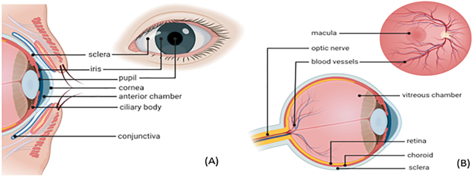
Fig 4: Schematic representation of the (A) anterior ocular anatomy (B) posterior ocular anatomy [28]

    1. RECTAL SYSTEM

Rectal anatomy and scope of drug selection

Anatomically, the rectum forms the distal part of the large intestine. A normal adult human rectum is about 12–15 cm long and deviates in three lateral curves: upper, middle and lower.

It is considered a cylindrical organ that does not contain villi or microvilli on the luminal surface and is mainly associated with water absorption and re-absorption from the gastro intestinal (GI) contents. Average rectal surface area is reported to be around 200–400 cm2 and the pH is 7.2–7.4,

The rectum is structured with columnar epithelial cells along with goblet cells that are responsible for mucous secretion. The mucous forms a layer approximately 100 mm that acts as a protectant to the rectal epithelia and is also a barrier for drug absorption [29-30].

Dosage forms for rectal route

Solid rectal dosage forms, Suppositories, Rectal capsules, tablets or powder for reconstitution, Liquid rectal dosage forms, Semi-solid rectal dosage forms.

    1. NASAL ROUTE

The nasal cavity is one of the most easily accessible and well tolerated drug delivery region in the body. It has the advantages like high surface area (approximately 150 cm2 ) with very rich blood vessels and good nasal cavity volume (approximately 15-20 ml). The mucus layer is regularly renewed every 15-20 min.

The turbinate region is the middle and the most important region of the nasal cavity. This area is the main area for the delivery of different formulations. It is composed of Superior, Middle and Inferior turbinate. The entire turbinate region is richly supplied with blood vessels and lymph vessels. The epithelial cells present in these regions are mainly ciliated/non-ciliated, basal and mucus secreting goblet cells. The ciliated cells are responsible for secretion of mucus, the ciliated and nonciliated cells have microvilli which provide very high surface area required for improved drug absorption. [31-33].

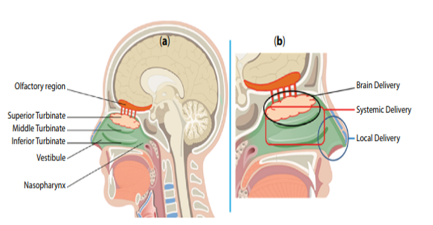
Fig 5: Illustration of the (a) Areas of Nasal Cavity (b) Possible drug delivery through nasal route [32]

Nasal Route for Brain Drug Delivery

Superior Turbinate region has a very rich supply of olfactory nerves and blood vessels. It is reported that the olfactory nerves reaching olfactory mucosa pass through cribriform plate of ethmoid bone. These nerves pass through the cribriform plate along with the arachnoid membrane which is filled with cerebrospinal fluid (CSF). The drugs to be delivered to the Central Nervous System (CNS), if delivered through nasal route, get adhered and lined to the olfactory mucosa of the Superior Turbinate region [34- 35].

Nasal Route for Local and Systemic Drug Delivery

The turbinate region of the nose is sectored as Superior, Middle and Inferior Turbinate. The whole turbinate area is richly supplied with blood and lymph vessels and thus is an important area for systemic drug and vaccine delivery. The cells present in turbinate region are mainly of four types: basal cells, ciliated cells, nonciliated cells, and goblet cells which are present on basement membrane and are richly supplied with blood vessels and thickly covered with mucus and pericillary fluid [36].

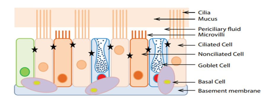
Fig 6: Cells in human nasal epithelium [32]

There are mainly four types of cells present in the nasal epithelium attached to basement membrane: Basal cells, goblet cells, ciliated cells, and nonciliated cells. Out of which goblet cells, ciliated cells and nonciliated cells are attached to each other through tight junction between cells (shown by blue star). The mucus layer is present over the periciliary fluid which acts as lubricating layer and provides sliding movement to mucus layer in ciliary clearance mechanism. These cells and tight junction also contribute to different drug absorption mechanisms [37].

TABLE 1: STUDIES ON NATURAL MUCILAGES AND GUMS [38-74]

Mucilage/ gums

Botanical name

Family

Pharmaceutical Application

Reference

Lipidium

sativum

Lepidum sativum

Cruciferae

Suspending agent, emulsifying agent,

38-39

Cordia gum

Cordia dichotoma

Boraginaecae

Sustained release matrix

40

Mucuna gum

Mucuna flagillepes

Papillionaceae

Microspheres

41

Okra mucilage

Hibiscus esculentus

Malvaceae

Hydrophilic matrix for controlled

42

Acacia

Acacia Senegal

Leguminosae

Osmotic drug delivery

43,44

Bhara gum

Terminalia bellirica

Combretaceae

Sustained release

45,46

Cactus

mucilage

Opuntia ficus-indica

Cactaceae

Gelling agent in sustained drug delivery

47

Guar gum

Cyamopsis

tetraganoloba

Leguminosae

Colon targeted drug delivery,

48-51

Locust bean

gum

Ceratania siliqua

Leguminosae

Controlled release agent

52

Okra gum

Abelmuschus

esculentus

malvaceae

suitability as suspending and disintegrating agent

53

Almond gum

Prunus communis

Rosaceae

emulsifier, thickener, suspending pharmaceutical, adhesive, glazing agent and stabilizer. Gum obtained from Almond as a binder in tablet formulations

54

Neem gum

Azadirachta indica

Meliaceae

Binder

55-57

Gellan gum

Pseudomonas elodea


Disintegrating agent

58

Aloe mucilage

Aloe barbadensis

Miller


A controlled delivery system

59

Albizia gum

Albizia zygia

Leguminosae

Binding agent, suspending agent

60

Karaya gum

Firmiana simplex

Malvaceae

gastric retentive dosage forms improve rate of dissolution of drug solid dispersions. Suspending agent Emulsifying agent. Dental adhesive Sustaining agent. Mucoadhesive

61

Tamarind gum

Tamarindus indica L.

Fabaceae

Drug release Biodegradable carrier for colon specific release

62

Grewia gum

Grewia mollis Juss

Malvaceae

Controlled release dose formulations. Suspending the agent increase. The release of cimetidine from tablets is controlled by the degree of packing and fluidity of the granules, which delay the release. Film making property

63-68

Cashew gum

Anacardium

occidentale L.

Anacardiaceae

As suspending agent

69

Cordia

mucilage

Cordia myxa

Boraginaceae

As enteric resistant and sustained release substantial

70

Fenugreek gum

Trigonella foenum graecum

Leguminosae

As an emulsifier and binding agent, suspending agent, mucoadhesive preparations.

71

Moi gum

Lannea coromandelica

Anacardiacea

As a microencapsulating agent, release rate control material.

72

Moringa gum

Moringa oleifera

Moringaceae

As a mucoadhesive agent, binder, disintegrant

73

Xanthan gum

Xanthomonas campestris

Xanthomonadacae

As a suspending agent, emulsifying agent,

74

1. Grafted Polymers

Definition: Grafted polymers are copolymers in which one or more types of polymer side chains (grafts) are chemically attached to a main polymer backbone, forming a branched structure with different polymers in the backbone and the branches.

TABLE 2: STUDIES ON GRAFTED POLYMERS

SR. NO.

Natural Gum / Mucilage

Source / Botanical Origin

Key Chemical Composition

Important Properties

Common Grafted Polymers (Graft Co-polymers)

Applications of Grafted Polymer

1

Lepidium sativum (Garden cress mucilage)

Seeds of Lepidium sativum

Polysaccharides (D-galactose, L-rhamnose, D-xylose), uronic acids

High viscosity, gel-forming, biodegradable

Polyacrylamide (PAM), Polyacrylic acid (PAA), Poly(N-vinyl-2-pyrrolidone) (PVP), Acrylic acid–Acrylamide copolymers

Hydrogels, drug delivery matrices, thickener

2

Cordia gum (Cordia dichotoma)

Fruits of Cordia dichotoma

L-arabinose, D-galactose, uronic acids

High water absorption, film-forming

Polyacrylamide, Polyacrylic acid, Methyl methacrylate (MMA) grafts

Controlled drug release, bioadhesive films

3

Mucuna gum (Mucuna pruriens)

Seeds of Mucuna pruriens

Galactomannans

High viscosity, stable over pH, biodegradable

Polyacrylamide, Polyacrylic acid, GMA (glycidyl methacrylate) grafts

Hydrogels, tablets, biomedical coatings

4

Okra mucilage (Abelmoschus esculentus)

Okra pods

Rhamnose, galactose, galacturonic acid

Excellent thickening, mucoadhesive, non-toxic

Polyacrylic acid, Acrylamide, Acrylic acid–Acrylamide copolymers

Wound dressing, oral drug delivery, hydrogels

5

Acacia gum (Gum Arabic)

Acacia senegal tree exudate

Arabinogalactan protein, rhamnose, glucuronic acid

Emulsifying, soluble, film-forming

Acrylamide, Acrylic acid, N-vinyl pyrrolidone

Nanocomposites, controlled-release systems

6

Bhara gum (Butea monosperma gum)

Exudate of Butea monosperma tree

Galactose, arabinose, rhamnose

Swelling, gel-forming, biodegradable

Polyacrylamide, Polyacrylic acid, Methyl methacrylate

Drug release matrices, hydrogel films

7

Cactus mucilage (Opuntia species)

Opuntia ficus-indica and others

Arabinose, xylose, galactose, pectic substances

Water retention, flocculating, eco-friendly

Polyacrylic acid, Acrylamide, Acrylic acid–Acrylamide

Water treatment hydrogels, superabsorbents

8

Guar gum

Seeds of Cyamopsis tetragonoloba

Galactomannan

Excellent thickener, stable, high viscosity

Polyacrylamide, Polyacrylic acid, Polyvinyl alcohol (PVA), GMA

Superabsorbents, drilling fluids, drug carriers

9

Locust bean gum (Carob gum)

Seeds of Ceratonia siliqua

Galactomannan (mannose:galactose 4:1)

Synergy with other gums, gel-enhancing

Polyacrylamide, Polyacrylic acid, Acrylamide-acrylate copolymers

Hydrogels, packaging films, biomedical gels

10

Okra Gum (Okra Mucilage)

Pods of Abelmoschus esculentus

Rhamnose, galactose, galacturonic acid

Thickener, mucoadhesive, biodegradable

Acrylic acid (AA), Acrylamide (AAm), Polyacrylic acid (PAA), N-vinyl pyrrolidone (NVP)

Hydrogels, wound dressing, controlled drug release

11

Almond Gum

Exudates of Prunus amygdalus

Arabinogalactans, glucose, uronic acids

Film-forming, emulsifying

Polyacrylamide, Polyacrylic acid, Methyl methacrylate (MMA)

Drug carriers, biodegradable films

12

Neem Gum

Exudate of Azadirachta indica

Arabinose, galactose, fucose

Swellable, stable

Polyacrylamide, Acrylic acid, AAm-AA copolymers

Hydrogel beads, heavy metal adsorption

13

Gellan Gum

Produced by Sphingomonas elodea

Glucose, rhamnose, glucuronic acid

Strong gel former, thermostable

Polyacrylamide, PEG-based grafts, Polyacrylic acid

Tissue engineering, drug release

14

Aloe Mucilage (Aloe vera Gel)

Leaves of Aloe barbadensis

Acetylated polysaccharides (acemannan)

Hydrating, bioactive

Polyacrylamide, Acrylic acid, Chitosan-graft polymers

Wound healing gels, moisturizing hydrogels

15

Albizia Gum

Albizia zygia exudate

Galactose, arabinose, rhamnose

Thickening, bioadhesive

Polyacrylic acid, Polyacrylamide

Tablets, sustained release systems

16

Karaya Gum (Sterculia Gum)

Exudate from Sterculia urens

Galactose, rhamnose, galacturonic acid

Highly swellable, bioadhesive

Polyacrylamide, Acrylic acid, GMA grafts

Superabsorbents, colon-targeted drug delivery

17

Tamarind Gum (Tamarind Kernel Powder)

Seeds of Tamarindus indica

Xyloglucan

Gel-forming, stable viscosity

Acrylic acid, Acrylamide, Polyacrylic acid, MMA

Hydrogels, sustained release tablets

18

Grewia Gum (Grewia Polysaccharide)

Bark of Grewia mollis or Grewia gumifera

Rhamnogalacturonan, arabinose, galactose

Thickener, mucoadhesive

Polyacrylamide, Acrylic acid, GMA

Drug release beads, hydrogels

19

Cashew Gum

Exudate of Anacardium occidentale

Arabinogalactan, glucose, glucuronic acid

Soluble, emulsifying

Polyacrylamide, Polyacrylic acid, MMA

Nanoparticles, drug delivery, coatings

20

Cordia Mucilage (Cordia gum)

Fruits of Cordia myxa / Cordia dichotoma

L-arabinose, D-galactose, uronic acids

Water absorption, film-forming

Polyacrylamide, Polyacrylic acid, MMA

Bioadhesive films, hydrogels

21

Fenugreek Gum (Fenugreek Galactomannan)

Seeds of Trigonella foenum-graecum

Galactomannan (mannose:galactose 1:1)

Thickener, stabilizer

Acrylic acid, Acrylamide, NVP, GMA

Superabsorbents, drug delivery

22

Moi Gum (Moi seed gum)

Seeds of Bassia latifolia (also known as Mahua tree)

Galactomannans (mannose + galactose), uronic acids, minor proteins

High viscosity, strong gel-former, biodegradable

Acrylic acid (AA), Acrylamide (AAm), Polyacrylamide (PAM), Methyl methacrylate (MMA)

Superabsorbent hydrogels, drug controlled-release matrices, water retention polymers

23

Moringa Gum

Exudate from bark of Moringa oleifera

Arabinose, galactose, xylose, rhamnose, uronic acids

Thickener, emulsifier, bioadhesive, stable

Polyacrylamide (PAM), Polyacrylic acid (PAA), N-vinyl pyrrolidone (NVP), Acrylamide-acrylate copolymers

Bioadhesive tablets, hydrogels, flocculants, biomedical films

24

Xanthan Gum

Fermentation gum produced by Xanthomonas campestris

Glucose, mannose, glucuronic acid; cellulose-like backbone

High viscosity at low concentrations, shear thinning, stable across pH & temperature

Polyacrylamide, Polyacrylic acid, GMA (glycidyl methacrylate), MMA, NVP

Drug delivery hydrogels, sustained-release matrices, tissue engineering scaffolds, viscosity modifiers

1.7 APPLICATION OF POLYMERS

  1. Formulations for spermicidal action and combating STD: Condom use has surged as a result of STDs, particularly infections with HIV. By combining a condom with a spermicidal composition has been shown to work synergistically to prevent these illnesses more effectively than just provide contraception [75].
  2. Using gums, hydrogel and polymeric film formulations have been created for topical use [76]
  3. Mucilages are often used in a variety of pharmaceutical preparations as adjuvants, thickening, binding, suspending, dissolving, emulsifying, gelling, stabilizing agents, etc. [77]
  4. Because of their bioactivity, biodegradability, hydrophilicity, and superabsorbent qualities, natural gums have the potential to replace synthetic polymers used in pharmaceutical and biomedical technologies. These gums can also yield hydrogels [78].
  5. Cell proliferation scaffolds are another use for mucilage. Hilary Urena-Saborio [79] reported on recent study that created electro spun nanofibers (ESNFs) using mucilage extracted from chan and linaza beans and mozote stem that is commercially accessible in Costa Rica.
  6. The application of gum in wound healing has significantly increased in recent years. Quince seed mucilage (QSM) has been used to heal burns and wounds in traditional Iranian medicine [80].

1.8 ADVANTAGES

  1. Local availability: Since guar gum and tragacanth are grown in the majority of poor nations, they will be readily available.
  2. Non-toxic and biocompatible: Almost majority of these plant components are carbohydrates in chemical form, made up of units of sugar that repeat. Hence, they are non-poisonous.
  3. Little cost: Using natural sources is constantly less expensive. In addition, manufacturing cost is suggestively less than that of synthetic materials. India and many other emerging nations rely heavily on agriculture.
  4. Biodegradable: Ev ery living thing produces naturally occurring biodegradable polymers. They are a really renewable resource and don't harm people or the environment in any way (e.g., causing skin and eye discomfort).
  5. Processing that is environmentally friendly – Because the manufacturing procedures are straightforward, it is possible to gather gums and mucilage’s from numerous springs in diverse seasons in big numbers [81].
    1. DISADVANTAGES
  1. Due to their carbohydrate composition and usually 10% greater moisture content, gum and mucilage may contain microbial contamination [82-84].
  2. Decreased viscosity during storage – Typically, formulations become more viscous when water comes into contact with gums and mucilages. It has been discovered that there is a decrease in viscosity after storage because gums and mucilages are complex substances.
  3. Batch-to-batch variation: While the production of gums and mucilages is dependent on periodic and environmental conditions, artificial manufacture follows a controlled process with fixed amounts of components.
  4. Unrestrained rate of hydration: The percentage of chemical elements present in a particular substance might change owing to variations in the gathering of natural materials at diverse periods, and variations in area, classes, and weather conditions. Appropriate monographs on the various gums and mucilage on the market must be created [81].
    1. MECHANISM OF ACTION

The mechanism of action of gums and mucilage on the vaginal route primarily involves their ability to provide lubrication, hydration, mucoadhesion, and potentially act as carriers for drug delivery systems.

Hydration and Lubrication: Gums and mucilage possess hydrophilic properties, enabling them to absorb water and form a gel-like consistency. When applied vaginally, they help hydrate the vaginal mucosa and provide lubrication, reducing friction and discomfort. This is particularly beneficial in cases of vaginal dryness [85].

Mucoadhesion: Gums and mucilage can adhere to the mucosal surfaces of the vagina dueto their adhesive properties. This mucoadhesive effect prolongs the residence time of drugs or formulations on the vaginal mucosa, enhancing their therapeutic efficacy [86].

Protective Barrier: Gums and mucilage may form a protective barrier on the vaginal mucosa, protecting it from irritants, pathogens, and other external agents. This function helps maintain the integrity of the vaginal epithelium and reduces the risk of infections or inflammation [87,88].

1.11 MECHANISM OF BIOADHESION

The mucus is secreted by the goblet cells. It is a highly viscous liquid covering the internal tract of the body. Its role is to protect the mucosa against various aggressions. Its exact composition varies, depending on its source and location. However, its major components are mucin, electrolytes, lipids, enzymes, sloughed epithelial cells, bacterial products and water (95%) [89].

Mucin is a glycoprotein in which 160-200 oligo-saccharides side-chains of the glycosylated regions

represent 50-60% of its weight. The mucus gel structure results from the intermolecular association of glycoproteins (mucin) in a polymeric network. The polymer is probably a terminally linked chain with numerous cross-linkings. A large proportion of the glycop~tein is not incorporated in the network, but present as a soluble fraction, enhancing the viscosity of the interstitial fluid.

Besides its protective role against chemical (pH) or enzymatic aggression, the mucus constitutes a diffusion barrier for molecules, not only against drug absorption but also against bioadhesive polymer molecular chain interdiffusion. Diffusion through the mucus layer depends largely on the physico- chemical characteristics of the diffusive molecule: molecule charge, hydration radius, ability to form hydrogen bonds and molecular weight [90].

1. Topical Bioadhesive Drug Delivery Systems (DDS)

Topical DDS aim to enhance drug residence time on the skin or mucosal surfaces (buccal, nasal, ocular, vaginal). Natural polymers provide adhesion, controlled release, and biocompatibility.

Common Natural Bioadhesive Polymers Used

  • Chitosan
  • Pectin
  • Sodium alginate
  • Hyaluronic acid
  • Gelatin
  • Guar gum
  • Xanthan gum

Topical DDS Types & Their Features

a) Bioadhesive Gels

  • Polymers: chitosan, carbopol + natural gums
  • Advantages: easy application, good spreading, sustained drug release
  • Applications: anti-inflammatory drugs, antifungals, wound healing

b) Bioadhesive Creams and Ointments

  • Enhanced retention on skin/mucosa
  • Used for: corticosteroids, antimicrobials, analgesics

c) Bioadhesive Patches/Films

  • Thin films for buccal, nasal, or transdermal delivery
  • Benefits: precise dosing, prolonged adhesion
  • Example: chitosan or alginate-based buccal films for systemic delivery (e.g., nicotine, analgesics)

d) Bioadhesive Nanoparticles

  • Chitosan nanoparticles enhance mucosal uptake
  • Used in: intranasal vaccines, ocular drug delivery

e) Wound Dressings/Hydrogels

  • Polymers: alginate, hyaluronic acid, chitosan
  • Provide moisture, adhesion, antimicrobial properties
  • Applications: chronic wounds, burns

2. Gastrointestinal (GI) Bioadhesive Drug Delivery Systems (DDS)

GI DDS use mucoadhesive materials to prolong drug residence time in stomach or intestines, enhance absorption, and target specific regions.

Natural Bioadhesive Polymers Used

  • Chitosan
  • Pectin
  • Guar gum
  • Alginate
  • Locust bean gum
  • Gum karaya
  • Tragacanth gum

GI DDS Types & Characteristics

a) Mucoadhesive Tablets

  • Polymers form strong adhesion to GI mucosa
  • Prolonged gastric retention
  • Used for: antihypertensives, antidiabetics, antibiotics

b) Mucoadhesive Microspheres/Nanospheres

  • Made from chitosan, alginate
  • Advantages:
    • Large surface area
    • Improved absorption
    • Controlled release
  • Applications: insulin, peptides, antiulcer drugs

c) Floating Bioadhesive Systems

  • Combine buoyancy + mucoadhesion for gastric retention
  • Polymers: chitosan, alginate
  • Used for: drugs absorbed in stomach (e.g., amoxicillin for H. pylori)

d) Colon-Targeted Mucoadhesive Systems

  • Natural polymers degraded by colonic bacteria:
    • pectin
    • guar gum
    • xanthan gum
  • Applications: inflammatory bowel disease (IBD), colon cancer drugs

e) In-situ Gelling Systems

  • Liquid dosage forms that gel upon contact with gastric pH or ions
  • Example: alginate gels in presence of Ca²?
  • Used for: antacids, local antibiotics

f) Bioadhesive Films for GI Delivery

  • Swell and adhere to mucosa
  • Good for peptides and proteins
  • Polymers: chitosan, gelatin

Five Drug Properties Important for Bioadhesive DDS

1. Drug Solubility

  • Drugs should have adequate solubility at the site of application (skin or mucosa).
  • Poorly soluble drugs benefit from sustained release, but extremely insoluble drugs may not diffuse from the bioadhesive matrix.

2. Molecular Weight

  • Low to moderate molecular weight drugs penetrate mucosa more easily.
  • High molecular weight drugs (like peptides) may require permeation enhancers.

3. Partition Coefficient (Lipophilicity)

  • A balanced hydrophilic–lipophilic nature is required.
  • Too hydrophilic → poor permeability.
  • Too lipophilic → slow release from polymer.

4. Stability at the Application Site

  • Drug must remain stable at local physiological conditions:
    • pH of buccal, nasal, vaginal, or GI mucosa
    • Presence of enzymes
  • Unstable drugs degrade before absorption.

5. Dose Requirement

  • Drugs with low dose (milligrams or micrograms) are ideal.
  • High-dose drugs make films/gels too large or uncomfortable for topical/mucosal application.

Effect of Polymer Properties on Mucoadhesive Drug Delivery Systems

The performance of any mucoadhesive drug delivery system depends strongly on the physicochemical properties of the polymer used. These properties influence adhesion strength, swelling, drug release, residence time, and overall therapeutic effect.

1. Molecular Weight of Polymer

  • Higher molecular weight polymers (e.g., chitosan, carbopol) exhibit stronger mucoadhesion because longer polymer chains interpenetrate better with mucin.
  • Very high molecular weight may reduce solubility and spreadability.

Effect:
? Strong adhesion, ? sustained release, ? slower hydration.

2. Degree of Crosslinking

  • Lightly crosslinked polymers swell more and form stronger adhesive bonds.
  • Highly crosslinked polymers have limited swelling → lower mucoadhesion.

Effect:
? Controls drug release rate, ? affects adhesive strength, ? excessive crosslinking reduces mucoadhesion.

3. Polymer Charge (Ionic Character)

  • Cationic polymers (e.g., chitosan) interact strongly with negatively charged mucin → excellent mucoadhesion.
  • Anionic polymers (pectin, alginate) adhere in acidic pH but lose adhesion at higher pH.
  • Non-ionic polymers (HPMC) rely on hydrogen bonding.

Effect:
? Strong bonds with mucin, ? pH-dependent adhesion.

4. Functional Groups

Presence of –COOH, –OH, –NH2 enhances adhesion.

  • Carboxyl groups (in pectin, alginate) form hydrogen bonds.
  • Amino groups (chitosan) form electrostatic bonds.

Effect:
? Increases chemical bonding with mucosal surface.

5. Swelling Ability

  • Swelling allows polymer chains to relax, spread, and interpenetrate mucin.
  • Too much swelling → gel erodes quickly.
  • Too little swelling → weak adhesion.

Effect:
? Improves adhesion and drug release, ? excessive swelling reduces retention.

6. Polymer Concentration

  • Higher concentration increases viscosity → improves adhesion.
  • Very high concentration makes the formulation too thick, reducing spreadability.

Effect:
? Optimal concentration needed for balance between viscosity and adhesion.

7. Polymer Flexibility

  • Flexible polymer chains penetrate mucin more easily.
  • Rigid polymers show poor mucoadhesion.

Effect:
? Improved chain interpenetration → stronger adhesion.

8. Hydrophilicity

  • Hydrophilic polymers absorb water → swell → promote adhesion.
  • Excessive hydrophilicity causes fast dissolution.

Effect:
? Facilitates hydration, swelling, and adhesion.

9. pH Sensitivity

  • Some natural polymers show pH-dependent charge and swelling.
    • Example: Pectin swells more at higher pH (intestinal).
    • Chitosan adheres at acidic pH (stomach).

Effect:
? Determines site-specific adhesion in GI tract.

1. Adsorption Theory

Adsorption theory states that bioadhesion occurs due to the formation of weak chemical bonds between the polymer and the biological surface after initial contact. These include:

  • Hydrogen bonds
  • Van der Waals forces
  • Electrostatic interactions

Key point: Adhesion results from secondary chemical interactions between polymer chains and mucin.

2. Mechanical Theory

Mechanical theory explains that bioadhesion occurs when the polymer penetrates into surface irregularities or pores of the mucosal membrane.
After penetration, the polymer locks into the surface, creating mechanical interlocking.

Key point: Adhesion results from physical interlocking between polymer and mucosa.

3. Cohesive Theory

Cohesive theory states that the internal strength of the polymer itself plays a major role in adhesion.
If the polymer has strong cohesive forces (good viscoelasticity and chain entanglement), it maintains integrity and adheres better.

Key point: Adhesion depends on internal strength and chain entanglement of the polymer.

What kinds of bioadhesive formulations exist (or have existed) on the market or in clinical/research use

• Buccal Films / Tablets / Patches

  • There have been commercial buccal mucoadhesive products — such as buccal tablets/films delivering systemic or local drugs.
  • For example, older mucoadhesive oral pastes/patches like Orabase used natural polymers (blends including pectin, gelatin) as the mucoadhesive component.
  • Other types: combination of natural polymers (e.g., gums, gelatin) with synthetic/semi-synthetic for mucoadhesive buccal films, often in research — e.g. films combining natural protein (gelatin, albumin, casein) with synthetic polymer to deliver drugs such as Glipizide through the buccal mucosa.
  • Bioadesive Gels / Hydrogels (Oral, Mucosal, Topical)
  • Gel or hydrogel formulations using natural polymers (e.g. chitosan, alginate, gums) have been studied for mucoadhesive delivery (oral cavity, nasal, etc.).
  • For example, natural-gum or chitosan-based mucoadhesive gels containing antimicrobial or other drugs have been formulated for local or systemic mucosal delivery.

• Microspheres / Nanoparticles / Particulate Systems with Mucoadhesive Polymers

  • Research formulations have used natural polymer-based microspheres or nanoparticles for mucoadhesion — though as of now these are mostly at lab/preclinical stage rather than wide commercial use.

TABLE 2: APPLICATIONS OF NATURAL POLYMERS IN DRUG DELIVERY [91,92]

Mucilage/ Gum

Dosage form

Disease/ Disorder

Application

Xanthine

Pellets

Kidney stone

Controlled drug delivery

Tamarind

Hydrogel

Dry eye, glaucoma

Ocular mucoadhesive drug delivery

Pectin

Beads, floating beads

Colon cancer, chron’s disease

Collon drug delivery

Luciana seed gum

Tablet

Constipation and diarrhea

Emulsifying agent, suspending agent

Agar

Gelletin agent in suppository

Diarrhea, Laxative, purgative

Suspending agent, emulsifying agent

Gum ghatti

Tablets, Emulsion and suspension

Constipation and diarrhea

Binder, emulsifying and suspending agent

Acacia

Tablet

Throat and stomach inflammation

Binder

Albizia

Tablet

Arthritis and Burn

Binder

Galan

Hydrogel, beads

Correct vision

Opthalmic drug delivery system

Isapgol

Powder, tablet

Diarrhea

Colon drug delivery and gastroretentive drug delivery

Evaluation Parameters for Mucoadhesive Dosage Forms

In?Vitro Evaluation Parameters

  1. Mucoadhesive Strength / Force of Adhesion – Measured using texture analyzer, modified balance, or tensile strength apparatus.
  2. Residence Time –   Determined using USP disintegration apparatus or modified flow?through methods.
  3. Swelling Index –    Percentage weight/volume increase after hydration in simulated biological fluids.
  4. In?Vitro Drug Release Studies – Performed using USP dissolution apparatus (Type I, II, or modified).
  5. Surface pH –        Ensures compatibility with mucosal tissue.
  6. Viscosity / Rheological Studies – Important for gels and liquid formulations.
  7.  Bioadhesion Time – Time for which dosage form remains adhered to mucosal tissue. 
  8. Ex?Vivo Mucoadhesion Study – Using excised animal mucosa (goat, sheep, porcine).
  9. Mechanical Properties – Tensile strength, folding endurance (films/patches).
  10. Drug Content Uniformity – Ensures dose accuracy.

In?Vivo Evaluation Parameters

  1. Residence Time in Biological Site – Evaluated in animals or human volunteers.
  2. Pharmacokinetic Studies – Cmax, Tmax, AUC for bioavailability assessment.
  3. Pharmacodynamic Studies – Therapeutic efficacy.
  4. Irritation / Histopathological Studies – Safety assessment on mucosal tissues.
  5. In?Vivo Imaging Techniques – Gamma scintigraphy, fluorescence imaging.
  6. Clinical Performance – Patient acceptability and compliance.

Different Dosage Forms for Bioadhesive Drug Delivery Systems

Sr. No.

Polymer Used

Model Drug

Dosage Form

Result

Reference

1

Carbopol 934

Metronidazole

Buccal Gel

Prolonged mucosal residence time

Lachman et al., Theory & Practice of Industrial Pharmacy

2

Chitosan

Insulin

Nasal Microspheres

Enhanced bioavailability

Ahuja et al., Int. J. Pharm.

3

HPMC

Propranolol HCl

Buccal Tablet

Sustained drug release

Banker & Rhodes

4

Sodium Alginate

Diclofenac Sodium

Mucoadhesive Gel

Improved retention at site

Remington

5

Polycarbophil

Lidocaine

Buccal Patch

Increased contact time

Smart JD, J. Pharm. Pharmacol.

6

Xanthan Gum

Theophylline

Mucoadhesive Tablet

Controlled drug release

USP Monograph

7

Pectin

Clotrimazole

Vaginal Gel

Improved therapeutic efficacy

Martin’s Physical Pharmacy

8

Guar Gum

Atenolol

Oral Bioadhesive Tablet

Enhanced absorption

Indian Journal of Pharmaceutical Sciences

9

Gelatin

Amoxicillin

Microspheres

Sustained drug delivery

International Journal of Pharmaceutics

10

Carbopol 971P

Fluconazole

Buccal Film

Increased residence time

European Journal of Pharmaceutical Sciences

11

Chitosan–Alginate

Insulin

Nanoparticles

Improved permeation

Journal of Controlled Release

12

HPMC–PVA

Nicotine

Buccal Patch

Reduced dosing frequency

Drug Development and Industrial Pharmacy

13

Tragacanth Gum

Metformin

Mucoadhesive Tablet

Prolonged drug release

Asian Journal of Pharmaceutics

14

Sodium CMC

Ibuprofen

Oral Gel

Improved patient compliance

Pharmaceutical Development and Technology

15

Aloe Vera Gel

Acyclovir

Mucoadhesive Gel

Enhanced healing and retention

Herbal Drug Research Review

CONCLUSION

Natural polymers, gums, and mucilages represent a versatile and promising class of bioadhesive components in drug delivery systems across various routes, including oral, nasal, transdermal, and ocular and vaginal applications. Their biocompatibility, biodegradability, and inherent adhesive properties make them ideal candidates for enhancing drug retention and absorption, ultimately improving therapeutic efficacy and patient compliance. As research continues to elucidate the mechanisms behind their bioadhesive behavior and optimize their formulation strategies, these natural materials are poised to play an increasingly vital role in pharmaceutical innovations.

Moreover, the shift towards sustainable and eco-friendly materials in drug delivery highlights the importance of leveraging these natural resources. The utilization of gums and mucilage as bioadhesive agents in the various route holds significant promise for enhancing drug delivery systems and improving patient healthcare, it is important to acknowledge the challenges associated with formulation development, including stability, consistency and scalability. These challenges necessitate ongoing research and optimization efforts to ensure the reliability and efficacy of different drug delivery systems containing gums and mucilage. Looking ahead continued research endeavours are expected to further refine the use of gums and mucilage in drug delivery. This includes exploring novel natural polymers, improving bioadhesive properties and advancing formulation technologies. ultimately, these efforts have the potential to enhance patient healthcare.

REFERENCES

  1. Kumar L, Verma S, Vaidya B, Gupta V. Bioadhesive Polymers for Targeted Drug Delivery. Nanotechnology-Based Approaches for Targeting and Delivery of Drugs and Genes. Elsevier Inc.; 2017. 322–362 p. http://dx.doi.org/10.1016/B978-0-12-809717-5.00012-9.
  2. Eneko Larrañeta, Ryan F. Donnelly , Bioadhesive Polymers for Drug Delivery; 30 june 2017: chapter-18, 559-561.
  3. Jimenez?Castellanos, M.R., Zia, H., Rhodes, C.T. Mucoadhesive drug delivery systems. Drug Development and Industrial Pharmacy, 1993;19:143–194
  4. Good, W.R. Transder®?Nitro controlled delivery of nitro glycerin via the transdermal route. Drug Development and Industrial Pharmacy, 1983;647–670
  5. Huntsberger, J.R. Mechanisms of adhesion. Journal of Paint Technology, 1967;39:199–211.
  6. Ingrid Henriksen, Keith green; Bioadhesion of hydrated chitosan: an in vitro and in vivo study;1996;231-240.
  7. T. Nagai and Y. Machida, Pharm. Int., 6, 196 (1985).
  8. K. R. Kamath and K. Park, Mucosal adhesive preparations, in Encyclopaedia of Pharmaceutical Technology, Vol. 10 (J. Swarbrick and J. C. Boylan, eds.), Marcel Dekker, New York, 1994, p. 133
  9. Pritchard, W.H. The role of hydrogen bonding in adhesion. Aspects of Adhesion, 1971;6:11–23.
  10. Deraguin, B.V., Smilga, V.P. Adhesion: Fundamentals and Practice. McLaren, London, 1969.
  11. D. Duchene, F. Touchard, and N. A. Peppas, Drug Dev. Ind. Pharm., 14, 283
  12. Ahuja, A., Khar, R.K., Ali, J. Mucoadhesive drug delivery systems. Drug Development and Industrial Pharmacy, 1997;23:489–515
  13. R. Jimenez-Castellanos, H. Zia, and C. T. Rhodes, Drug Dev. Ind. Pharm., 19(1&2), 143
  14. K. R. Kamath and K. Park, Mucosal adhesive preparations, in Encyclopedia of Pharmaceutical Technology, Vol. 10 (J. Swarbrick and J. C. Boylan, eds.), Marcel Dekker, New York, 1994, p. 133
  15. R. B. Gandhi and J. R. Robinson, Indian J. Pharm. Sci., 50, 145
  16. D. Duchene, F. Touchard, and N. A. Peppas, Drug Dev. Ind. Pharm., 14, 283
  17. J. L. Richardson and L. Illum, Adv. Drug. Delivery Rev., 8, 341
  18. Pallavi bassi, Gurpreet kaur ;Innovations in bioadhesive vaginal drug delivery system;2012;1019- 1031
  19. Hussain A, Ahsan F. The vagina as a route for systemic drug delivery. J Control Release 2005;103:301-313
  20. Vermani K, Garg S. The scope and potential of vaginal drug delivery. PSTT 2000; 3:359-64
  21. Linhares, I.M.; Sisti, G.; Minis, E.; de Freitas, G.B.; Moron, A.F.; Witkin, S.S. Contribution of Epithelial Cells to Defense Mechanisms in the Human Vagina. Curr. Infect. Dis. Rep. 2019, 21, 30
  22. Cook MT, Brown MB. Polymeric gels for intravaginal drug delivery. J Control Release. 2018; 270:145–57. http://dx.doi.org/10.1016/j.jconrel.2017.12.004
  23. Shojaei Amir H, Buccal Mucosa As A Route For Systemic Drug Delivery: A Review;J Pharm Pharmaceut Sci (www.ualberta.ca/~csps) 1998;1 (1):15-30
  24. Woodley, J. (2001). Bioadhesion: new possibilities for drug administration? Clinical Pharmacokinetics, 40(2), 77–84.
  25. Gandhi, R. E., & Robinson, J. R. (1988). Bioadhesion in drug delivery. Indian Journal of Pharmaceutical Sciences, 50, 145–152
  26. Ahuja, A., Khar, R. K., & Ali, J. (1997). Mucoadhesive drug delivery systems. Drug Development and Industrial Pharmacy, 23(5), 489– 515
  27. Guo, J. H., & Cooklock, K. M. (1996). The effects of backing materials and multilayered systems on the characteristics of bioadhesive buccal patches. Journal of Pharmacy and Pharmacology, 48, 255– 257.
  28. Ridhdhi S. Dave, Taylor C. Goostrey, Maya Ziolkowska, Sofia Czerny-Holownia, Todd Hoare, Ocular drug delivery to the anterior segment using nanocarriers: A mucoadhesive/mucopenetrative perspective, Journal of Controlled Release, 336, 2021, 71-88.
  29. Batchelor H. 2014. Pediatric formulations. New York, NY: Springer. Chapter 20, Rectal drug delivery; p. 303–310.
  30. Jannin V, Lemagnen G, Gueroult P, Larrouture D, Tuleu C. 2014. Rectal route in the 21st Century to treat children. Adv Drug Deliv Rev. 73:34–49.
  31. D. Marx, G. Williams, and M. Birkhoff, Intranasal drug administration — An attractive delivery route for some drugs, in: Drug Discovery and Development from Molecules to Medicine. O. Vallisuta and S. Olimat (Eds.), pp 299-320, Intech Open Publishing, London, UK, (2015).
  32. Anatomy and physiology of nose, downloaded on July 11, 2019. Web address - www.getbodysmart.com/nasal-cavity/turbinate-nasal-conchae.
  33. S.B. Bhise, A.V. Yadav, A.M. Avachat and R. Malayandi, Bioavailability of intranasal drug delivery system. Asian J. Pharmaceutics, 2, 201-215 (2008)
  34. A.R. Khan, M. Liu, M.W. Khan, and G. Zhai, Progress in brain targeting drug delivery system by nasal route. J Controlled Release. 268, 364-389 (2017).
  35. S. Cunha, M.H. Amaral, J.M Sousa Lobo, and A.C. Silva, Lipid nanoparticles for nasal/intranasal drug delivery. Critical Review in Therapeutic Drug Carrier Systems, 34, 257-282 (2017).
  36. M.A. Selimoglu, E. Selimoglu, and A. Kurt, Nasal mucociliary clearance and nasal and oral pH in patients with insulin-dependent diabetes. Ear Nose Throat J. 78, 585-588 (1999).
  37. K.J. Travaglini and M.A. Krasnow, Profile of an unknown airway cell. Nature, 560, 313-314 (2018).
  38. Patel MM, Chauhan GM, Patel LD. Mucilage of lepidium sativum, Linn (Asario) and ocimumcanum, sims. (Bavchi) as emulgents. Indian J Hosp Pharm, 1987; 24: 200-202
  39. Qudiha Pankaj. Medicinal plant- Chandrasoor, National Scientist Academy of India, Agricultural Department, 1998, pp. 44.
  40. Mukherjee B, Dinda SC, Barik BB. Gum Cordia: A novel matrix forming material for enteric resistant and sustained drug delivery - A Technical Note. AAPS Pharm Sci Tech, 2008, 9(1):330-333
  41. Anthony AA, Nwabunze OJ. Mucuna gum microspheres for oral delivery of glibenclamide: In vitro evaluation. Acta Pharm, 2007; 57: 161–171.
  42. Kalu VD, Odeniyi MA, Jaiyeoba KT. Matrix properties of a new plant gum in controlled drug delivery. Arch Pharm Res, 2007; 30: 884-889
  43. Beneke, C.E., Viljoen, A. M, Hamman, J. H. Polymeric Plant-derived Excipients in Drug Delivery. Molecules. 2009; 14, 2602-2620
  44. Lu, E. X., Jiang. Z. Q., Zhang. Q. Z. A water-insoluble drug monolithic osmotic tablet system utilizing gum Arabic as an osmotic, suspending and expanding agent. J. Cont. Rel. 2003; 92, 375-382.
  45. Shankar, N.B.; Kumar, N.U.; Balakrishna, P.K.; Kumar, R.P. Design and evaluation of controlled release bhara gum microcapsules of famotidine for oral use. Res. J. Pharm. Technol. 2008, 1, 433–437.
  46. Mate, C.J.; Mishra, S. Exploring the potential of moi gum for diverse applications: A Review. J. Polym. Environ. 2020, 28, 1579–1591.
  47. Cárdenas, A., Higuera-Ciapara, I., Goycoolea, F. M. Rheology and aggregation of Cactus (Opuntia ficusindica) mucilage in solution. J PACD. 1997; 152- 159.
  48. Krishnaiah, Y. S. R., Devi, S., Rao, N., Karthikeyan, R.S., Bhaskar, P., Satyanarayana, V. Guar gum as a carrier for colon specific delivery; influence of Metronidazole and Tinidazole on In Vitro release of Albendazole from Guar gum matrix tablets. Pharm. Pharmaceut. Sci. 2001; 4, 235–243.
  49. Krishnaiah, Y.S.R., Latha, K., Rao, N., Karthikeyan, R.S., Bhaskar, P., Satyanarayana, V. Development of colon targeted oral Guar gum matrix tablets of Albendazole for the treatment of helminthiasis. Ind. J. Pharm. Sci. 2003; 65: 378–385
  50. Chourasia, M. K., Jain, S. K. Pharmaceutical approaches to colon targeted drug delivery systems. J. Pharm. Pharm. Sci. 2003; 6, 33–66.
  51. Chourasia, M. K., Jain, S. K., Potential of guar gum microspheres for target specific drug release to colon. J. Drug Target. 2004; 12, 435-442.
  52. Xiaohong, M. G., Michae, J. T., John, N. S. Influence of physiological variables on the in-vitro drug-release behavior of a polysaccharide matrix controlledrelease system. Drug Dev. Ind. Pharm. 2003; 29, 19- 29.
  53. Ashton WA, Jefferies M, Morley RG, Pass G, Phillips G O, Power DMJ. Physical properties and applications of aqueous solutions of Albizia zygia gum. J Sci Food Agric 1975; 26:697–704.
  54. S. Sarojini, Deepthi S. Kunam, R. Manavalan and B. Jayanthi. Effect of natural gum as a binder in the formulation of diclofenac sodium tablets.International journal of pharmaceutical sciences and research (2010);1(3):55-60.
  55. Kokate CK, Purohit AP and Gokhale SB. Pharmacognosy. 11th ed.; Pune; Nirali Prakashan; 1999:498
  56. Gangurde A. B., Malode S. S. and Bhambar R. S. Preliminary Evaluation of Neem Gum as Tablet Binder. Indian Journal of Pharmaceutical Education & Research 2008; 42(4):344-347.
  57. Hindustan Abdul Ahad, Chitta Suresh Kumar, Anil Kumar B. Fabrication and evaluation of Nimesulide Azadirachta indica fruit mucilage based sustained release matrix tablets. International Journal of ChemTech Research 2010; 2(2): 937-941. L.Naga Vamsi Krishna et al. / IJDFR volume 2 Issue 6, Nov.-Dec.2011 66 L.Naga Vamsi Krishna et al. / IJDFR volume 2 Issue 6, Nov.-Dec.2011
  58. Kuo MS, Dell A, Mort AJ. Identification and location of L-glyceratel56, an unusual substituent in gellan gum. Carbohydrate Res 1986; 156: 173-187
  59. Vazquez B, Avila G, Segura D, Escalante B. Antiinflammatory activity of extracts from Aloe vera gel. J Ethnopharmacol1996; 55:69-75.
  60. Ashton WA, Jefferies M, Morley RG, Pass G, Phillips G O, Power DMJ. Physical properties and applications of aqueous solutions of Albizia zygia gum. J Sci Food Agric 1975; 26:697–704.
  61. Kumar, S.; Gupta, S.K. Natural polymers, gums and mucilages as excipients in drug delivery. Polim. Med. 2012, 42, 191–197.
  62. Ashton WA, Jefferies M, Morley RG, Pass G, Phillips G O, Power DMJ. Physical properties and applications of aqueous solutions of Albizia zygia gum. J Sci Food Agric 1975; 26:697–704
  63. Nep, E.; Kaur, N.; Shaboun, S.; Adebisi, A.; Smith, A.; Conway, B.; Asare-Addo, K. Mechanical and release behaviour of theophylline from matrix tablets containing psyllium powder in combination with grewia polysaccharides. Coll. Surf. B Biointerfaces 2020, 188, 110809.
  64. Saha, T.; Masum, Z.; Mondal, S.; Hossain, M.; Jobaer, M.; Shahin, R.; Fahad, T. Application of natural polymers as pharmaceutical excipients. Global J Life Sci. Biol. Res. 2018, 4. [CrossRef]
  65. Oke, E.O.; Adeyi, O.; Adeyi, A.J.; Adekunle, K.F. Modelling of Grewia mollis stem bark gum extraction yield using neuro-fuzzy technique. Proc. Int. J. Eng. Res. Afr. 2018, 34, 70–80. [CrossRef]
  66. Martins, E.; Christiana, I.; Olobayo, K. Effect of Grewia gum on the mechanical properties of Paracetamol tablet formulations. Afr. J. Pharm. Pharmacol. 2008, 2, 001–006.
  67. Nep, E.; Conway, B.R. Polysaccharide gum matrix tablets for oral controlled delivery of cimetidine. J. Pharm. Sci. Res. 2010, 2, 708–716.
  68. Ogaji, I.; Okafor, I.S. Potential of Grewia gum as film coating agent: Some physicochemical properties of coated praziquantel tablets. Int. J. Pharm. Res. 2011, 3, 16–19.
  69. Pontes UR (1971) Determination of HLB of Anacardium gum. Rev Farm BioquimUniv Sao Paulo 2:83–91
  70. Subas CD, Biswajit M (2009) Formulation and evaluation of gum cordia as an enteric resistant and sustained release material in microencapsulated matrix tablet formulations. Int J Pharm Sci Technol 2(1):37– 41.
  71. Subas CD, Biswajit M (2009) Formulation and evaluation of gum cordia as an enteric resistant and sustained release material in microencapsulated matrix tablet formulations. Int J Pharm Sci Technol 2(1):37– 41.
  72. Amelia MA, Rakesh RD, Shilpa NS (2011) Recent investigations of plant based natural gums, mucilages and resins in novel drug delivery systems. Indian J Pharm Educ Res 45(1):86–99.
  73. Mitul TP, Jitendra KP, Umesh MU (2012) Assessment of various pharmaceutical excipients properties of natural Moringa oleifera gum. Int J Pharm Life Sci 3(7):1833–1847.
  74. Dhopeshwarkar,ZatzAL(1993)Evaluation of xanthan gum in the preparation of sustained release matrix tablets. Drug Dev Ind Pharm 19(9):999–1017.
  75. Laboratories Innothera societe anonyme Arcueil. Unit galenical formulation for vaginal use as local contraceptive and/or to combat sexually transmitted diseases and/or HIV. US6664296B1; 2003
  76. S. Ahmad et al.A review on latest innovations in natural gums-based hydrogels: Preparations & applications:2019
  77. Guo J, Skinner GW, Harcum WW, Barnum P.E. Pharmaceutical applications of naturally occurring water-soluble polymers. Pharm Sci Technol Today 1998;1: 254-261.
  78. A.M. Slavutsky, M.A. Bertuzzi, Formulation and characterization of hydrogel based on pectin and brea gum, Int. J. Biol. Macromol. 123 (2019) 784–791
  79. Urena-Saborio H., Alfaro-Viquez E., Esquivel-Alvarado D., Madrigal-Carballo S., Gunasekaran S. Electrospun plant mucilage nanofibers as biocompatible scaffolds for cell proliferation. Int. J. Biol. Macromol. 2018; 115:1218–1224.
  80. Tamri P., Hemmati A., Boroujerdnia M.G. Wound healing properties of quince seed mucilage: In vivo  evaluation  in  rabbit  full-thickness  wound  model. Int.  J.  Surg. 2014;  12:843–847. doi: 10.1016/j.ijsu.2014.06.016
  81. Girish JK, Dhiren SP, Vipul PD and Vineet JC. Gums andmucilages: versatile excipients for pharmaceutical formulations. Asian J Pharm Sci 2009; 4:309-323
  82. Barani M., Torkzadeh-Mahani M., Mirzaei M., Nematollahi M.H. Comprehensive evaluation of gene expression in negative and positive trigger-based targeting niosomes in HEK-293 cell line. Iran. J. Pharm. Res. IJPR. 2020;19:166.
  83. Bilal M., Barani M., Sabir F., Rahdar A., Kyzas G.Z. Nanomaterials for the treatment and diagnosis of Alzheimer’s disease: An overview. NanoImpact. 2020;20:100251.
  84. Das S.S., Bharadwaj P., Bilal M., Barani M., Rahdar A., Taboada P., Bungau S., Kyzas G.Z. Stimuli-responsive  polymeric  nanocarriers  for  drug  delivery,  imaging,  and theragnosis. Polymers. 2020;12:1397. doi: 10.3390/polym12061397.
  85. Nair, R., & Maa, Y. F. (2008). Polymers in controlled drug delivery systems. In Controlled drug delivery (pp. 107-130). Springer, New York, NY.
  86. Smart, J. D. (2005). The basics and underlying mechanisms of mucoadhesion. Advanced Drug Delivery Reviews, 57(11), 1556-1568.
  87. Owen, D. H., & Katz, D. F. (1999). A vaginal fluid simulant. Contraception, 59(2), 91-95.
  88. Robert, C. and Buri, P., Le mucus et son rôle dans L'absorption des médicaments, Pharm, Acts Helv.1986, 61, 210-214.
  89. Gu, J.-M., Robinson, J.R. and Leung, S,-H.S., Binding acrylic polymers to mucin-epithelial surfaces, structure/property relationship, CRC Crit. Rev. Ther. Drug Carrier Sys. 1988, 5(l), 21-67.
  90. D. Mahidhar Reddy, Sk. Razia Begum, K. Jyothirmai, G.V. Dileep Kumar, S. Jayasree, J. Jeevankumar, A.V.S. Gita Samira. "A Novel Review on Natural Polymers Used In Formulation of Pharmaceutical Dosage Forms." International Journal of Pharmacy and Natural Medicines., 2013; 1(1): 71-78.
  91. Rishi Kumar, Rishabha Malviya and Promod Kumar Sharma. "Pharmaceutical Applications and Patents in Natural Polymer Based Drug Delivery System." Advances in Biological Research., 2015; 9(1): 24-32.

Reference

  1. Kumar L, Verma S, Vaidya B, Gupta V. Bioadhesive Polymers for Targeted Drug Delivery. Nanotechnology-Based Approaches for Targeting and Delivery of Drugs and Genes. Elsevier Inc.; 2017. 322–362 p. http://dx.doi.org/10.1016/B978-0-12-809717-5.00012-9.
  2. Eneko Larrañeta, Ryan F. Donnelly , Bioadhesive Polymers for Drug Delivery; 30 june 2017: chapter-18, 559-561.
  3. Jimenez?Castellanos, M.R., Zia, H., Rhodes, C.T. Mucoadhesive drug delivery systems. Drug Development and Industrial Pharmacy, 1993;19:143–194
  4. Good, W.R. Transder®?Nitro controlled delivery of nitro glycerin via the transdermal route. Drug Development and Industrial Pharmacy, 1983;647–670
  5. Huntsberger, J.R. Mechanisms of adhesion. Journal of Paint Technology, 1967;39:199–211.
  6. Ingrid Henriksen, Keith green; Bioadhesion of hydrated chitosan: an in vitro and in vivo study;1996;231-240.
  7. T. Nagai and Y. Machida, Pharm. Int., 6, 196 (1985).
  8. K. R. Kamath and K. Park, Mucosal adhesive preparations, in Encyclopaedia of Pharmaceutical Technology, Vol. 10 (J. Swarbrick and J. C. Boylan, eds.), Marcel Dekker, New York, 1994, p. 133
  9. Pritchard, W.H. The role of hydrogen bonding in adhesion. Aspects of Adhesion, 1971;6:11–23.
  10. Deraguin, B.V., Smilga, V.P. Adhesion: Fundamentals and Practice. McLaren, London, 1969.
  11. D. Duchene, F. Touchard, and N. A. Peppas, Drug Dev. Ind. Pharm., 14, 283
  12. Ahuja, A., Khar, R.K., Ali, J. Mucoadhesive drug delivery systems. Drug Development and Industrial Pharmacy, 1997;23:489–515
  13. R. Jimenez-Castellanos, H. Zia, and C. T. Rhodes, Drug Dev. Ind. Pharm., 19(1&2), 143
  14. K. R. Kamath and K. Park, Mucosal adhesive preparations, in Encyclopedia of Pharmaceutical Technology, Vol. 10 (J. Swarbrick and J. C. Boylan, eds.), Marcel Dekker, New York, 1994, p. 133
  15. R. B. Gandhi and J. R. Robinson, Indian J. Pharm. Sci., 50, 145
  16. D. Duchene, F. Touchard, and N. A. Peppas, Drug Dev. Ind. Pharm., 14, 283
  17. J. L. Richardson and L. Illum, Adv. Drug. Delivery Rev., 8, 341
  18. Pallavi bassi, Gurpreet kaur ;Innovations in bioadhesive vaginal drug delivery system;2012;1019- 1031
  19. Hussain A, Ahsan F. The vagina as a route for systemic drug delivery. J Control Release 2005;103:301-313
  20. Vermani K, Garg S. The scope and potential of vaginal drug delivery. PSTT 2000; 3:359-64
  21. Linhares, I.M.; Sisti, G.; Minis, E.; de Freitas, G.B.; Moron, A.F.; Witkin, S.S. Contribution of Epithelial Cells to Defense Mechanisms in the Human Vagina. Curr. Infect. Dis. Rep. 2019, 21, 30
  22. Cook MT, Brown MB. Polymeric gels for intravaginal drug delivery. J Control Release. 2018; 270:145–57. http://dx.doi.org/10.1016/j.jconrel.2017.12.004
  23. Shojaei Amir H, Buccal Mucosa As A Route For Systemic Drug Delivery: A Review;J Pharm Pharmaceut Sci (www.ualberta.ca/~csps) 1998;1 (1):15-30
  24. Woodley, J. (2001). Bioadhesion: new possibilities for drug administration? Clinical Pharmacokinetics, 40(2), 77–84.
  25. Gandhi, R. E., & Robinson, J. R. (1988). Bioadhesion in drug delivery. Indian Journal of Pharmaceutical Sciences, 50, 145–152
  26. Ahuja, A., Khar, R. K., & Ali, J. (1997). Mucoadhesive drug delivery systems. Drug Development and Industrial Pharmacy, 23(5), 489– 515
  27. Guo, J. H., & Cooklock, K. M. (1996). The effects of backing materials and multilayered systems on the characteristics of bioadhesive buccal patches. Journal of Pharmacy and Pharmacology, 48, 255– 257.
  28. Ridhdhi S. Dave, Taylor C. Goostrey, Maya Ziolkowska, Sofia Czerny-Holownia, Todd Hoare, Ocular drug delivery to the anterior segment using nanocarriers: A mucoadhesive/mucopenetrative perspective, Journal of Controlled Release, 336, 2021, 71-88.
  29. Batchelor H. 2014. Pediatric formulations. New York, NY: Springer. Chapter 20, Rectal drug delivery; p. 303–310.
  30. Jannin V, Lemagnen G, Gueroult P, Larrouture D, Tuleu C. 2014. Rectal route in the 21st Century to treat children. Adv Drug Deliv Rev. 73:34–49.
  31. D. Marx, G. Williams, and M. Birkhoff, Intranasal drug administration — An attractive delivery route for some drugs, in: Drug Discovery and Development from Molecules to Medicine. O. Vallisuta and S. Olimat (Eds.), pp 299-320, Intech Open Publishing, London, UK, (2015).
  32. Anatomy and physiology of nose, downloaded on July 11, 2019. Web address - www.getbodysmart.com/nasal-cavity/turbinate-nasal-conchae.
  33. S.B. Bhise, A.V. Yadav, A.M. Avachat and R. Malayandi, Bioavailability of intranasal drug delivery system. Asian J. Pharmaceutics, 2, 201-215 (2008)
  34. A.R. Khan, M. Liu, M.W. Khan, and G. Zhai, Progress in brain targeting drug delivery system by nasal route. J Controlled Release. 268, 364-389 (2017).
  35. S. Cunha, M.H. Amaral, J.M Sousa Lobo, and A.C. Silva, Lipid nanoparticles for nasal/intranasal drug delivery. Critical Review in Therapeutic Drug Carrier Systems, 34, 257-282 (2017).
  36. M.A. Selimoglu, E. Selimoglu, and A. Kurt, Nasal mucociliary clearance and nasal and oral pH in patients with insulin-dependent diabetes. Ear Nose Throat J. 78, 585-588 (1999).
  37. K.J. Travaglini and M.A. Krasnow, Profile of an unknown airway cell. Nature, 560, 313-314 (2018).
  38. Patel MM, Chauhan GM, Patel LD. Mucilage of lepidium sativum, Linn (Asario) and ocimumcanum, sims. (Bavchi) as emulgents. Indian J Hosp Pharm, 1987; 24: 200-202
  39. Qudiha Pankaj. Medicinal plant- Chandrasoor, National Scientist Academy of India, Agricultural Department, 1998, pp. 44.
  40. Mukherjee B, Dinda SC, Barik BB. Gum Cordia: A novel matrix forming material for enteric resistant and sustained drug delivery - A Technical Note. AAPS Pharm Sci Tech, 2008, 9(1):330-333
  41. Anthony AA, Nwabunze OJ. Mucuna gum microspheres for oral delivery of glibenclamide: In vitro evaluation. Acta Pharm, 2007; 57: 161–171.
  42. Kalu VD, Odeniyi MA, Jaiyeoba KT. Matrix properties of a new plant gum in controlled drug delivery. Arch Pharm Res, 2007; 30: 884-889
  43. Beneke, C.E., Viljoen, A. M, Hamman, J. H. Polymeric Plant-derived Excipients in Drug Delivery. Molecules. 2009; 14, 2602-2620
  44. Lu, E. X., Jiang. Z. Q., Zhang. Q. Z. A water-insoluble drug monolithic osmotic tablet system utilizing gum Arabic as an osmotic, suspending and expanding agent. J. Cont. Rel. 2003; 92, 375-382.
  45. Shankar, N.B.; Kumar, N.U.; Balakrishna, P.K.; Kumar, R.P. Design and evaluation of controlled release bhara gum microcapsules of famotidine for oral use. Res. J. Pharm. Technol. 2008, 1, 433–437.
  46. Mate, C.J.; Mishra, S. Exploring the potential of moi gum for diverse applications: A Review. J. Polym. Environ. 2020, 28, 1579–1591.
  47. Cárdenas, A., Higuera-Ciapara, I., Goycoolea, F. M. Rheology and aggregation of Cactus (Opuntia ficusindica) mucilage in solution. J PACD. 1997; 152- 159.
  48. Krishnaiah, Y. S. R., Devi, S., Rao, N., Karthikeyan, R.S., Bhaskar, P., Satyanarayana, V. Guar gum as a carrier for colon specific delivery; influence of Metronidazole and Tinidazole on In Vitro release of Albendazole from Guar gum matrix tablets. Pharm. Pharmaceut. Sci. 2001; 4, 235–243.
  49. Krishnaiah, Y.S.R., Latha, K., Rao, N., Karthikeyan, R.S., Bhaskar, P., Satyanarayana, V. Development of colon targeted oral Guar gum matrix tablets of Albendazole for the treatment of helminthiasis. Ind. J. Pharm. Sci. 2003; 65: 378–385
  50. Chourasia, M. K., Jain, S. K. Pharmaceutical approaches to colon targeted drug delivery systems. J. Pharm. Pharm. Sci. 2003; 6, 33–66.
  51. Chourasia, M. K., Jain, S. K., Potential of guar gum microspheres for target specific drug release to colon. J. Drug Target. 2004; 12, 435-442.
  52. Xiaohong, M. G., Michae, J. T., John, N. S. Influence of physiological variables on the in-vitro drug-release behavior of a polysaccharide matrix controlledrelease system. Drug Dev. Ind. Pharm. 2003; 29, 19- 29.
  53. Ashton WA, Jefferies M, Morley RG, Pass G, Phillips G O, Power DMJ. Physical properties and applications of aqueous solutions of Albizia zygia gum. J Sci Food Agric 1975; 26:697–704.
  54. S. Sarojini, Deepthi S. Kunam, R. Manavalan and B. Jayanthi. Effect of natural gum as a binder in the formulation of diclofenac sodium tablets.International journal of pharmaceutical sciences and research (2010);1(3):55-60.
  55. Kokate CK, Purohit AP and Gokhale SB. Pharmacognosy. 11th ed.; Pune; Nirali Prakashan; 1999:498
  56. Gangurde A. B., Malode S. S. and Bhambar R. S. Preliminary Evaluation of Neem Gum as Tablet Binder. Indian Journal of Pharmaceutical Education & Research 2008; 42(4):344-347.
  57. Hindustan Abdul Ahad, Chitta Suresh Kumar, Anil Kumar B. Fabrication and evaluation of Nimesulide Azadirachta indica fruit mucilage based sustained release matrix tablets. International Journal of ChemTech Research 2010; 2(2): 937-941. L.Naga Vamsi Krishna et al. / IJDFR volume 2 Issue 6, Nov.-Dec.2011 66 L.Naga Vamsi Krishna et al. / IJDFR volume 2 Issue 6, Nov.-Dec.2011
  58. Kuo MS, Dell A, Mort AJ. Identification and location of L-glyceratel56, an unusual substituent in gellan gum. Carbohydrate Res 1986; 156: 173-187
  59. Vazquez B, Avila G, Segura D, Escalante B. Antiinflammatory activity of extracts from Aloe vera gel. J Ethnopharmacol1996; 55:69-75.
  60. Ashton WA, Jefferies M, Morley RG, Pass G, Phillips G O, Power DMJ. Physical properties and applications of aqueous solutions of Albizia zygia gum. J Sci Food Agric 1975; 26:697–704.
  61. Kumar, S.; Gupta, S.K. Natural polymers, gums and mucilages as excipients in drug delivery. Polim. Med. 2012, 42, 191–197.
  62. Ashton WA, Jefferies M, Morley RG, Pass G, Phillips G O, Power DMJ. Physical properties and applications of aqueous solutions of Albizia zygia gum. J Sci Food Agric 1975; 26:697–704
  63. Nep, E.; Kaur, N.; Shaboun, S.; Adebisi, A.; Smith, A.; Conway, B.; Asare-Addo, K. Mechanical and release behaviour of theophylline from matrix tablets containing psyllium powder in combination with grewia polysaccharides. Coll. Surf. B Biointerfaces 2020, 188, 110809.
  64. Saha, T.; Masum, Z.; Mondal, S.; Hossain, M.; Jobaer, M.; Shahin, R.; Fahad, T. Application of natural polymers as pharmaceutical excipients. Global J Life Sci. Biol. Res. 2018, 4. [CrossRef]
  65. Oke, E.O.; Adeyi, O.; Adeyi, A.J.; Adekunle, K.F. Modelling of Grewia mollis stem bark gum extraction yield using neuro-fuzzy technique. Proc. Int. J. Eng. Res. Afr. 2018, 34, 70–80. [CrossRef]
  66. Martins, E.; Christiana, I.; Olobayo, K. Effect of Grewia gum on the mechanical properties of Paracetamol tablet formulations. Afr. J. Pharm. Pharmacol. 2008, 2, 001–006.
  67. Nep, E.; Conway, B.R. Polysaccharide gum matrix tablets for oral controlled delivery of cimetidine. J. Pharm. Sci. Res. 2010, 2, 708–716.
  68. Ogaji, I.; Okafor, I.S. Potential of Grewia gum as film coating agent: Some physicochemical properties of coated praziquantel tablets. Int. J. Pharm. Res. 2011, 3, 16–19.
  69. Pontes UR (1971) Determination of HLB of Anacardium gum. Rev Farm BioquimUniv Sao Paulo 2:83–91
  70. Subas CD, Biswajit M (2009) Formulation and evaluation of gum cordia as an enteric resistant and sustained release material in microencapsulated matrix tablet formulations. Int J Pharm Sci Technol 2(1):37– 41.
  71. Subas CD, Biswajit M (2009) Formulation and evaluation of gum cordia as an enteric resistant and sustained release material in microencapsulated matrix tablet formulations. Int J Pharm Sci Technol 2(1):37– 41.
  72. Amelia MA, Rakesh RD, Shilpa NS (2011) Recent investigations of plant based natural gums, mucilages and resins in novel drug delivery systems. Indian J Pharm Educ Res 45(1):86–99.
  73. Mitul TP, Jitendra KP, Umesh MU (2012) Assessment of various pharmaceutical excipients properties of natural Moringa oleifera gum. Int J Pharm Life Sci 3(7):1833–1847.
  74. Dhopeshwarkar,ZatzAL(1993)Evaluation of xanthan gum in the preparation of sustained release matrix tablets. Drug Dev Ind Pharm 19(9):999–1017.
  75. Laboratories Innothera societe anonyme Arcueil. Unit galenical formulation for vaginal use as local contraceptive and/or to combat sexually transmitted diseases and/or HIV. US6664296B1; 2003
  76. S. Ahmad et al.A review on latest innovations in natural gums-based hydrogels: Preparations & applications:2019
  77. Guo J, Skinner GW, Harcum WW, Barnum P.E. Pharmaceutical applications of naturally occurring water-soluble polymers. Pharm Sci Technol Today 1998;1: 254-261.
  78. A.M. Slavutsky, M.A. Bertuzzi, Formulation and characterization of hydrogel based on pectin and brea gum, Int. J. Biol. Macromol. 123 (2019) 784–791
  79. Urena-Saborio H., Alfaro-Viquez E., Esquivel-Alvarado D., Madrigal-Carballo S., Gunasekaran S. Electrospun plant mucilage nanofibers as biocompatible scaffolds for cell proliferation. Int. J. Biol. Macromol. 2018; 115:1218–1224.
  80. Tamri P., Hemmati A., Boroujerdnia M.G. Wound healing properties of quince seed mucilage: In vivo  evaluation  in  rabbit  full-thickness  wound  model. Int.  J.  Surg. 2014;  12:843–847. doi: 10.1016/j.ijsu.2014.06.016
  81. Girish JK, Dhiren SP, Vipul PD and Vineet JC. Gums andmucilages: versatile excipients for pharmaceutical formulations. Asian J Pharm Sci 2009; 4:309-323
  82. Barani M., Torkzadeh-Mahani M., Mirzaei M., Nematollahi M.H. Comprehensive evaluation of gene expression in negative and positive trigger-based targeting niosomes in HEK-293 cell line. Iran. J. Pharm. Res. IJPR. 2020;19:166.
  83. Bilal M., Barani M., Sabir F., Rahdar A., Kyzas G.Z. Nanomaterials for the treatment and diagnosis of Alzheimer’s disease: An overview. NanoImpact. 2020;20:100251.
  84. Das S.S., Bharadwaj P., Bilal M., Barani M., Rahdar A., Taboada P., Bungau S., Kyzas G.Z. Stimuli-responsive  polymeric  nanocarriers  for  drug  delivery,  imaging,  and theragnosis. Polymers. 2020;12:1397. doi: 10.3390/polym12061397.
  85. Nair, R., & Maa, Y. F. (2008). Polymers in controlled drug delivery systems. In Controlled drug delivery (pp. 107-130). Springer, New York, NY.
  86. Smart, J. D. (2005). The basics and underlying mechanisms of mucoadhesion. Advanced Drug Delivery Reviews, 57(11), 1556-1568.
  87. Owen, D. H., & Katz, D. F. (1999). A vaginal fluid simulant. Contraception, 59(2), 91-95.
  88. Robert, C. and Buri, P., Le mucus et son rôle dans L'absorption des médicaments, Pharm, Acts Helv.1986, 61, 210-214.
  89. Gu, J.-M., Robinson, J.R. and Leung, S,-H.S., Binding acrylic polymers to mucin-epithelial surfaces, structure/property relationship, CRC Crit. Rev. Ther. Drug Carrier Sys. 1988, 5(l), 21-67.
  90. D. Mahidhar Reddy, Sk. Razia Begum, K. Jyothirmai, G.V. Dileep Kumar, S. Jayasree, J. Jeevankumar, A.V.S. Gita Samira. "A Novel Review on Natural Polymers Used In Formulation of Pharmaceutical Dosage Forms." International Journal of Pharmacy and Natural Medicines., 2013; 1(1): 71-78.
  91. Rishi Kumar, Rishabha Malviya and Promod Kumar Sharma. "Pharmaceutical Applications and Patents in Natural Polymer Based Drug Delivery System." Advances in Biological Research., 2015; 9(1): 24-32.

Photo
Omkar Joshi
Corresponding author

Samarth Institute of Pharmacy, Belhe, Junnar, Maharashtra, India

Photo
Dr. Kuldeep Ramteke
Co-author

Samarth Institute of Pharmacy, Belhe, Junnar, Maharashtra, India

Photo
Dr. Sachin Bhalekar
Co-author

Samarth Institute of Pharmacy, Belhe, Junnar, Maharashtra, India

Photo
Dr. Rahul Lokhande
Co-author

Samarth Institute of Pharmacy, Belhe, Junnar, Maharashtra, India

Photo
Ganesh Lamkhade
Co-author

Samarth Institute of Pharmacy, Belhe, Junnar, Maharashtra, India

Omkar Joshi, Dr. Kuldeep Ramteke, Dr. Sachin Bhalekar, Dr. Rahul Lokhande, Ganesh Lamkhade, Bioadhesive Properties of Natural Polymer in Drug Delivery: An Overview, Int. J. of Pharm. Sci., 2026, Vol 4, Issue 2, 958-985. https://doi.org/10.5281/zenodo.18509299

More related articles
Integrating Morphological Traits, Phytochemistry, ...
Prajakta Devkate, Dr. Kiran Wadkar, Dr. Sandeep Patil, Shankar Jo...
Post Traumatic Stress Disorder and Associated Psyc...
Abhirami Jayaram, K. Vineetha, A. R. Shabaraya, ...
Tectona grandis L. F. (Teak): A Comprehensive Revi...
Rahul Kashid, Vikas Nale, Vaibhav Karande, N. G. Shinde, ...
Medicinal Properties and Phytoconstituents of Alstonia Scholaris: A Comprehensiv...
Dr. Pranay Soni , Dr. Akhilesh Tiwari, Dr. Sunita Minz, Aniket Kumar, Narendra Bankira, ...
A Comprehensive Review On Technology Transfer Of Pharmaceuticals...
Mayuri bhokre, Pratiksha Ashok Udawant, Ige Pradyumna , ...
Prenatal Fear Of Child Birth...
Sneha Bhoir, Saurabh Borde, Saloni Bhoir, Sidra Choudhary, ...
Related Articles
Formulation And Evaluation of Eczema Cream...
Srushti Mitkari, Shraddha Sonune, Swati Tijara, Snehal Khiradkar, Shraddha Chikte, Samiksha Jaiswal,...
Formulation Development & Invitro Evaluation of Anti-Aging Polyherbal Cosmetic F...
T. Mathesvaran, E. Aravind, T. Aruna, S. Dineshkumar, M. Durgadevi, P. Thilagavathy, ...
Microballoons- Novel Approach in Floating Drug Delivery System ...
Gurpreet Kaur, Ashita Pawaiya, Damandeep Kaur, Rajat Kumar Sharma, ...
Integrating Morphological Traits, Phytochemistry, Traditional Uses, and Pharmaco...
Prajakta Devkate, Dr. Kiran Wadkar, Dr. Sandeep Patil, Shankar Joshi, Pranav Ghatte, Pranali Bhagate...
More related articles
Integrating Morphological Traits, Phytochemistry, Traditional Uses, and Pharmaco...
Prajakta Devkate, Dr. Kiran Wadkar, Dr. Sandeep Patil, Shankar Joshi, Pranav Ghatte, Pranali Bhagate...
Tectona grandis L. F. (Teak): A Comprehensive Review of Its Phytochemistry, Phar...
Rahul Kashid, Vikas Nale, Vaibhav Karande, N. G. Shinde, ...