Loknete Dr.J . D. Pawar college of Pharmacy Kalwan-423501, Nashik, Maharashtra, India.
The C-Turmeric ocular wipes are formulated using turmeric and chloramphenicol to provide an effective and convenient method for managing eye infections, particularly conjunctivitis. Turmeric (Curcuma longa), known for its active compound curcumin, possesses antimicrobial, anti-inflammatory, antioxidant, wound-healing properties. With bacterial resistance to conventional antibiotics increasing, there is a growing need for therapies that can enhance treatment outcomes while minimizing the development of resistance. Reliance on synthetic antibiotics alone has become challenging due to the rise of resistant strains. Consequently, combining natural compounds with standard antibiotics has gained attention as a promising way to improve antimicrobial effectiveness. Turmeric, with its wide therapeutic benefits, offers strong potential for such combination approaches, while Chloramphenicol is a broad-spectrum antibiotic frequently used in ophthalmic formulations, is effective against many bacterial eye infections, including conjunctivitis. However, extensive use has led to bacterial resistance, reducing its standalone efficacy. By combining chloramphenicol with curcumin, this study aims to evaluate the synergistic potential of both agents to enhance antibacterial activity and promote faster healing of the ocular surface. The development of these herbal ocular wipes focuses on reducing bacterial growth and preventing infection while offering a simple, non-invasive, and user-friendly alternative to traditional eye drops or ointments. This formulation may be particularly useful in environments with poor hygiene or limited access to sterile application methods. If successful, turmeric-based ocular wipes could offer a significant advancement in the treatment of conjunctivitis and other bacterial eye conditions, providing both preventive and therapeutic benefits while helping address the challenge of antibiotic resistance.
Conjunctivitis, often referred to as "pink eye" is a highly common eye condition that can affect individuals of any age worldwide. It results from inflammation or infection of the conjunctiva the clear, thin tissue that lines the inside of the eyelids and extends over the white part of the eye. When this membrane becomes irritated or infected, the blood vessels within it may become more visible, causing the eye to appear red or pink, which explains the origin of the term "pink eye." In addition to the redness, individuals often experience irritation, itching, a gritty sensation, and an increased sensitivity to light. The inflammation may also lead to excessive tearing or the production of eye discharge, which can range from clear and watery thick, yellowish, or greenish, depending on whether the cause is viral, bacterial, or allergic in nature. Some people also report burning sensations, mild pain, or swelling of the eyelids, making it not only physically uncomfortable but also disruptive to daily routines such as reading, working, or attending school. Due to its high frequency and noticeable symptoms, conjunctivitis is one of the leading reasons individuals seek medical attention for eye-related issues. Interestingly, more than 80% of conjunctivitis cases are diagnosed and managed not by eye specialists but by general healthcare providers, such as pediatricians, family doctors, internists, and nurse practitioners. This highlights the importance of ensuring that all healthcare professionals regardless of their specialty are trained to recognize the signs and symptoms of conjunctivitis, understand its different causes, and provide appropriate treatment or referrals when necessary. (1)
Beyond the personal discomfort it causes, conjunctivitis also possess a substantial burden on healthcare systems worldwide. High patient volumes, school and work absences, and the potential for misdiagnosis or inappropriate antibiotic use all contribute to the overall impact of this seemingly minor condition. Clinically, any inflammation or infection of the conjunctival membrane is categorized as conjunctivitis, and it remains the most common cause of what is broadly referred to as "Red Eye." It’s prevalence and potential to spread especially in group settings like schools, daycare centers, and offices make prompt identification, treatment, and public health education critical in managing its effects and preventing outbreaks. In most cases, a comprehensive medical history combined with a careful eye examination is adequate to identify the cause of conjunctivitis. However, when symptoms are atypical or the condition does not improve with initial treatment, additional diagnostic tools such as bacterial or viral cultures, or polymerase chain reaction (PCR) testing may be required to confirm the diagnosis and assist in selecting appropriate antimicrobial therapy. Viruses and bacteria are the primary causes of conjunctivitis. Viral conjunctivitis most frequently linked to adenoviruses is highly contagious and usually presents with symptoms such as eye redness, irritation, and a watery discharge. It often starts in one eye and can quickly spread to the other. In contrast, bacterial conjunctivitis typically produces a thicker, pus-like discharge and may affect both eyes at the same time. Special consideration should be given to certain high-risk groups. In neonates and sexually active adults, the sudden onset of acute, bilateral, purulent conjunctivitis particularly within three to five days after birth raises strong suspicion for infection with Neisseria gonorrhoeae. This is a medical emergency due to the risk of corneal perforation and permanent vision loss. Prompt diagnosis and aggressive systemic antibiotic therapy, typically with intravenous or intramuscular ceftriaxone, are critical in such cases. Although bacterial conjunctivitis caused by common pathogens such as Staphylococcus aureus, Streptococcus pneumoniae, and Hemophilus influenzae often resolves on its own within one to two weeks, certain infections demand more aggressive treatment. Cases involving Chlamydia trachomatis or Neisseria gonorrhoeae require specific and intensive therapy due to the potential for serious complications and higher transmission risk. In particular, chlamydial conjunctivitis tends to have a more subtle onset and typically requires systemic antibiotics in addition to topical treatment. Chronic or recurrent conjunctivitis is frequently associated with underlying eyelid conditions such as blepharitis or meibomian gland dysfunction. These conditions can lead to chronic inflammation and bacterial colonization of the eyelid margins, contributing to recurrent styes and persistent conjunctival irritation. Effective management involves meticulous eyelid hygiene including warm compresses and lid scrubs as well as the judicious use of topical or systemic antibiotics guided by culture and sensitivity results. Allergic conjunctivitis is a frequently encountered type of eye inflammation marked by significant itching, watery eyes, and swelling of the conjunctiva. It typically affects both eyes and is triggered by allergens such as pollen, dust mites, or animal dander. While seasonal allergic conjunctivitis appears during certain times of the year, perennial forms can persist throughout the year. Management includes avoiding known allergens, applying antihistamine or mast cell stabilizer eye drops, and in some cases, using oral antihistamines for symptom relief. Top of FormBottom of FormIn all forms of conjunctivitis, patient education on hygiene practices including frequent handwashing and avoidance of eye rubbing or sharing personal items is crucial in preventing the spur Topical antihistamines, mast-cell stabilizers, or anti-inflammatory drugs are commonly used to treat conjunctivitis, an inflammation or irritation of the conjunctiva. This condition can be caused by allergic reactions, bacterial or viral infections, or environmental irritants. These medications help to reduce itching, redness, swelling, and other symptoms associated with the condition by either blocking the action of histamines, stabilizing mast cells to prevent the release of inflammatory chemicals, or reducing inflammation directly. The conjunctiva is a thin, clear, and flexible mucous membrane that serves essential functions in eye protection and lubrication. It consists of two primary sections the palpebral and bulbar conjunctiva. The palpebral portion lines the inner surfaces of the eyelids, reducing friction during blinking by providing a smooth interface. In contrast, the bulbar conjunctiva extends over the front surface of the sclera the white part of the eye stopping at the corneal margin without covering the cornea itself. Beneath the conjunctiva are deeper occular structures, including the sclera, episcleral, and uveal tract. The sclera is the tough, fibrous outer layer of the eye that provides structural support and protection. The episcleral lies just above the sclera and contains blood vessels that can become inflamed during certain eye conditions, such as episcleritis. The uveal tract, which consists of the iris, ciliary body, and choroid, is the vascular layer of the eye responsible for supplying nutrients and oxygen to the eye tissues. Together, these structures contribute to maintaining eye health, and any inflammation or infection involving the conjunctiva can impact them, highlighting the importance of prompt and appropriate treatment. Proper diagnosis and tailored management ensure optimal outcomes and help to prevent complications.
Table 1. Key features of viral, bacterial, allergic, chlamydial, and gonococcal conjunctivitis.
|
Aetiology |
History |
Examination |
Investigations |
Management |
|
Viral |
? Red, irritated eyes with tearing and burning ? Watery or sticky discharge, especially on waking ? Recent cold or contact with someone infected (3–5 days ago) ? Can spread from one eye to the other (2–3 days) |
? May develop pseudo membranes (false membranes)
? Swollen lymph nodes in front of the ears are common
|
Take a viral swab for PCR testing make sure adenovirus is checked |
Symptomatic Specialist referral if significant pseudo membrane formation Limit follow-up as highly contagious If ongoing symptoms in 2–3 weeks, re?review with GP/optometrist and refer if concerned |
|
Bacterial |
? Red eye in one or both eyes ? Sudden and fast-developing, or slower with moderate to severe symptoms ? Key signs to watch for:
|
Hyperaemia chemosis |
? Take a bacterial swab of the eye discharge for testing
? If symptoms last longer than usual, test for chlamydia using PCR
|
? Treat symptoms to provide relief
? Use broad-spectrum antibiotic eye drops (like chloramphenicol) following local advice
? Refer contact lens wearers to a specialist to rule out serious eye infections
|
|
Allergic |
? Often have a history of allergies or atopic conditions ? Itching is a key sign ? Redness, watery eyes, and discharge are common ? May experience discomfort or eye pain |
? Swollen eyelids with overall redness and mild puffiness .
? Eye exam may show small bumps (papillae) on the conjunctiva .
|
Nil specific |
? Manage symptoms with antihistamines (eye drops like olopatadine or ketotifen, or oral options) ? Avoid known allergens whenever possible ? Refer to a specialist if symptoms don’t improve with both eye drops and oral antihistamines. |
|
Chlamydial |
? Conjunctivitis that lasts longer than usual ? Usually affects one eye ? May have current or past exposure to a sexually transmitted infection (STI) |
Clear, watery eye discharge |
Chlamydia PCR |
Use oral antibiotics like azithromycin or doxycycline, following local treatment guidelines. |
|
Gonococcal |
Sudden onset of very thick, pus-like eye discharge (hyper-purulent conjunctivitis) |
Large amounts of thick, yellow or greenish eye discharge |
Gram stain |
Give 1g of ceftriaxone by IV or IM right away Also treat for other possible STIs based on clinical judgment |

Figure 1. Breakdown of eye-related bacterial samples by site (A), and number of samples collected from March 2011 to March 2022 (B).[3]

Flowchart 1. Analyzing the symptoms of conjunctivitis
1.2 Symptoms of Pink Eye
A] Redness in one or both eyes
B] Discomfort or irritation in the eye
C] A sandy or gritty sensation
D] Overnight crust-forming eye discharge
E] Excessive tearing
1.3 Causes of Pink Eye (Conjunctivitis)
Viral Conjunctivitis-The most prevalent type of pink eye is viral conjunctivitis, often caused by viruses similar to those that trigger the common cold. It frequently occurs after an upper respiratory infection like a cold or cough. Due to its highly contagious nature, viral conjunctivitis can spread quickly through direct contact with infected eye secretions. Transmission happens by touching the eyes or face, coming into contact with contaminated surfaces, or through airborne droplets released when an infected person sneezes or coughs. Outbreaks are common in places where people gather closely, such as schools, daycare centers, and workplaces.
Bacterial Conjunctivitis-Bacterial conjunctivitis results from infection by various bacteria that invade the conjunctiva the transparent membrane covering the white of the eye and the inner eyelids. In contrast to viral conjunctivitis, bacterial infections usually cause a thicker, colored discharge (yellow, green, or white) that can lead to the eyelids sticking together, particularly upon waking.
Allergic Conjunctivitis- Allergic conjunctivitis happens when the eyes respond to allergens like pollen, dust mites, mold spores, pet dander, or certain chemicals. Unlike viral or bacterial conjunctivitis, it is not contagious. This condition typically affects both eyes at the same time and is marked by severe itching, redness, swelling, and watery discharge. People with other allergic conditions like hay fever, asthma, or eczema are more prone to this type of conjunctivitis. Symptoms often worsen during certain seasons when pollen counts are high or in environments with high allergen exposure.
Eye Irritation from Chemical Splashes- When harsh chemicals such as those found in cleaning products, pool chlorine, or industrial solvents come into contact with the eye, they can cause irritation and inflammation of the conjunctiva (conjunctivitis). Immediate rinsing of the eye with plenty of water is crucial to minimizing damage. In some cases, medical treatment may be necessary. Foreign particles in the eye small particles like dust, sand, or an eyelash can irritate the conjunctiva, causing inflammation.
Risk Factors for Pink Eye (Conjunctivitis) [6]- Conjunctivitis, commonly referred to as pink eye, is a widespread eye condition marked by inflammation and redness of the conjunctiva the clear membrane that lines the eyelids and covers the white portion of the eye. Being aware of the factors that contribute to the development of this condition is essential for effective symptom management and limiting its transmission.
1]Close contact with infected individuals-pink eye caused by viral or bacterial agents is very contagious. Close contact with an infected individual greatly raises the chance of catching the infection. The germs can be transmitted directly through contact with the infected person’s eye fluids or indirectly by touching contaminated surfaces or objects. Places like homes, workplaces, and particularly schools where close interaction among children is common are frequent sites of transmission.
2]Exposure to allergens- Allergic conjunctivitis is a type of pink eye triggered by allergens rather than infection. When substances such as mold spores, dust mites, pet dander, or pollen come into contact with the eyes, they can cause an allergic reaction leading to redness, itching, and swelling. Individuals who suffer from seasonal allergies may notice that their pink eye symptoms worsen during certain times of the year, especially in spring and fall when pollen counts are high. When allergens come into contact with the conjunctiva (the membrane covering the white part of the eye), they can trigger an immune response, leading to inflammation.
3]Environmental Factor -Certain environmental conditions can also increase the risk of pink eye. Exposure to smoke, chemical fumes, or air pollution can irritate the eyes and make them more susceptible to inflammation. Dry environments or prolonged use of contact lenses without proper hygiene can also contribute to the development of conjunctivitis. Air Pollution Sources: Vehicle emissions, industrial pollutants, cigarette smoke, smog. Effect: Irritants in polluted air can inflame the conjunctiva, leading to irritant or allergic conjunctivitis.
4)Poor hygiene practices-Poor hand hygiene and rubbing or touching the eyes with dirty hands can transfer infectious agents to the eyes. Practicing proper hygiene, including regular handwashing and refraining from touching the eyes, is essential to lower the chances of developing pink eye.
AIM AND OBJECTIVE
Aim- To prepare and evaluate C-turmeric ocular wipes.
Objective-
To collect materials from their authentic sources.
To prepare Curcumin solution (0.5 %) and Chloramphenicol solution (0.5%).
To prepare C-Turmeric ocular wipes and their evaluation.
To perform stability study of C-Turmeric ocular wipes.
RATIONALE OF THE STUDY
In the season of monsoon, there is increasing viral and bacterial infection to eye, while in summer season owing to more pollen counts leads to chances of eye infection such as conjunctivitis. Bacteria such as Staphylococcus aureus, Staphylococcus pneumoniae and Haemophiles influenzae causes the conjunctivitis in the seasons.
In ancient time peoples were using the handkerchief soaked in turmeric solution for the treatment of conjunctivitis. Due to repeatedly use of handkerchief, there may be chances of cross contamination thus delaying in the recovery of eye infection and might be transmission of infection person to person results in to epidemics. This traditional knowledge of remedy related to conjunctivitis, prompted us to develop disposable handkerchief or wipes (C-termeric Occular Wipes) impregnated with turmeric and chloramphenicol solution to prevent and cure eye infections. Turmeric is used as antiseptic and anti-inflammatory while chloramphenicol used as broad spectrum antibiotics.
REVIEW OF LITERATURE
|
Sr.no |
Year of Publication |
Author(s) |
Investigation |
|
1 |
1998 |
Morrow GL, Abbott RL. |
In their study presented the diagrams of conjunctivitis. |
|
2 |
2004 |
Chattopadhyay I, |
Turmeric and Curcumin: Biological actions and medicinal applications were studied. Current science. |
|
3 |
2013 |
Azari AA, Barney NP. |
A systematic review of diagnosis and treatment of Conjunctivitis was done. |
|
4 |
2018 |
Dogan U, Agca S. |
To analysed the possible risk factors in the development of seasonal allergic conjunctivitis (SAC) through an evaluation of skin allergy tests and data obtained from questionnaires. |
|
5 |
2019 |
Radomska-Le?niewska DM |
Therapeutic potential of Curcumin in eye diseases was studied. |
|
6 |
2023 |
Manasa PS |
A comprehensive review on various extraction techniques of Curcumin was done. |
|
7 |
2024 Feb |
Gin, C., Crock, C., & Wells, K. |
Overview study of conjunctivitis was done. |
|
8 |
2024 March |
Jasim HZ |
A systematic study of symptoms, and causes were carried out. |
DRUG PROFILE
Turmeric (Curcuma longa L.)

Figure 2. Turmeric
India has a rich and ancient tradition of using plants for medicinal purposes, deeply rooted in systems like Ayurveda, Unani, and Siddha medicine. One of the most valued and widely used plants in these traditions is turmeric (Curcuma longa L.), renowned for Its therapeutic, Botanically, turmeric belongs to the Zingiberaceae family, making it closely related to ginger. It is a perennial herbaceous plant characterized by a short, thick stem, broad oblong leaves, and distinctive rhizomes. These rhizomes the underground parts of the plant are typically ovate, pyriform (pear-shaped) or oblong, often branched, and have a brownish-yellow coloration due to the presence of curcuminoids, the active constituents of turmeric. Traditionally, turmeric has been used as a natural food preservative, and a coloring agent, particularly in South Asian countries such as India, China, and throughout Southeast Asia. Beyond its culinary value, turmeric holds a sacred place in Indian culture and religion. It is often used in rituals and ceremonies, symbolizing purity, prosperity, and protection. In ancient Hindu medicine, turmeric was commonly applied as a topical treatment for sprains, swellings, and injuries, due to its anti-inflammatory and healing properties. Over time, its uses expanded significantly. In modern traditional medicine, turmeric powder is used to treat a variety of conditions including:
In Traditional Chinese Medicine (TCM), turmeric is also used, particularly for ailments related to abdominal pain and digestive issues. The coloring principle in turmeric, primarily curcumin, is not only responsible for its deep yellow color but is also the major compound associated with its anti-inflammatory, antioxidant, and antimicrobial properties. Curcumin has been the subject of numerous scientific studies investigating its potential in preventing and treating chronic diseases. The scientific classification of turmeric, is as follows:
It’s important to note that turmeric exists in more than one form. The wild variety is known as Curcuma aromatica, which is typically more aromatic and less intensely colored than the cultivated, domesticated species, Curcuma longa, which is more commonly used in medicine and cuisine.
Phenolic compounds and their importance (7) -Phenolic compounds are found in many foods, herbs, and dietary supplements. Their structure, characterized by one or more hydroxyl groups bonded to aromatic rings, enables them to scavenge free radicals and influence several biochemical processes in the body. Recently, these compounds have attracted interest for their potential role in preventing and managing chronic illnesses, including cardiovascular and neurodegenerative diseases. Turmeric is a versatile and sacred plant that has served both therapeutic and cultural purposes for centuries. Its widespread use in traditional medicine across Asia, combined with modern scientific validation, underscores its enduring importance as a natural remedy and health-promoting herb.
Chemical composition of turmeric -Turmeric is composed of approximately 6.3% protein, 5.1% fat, 3.5% minerals, 69.4% carbohydrates, and 13.1% moisture. The essential oil, which makes up about 5.8% of turmeric and is extracted through steam distillation of the rhizomes, contains various components: α-phellandrene (1%), sabinene (0.6%), cineol (1%), borneol (0.5%), zingiberene (25%), and a mixture of sesquiterpenes (53%). Curcumin, also known as diferuloylmethane, is the compound responsible for turmeric’s characteristic yellow hue, comprising roughly 3–4% of its content. This includes curcumin I (94%), curcumin II (6%), and curcumin III (0.3%). Additionally, demethoxycurcumin and bisdemethoxycurcumin—structural derivatives of curcumin have also been identified.
Biological Activity of Turmeric and Its Compounds - Turmeric (Curcuma longa) is widely known not only as a culinary spice but also for its diverse range of biological and pharmacological activities. These effects are largely attributed to its active compounds, including curcumin, curcuminoids, essential oils, and various extracts (ethanolic, petroleum ether, aqueous, etc.).
Uses of Turmeric-
A] Wound healing properties
B] Anti-inflammatory
C] Hepatoprotective
D] Antimicrobial
E] Antioxidant
F] Anticancer and antitumor activity.
Chloramphenicol-
Chloramphenicol is a broad-spectrum antibiotic belongs to Amphenicol antibiotics. It has shown bacteriostatic action or bactericidal at high concentrations.
Use: systemic and topical (especially ophthalmic).
Structure of chloramphenicol

Mechanism of Action
PLAN OF WORK
|
Month required |
Work |
|
July - August |
Collection of materials |
|
September - October |
Analysis of material, Preparation of c-turmeric Occular wipes |
|
November - December |
Evaluation and stability study of c-turmeric Occular wipes. |
METHODOLOGY
MATERIAL AND METHOD
The wipes taken here are procured from local market from reputed shops, the turmeric is also taken from grocery store, chloramphenicol from medical store which was already available.
Method of Extraction-Fresh turmeric rhizomes (Curcuma longa) were selected as the raw material for the extraction process. Initially, the rhizomes were thoroughly washed under running tap water to remove any adhering soil or debris. They were then sliced into smaller pieces to facilitate uniform drying. The sliced turmeric was dried in a hot air oven for three hours at a temperature of 105?°C to remove moisture content efficiently. This high temperature drying ensured rapid dehydration while minimizing potential degradation of active compounds. Once the drying process was complete, the dehydrated turmeric rhizomes were ground into a fine and uniform powder using a mechanical grinder. The resulting turmeric powder was then sieved through a mesh to obtain consistent particle size, which enhances the efficiency of solvent extraction. To prevent the powder from absorbing atmospheric moisture and to preserve the chemical stability of its bioactive components, the powdered sample was stored in an airtight container and kept in a refrigerator at 4?°C until further use.
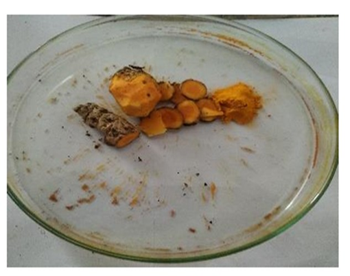
Figure 3. Turmeric
For the extraction of curcuminoids, particularly curcumin, a Soxhlet extraction method was employed. Precisely 10 grams of the refrigerated turmeric powder was weighed using an analytical balance and carefully loaded into a cellulose thimble. The thimble was then placed in the main chamber of the Soxhlet extractor. Acetone, a polar aprotic solvent known for its high affinity for curcuminoids, was used as the extraction solvent. The Soxhlet apparatus was gradually filled with acetone, and the extraction process which was performed at a controlled temperature of 60?°C for a total duration of eight hours. During this period, the acetone repeatedly percolated through the turmeric powder, facilitating the exhaustive extraction of curcumin and other soluble components. Upon completion of the extraction cycle, the acetone extract was collected and subjected to solvent removal using a rotary evaporator. The evaporation was performed under reduced pressure at a temperature of 35?°C to ensure gentle removal of acetone without causing thermal degradation of the curcumin compounds. The resulting concentrated extract, now free of solvent, was collected and further dried in a desiccator to eliminate any residual solvent traces. The final dried extract was weighed to determine yield. To confirm the presence of curcumin in the extract, Thin Layer Chromatography (TLC) analysis was performed. A small quantity of the dried extract was spotted onto a silica gel-coated TLC plate and developed in an appropriate solvent system. The chromatogram was visualized under UV light, and the Rf value of the detected spots was compared with that of a standard curcumin sample to verify its presence.
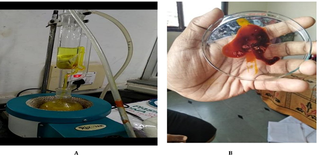
Figure 4. Soxhlet apparatus and extracted curcumin
Preparation of Curcumin Solution (0.5%) Dissolved 0.5 g% of curcumin in15 ml ethanol, stirred it and shaken until fully dissolved. Transferred this solution to measuring cylinder. Added distilled water to make up the final volume of 100ml.Mixed the solution well until it dispersed uniformly. Stored the solution in amber color bottle to protect from the sunlight.
Preparation of Chloramphenicol Solution (0.5%) About 0.5 gm Chloramphenicol dissolved in 15 ml distilled water and transferred it in to volumetric flask and make up final volume of 100 ml. This is the most common concentration for eye drops and is equivalent to 5 mg of Chloramphenicol per 1 milliliter (ml) of solution.
Composition
|
Sr.no |
Ingredients |
Quantity taken |
|
1 |
Chloramphenicol |
50mg/10ml |
|
2 |
Curcumin |
50mg/10ml |
|
3 |
NaCl (Isotonic agent) |
90mg/10ml |
|
4 |
SCMC (Sodium Carboxymethyl Cellulose) |
20mg/10ml |
|
5 |
Glycerine |
Q.S. |
|
6 |
Tissue paper |
An appropriate size |
7.1 Procedure
Selection of Tissue Paper-
We began the process by choosing a commercially available tissue paper that is soft, absorbent, and safe for use around the eyes. The chosen tissue was then studied for its physical properties, such as texture, absorbency, strength, and compatibility with medicinal solutions, to ensure it would be suitable as a base for ocular wipes.
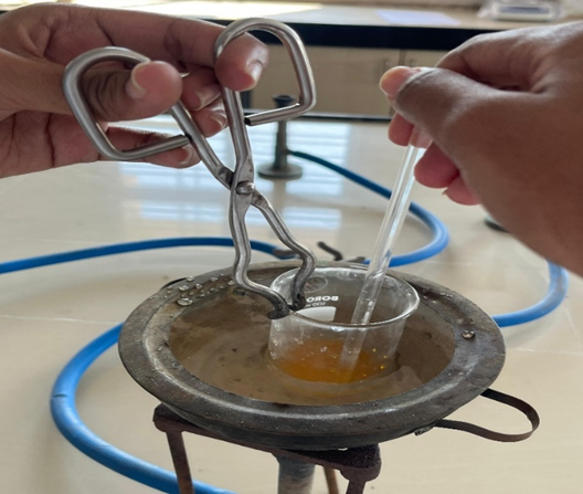
Figure 5.Preparation of Curcumin
Incorporation of Turmeric and chloramphenicol:
Infusion of Tissue Paper-The prepared tissue papers were then soaked in the turmeric and chloramphenicol solution. This allowed the tissues to absorb the medicated mixture uniformly, transforming them into medicated ocular wipes.
Drying Process-After the tissues were fully soaked, they were placed in a hot air oven and dried under controlled conditions. This step ensured the wipes were free from excess moisture, making them easier to handle and extending their shelf life.
Packaging-Once dried, the wipes were carefully packed in sterile, airtight packaging to prevent any contamination. Proper packaging is essential to maintain the hygiene and effectiveness of the ocular wipes until they are used.
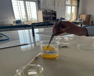
Figure 6. Dipping of tissues in the solutions of Chloramphenicol and Turmeric.
Evaluation Parameters-
1) Draize test- A chemical is applied to a rabbit's skin or eye in the contentious Draize test to determine whether it irritates or damages the animal. For the protection of the eyes or skin, it entails securing the animal in a restraint, administering the drug, and then monitoring it for symptoms of irritation, opacity, and inflammation over a few days. Animal welfare concerns and doubts over the test's ability to accurately predict human reactions make it relevant. [12
2) Physical Stability
3) Packaging Compatibility
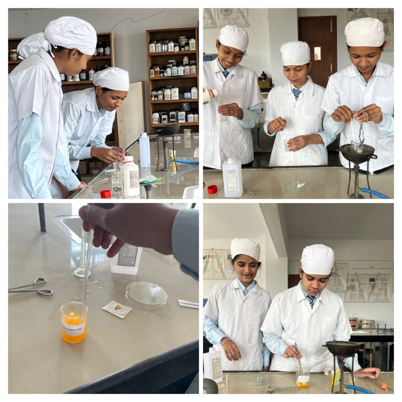
Photos : Preparation C-Turmeric occular wipes were carried out in Pharmacognosy Laboratory.
RESULT AND DISCUSSION
The C-Turmeric occular wipes offer a practical and gentle way to help , manage and treat conductivities. By keeping the eye area clean and reducing the build-up of irritants and microbes, these wipes can support the healing process and improve overall comfort. Chloramphenicol plays a helpful role in broad-spectrum antibiotics by reducing the presence of harmful microorganisms on the occular surface, allowing the treatment to work more effectively.
CONCLUSION
The newly formulated ocular wipes are effective against conjunctivitis and other eye infections. It is safe, eye-friendly, biodegradable, eco-friendly and easily affordable to common man. This product may be beneficial for both preventative and therapeutic purposes. It helps to prevent cross infection of conjunctivitis ultimately stop their epidemics in society.
REFERNCES


Priyanka Aher, Sakshi Aher, Gauri Ahirrao, Gauri Bhalerao, Bhagyashri Bhamare, Santosh Surana, Design and evaluation of C-Turmeric Occular Wipes, Int. J. of Pharm. Sci., 2026, Vol 4, Issue 4, 1980-1993, https://doi.org/10.5281/zenodo.19550199
 10.5281/zenodo.19550199
