
We use cookies to ensure our website works properly and to personalise your experience. Cookies policy
Department of Pharmaceutics, Oriental College of Pharmacy, Navi Mumbai, Maharashtra, India.
Electrospun nanofibers have attracted growing interest due to their high surface area, adjustable structure, and diverse material options, making them suitable for applications in biomedicine, energy, and environmental fields. This review outlines recent progress in electrospinning techniques, emphasizing their use in smart textiles, drug delivery, tissue engineering, and environmental cleanup. It also explores the impact of polymer blends, surface modifications, and functional materials on nanofiber performance. Challenges in scaling up production and integrating nanofibers into commercial products are addressed. Additionally, the review highlights current trends, such as sustainable electrospinning, multifunctional fiber development, and applications in flexible electronics and advanced filtration. Finally, it offers insights into future directions, focusing on innovation and cross-disciplinary research to expand the potential of electrospun nanofibers in various sectors.
One new and exciting paradigm in biomedical research is nanotechnology, or the utilisation of nanomaterials for biomedical purposes. In order to accomplish the intended therapeutic efficacy, nanomaterials with remarkable physiochemical characteristics, biocompatibility, and low biological toxicity can sense their local biological surroundings and start cellular level reprogramming. Current applications for diagnosis, imaging, and therapy include a variety of zero-dimensional (quantum dots, carbon dots, graphene quantum dots), one-dimensional (nanorods, nanowires, nanotubes, nanofibers), and two-dimensional (graphene oxide, transition metal dichalcogenides, transition metal oxide etc.) nanomaterials and nanosized particles. Nanofibers are nanostructures that resemble fibers and usually have two nanoscale dimensions. Nanofibers are easily functionalised with biological molecules and have a high surface area-to-volume ratio with adjustable porosity. Nanofibers are a strong and appealing option for many cutting-edge biomedical applications because they can be made from a wide range of materials, including natural and synthetic polymers, inorganic nanomaterials, composites, and biomolecules as medications Because of their exceptional qualities, nanofibers are the perfect nanomaterial for applications in biomedical engineering, healthcare, water and environmental treatment, and energy generation and storage [1].
Nanocatalysis, tissue scaffolds, protective apparel, filtration, and nano-electronics are among the uses for nanofibers. Phase separation and template synthesis are two other techniques for creating nanofibers, but few, if any, can compare to electrospinning in terms of adaptability, flexibility, and ease of fiber manufacturing [2] Because of its easy handling, low solution consumption, controlled diameter, and affordability, electrospinning is the primary method of choice for producing nanofibers on a wide scale. Numerous biological uses for electrospun nanofibers exist, including wound dressings, medication and gene delivery devices, sensors, and catalysts. The use of electrospun nanofibers in therapeutics (drug/gene) delivery is the main topic of this review. For effective loading, release, and accumulation of the therapies into the target region, a variety of therapeutic delivery systems have been studied. But when it comes to choosing various substances, medications, and genes (DNA, RNA, etc.) for therapeutic uses, electrospinning offers a great deal of freedom [1]. This review aims to provide a comprehensive exploration of the latest advancements in electrospun nanofibers, focusing on fabrication techniques, material innovations, and diverse applications. In particular, this review includes a detailed discussion on the use of electrospun nanofibers in smart textiles, filtration systems, tissue engineering, and water purification. Additionally, it covers the challenges of scaling production, improving material properties through surface modifications, and integrating electrospun nanofibers into commercial products. By highlighting both current trends and future directions, this review aims to offer valuable insights into the potential and ongoing developments in the field of electrospun nanofibers.
Electrospinning Technology
Electrospinning is a voltage-driven technique in which a liquid droplet is electrified to create a jet, which is then stretched and elongated to create fibers. The main setup for electrospinning, includes a spinneret (syringe needle) linked to a high-voltage (5 to 50 kV) source make up the basic electrospinning setup. Surface tension causes the liquid to protrude from the spinneret during electrospinning, creating a pendant droplet. A charged jet is emitted from an electrified droplet when it is deformed into a conical shape called the Taylor cone by electrostatic repulsion between surface charges of the same sign. A charged solution jet is released from the tip of the Taylor cone as soon as the electric field reaches a critical value, which is the point at which the repulsive electric forces surpass the surface tension forces. As the jet is charged, its path can be regulated by an electric field. A charged polymer fibre is left behind when the solvent evaporates as the jet passes through the air [3,4]. A number of variables affect the electrospinning procedure. Researchers investigated how the electric current and charge density of the polymer jet during electrospinning were affected by the controlling factors, voltage, solution flow rate, concentration, molecular weight, distance, and solvent grade. It has been discovered that the viscosity of the solution affects the fiber diameter, which is correlated with the molecular weight and concentration of the polymer. Larger-diameter fiber production has been associated with increasing the viscosity of the solution [5,6]. The influence on fiber diameter and voltage are also related to solution conductivity; thin fibres produce high solution conductivity [4, 7].
Basics of Electrospinning
Electrospinning is widely used in labs and enterprises worldwide, and it has been recognized as an old nanotechnology that has been rediscovered for contemporary needs. Appropriate control of the electrospinning process is essential for producing nanofiber-based textiles in large quantities from electrospun nanofibers. Although electrospinning is a fairly straightforward and economical technique, it is a complex procedure that heavily relies on a number of key technical and processing aspects [8].
To control the development of electrospun fibers and prevent defects like beaded fibers or sprayed droplets, an optimal concentration of polymer solution is required. It is noted that the creation of sprayed droplets occurs because the polymer chain entanglements are insufficient to stabilize the Coulombic repulsion within the ejected jet when the concentration of the optimized spinning solution is below it. In contrast, the typical spin solution feed rate used in the electrospinning process is between 0.5 and 2.0 mL/h. In order to prevent droplet formation, the feed rate and voltage are really worked together to create a consistent Taylor cone shape.
Since a larger volume of solution drawn will just take longer for the solvent to dry selected a lower feed rate for spinning for creating polysulfone electrospun fibers. If not, the fibers may fuse when they come into contact with the collector due to the residual solvents, which would result in an unsatisfactory fibrous structure [8].
Standard Electrospinning Setup
Generally, a typical electrospinning setup consists of three primary components: a high-voltage power supply to charge polymer solution, a syringe with pumps from which the polymer solution is fed through a capillary connected to a syringe filled with the polymer solution, and a grounded collector where nanofibers are deposited. In this process a syringe pump with a constant rate is used to extract the solution (particulate suspension, polymeric or molten solution) from the needle tip of a syringe containing the solution. The main differences between the upward and the downward electrospinning (DWEs) setups are the orientation of fibers and the number of beads. As opposed to the descending method's unpredictable direction, the upward electrospinning (UWEs) setup's fibers gathering will follow a consistent pattern. However, compared to the descending process, there will be less bead creation in the upward setup [9]. While the upward setup has been chosen for the industrial production scale because mass production of the nanofibers using the traditional single needle is difficult and challenging, the downward setup of electrospinning is the most advantageous for small laboratory scale use due to the straightforward optimization and operational monitoring. Conventional fibers spinning methods (dry, wet, melt, or gel spinning) can be used to create polymeric fibers with a diameter of a few micrometers. In dry spinning, the polymer will be dissolved using a specific solvent or a combination of solvents. Because of its low boiling point, this solvent will evaporate, leaving the polymer in fibers form. Due to the additional equipment needed to create the combination and the solvent recovery procedure, dry spinning is more costly than melt spinning. On the other side, the wet spinning method uses the same dry spinning concept. Here, the sole distinction is that the solvent is leached in a coagulation bath as opposed to being evaporated. In addition, wet spinning is more difficult than dry spinning because it requires the right coagulant to precipitate the solvent in a coagulating bath. Furthermore, the gel spinning process has been covered in numerous publications [10]. Gel spinning, which involves spinning a gel-like polymer and cooling it to harden, can produce fibers with greater strength. Fig. 1 and 2 shows a schematic view of the conventional horizontal and vertical electrospinning setup [11].
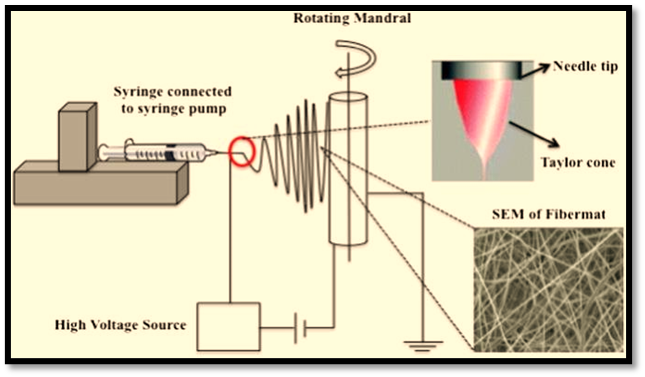
Fig. 1 Schematic view of the conventional horizontal electrospinning setup.
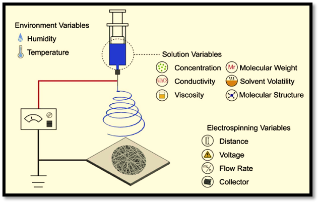
Fig. 2 schematic view of the conventional vertical electrospinning setup.
Production Methods of Nanofibers Mats
Production Methods of Nanofibers Mats Interest in electrospinning and nanofiber mats is increasing year by year, and various methods for fabricating nanofibers are described in the literature, including melt electrospinning, coaxial electrospinning, multi-jet electrospinning, needleless electrospinning, bubble electrospinning, electro-blowing, cylindrical porous hollow tube electrospinning, self-bundling electrospinning, and charge injection electrospinning. Among all of these techniques, electrospinning is the most widely used and represents the simplest method for producing nanofibers 12-13.
Types of electrospinning
Generally speaking, the kind of ES technique used determines how flexible electrospun NFs are made. In this instance, emulsion electrospinning, melt electrospinning, gas jet electrospinning, blend electrospinning, coaxial electrospinning and other frequent forms of ES are reported. [14].
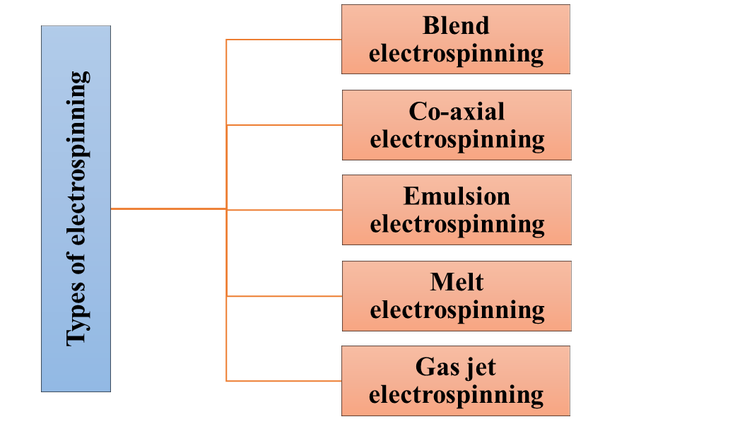
Fig. 3 Types of electrospinning
1.Blend electrospinning: This commonly used and traditional ES method is called blend electrospinning. Its foundation is the mixture of medication or bioactive chemicals that are dissolved or distributed in the ES polymeric solution before being spun to create fibres (Fig. 4). These NFs are manufactured so that the medicine or bioactive ingredient is evenly dispersed throughout them. However, the use of a solvent during the blend, dispersion, or solution preparation process may result in bioactive denaturation, which causes the molecules (drugs/bioactive) to burst out.

Fig. 4 Blend electrospinning
2. Co-axial electrospinning: This method gets around the problems with mix electrospinning. Its main components are two nozzles that are connected to a high voltage and two dissimilar solutions (drugs, bioactive molecules, or natural/synthetic polymers) that are poured into each nozzle (Fig. 5). In a nutshell, the polymeric solution is poured into the outer jet (co-spun) and the bioactive or medication is poured into the inner get of ES. The prolonged release of actives is thus provided by the active that is trapped in the core of NFs and polymeric solutions, which cover the core (shell) [15, 16]. Consequently, the denaturation of bioactive or proteins is prevented or mitigated by this ES approach.
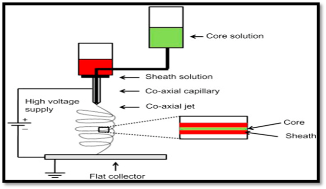
Fig. 5 Co-axial electrospinning
3. Emulsion electrospinning Blend electrospinning is comparable to emulsion ES.
It usually depends on the makeup of two immiscible solvent systems, such as an oil/water emulsion. The core and shell of spinning NFs are visible in the resultant emulsion. To put it briefly, the active and surfactant combine to form the W/O emulsion, which is then combined with appropriate natural or synthetic polymers and spun to create fibers (Fig. 6). In the axial area, it then transforms into the droplet enrichment and viscous gradient. As a result, the generated NFs provide a persistent release of active and overcome the initial burst release [17,18].

Fig. 6 Emulsion electrospinning
4. Melt electrospinning: The traditional ES method is called melt electrospinning. In short, melt electrospinning is the process of melting a polymer (natural or synthetic), medication, and additional excipients into a capillary and then extruding it (Fig. 7).

Fig. 7 Melt electrospinning
5. Gas jet electrospinning: In general, the gas jet ES type offers a respectable enhancement above traditional melt ES. It gets around melt ES's temperature-based restrictions.
In short, the gas jet assembly is coupled to the ES configuration (Fig. 8). Here, the NFs close to the nozzle get enough heat from the heated gas covering the coaxial jet to cause a delay in their solidification [19, 20].
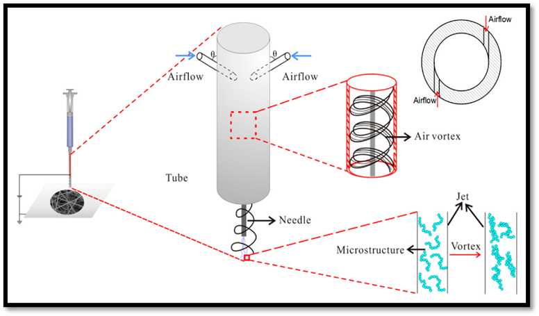
Fig. 8 Gas jet electrospinning
MEDICAL APPLICATIONS OF ELECTROSPINNING
Electrospun Nanofibers in Wound Dressing
Electrospun nanofibers exhibit exceptional qualities in accelerating the healing of wounds. Their morphology closely resembles the extracellular matrix (ECM) structure of the human body, which promotes cell adhesion, growth, and proliferation. Simultaneously, the high permeability and absorption rate helps keep the healing environment moist by absorbing the exudate that forms on the wound surface. Furthermore, loading and delivering bioactive components like medications and growth hormones is made easier by the increased surface area [21, 22]. As a result, electrospun nanofiber materials are thought to be the best option for treating wounds.
Currently, drug carriers can be successfully prepared by electrospinning hundreds of different polymers. Numerous studies on electrospinning wound dressings have made extensive use of both natural and manmade polymers.
Electrospun fibers are based on polymers, which also greatly influence their elongation behavior, wettability, cell adherence, and release of integrated active substances [23]. Synthetic and natural polymers are two types of polymers that can be used separately or in combination. Synthetic polymers include electrospun polyethylene oxide (PEO), polycaprolactone (PCL), and poly(lactic-co-glycolic acid) (PLGA). Polyvinyl alcohol (PVA) is often easy to electrospin, but requires organic solvents to dissolve. As an alternative, so-called "generally recognised as safe" (GRAS) solvents are utilized, which facilitate easier commercialization of the fibers systems later on. Environmentally friendly, safe to handle, and non-toxic solvents are referred to as GRAS solvents. Water, acetone, ethanol (EtOH), and dimethyl sulfoxide (DMSO) are examples of solvents that are rarely or never harmful to human health, whereas trifluoroethanol (TFE), dimethylformamide (DMF), and chloroform are hazardous. Although they require fewer harsh solvents, natural polymers including collagen, elastin, chitosan, gelatin, and alginates are more challenging to electrospin and occasionally can only be done with synthetic co-polymers.
Antibacterial Nanofibers for Wound Dressing
The use of antibacterial materials is crucial because bacterial infections are one of the main causes of chronic infections and they fester in wounds that already exist at a high rate. Antibacterial nanofibers enable the effective incorporation of antibacterial chemicals due to their enormous surface area. Nanotechnology has developed at an accelerated rate in recent years. Additionally, the fields of study under nanotechnology are growing at an exponential rate. Nanomedicine is one of the scientific fields covered by this revolution, and in recent decades, it has demonstrated a strong potential to grow into a significant area of study. Human health has significantly improved as a result of this field's research [24.25]. Nanofibers have been created using a variety of methods, including electrospinning, melt spinning, chemical vapour deposition, sintering, and solution spinning.
It has been found that the electrospinning approach is the most economical way to create continuous nanofibers from a variety of polymers or chemicals. The electrospun nanofibers have a high porosity, a large specific area, and are highly desirable for use in wound dressing, tissue engineering, and regenerative medicine.
BIOLOGICAL PRODUCTS IN NANOFIBERS
Bacteria
Probiotics are often bacteria that have been integrated into electrospun nanofibers. Probiotics are live microorganisms that, when given in sufficient quantities, can help the host's health. A barrier to their broader use is their poor survivability and great sensitivity to environmental influences. Therefore, novel probiotic delivery methods are required [26]. Probiotics have been included using various electrospun nanofibers, which have preserved their vitality and have made arrangements for their delivery. In addition to being utilized therapeutically, non-probiotic bacteria have also been integrated into nanofibers and utilized as biosensors, biocatalysts, and in food and agriculture. The microorganisms that have been integrated into various nanofibers and their uses are listed in Table 1. The most prevalent genus of probiotic bacteria that have been integrated into nanofibers is Lactobacillus. The typical vaginal flora contains Lactobacillus acidophilus, which can stop harmful microbes from colonizing and growing. The viability of L. acidophilus has been increased for 78.6 – 90% when incorporated into agriculture waste-based nanofibers combined with PVA; i.e. soluble dietary fiber from okara (soybean solid waste), oil-palm trunks, and oil palm fronds [27]. Several probiotic bacterican produce antibacterial substances that are active against pathogenic bacteria. Plantaricin 423 is a bacteriocin produced by Lb. plantarum. Incorporation of bacteriocin producing cells into electrospun PEO nanofibers represents a drug-delivery system for bacteriocins and probiotic lactic acid bacteria [28]. The formation of biofilms can improve the stability of probiotics and other microbes. Microorganisms can adopt a multicellular behavior and extend their lifespan in harsh environments by forming biofilms, which are an alternate lifestyle. Microorganisms in biofilms are shielded by the extracellular matrix, which is made up of proteins and polysaccharides. Single-species Lb. plantarum biofilms have been scaffolded using electrospun cellulose acetate nanofibers. The Lb. plantarum outlived the planktonic Lb. plantarum because the probiotic biofilms on these nanofibers were robust to the gastrointestinal environment. Compared to just 0.77% of planktonic cells, 27.40% of biofilm cells survived after three weeks of storage [29].
Table 1 Bacteria in nanofibers
|
Bacteria |
Polymer |
Purpose |
|
Lb. plantarum |
Polyethylene oxide |
Delivery system for probiotics |
|
Lb. plantarum |
Cellulose acetate |
Food technology (starter culture) |
|
Lb. acidophilus |
Soluble dietary fiber, oil-palm trunk + oil-palm fronds, with polyvinyl alcohol |
Encapsulation of probiotic bacteria |
|
Spirulina sp. |
Polyethylene oxide |
Tissue engineering scaffold |
|
P. agglomerans |
Polyvinyl alcohol |
Agriculture (seed coating) |
|
G. oxydans |
Polycaprolactone/polyethyleneimine |
Biosensing (glucose) |
|
S. epidermidis |
Carboxymethyl cellulose/Polyethylene oxide |
Delivery system for probiotics (diabetic foot) |
|
Bacillus sp |
Polyethylene oxide/chitosan + polyethylene oxide |
Delivery system for probiotics (periodontal disease) |
Fungi
The incorporation of fungi into nanofibers has focused on exploiting their biocatalytic properties rather than their biotherapeutic properties, as shown in Table 2. Candida tropicalis is one of the most virulent fungi in the Candida genus and can cause several human diseases. However, their degradation of a wide range of environmental pollutants has increased their use for purification of waste waters. Water-soluble nanofibers cannot be used in aqueous environments, such as wastewaters, and therefore coaxial electrospinning has been used to form nanofibers with a water-soluble core and a hydrophobic shell. C. tropicalis was successfully encapsulated within nanofibers consisting of a PVP core and a shell of polyvinylidene fluoride and hexafluoropropylene using coaxial electrospinning. The fungus has introduced degraded phenols and ethanol fermented with olive wastewater, and also shows toxicity to Escherichia coli [30]. Other types of fungi that have been proposed for water purification include Kluyveromyces lactis and Saccharomyces cerevisiae. When these two heat-inactivated fungi were immobilized in PVA-cellulose acetate hybrid nanofibers, they provided improved removal of aflatoxin B2 from contaminated water by binding it to their surface. The resulting water was found to have low cytotoxicity towards human fibroblast cells. Trichoderma viride spores were incorporated into electrospun nanofibers composed of PEO, polyacrylamide, and chitosan, and their viability was preserved. The mushroom dispute inhibited the growth of plant pathogen (that is, Fusalium, Alternalia) [31].
Table 2 Fungi in nanofibers
|
Fungus |
Polymer |
Purpose |
|
C. tropicalis |
Polyvinyl pyrrolidone/ polyvinylidene fluoride + hexafluoropropylene |
Wastewater treatment (biocatalysis) |
|
K. lactis |
Polyvinyl alcohol/cellulose acetate |
Wastewater treatment (aflatoxin binding) |
|
S. cerevisiae EBY100 |
Polyvinyl alcohol |
Biocatalysis |
|
T. viride |
Chitosan/polyethylene oxide/polyacrylamide |
Agriculture (plant protection) |
Stem cells
Stem cells can differentiate into other cell types and build any tissue in the body. These characteristics mean that they have many therapeutic potentials and can be used for a variety of purposes. However, stem cells have limited viability and are difficult to proliferate, which impedes their wider use for a variety of potential therapeutic benefits. Combinations of stem cells and electrospun nanofibers have two main advantages. Firstly, nanofibers act as favorable support for the maintenance of stem cells by modifying their chemical properties nanofibers to improve the interaction with stem cells. Secondly, nanofibers act as a system to deliver stem cells to specific tissues or organs for tissue engineering and wound healing. PLGA is the most commonly used polymer for the production of nanofibers. Stem cells. PLGA is a biocompatible polymer that is useful for stem cell proliferation and their multilineage differentiation [32]. The types of stem cells seeded onto these nanofiber scaffolds include MSCs (bone marrow derived) and adipose tissue derived stem cells (Table 3) Of these, each type has different potential clinical applications [33]. For example, MSCs, together with other cell lines (e.g., fibroblasts, keratinocytes, endothelial cells), play a crucial role in wound healing: MSCs can stimulate the formation of new blood vessels, regulate inflammatory responses, promote keratinocyte migration, and enhance the production of extracellular matrix [34]. Stem cell growth on nanofibers has a positive effect on their viability. Poly-L-lactic acid nanofibers have been shown to be compatible with MSCs as these cells showed high cell viability and high proliferation rates, while their normal morphology characteristics were maintained [35]. Adipose derived stem cells were combined with platelet-derived growth factor BB (PDGF-BB) and incorporated into a heparin/fibrin-based delivery system hydrogel, which was then layered with PLGA nanofibers. The seeded cells remained viable and PDGFBB showed sustained release. Therefore, nanofiber scaffolds have been proposed as a potential delivery system for tendon repair [36]. MSCs seeded on PLGA and chitosan nanofibers showed better cell engraftment and neuroprotective effects in rats with spinal cord injury compared with injection of MSCs into the affected area [37]. MSCs have also been used to treat autoimmune diseases. Due to their immunomodulatory effects, MSCs seeded on PLGA nanofibers inhibited T-cell proliferation and suppressed the systemic inflammatory response leading to the inhibition of arthritis and bone destruction. MSCs were also successfully grown on collagen/poly(L-lactic acid)–co-polycaprolactone nanofiber scaffolds, where they differentiated into keratinocytes and expressed keratin 10 and filaggrin [38].
Table 3 Stem cells in nanofibers
|
Steam cells |
Polymer |
Purpose |
|
Adipose-derived stem cells + platelet-derived growth factor BB |
Poly (lactic-co-glycolic acid) |
Tissue regeneration (tendon repair) |
|
Mesenchymal stem cells |
Poly (lactic-co-glycolic acid)/chitosan Poly (lactic-co-glycolic acid) Collagen/poly (L-lactic acid)–co–polycaprolactone |
Tissue regeneration (spinal cord injury Immuno-regulation (arthritis) Tissue regeneration (wound healing) |
|
Adipose-derived stem cells |
Poly L-lactic acid |
Compatibility testing |
Viruses
Although viruses are generally considered to be pathogens, when delivered in the right amounts to the right tissue or organ, they can have a therapeutic effect. Embedding viruses into nanofibers can maintain their viability for a long time. Nanofibers with embedded viruses can serve as an alternative means of delivering viruses and can be used as antibacterial or cancer therapy, as well as for gene delivery (Table 4). Viruses with antibacterial properties are called bacteriophages. Bacteriophage vB_Pae_Kakheti25 capsids immobilized on electrospun PCL nanofibers showed antibacterial activity against Pseudomonas aeruginosa, a pathogen that causes acute and chronic skin infections [39]. Bacteriophages T7, T4, and λ maintained their viability when encapsulated in PVA nanofibers for 3 months at −4 ?C, −20 ?C, and −55 ?C [51]. Vaccinia virus has oncolytic activity against colorectal cancer; however, its administration to humans remains difficult due to its immunogenicity and spread to other organs, leading to health problems. Vaccinia virus incorporated into electrospun PLGA nanofibers has proven to be a promising alternative for local virus delivery and has demonstrated enhanced apoptosis of colon cancer cells. Viruses enable transduction of specific cells with nucleic acids, which may lead to gene delivery or therapy; the latter can be improved by using an efficient viral delivery system. Adeno-associated viruses are safe, non-pathogenic viruses and a highly efficient method of delivering specific genes. Adeno-associated viruses encapsulated in nanofibers composed of a mixture of elastin-like polypeptides and PCL have been shown to promote efficient transduction of fibroblast cells [40]. Efficient transduction of the green fluorescent protein (GFP) gene was also achieved in HEK 293 cells when they were cultured on PCL nanofibers containing an engineered adenovirus [41].
Table 4 Viruses in nanofibers
|
Viruses |
Polymer |
Purpose |
|
Bacteriophage vB_Pae_Kakheti25 |
Polycaprolactone |
Antibacterial (textile protection) |
|
Bacteriophages T7, T4, λ |
Polyvinyl alcohol |
Antibacterial (phage therapy |
|
Adeno-associated virus |
Elastin-like polypeptides/polycaprolactone |
Gene delivery |
|
Adenovirus |
Polycaprolactone |
Gene delivery |
|
Vaccinia virus |
Poly (lactic-co-glycolic acid) |
Anticancer (colon cancer therapy) |
Nucleic acids
Nucleic acids, such as DNA and RNA, are integrated into electrospun nanofibers. Despite the fact that nucleic acids are useful in treating a variety of diseases, delivery is challenging due to their toxicity, immunogenicity, and instability. Nanofibers offer a potential solution by protecting nucleic acids from degradation and providing a regulated release mechanism. The text discusses the use of nanofibers to transfer plasmid DNA, which can be used for non-viral gene delivery. PLGA, PLA-PEG, PCL, and poly (DL-lactide)-poly (ethylene glycol) are among the polymers that have been used to encapsulate and used to encapsulate and deliver plasmid DNA to cells [42].
Table 5 Nucleic acids in nanofibers
|
Nucleic acid |
polymer |
Purpose |
|
Plasmid (β-galactosidase gene) |
Poly (lactic-co-glycolic acid) + poly (lactic-co-glycolic acid)/polyethylene glycol |
Simulating gene delivery |
|
Plasmid (eGFP gene) |
Polycaprolactone/polyethylene glycol |
Simulating gene delivery |
|
Plasmids (vascular endothelial growth factor/fibroblast growth factor genes) |
Poly (DL-lactide)-poly (ethylene glycol) |
Gene delivery (regeneration of blood vessels) |
|
MicroRNA (miRNA-222) |
Polycaprolactone-co-ethyl ethylene phosphate |
Gene silencing of NTF3 |
|
MicroRNAs (miRNA-219, miRNA-338) |
Polycaprolactone |
Gene silencing of PDGFR-α, Sox6, Hes5, FoxJ3, ZFP238 |
|
siRNA |
Polycaprolactone or polycaprolactone/polyethylene glycol |
Gene silencing of GAPDH |
Application in drug delivery system
Numerous drug delivery methods, including nanoscale formulations (such as liposomes), polymer micelles, certain complexes, and nanofibers, have been created or are presently being studied. Due to their potential to enhance therapeutic effects and lessen the toxicity of traditional dose forms, they have garnered particular interest in recent decades. High loading capacity, high encapsulation efficiency, simultaneous delivery of multiple therapies, cost-effectiveness and ease of use, whether for immediate or extended release, wound dressing, and local chemotherapy are some of the desirable characteristics of the perfect drug delivery system. However, because the electrospun mats are simple to insert into the patient's tongue, administering the medication to a patient is simpler than with other techniques. Considering everything mentioned above, electrospinning is a technique that is appealing for the field of drug delivery system [43].
Drugs loaded in electrospun nanofibers
Anti-inflammatory Drugs
Anti-inflammatory agents are substances that can reduce swelling and inflammation symptoms and may also have analgesic and antipyretic properties. Researchers are very interested in loading anti-inflammatory medicines onto nanofibers because of the molecules' limited water solubility and the desired, nearly instantaneous alleviation. In order to reduce the immune response in tissue engineering, nanofiber-based scaffolds were made using a polymeric combination (PLA and PCL) and the steroid anti-inflammatory medication dexamethasone. In vitro, they demonstrated a steady release of dexamethasone, and stem cells were able to adhere and multiply on the nanofibers [43]. In a different study, PCL and a mixture of polyvinyl alcohol (PVA), collagen, and Ibuprofen (Ibu) were used to create electrospun nanofibers as the Guided Tissue Regeneration (GTR) membrane [44]. Ibu release was sustained and regulated, and the membrane showed satisfactory mechanical characteristics. In a recent study, electrospun nanofibers were prepared using Eudragit® L100-55 (EL100-55), a pH-sensitive polymer, and poly (N-isopropylacrylamide) (PNIPAAm), a thermosensitive polymer, to produce pH and thermoresponsive release of ketoprofen. They demonstrated how temperature and pH affected the release of ketoprofen [45].
Antibiotics
Microbial infection is considered one of the most complex problems in medicine, which currently threatens global health. In addition, antimicrobial resistance is accelerating due to the overuse of antibiotics. Therefore, there is a need to develop effective drug delivery systems for antibiotics that act selectively at the site of infection to prevent overdose and antimicrobial resistance. In preparation Electrospun nanofibers made of polylactic glycolacid (PLGA) and PCL loaded with linezolid for the prevention of infections associated with artificial scaffolds. They showed that the nanofibers effectively improved wound healing and delivered optimal dosage to the infected area in a rat model of the drug at the targeted location. By enabling controlled release of drugs, the need for antibiotic administration can be reduced by approximately 37-fold compared to conventional methods. This approach is a cost-effective treatment that can prevent the progression of antibiotic resistance [46] . In a recent study, nanofibers were produced by electrospinning a mixture of PVA and chitosan containing moxifloxacin. According to the results obtained, the prepared nanofibers were able to significantly inhibit the growth of Staphylococcus aureus and Pseudomonas aeruginosa. They had better antibacterial activity than the control group [47].
Anticancer Drugs
Even with all of the advancements in oncology, cancer remains one of the world's most deadly illnesses. Chemotherapy for cancer often involves administering cytotoxic chemicals to stop the growth of malignant cells. It is obvious that administering cytotoxic medications might have serious side effects. Therefore, by limiting the action of medications primarily to tumor tissues and minimizing systemic side effects, creating a localized drug delivery system may be a potential strategy to increase the effectiveness of traditional chemotherapeutic agents.
Certain characteristics of tumors can be taken into account when creating delivery methods that are very selective to cancer cells. For example, the tumor microenvironment is acidic in comparison to normal tissues because of the elevated metabolic rate in tumor cells and the significant lactic acid production brought on by the glycolytic pathway. Therefore, the variation in pH can be taken into account for creating responsive materials that can only release loaded cargos in acidic environments, like tumors. A pH-responsive scaffold for 5-fluorouracil (5-FU) was designed using electrospun nanofibers. 5-FU was covalently bonded to keratin to produce a polymer that could be electrospun and used to create nanofibrous scaffolds for localized 5-FU distribution. This allowed for pH-responsive drug release. After a quick drug release in the initial hours, scaffolds that were manufactured demonstrated strong anticancer effects. S. It was unable to deliver the drug's continuous release over an extended period of time, though. Notably, in order to remove the tumor, the cargo must be released over an extended period of time at the tumor location [48].
It may be possible to use carbon nanotubes (CNTs) to enhance medication delivery, mechanical properties, and structural integrity. It was unable to deliver the drug's continuous release over an extended period of time, though. Notably, in order to remove the tumour, the cargo must be released over an extended period of time at the tumour location [58]. There is promise for using carbon nanotubes (CNTs) to enhance medication delivery, mechanical properties, and structural integrity. MWCNTs were incorporated into PLGA nanofibers to improve their mechanical qualities in addition to maintaining the nanofibers' original form. Furthermore, over a 42-day period, this hybrid system may reduce burst DOX release while providing steady DOX release. Hyperthermia, or raising the temperature locally in the tumor tissue, is one method of killing cancer cells. Magnetic nanoparticles (NPs), which fall into two categories—magnetic alloy NPs (MANPs) and magnetic metal oxide NPs (MMONPs)—can be used to do this. Under a magnetic field, these NPs can build up in the tumor tissue and generate heat [49]. A recent study developed an implantable hybrid magnetic nanofiber device that can be utilized for both pH-dependent anticancer medication release in the tumor and magnetic hyperthermia [50]. To do this, a PLGA solution was combined with Fe3O4 NPs to create the electrospinning solution. Following electrospinning, the surface of the generated magnetic nanofibers was simply submerged to grow a shell of polydopamine. The polydopamine-based shell that included many catechol moieties on the nanofiber surface was able to bind to the anticancer drug bortezomib (BTZ). Based on the findings, they concluded that this clever system was very advantageous because of its increased therapeutic efficacy, low toxicity to healthy cells, and the possibility of using magnetic nanoparticles for controlled drug release in tumor tissue and repeated applications of hyperthermia.
Cardiovascular Drugs
The biggest cause of death worldwide is cardiovascular disease, which includes heart failure, stroke, and hypertensive heart disease. For the treatment of cardiovascular disorders, several therapeutic modalities have been developed. For instance, the most common treatment for coronary heart disease is arterial stents. In order to maintain constant blood flow, arterial stents are used to keep the artery open and stop blockage [51]. The hybrid electrospun nanofibers loaded with simvastatin to increase the effectiveness of this strategy to include NiTiNOL stents was recently reported. A polymeric combination including chitosan and β-cyclodextrin (CD) was electrospun to create nanofibers, which formed a polyelectrolyte complex for simvastatin loading. These nanofibers demonstrated superior mechanical qualities in addition to a high drug loading capacity. To examine the biocompatibility of these implants, additional preclinical testing is required [52].
A vasodilator called nicorandil can be used to treat angina pectoris, a chest ache brought on by brief episodes of myocardial ischaemia. In order to reduce mucosal ulceration—the primary side effect of sublingual nicorandil delivery—and increase the drug's absorption, this medication was recently packaged into electrospun nanofibers for sublingual administration. Polymeric nanofibers were made using vitamin B12, PVA, and hyaluronic acid. Vitamin B12 was used in this study because of its encouraging beneficial effects on mucosal ulceration [53], and hyaluronic acid was used because of its viscoelastic qualities, which guarantee the sustained release of a loaded drug with an extended retention time at the site of administration [54]. Histopathological findings showed no signs of mucosal ulcers at the application location, and the produced nanofibers' preclinical safety was established. The findings indicated that these biocompatible nanofibers enhanced the limited bioavailability of nicorandil and had a great potential for sublingual delivery [55].
Table VI Examples of drugs loaded in electrospun nanofibers
|
Drug |
class |
Poymer |
Electrospinning method |
Purpose |
|
Ibuprofen |
Anti inflammatory |
Poly (N-isopropyl acrylamide)/PCL |
Blending |
Inflammatory disorders |
|
Ibuprofen |
Anti inflammatory |
PCL/PVA/COll |
Blending |
Guided tissue engineering |
|
Dexamethasone |
Anti inflammatory |
PLA/PCL |
Blending |
Tissue engineering |
|
7-Ethyl-10-amino-hydroxy camptothecin |
Anti inflammatory |
PCL/gelatin |
Blending |
Glioma |
|
Cisplatin |
Anticancer |
PCL/CS |
Blending |
Cervical cancer |
|
Doxorubicin hydrochloride |
Anticancer |
PLA/PEO |
Blending |
Hepatic cancer |
|
PTX and DOX hydrochloride |
Anticancer |
PEG/PLA |
|
Glioma |
|
Camptothecin and DOX |
Anticancer |
PLGA/gelatin |
Blending |
Liver cancer |
|
5-FU |
Anticancer |
PLA/Keratin |
Blending |
Colorectal cancer |
|
Ciprofloxacin |
Antibiotic |
PVA/Alginate |
Blending |
|
|
Ofloxacin |
Antibiotic |
Gellan/PVA |
Blending |
Gastroretentive sustained drug release system for ofloxacin |
|
Linezolide |
Antibiotic |
PLGA/PCL |
Blending |
Prophylaxis of skeletal prosthesis-related infections |
|
Moxifloxacin |
Antibiotic |
PVA/CS |
Blending |
Potential as antibacterial materials |
|
Doxycycline |
Antibiotic |
CS/PVA |
Blending |
Diabetic wounds |
|
Vancomycin |
Antibiotic |
PVA/graphene oxide sheets/PCL |
Coaxial electrospinning |
Antibacterial material with time-programmed, biphasic release behavior |
REFERENCES


Tanaya Bhoir, Abhijit Kanavaje, Anjali Gupta, Priyanka Yadav, Vipul Sansare, Electrospun Nanofibers: Advancements in Fabrication Techniques and Applications, Int. J. of Pharm. Sci., 2025, Vol 3, Issue 7, 2719-2738. https://doi.org/10.5281/zenodo.16152581
 10.5281/zenodo.16152581