Arvind Gavali College of Pharmacy, Satara, Maharashtra- 415004 (India).
Gastric ulcer is a major gastrointestinal disorder characterized by mucosal erosion caused by oxidative stress, inflammation, and reduced cytoprotective mechanisms. The limitations and side effects associated with prolonged use of synthetic anti-ulcer drugs have promoted interest in plant-based alternatives. This study aimed to evaluate the anti-ulcer potential of the aqueous extract of Vigna unguiculata (L.) Walp leaves in Indomethacin-induced gastric ulcer models in Wistar rats. The plant material was authenticated through herbarium preparation and processed using Soxhlet extraction with water as the solvent. Qualitative phytochemical screening revealed the presence of flavonoids, phenols, alkaloids, saponins, tannins, and carbohydrates. Acute toxicity testing following OECD-423 guidelines confirmed the extract’s safety up to 2000 mg/kg. Three doses (800, 1000, and 1200 mg/kg) were selected for in-vivo evaluation. Anti-ulcer activity was assessed using parameters such as gastric pH, ulcer index, oxidative stress markers (MDA, GSH), inflammatory mediators (TNF-?, PGE?), and histopathological examination. The extract exhibited significant dose-dependent gastroprotective effects, with the highest dose (1200 mg/kg) showing marked improvement in gastric pH (~5), reduced ulcer index, elevated mucosal GSH and PGE? levels, and decreased MDA and TNF-? levels. Histopathology confirmed restoration of mucosal integrity, reduced inflammatory cell infiltration, and enhanced epithelial regeneration comparable to the standard drug, Omeprazole. Overall, the findings validate the traditional use of Vigna unguiculata leaves in gastrointestinal disorders and highlight its potential as a safe, cost-effective natural therapy for gastric ulcers. Further studies are required to isolate active phytoconstituents, define mechanisms of action, and assess long-term therapeutic applications.
Gastric ulcer remains one of the most prevalent gastrointestinal disorders worldwide, resulting from an imbalance between aggressive factors such as gastric acid, pepsin, reactive oxygen species, and inflammatory mediators,1 and the defensive mechanisms of the gastric mucosa. Although several synthetic drugs including proton pump inhibitors, H? blockers, and prostaglandin analogues are routinely used for ulcer management, their long-term use is often associated with adverse effects,2,3 high cost, drug resistance, and recurrence. These limitations have strengthened the global interest in natural, plant-based therapies that offer safer and more sustainable alternatives for gastrointestinal protection.4,5
Vigna unguiculata (L.) Walp, commonly known as cowpea, is a widely cultivated legume valued for its nutritional, medicinal, and agronomic significance. While its pods, seeds, and stems are extensively studied, the therapeutic potential of its leaves remains largely unexplored. Traditionally, different parts of Vigna unguiculata have been used for managing inflammation, infections, and metabolic disorders, suggesting the presence of bioactive phytochemicals. Leaves of this plant are particularly rich in flavonoids, phenols, alkaloids, tannins, and saponins—phytochemicals known for their antioxidant, anti-inflammatory, and cytoprotective properties. These constituents may play a crucial role in counteracting gastric mucosal injury by enhancing defensive mechanisms, reducing oxidative stress, and modulating inflammatory pathways.6-8 Indomethacin-induced gastric ulcer models offer a reliable and reproducible method to study ulcer pathology and evaluate potential anti-ulcer agents. Nonsteroidal anti-inflammatory drugs (NSAIDs) like indomethacin are known to cause gastric damage by inhibiting prostaglandin synthesis, increasing oxidative stress, and impairing mucosal integrity. This makes them ideal for assessing the therapeutic efficacy of natural extracts with possible gastroprotective properties. Given the phytochemical richness and traditional use of Vigna unguiculata, investigating its leaves extract against indomethacin-induced ulceration can provide scientific evidence supporting its medicinal value.9-10
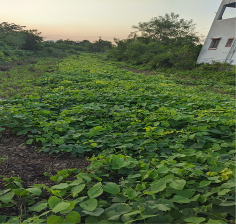
Figure 1: Vigna unguiculata leaves
The present research was designed to explore the anti-ulcer potential of the aqueous extract of Vigna unguiculata leaves. The study involved authentication of plant material, extraction using Soxhlet apparatus, qualitative phytochemical screening, acute toxicity evaluation, and in-vivo assessment in Wistar rats. Key parameters, including gastric pH, ulcer index, oxidative stress markers (MDA, GSH), inflammatory markers (TNF-α, PGE?), and histopathological changes, were analyzed to determine the extract’s efficacy and mechanism of action. By integrating biochemical, physiological, and tissue-level assessments, this study provides a comprehensive evaluation of the plant’s gastroprotective potential.11-12
Overall, this research aims to scientifically validate the traditional knowledge associated with Vigna unguiculata and highlight its significance as a cost-effective, natural alternative to synthetic anti-ulcer therapies. The findings may contribute to the development of herbal formulations that are safer, more accessible, and environmentally sustainable for the management of gastric ulcers.
MATERIALS AND METHODS:
Authentication of Plant Material:
Fresh plant specimens of Vigna unguiculata (approximately 10 inches in height and 7–8 inches in width), including stem, leaves, flowering buds, and cowpea pods, were collected for authentication. The plant specimen was manually pressed between sheets of paper and dried under direct sunlight for 7–8 days to eliminate residual moisture. After complete drying, the specimen was mounted on a standard herbarium sheet measuring 41 × 29 cm using adhesive. The prepared herbarium was submitted to the Botany Department of Y.C. College, Satara, for taxonomic identification. The plant was authenticated and assigned the Accession Number RAW001, with the confirmed botanical identity Vigna unguiculata (L.) Walp.
Preparation of Plant Extract
Drying of Leaves
Following authentication, collected leaves were further dried in a hot air oven at 65°C for 20 minutes to remove moisture and prepare them for extraction. Drying enhances extraction efficiency by improving solvent penetration, preventing enzymatic degradation, increasing phytochemical concentration, and providing a consistent and stable starting material for further procedures.13
Soxhlet Extraction
A Soxhlet extractor was employed to obtain the aqueous extract of Vigna unguiculata leaves. Dried leaves were coarsely powdered and packed into a filter-paper thimble placed inside the extraction chamber. Distilled water was used as the extraction solvent and filled into a round-bottom flask connected to the Soxhlet apparatus and a condenser. Upon heating, water vapors traveled through the condenser, condensed, and percolated through the leaf powder, dissolving water-soluble phytoconstituents. Once the chamber filled, the extract was automatically siphoned back into the flask, allowing repeated cycles of evaporation, condensation, and extraction. The process was maintained at 80°C for several hours until sufficient extract was collected. From 500 g of dried leaves, approximately 470 mL of aqueous extract was obtained.14,15
Hot Plate Drying
The aqueous extract obtained from Soxhlet extraction was subjected to hot plate drying to remove excess water and concentrate the bioactive constituents. The extract was transferred to shallow porcelain dishes and heated at 100–150°C on a controlled hot plate. Gradual evaporation of water resulted in a semi-solid, concentrated mass rich in phytochemicals. This method ensured efficient drying, improved stability, and produced an extract suitable for further phytochemical screening and pharmacological evaluation.16
Percentage Yield of Extract
For yield determination, 20 g of dried leaf powder was subjected to aqueous Soxhlet extraction, producing approximately 240 mL of extract. Two porcelain dishes were filled with 120 mL of extract each and dried to obtain a thick, semi-solid residue.17 The average initial dish weight was 62.2 g, while the final weight after drying was 64.1 g. Thus, each dish yielded:
Yield = (64.1 g – 62.2 g) × 2 = 3.8 g
Therefore, 20 g of dried leaves produced approximately 3.8 g of concentrated aqueous extract.
Phytochemical Screening
Qualitative Analysis
Preliminary phytochemical screening of the aqueous extract was carried out to identify major classes of bioactive compounds. This analysis is essential to confirm the presence of pharmacologically important constituents such as alkaloids, flavonoids, phenols, tannins, steroids, and saponins. Qualitative screening validates traditional therapeutic claims, serves as a foundation for drug discovery, ensures safety by detecting toxic metabolites, and aids in the standardization of herbal formulations. The results also guide dose selection for subsequent in vivo anti-ulcer studies and support quality control requirements.18,19
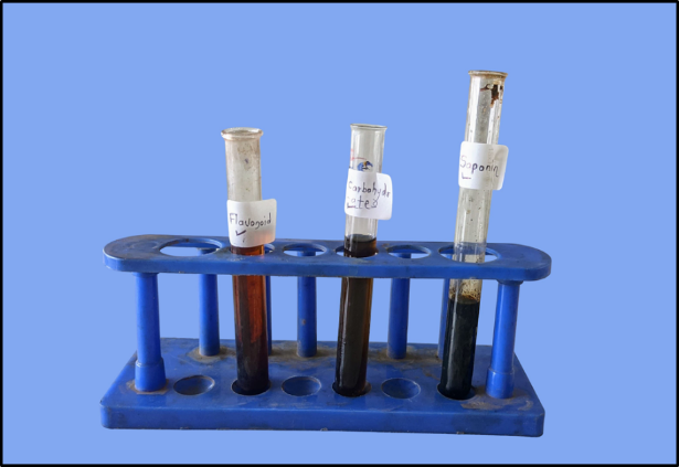
Figure 2: Qualitative Analysis of extract
Identification and Isolation of Phytochemicals
Thin Layer Chromatography (TLC)
To identify and isolate key phytochemical groups, especially flavonoids, TLC analysis was performed. The mobile phase used for flavonoid identification was Ethyl acetate:Water:Methanol (100:10:13.5), which produced distinct spots under UV light at 265 nm and 365 nm, confirming the presence of flavonoids. For isolation, multiple TLC plates were developed using a refined mobile phase consisting of Ethyl acetate:Glacial acetic acid:Formic acid:Water (100:11:11:27). Bands corresponding to flavonoids were visualized under UV illumination, scraped from the plates, and dissolved in ethanol. The mixture was filtered to remove silica gel, and the filtrate was concentrated. Recrystallization was performed by adding minimal hot solvent and allowing the solution to cool slowly, leading to the formation of purified flavonoid crystals. This procedure enabled the separation and isolation of specific flavonoid constituents responsible for the plant’s potential anti-ulcer activity.20,21
Animal Studies
Animal Selection and Housing
Albino Wistar rats were selected for the present study due to their well-established physiological stability, genetic homogeneity, and predictability in biomedical research. Their consistent biological responses make them particularly suitable for evaluating pharmacological interventions such as anti-ulcer therapeutics. Healthy adult rats were sourced from Krantisinh Nana Patil College of Veterinary Science & Animal and Fishery Sciences University, Nagpur, located at Shirwal, Satara, Maharashtra.
All experimental procedures were conducted following approval from the Institutional Animal Ethics Committee (IAEC No.: AGCOP/PHARMA/06/2025). The animals were housed in polypropylene cages under well-maintained laboratory conditions, including a controlled temperature of 24 ± 1°C and relative humidity of 55–65%. The facility operated under CPCSEA registration (No. 1874/PO/Re/S/16/CPCSEA). Rats were provided with a standardized pelleted diet and unrestricted access to clean drinking water throughout the study period to ensure optimal physiological conditions.
Toxicity Study
An acute oral toxicity assessment of the aqueous leaf extract of Vigna unguiculata was conducted in accordance with OECD Guideline 423. Groups of three Wistar rats were administered progressively increasing doses of the extract (5, 50, 300, and 2000 mg/kg body weight) to evaluate potential toxicity. The animals were continuously monitored for behavioral changes, physiological abnormalities, and mortality.
No signs of toxicity, morbidity, or mortality were observed at any of the administered doses, including at the limit dose of 5000 mg/kg. The absence of adverse effects confirmed the wide safety margin of the extract, allowing subsequent pharmacological evaluation using doses within the therapeutic range.22
Experimental Design for Anti-Ulcer Activity
The anti-ulcer potential of the aqueous extract of Vigna unguiculata was investigated using six experimental groups, each comprising six animals. Ulcers were induced using a single oral dose of indomethacin (15 mg/kg), except in the normal control group. Treatment was administered for 14 consecutive days in both the standard and test groups. At the end of the study, animals were anaesthetized, and biological samples such as blood, gastric contents, and stomach tissues were collected for analytical evaluation.23
Table 1: Experimental Design for Anti-Ulcer Activity
|
Group No. |
Group Name |
Treatment Administered |
Dose / Duration |
|
Group 1 |
Normal Control |
Saline (oral) |
Once daily for 14 days |
|
Group 2 |
Disease Control |
Indomethacin (oral) |
15 mg/kg, single dose |
|
Group 3 |
Standard Control |
Omeprazole (oral) |
5 mg/kg daily for 14 days |
|
Group 4 |
Test Group I |
Vigna unguiculata aqueous extract |
800 mg/kg daily for 14 days |
|
Group 5 |
Test Group II |
Vigna unguiculata aqueous extract |
1000 mg/kg daily for 14 days |
|
Group 6 |
Test Group III |
Vigna unguiculata aqueous extract |
1200 mg/kg daily for 14 days |
Evaluation Parameters
Gastric Ulcer Index
The ulcer index was assessed to determine the extent and severity of gastric mucosal injury. Following dissection, each stomach was opened along the greater curvature, rinsed with saline, and examined under adequate illumination. The number, size, and appearance of ulcer lesions were scored according to a standardized scale. This evaluation provided quantitative data to compare the protective efficacy of the extract against induced ulcers.24,25
Gastric Juice pH
After sacrifice, the gastric contents were collected and centrifuged to obtain a clear supernatant. The pH of the gastric juice was determined using a calibrated digital pH meter. An increase in gastric pH indicated a reduction in gastric acidity, which is a key parameter in assessing anti-ulcer activity.26,27,28

Figure 3: Blood samples & Isolated gastric content of each group individually
Inflammatory Markers
TNF-α Estimation
Tumor Necrosis Factor-α (TNF-α), a crucial inflammatory mediator, was measured using Western blot analysis. Protein expression levels served as indicators of inflammatory response and mucosal damage within gastric tissue.29,30
Prostaglandin E? (PGE?) Measurement
PGE? levels were quantified using a rat-specific enzyme-linked immunosorbent assay (ELISA) kit. The absorbance values obtained from the colorimetric reaction were measured spectrophotometrically, providing insights into gastric mucosal protection and prostaglandin activity.31,32,33
Oxidative Stress Markers
Malondialdehyde (MDA)
MDA levels, indicative of lipid peroxidation and oxidative tissue injury, were measured using the thiobarbituric acid reactive substances (TBARS) assay. Elevated MDA levels reflected increased oxidative stress in the gastric mucosa.34, 35, 36
Gastric Glutathione (GSH)
Glutathione content was estimated to assess the antioxidant defense capacity of gastric tissues. Higher GSH levels suggested enhanced protection against oxidative damage and improved mucosal resilience following extract treatment.37, 38
Statistical Analysis
All experimental data were expressed as mean ± standard error of the mean (SEM). Statistical evaluation was carried out to determine the significance of differences between the control and treated groups. The results were analyzed using one-way Analysis of Variance (ANOVA), followed by Dunnett’s multiple comparison post-hoc test to compare each treatment group with the disease control group. A p-value of less than 0.05 (p < 0.05) was considered statistically significant, while p < 0.01 and p < 0.001 indicated highly significant and extremely significant differences, respectively. Statistical computations were performed using GraphPad Prism software.
RESULTS AND DISCUSSION:
Qualitative Phytochemical Analysis
The preliminary phytochemical screening of the aqueous leaf extract of Vigna unguiculata revealed the presence of several biologically important secondary metabolites. Carbohydrates, glycosides, tannins, saponins, alkaloids, flavonoids, phenols, and quinones tested positive in their respective qualitative tests, indicating a rich phytochemical profile responsible for potential therapeutic activity. In contrast, proteins, amino acids, and steroids were found to be absent, as indicated by negative test reactions. These findings suggest that the aqueous extract is particularly enriched with phenolic and flavonoid compounds, which may contribute to its antioxidant and anti-ulcer properties.
Table 2: Qualitative Phytochemical Analysis of Aqueous Extract of Vigna unguiculata Leaves
|
Phytochemical |
Test Performed |
Observation |
Result |
|
Carbohydrates |
Fehling’s Test / Benedict’s Test |
Brick-red / Reddish-brown colour |
+ |
|
Proteins |
Biuret Test |
No violet–pink colour |
– |
|
Amino Acids |
Ninhydrin Test |
No purple–blue colour |
– |
|
Glycosides |
Legal Test |
Dark red colour |
+ |
|
Tannins |
Acetic Acid Test |
Red-coloured solution |
+ |
|
Saponins |
Froth Test |
Persistent foaming layer |
+ |
|
Steroids |
Salkowski Test |
No red–yellow chloroform layer |
– |
|
Alkaloids |
Wagner’s Test |
Reddish-brown precipitate |
+ |
|
Flavonoids |
Sulfuric Acid Test |
Orange–red colour |
+ |
|
Phenols |
Acetic Acid Test |
Red coloration |
+ |
|
Quinones |
Sulfuric Acid Test |
Red precipitate |
+ |
Identification & Isolation of phytochemical
Preliminary Compound Screening
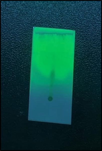
Figure 4: TLC for flavonoid under 265 Nm
TLC Screening for the Flavonoid group
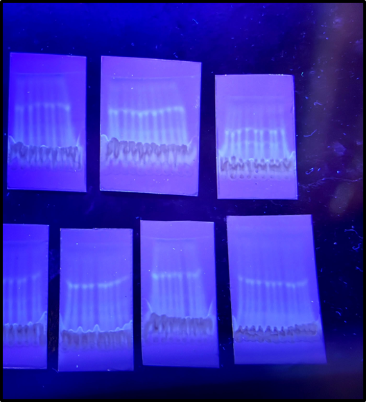
Figure 5: Group specified Flavonoids TLC 365 nm UV
Infrared radiation of isolated quercetin
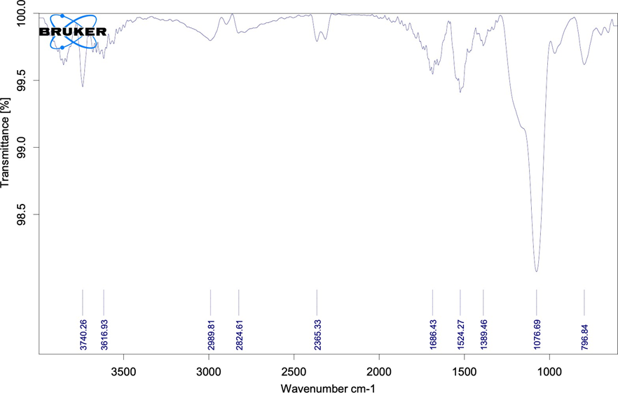
Figure 6: FT-IR of isolated Flavonoid
Table 3: FT-IR Spectra Observations-1
|
Name of constituent |
Functional Group |
Standard Value |
Observed Value |
|
Flavonoid |
-OH (Alcohol) |
3400-3500 cm-1 |
3616.93 cm-1 |
|
C=O (Ketone) |
1650-1700 cm-1 |
1686.43 cm-1 |
|
|
C=C (Alkene) |
1600-1650 cm-1 |
1542.27 cm-1 |
|
|
C-O (Carbonyl) |
1200-1300 cm-1 |
1389.46 cm-1 |
|
|
C-H |
1000-1200 cm-1 |
1076.69 cm-1 |
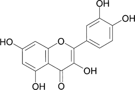
Figure 7: Structure of Quercetin
Toxicity Study
The acute oral toxicity study revealed that the aqueous extract of Vigna unguiculata leaves was safe up to a dose of 2000 mg/kg, with no signs of mortality or observable toxic effects in any treatment group. All animals remained active, with normal behavior and no physiological abnormalities, indicating a wide safety margin suitable for pharmacological evaluation.
Table 4: Acute Toxicity Study of Test Extract
|
Dose (mg/kg) |
No. of Animals |
% Mortality |
Side Effects |
|
5 |
3 |
0% |
None |
|
50 |
3 |
0% |
None |
|
300 |
3 |
0% |
None |
|
1000 |
3 |
0% |
None |
|
2000 |
3 |
0% |
None |
Body Weight Changes
Throughout the 14-day study, the normal control group showed a gradual increase in body weight, while the disease control group exhibited noticeable weight loss due to ulcer induction and associated stress. The standard group and higher-dose test groups (Test 2 and Test 3) demonstrated better weight maintenance, indicating improved physiological stability and protective effects of the treatments.
Table 5: Initial Body Weight of Rats (g)
|
Group |
1 |
2 |
3 |
4 |
5 |
6 |
|
Normal Control |
176.3 |
181.8 |
192.4 |
201.2 |
194.9 |
178.1 |
|
Disease Control |
168.2 |
180.3 |
172.7 |
190.2 |
175.2 |
183.4 |
|
Standard Control |
173.2 |
178.4 |
167.3 |
169.3 |
174.3 |
182.3 |
|
Test 1 |
183.2 |
180.4 |
192.3 |
173.3 |
182.3 |
179.2 |
|
Test 2 |
192.3 |
190.0 |
178.4 |
184.3 |
172.3 |
167.3 |
|
Test 3 |
183.3 |
164.3 |
181.3 |
192.3 |
203.2 |
187.4 |
Table 6: Final Body Weight Prior to Dissection (g)
|
Group |
1 |
2 |
3 |
4 |
5 |
6 |
|
Normal Control |
178.6 |
186.5 |
195.3 |
210.2 |
197.2 |
182.4 |
|
Disease Control |
159.2 |
171.2 |
162.2 |
182.3 |
164.2 |
167.2 |
|
Standard Control |
170.3 |
175.2 |
166.8 |
162.3 |
173.2 |
176.4 |
|
Test 1 |
179.3 |
171.7 |
186.5 |
167.8 |
173.9 |
171.4 |
|
Test 2 |
185.3 |
183.4 |
171.6 |
179.9 |
165.8 |
159.5 |
|
Test 3 |
177.4 |
161.9 |
173.7 |
185.0 |
195.6 |
181.3 |
EVALUATION PARAMETERS:
1. Gastric pH
A significant alteration in gastric acidity was observed among the experimental groups. The Normal Control group exhibited a high gastric pH (5.81 ± 0.22), confirming a healthy gastric environment. In contrast, the Disease Control group showed a markedly reduced pH (2.18 ± 0.21; p<0.0001), indicating successful ulcer induction. The Standard Control group significantly restored the gastric pH (5.20 ± 0.08; p<0.0001 vs. Disease Control). Among the test formulations, Test 1 showed moderate improvement (3.37 ± 0.25; p<0.001), while Test 2 (4.25 ± 0.20; p<0.0001) and Test 3 (4.74 ± 0.13; p<0.0001) exhibited considerably higher pH restoration, with Test 3 showing values closest to the Standard Control.
2. Ulcer Index
The ulcer index showed clear variability between groups. Normal Control animals displayed no ulceration (0.00 ± 0.00), whereas Disease Control animals demonstrated severe ulceration (38.75 ± 1.40; p<0.0001). Standard treatment significantly reduced ulcer index values (22.78 ± 1.09; p<0.0001). Test 1 showed minimal protection (36.08 ± 0.78), whereas Test 2 (33.83 ± 0.43; p<0.01) and Test 3 (32.42 ± 0.31; p<0.001) demonstrated better gastroprotective effects, with Test 3 again being the most effective among the test formulations.
3. TNF-α Levels
TNF-α levels increased significantly in the Disease Control group (792.3 ± 11.53 pg/g; p<0.0001), confirming a strong inflammatory response. Standard treatment markedly reduced TNF-α (391.4 ± 12.81 pg/g; p<0.0001). Test 1 (782.6 ± 8.04) and Test 2 (778.4 ± 10.85) showed little improvement. However, Test 3 presented a moderate but significant decrease (734.1 ± 6.12; p<0.01), demonstrating partial anti-inflammatory activity.
4. Prostaglandin E2 (PGE2)
PGE2 levels were significantly reduced in the Disease Control group (24.53 ± 1.25; p<0.0001), indicating compromised mucosal protection. The Standard Control group showed substantial restoration (34.70 ± 0.99; p<0.0001). Test 1 produced minimal recovery (22.05 ± 0.91), whereas Test 2 (28.03 ± 0.57; p<0.05) and Test 3 (29.18 ± 0.86; p<0.01) demonstrated better enhancement in mucosal prostaglandins, with Test 3 again outperforming the other test groups.
5. Malondialdehyde (MDA)
MDA, an indicator of lipid peroxidation, was significantly elevated in Disease Control animals (295.9 ± 3.55; p<0.0001). Standard treatment markedly reduced MDA (230.8 ± 2.13; p<0.0001). Test 1 showed almost no improvement (291.7 ± 2.39), while Test 2 (283.0 ± 4.05; p<0.05) and Test 3 (269.9 ± 2.51; p<0.01) displayed moderate reductions, suggesting antioxidant effects, with Test 3 proving most effective among the test samples.
6. Gastric Glutathione (GSH)
GSH levels were significantly depleted in the Disease Control group (169.1 ± 1.64; p<0.0001). Standard treatment effectively restored GSH concentrations (366.6 ± 4.03; p<0.0001). Test 1 showed minimal improvement (171.2 ± 2.67), whereas Test 2 (185.9 ± 3.71; p<0.05) and Test 3 (227.0 ± 2.77; p<0.01) demonstrated better enhancement, indicating antioxidant and cytoprotective effects, with Test 3 again being the most promising.
Table 7: Evaluation parameters
|
Parameter |
Normal Control |
Disease Control |
Standard Control |
Test 1 |
Test 2 |
Test 3 |
|
Gastric pH |
5.81 ± 0.22 |
2.18 ± 0.21 |
5.20 ± 0.08**** |
3.37 ± 0.25*** |
4.25 ± 0.20**** |
4.74 ± 0.13**** |
|
Ulcer Index (%) |
0.00 ± 0.00 |
38.75 ± 1.40 |
22.78 ± 1.09**** |
36.08 ± 0.78 |
33.83 ± 0.43** |
32.42 ± 0.31*** |
|
TNF-α (pg/g) |
238.2 ± 4.37 |
792.3 ± 11.53 |
391.4 ± 12.81**** |
782.6 ± 8.04 |
778.4 ± 10.85 |
734.1 ± 6.12** |
|
PGE2 (pg/g) |
40.27 ± 0.90 |
24.53 ± 1.25 |
34.70 ± 0.99**** |
22.05 ± 0.91 |
28.03 ± 0.57* |
29.18 ± 0.86** |
|
MDA (nmol/g) |
140.0 ± 2.31 |
295.9 ± 3.55 |
230.8 ± 2.13**** |
291.7 ± 2.39 |
283.0 ± 4.05* |
269.9 ± 2.51** |
|
GSH (mg/g) |
403.3 ± 4.78 |
169.1 ± 1.64 |
366.6 ± 4.03**** |
171.2 ± 2.67 |
185.9 ± 3.71* |
227.0 ± 2.77** |
Values are represented as mean ± SEM, n=6, *p<0.05, **p<0.01, ***p<0.001, ****p<0.0001, #p<0.0001 for disease & Normal control group.Values of Normal Control group and Disease control group were compared by Student’s t-test. Values of Standard control, Test 1, Test 2, Test 3 groups were compared with the Disease Control group by One-Way ANOVA followed by Dunnett’s multiple comparison test.
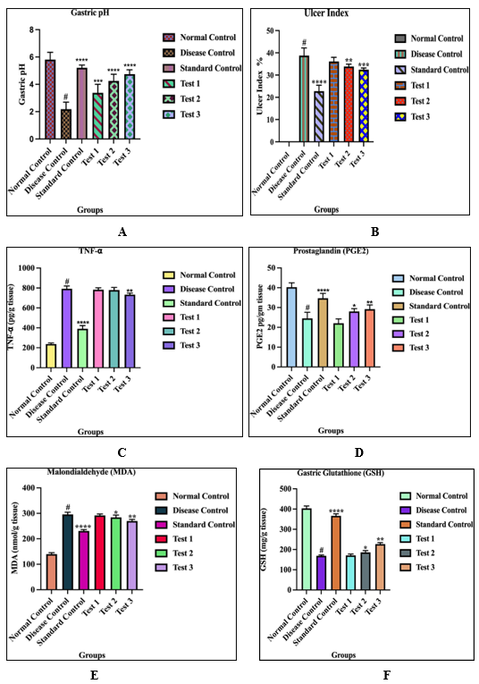
Figure 8: A) Graphical analysis of Gastric pH B) Graphical analysis of Ulcer Index C) Graphical analysis of TNF factor D) Graphical analysis of PGE2 E) Graphical analysis of MDA F) Graphical analysis of Gastric Glutathione
HISTOPATHOLOGICAL OBSERVATIONS
1. Normal Control Group
The normal control tissue shows intact gastric architecture with parallelly arranged gastric glands and a continuous epithelial lining. There is no evidence of ulceration, necrosis, hemorrhage, or inflammatory cell infiltration, indicating a completely healthy mucosal environment. Parietal and chief cells are clearly distinguishable, reflecting normal cellular differentiation and secretory function.
2. Diseased / Ulcer-Induced Group
The diseased group exhibits severe mucosal disruption characterized by complete loss of epithelial continuity, breakdown of glandular structure, and the presence of necrotic debris and vacuolated cells. A dense inflammatory infiltrate of neutrophils and mononuclear cells is observed, suggesting an acute inflammatory reaction due to mucosal injury. Edema and hemorrhage are evident in the lamina propria and submucosa, with vascular dilation and irregular eosinophilic patches. Cytoplasmic degeneration and pyknotic nuclei indicate extensive cellular death, and the damaged region shows features of ulcer crater formation.
3. Test Group 1 (Slight Healing)
The tissue from Test Group 1 demonstrates partial healing, with the villi and epithelial layers showing incomplete restoration. Although epithelial disruption persists, the overall damage is less severe than in the untreated ulcer group. The nuclei remain densely stained, and the epithelial arrangement is uneven, suggesting ongoing injury. A moderate reduction in inflammatory cell infiltration is visible, indicating the initial stages of healing. The submucosa appears dense and homogenous, likely due to residual edema or early fibrosis. At higher magnification, slight improvements in epithelial continuity and the presence of some goblet cells reflect early mucosal recovery.
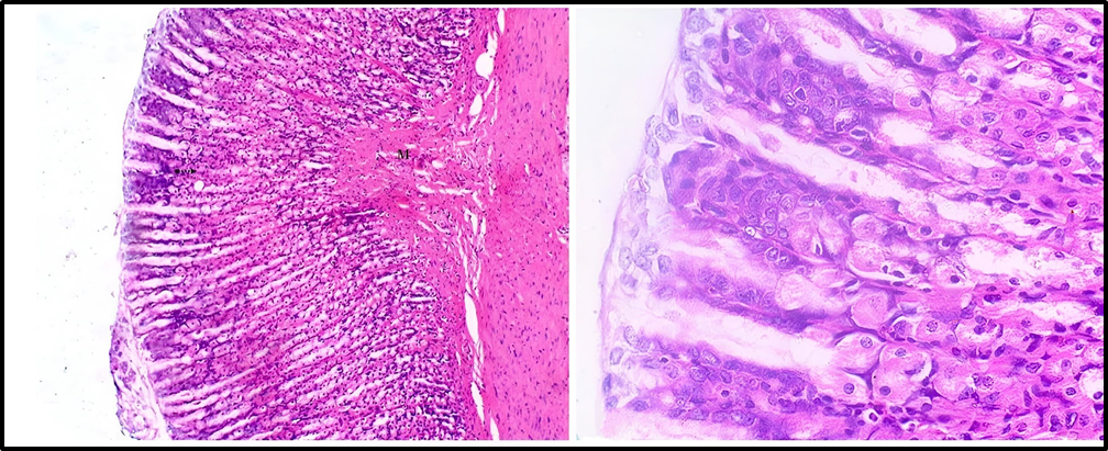
Figure 9: Test Group 1
4. Test Group 2 (Moderate Healing)
The tissue in Test Group 2 shows significant improvement, with a more continuous epithelial lining and well-organized villi indicating advanced re-epithelialization. Inflammatory cell infiltration is markedly reduced compared to the diseased and Test 1 groups, demonstrating a substantial decline in inflammation. The increased presence of goblet cells suggests enhanced mucus production, which contributes to mucosal protection and supports healing. The submucosa displays improved structural organization, indicating ongoing tissue remodeling.
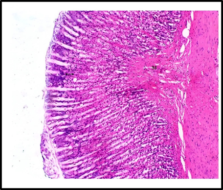
Figure 10: Test Group 2
5. Test Group 3 / Standard Treatment (Near-Complete Healing)
The Test Group 3 tissue displays nearly complete mucosal restoration, with a fully continuous epithelial layer and well-formed, uniform villi similar to healthy tissue. Inflammatory cells are virtually absent from the submucosa, confirming the resolution of inflammation. A higher number of goblet cells indicates robust mucus production and restored mucosal defense. The submucosa appears normal, without signs of edema or fibrosis, demonstrating complete tissue remodeling. Overall, this group represents near-complete healing of the ulcerated mucosa.
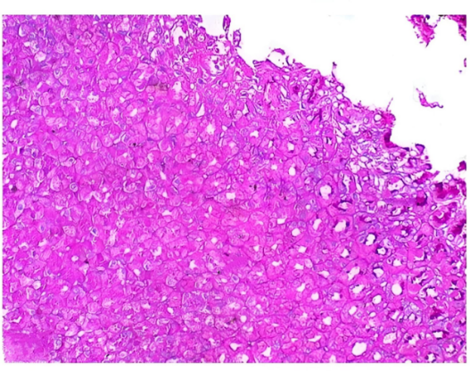
Figure 11: Test Group 3
DISCUSSION:
This study explored the anti-ulcer potential of Vigna unguiculata leaves as a natural, affordable, and widely available alternative to synthetic drugs. Since the plant is commonly consumed and rich in phytochemicals such as flavonoids, phenols, saponins, alkaloids, and carbohydrates, it was selected for evaluation against ulcerative conditions. Preliminary literature suggested anti-inflammatory properties in other plant parts, but the leaf activity was not fully established, prompting this investigation.
Qualitative screening confirmed the presence of key bioactive compounds, supporting its therapeutic relevance. Acute toxicity assessment following OECD 423 guidelines indicated that the extract was safe up to ≥5000 mg/kg, leading to the selection of 800, 1000, and 1200 mg/kg for in-vivo evaluation. Isolation and identification of quercetin further justified the possibility of significant anti-ulcer activity.
Indomethacin (15 mg/kg) was used to induce gastric ulcers, and omeprazole (5 mg/kg) served as the standard drug. The study assessed multiple parameters—gastric pH, ulcer index, TNF-α, PGE2, MDA, and GSH—to measure gastric protection and inflammation. As expected, ulcer induction increased TNF-α and MDA while reducing PGE2 and GSH levels.
The extract demonstrated a clear dose-dependent protective effect, improving gastric pH, reducing ulcer severity, lowering oxidative and inflammatory markers, and restoring protective factors. The highest dose (1200 mg/kg) produced results comparable to omeprazole, highlighting its strong gastroprotective capacity. Histopathological findings confirmed reduced epithelial damage and better mucosal preservation in treated groups, supporting the biochemical outcomes.
Although promising, the study is limited by the absence of detailed mechanistic exploration and long-term safety data. Future work should focus on isolating specific active compounds, clarifying molecular pathways, and evaluating the extract in additional ulcer models. Overall, Vigna unguiculata leaves show significant potential as an effective, safe, and economical anti-ulcer agent, strengthening the relevance of plant-based therapies in modern pharmacology.
CONCLUSION:
The present study concludes that the aqueous extract of Vigna unguiculata leaves exhibits significant dose-dependent anti-ulcer activity in Indomethacin-induced gastric ulcer models in Wistar rats. The plant material collected from Alandi (Pune, Maharashtra) was authenticated through a prepared herbarium specimen at Yashwantrao Chavan College, Satara, and the dried leaves were successfully extracted using a Soxhlet apparatus with water as the solvent.
The extract demonstrated marked gastroprotective effects by increasing gastric pH, reducing ulcer index, enhancing protective biomarkers such as GSH and PGE?, and lowering oxidative and inflammatory markers like MDA and TNF-α. The highest dose (1200 mg/kg) showed effects comparable to the standard drug, indicating strong therapeutic potential. Histopathological findings further confirmed reduced mucosal damage and improved tissue healing in treated groups.
These outcomes support the traditional use of Vigna unguiculata in gastrointestinal disorders and highlight its potential as a safe, cost-effective natural alternative for ulcer management. However, further investigations are required to isolate the active constituents, establish the precise mechanisms of action, and evaluate long-term safety to strengthen its applicability in clinical settings.
AKNOWLEDGMENT:
The authors sincerely thank the Department of Pharmacology, Arvind Gavali College of Pharmacy, Satara, Maharashtra–415004 (India) for providing the necessary facilities, guidance, and support to carry out this research work. Appreciation is also extended to the faculty members and laboratory staff for their valuable assistance throughout the study.
CONFLICT OF INTEREST:
The authors declare that there is no conflict of interest regarding the publication of this research work.
REFERENCES


Hrushikesh Warade*, Dr. Vasant Yashwant Lokhande, Pournima Shelar, Kirti Godse, Shravani Pawar, Evaluation of Anti-Ulcer Activity of Vigna Unguiculata Leaves Extract, Int. J. of Pharm. Sci., 2025, Vol 3, Issue 11, 2545-2560 https://doi.org/10.5281/zenodo.17637124
 10.5281/zenodo.17637124
