
We use cookies to ensure our website works properly and to personalise your experience. Cookies policy
1,2Department of Veterinary Pharmacology and Toxicology, Mumbai Veterinary College, Mumbai.
3Maharashtra Animal and Fishery Sciences University, Nagpur, Vetrina Healthcare Pvt Ltd, Pune, Maharashtra, India.
Lanthanum carbonate (Vetricare Renal (®), Vetrina Healthcare Pvt Ltd) is an effective non-calcium, non-resin phosphate binder for the treatment of hyperphosphataemia in chronic kidney disease (CKD). In this study, we used a rat model of chronic renal failure (CRF) to examine the effects of controlling serum phosphorus with lanthanum carbonate treatment on the biochemical and bone abnormalities. Rats were randomly divided into 7 groups (n=10). Chronic Kidney Disease was induced by Adenine @220 mg/kg/day orally for 14 days. Groups IV–VII received Lanthanum Carbonate at varying doses (150–750 mg/kg/day) from day 15 to 42. Adenine administration significantly reduced body weight, with values dropping from 172.5 ± 2.007 g on day 0 to 158 ± 2.494 on day 14. However, Lanthanum Carbonate treatment led to a marked improvement in body weight by day 42, particularly in groups receiving 500 mg/kg and 750 mg/kg doses. Adenine also caused a notable increase in serum phosphorus levels (18.36 ± 0.837 mg/dL on day 14 and 17.84 ± 0.811 mg/dL on day 42). Treatment with Lanthanum Carbonate significantly reduced biochemical markers such as Serum Phosphorus, Creatinine, Blood Urea Nitrogen and Alkaline Phosphatase. The higher doses (500 mg/kg and 750 mg/kg) were more effective than lower doses (150 mg/kg and 250 mg/kg). No significant changes were observed in serum calcium, Serum Glutamate Pyruvate Transaminase or Serum Glutamate Oxaloacetate Transaminase levels. Histopathological examination confirmed renal protection, while histomorphometry analysis indicated an osteoprotective effect. Treatment with lanthanum carbonate reduced the biochemical and bone abnormalities in a rat model of CRF. These findings suggest that Lanthanum Carbonate, particularly at higher doses, offers significant renal and skeletal protection in Chronic Kidney Disease induced by Adenine in rats.
Chronic kidney disease (CKD) is a prevalent global health issue marked by a progressive . loss of kidney function over time. Chronic Kidney Disease is a characterized by a gradual loss of renal function over time, often resulting from various infectious and non-infectious factors. The most common causes of Chronic kidney disease (CKD) include diabetes, hypertension, glomerulonephritis (Levey et al., 2005), exposure to toxins such as ethylene glycol, aminoglycoside antibiotics, hypercalcemia, haemoglobinuria, ingestion of grapes or raisins, and nonsteroidal anti-inflammatory drugs (NSAIDs). Other contributing factors include ischemia, parasitic infections like Babesia, Hepatozoon, and Ehrlichia, as well as bacterial infections such as leptospirosis and borreliosis (Aiello and Moses., 2016) Clinically, it is characterized by symptoms such as anorexia, dehydration, depression, oral ulceration, vomiting, diarrhoea, oliguria, and the presence of enlarged, painful kidneys. Chronic kidney disease (CKD) is particularly common among older animals, while congenital forms of the disease may cause a temporary increase in prevalence among animals younger than three years. In geriatric populations Chronic kidney disease (CKD) affects up to 10% of dogs and 35% of cats, although the prevalence in the broader small animal population is estimated to be lower, ranging between 1% and 3%. Heritable forms of Chronic kidney disease (CKD) have been identified in certain breeds, but there is no specific breed or sex predisposition for nonheritable forms of the disease in dogs and cats.( Brown., 2013). Vetricare Renal contains Lanthanum Carbonate a non-calcium-based compound has shown promise in managing hyperphosphatemia without contributing to calcium overload. Animal models, especially the Adenine induced chronic kidney disease model, have proven effective in mimicking human renal pathology and evaluating potential therapeutic agents. The objective of this study was to evaluate the effect of controlling serum phosphorus with lanthanum carbonate on biochemical and bone parameters in a rat model (adenine-induced). We also examined the effects of treatment with lanthanum carbonate on tissue lanthanum deposition and liver function.
MATERIAL AND METHODS:
The study titled “Evaluation of efficacy of Lanthanum Carbonate in rats with adenine induced chronic kidney disease” was conducted in 2024 at the Central Laboratory Animal House, Department of Veterinary Pharmacology and Toxicology, Mumbai Veterinary College (MVC), under MAFSU, Nagpur. The experimental protocol was approved by the IAEC (Approval No. MVC/IAEC/08/53/2024) in accordance with CCSEA guidelines.
Chemicals and Reagents
Experimental Animals
Seventy healthy Sprague–Dawley rats (150–200 g) were housed under standard lab conditions and acclimatized for one week. All animals received commercial pelleted feed and water ad libitum throughout the 42-day study.
Experimental Design
Rats were randomly divided into 7 groups (n=10). Chronic Kidney Disease was induced by Adenine @220 mg/kg/day orally for 14 days. Groups IV–VII received Lanthanum Carbonate at varying doses (150–750 mg/kg/day) from day 15 to 42. After one week of acclimatisation period, the rats underwent standard health assessments, after which seventy Sprague-Dawley (SD) rats were evenly distributed into seven groups (Groups I, II, III, IV, V, VI and VII), with each group consisting of ten animalsin each group. Group I acted as the normal control group, receiving a standard diet for 42 days. Group II was designated as the NRF group and was administered Lanthanum Carbonate @500 mg/kg body weight once daily for the 28-day experimental period. Group III functioned as the positive control group for chronic renal failure (CRF) and was given an Adenine @220 mg/kg body weight through oral gavage once daily for 14 days (Ashour et al., 2023). Group IV, group V, group VI and group VII were also given the Adenine @220 mg/kg body weight by gavage once daily for the first 14 days to induce Chronic Kidney Disease, after which they received Lanthanum Carbonate treatment @150 mg/kg, @250 mg/kg, @500 mg/kg and @750 mg/kg body was administered orally from day 15 to 42.
Induction of Chronic Kidney Disease: -
Rats were randomly selected and divided into seven groups (Groups I, II, III, IV, V, VI and VII), with each group containing ten rats. Group I served as the normal control group and received only a standard diet. Group II was designated as the NRF group and was administered Lanthanum Carbonate @500 mg/kg. Group III functioned as the positive control group for Chronic Kidney Disease (CKD), receiving Adenine @220 mg/kg for a duration of 14 days. The induction of Chronic Kidney Disease was also carried out in the rats of group IV, group V, group VI and group VII. All rats in the study were subjected to regular clinical examinations on a daily basis. Each rat was monitored for feed and water intake, general behaviour, alertness, urine output, and other clinical symptoms throughout the study period. To evaluate biochemical parameters, blood samples were drawn from the retro-orbital plexus of the rats under light anesthesia on days 0, 14, and 42. The serum was then separated and analyzed for renal biochemical parameters. Rats were humanely sacrificed under light anesthesia with Ketamine and Xylazine on 43rd day. The kidneys, liver, and bones were collected and preserved in 10% Neutral Buffered Formalin (NBF) for histopathological and histomorphometry analysis.
Histopathological Studies: -
Kidney and liver tissue samples were collected and preserved in 10% neutral buffered formalin (NBF), with subsequent processing using the routine paraffin embedding method outlined by Aziz and Zeman (2022). The resulting tissue sections, measuring 4 microns, were then stained following the standard Hematoxylin (H) and Eosin (E) protocol as recommended by Cardiff et. al.,1995.
Histomorphometry Of Bone: -
Bone specimens collected and fixed them immediately in 10% neutral-buffered formalin to preserve their structure. For paraffin embedding, decalcification of samples was done with formic acid. Decalcified bones were embedded in paraffin. Used a microtome for sectioning, paraffin sections of 5–7 μm thickness were done. Applied histological stains to enhance tissue contrast Hematoxylin and Eosin (H&E) for general morphology. Used a light microscope equipped with a camera to capture high resolution images.
Statistical analyses
All statistical analyses were conducted using SAS statistical software (SAS Institute, Cary, NC, USA). Multiple group comparisons were conducted using one-way (group) analysis of variance by either Duncan or Dunnett's procedure using the vehicle-fed CRF group as reference. Data collected at different time points in the study were analysed separately. Results are reported as mean ± standard error of the mean (SEM), unless otherwise stated.
RESULT
Body weight
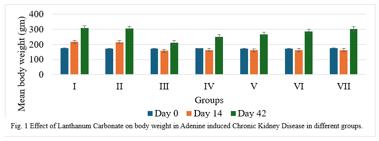
Body weight, a vital indicator of therapeutic efficacy, was recorded biweekly. The mean body weights on days 0, 14, and 42 are presented in Figure 1. Statistical analysis revealed significant differences across groups at different time points. On day 14, rats in the adenine-only group (Group III) showed a significant reduction in body weight (P ≤ 0.01), confirming disease induction. Groups IV–VII (Adenine + Lanthanum Carbonate) also exhibited weight loss, although less severe. By day 42, a dose-dependent improvement was noted, particularly in Groups V–VII, indicating a potential therapeutic effect of lanthanum carbonate.
Blood biochemistry was assessed before induction of Chronic kidney disease (Day 0), at the end of disease induction (Day 14) and at the end of treatment (Day 42). Serum phosphorus, Serum Creatinine, Blood urea nitrogen, Alkaline Phosphatase, Serum Glutamate Pyruvate Trasnaminase and Serum Glutamate Oxaloacetate Transaminase levels were measured on days 0, 14, and 42. On day 14, a significant increase (P ≤ 0.01) was observed in Adenine treated rats (Groups III–VII). Group III (Adenine alone) maintained elevated levels through day 42. However, Groups IV–VII, treated with Lanthanum Carbonate from days 15–42 at increasing doses (150–750 mg/kg), showed a dose-dependent reduction in Serum Phosphorus, Serum Creatinine, Blood urea nitrogen, Alkaline Phosphatase by day 42.
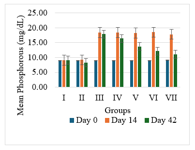
Fig. 2 Effect of Lanthanum Carbonate on serum Phosphorous in Adenine induced chronic kidney disease in different groups.
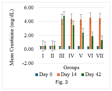
Fig. 3 Effect of Lanthanum Carbonate on serum Creatinine in Adenine induced Chronic Kidney Disease in different groups.
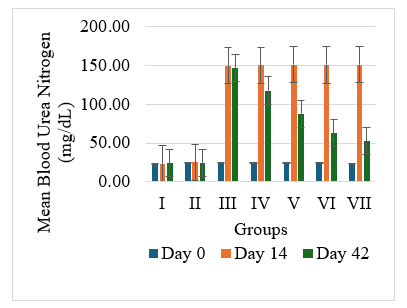
Fig. 4 Effect of Lanthanum Carbonate on Blood Urea Nitrogen in Adenine induced chronic kidney disease in different groups.

Fig. 5 Effect of Lanthanum Carbonate on Alkaline Phosphatase in Adenine induced chronic kidney disease in different groups.
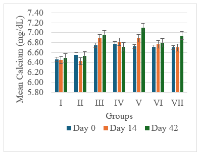
Fig. 6 Effect of Lanthanum Carbonate on serum Calcium in Adenine induced chronic kidney disease in different groups.
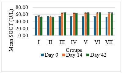
Fig. 7 Effect of Lanthanum Carbonate on Serum Glutamate-Pyruvate Transaminase in Adenine induced chronic kidney disease in different groups.
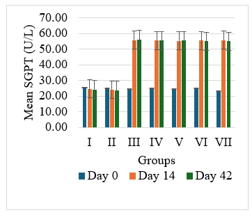
Fig. 8 Effect of Lanthanum Carbonate on Serum Glutamate Oxaloacetate Transaminase in Adenine induced chronic kidney disease in different groups.
Inflammatory markers
The mean serum Interleukin-1β (IL-1β) and Caspase-3 levels were evaluated on day 42. A significant increase (P ≤ 0.01) in IL-1β and Caspase-3 was observed in the adenine-only group confirming an inflammatory response due to chronic kidney disease. Groups IV–VII (Adenine + Lanthanum Carbonate) showed a dose-dependent reduction in IL-1β and Caspase-3 levels.
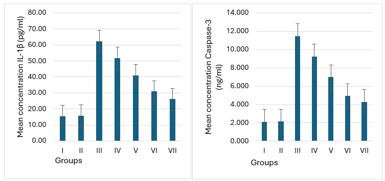
Fig. 9 & Fig. 10 Effect of Lanthanum Carbonate on interleukin-1β and caspase-3 respectively
Chronic kidney disease (CKD) is defined as any structural and/or functional abnormality in one or both kidneys that has been present in patients for at least three months. This condition is quite common among dogs, particularly in older animals, and it has a high prevalence within the canine population. The progression of CKD is typically slow, with affected dogs having a survival time that can range from several months to one or two years (Bartges, 2012; Davies, 2016; Polzin, 2011; Smets et al., 2010). In our study, administration of Adenine @ 220mg/kg orally resulted in CRD including decrease in body weight, elevation of Phosphorus, serum creatinine, BUN, alkaline phosphate and inflammatory biomarkers.. these changes were associated with increase levels of fibroisis in bone tissue. The similar observations were observed by Yang et al., (2013) who exhibited significant loss of body weight and this deerease was due to anorexia and potential renal failure. Chang et al. (2017) and Li et al., (2018) also observed similar findings that Adenine treated animals showed a significant decrease of body weight @220 mg/kg for 28 days, which might be due to the toxic effect of Adenine. Showed a significant decrease in serum phosphorus by 50% compared to Adenine induced group. Zhang et al. (2013), and Takahara et al. (2014) recorded that Lanthanum Carbonate on rats showed a significant decrease in serum phosphorus by 50% compared to Adenine induced group. The result of the study was in accordance with Abellán et al. (2019), Damment et al. (2011) and Chang et al. (2017) who observed hyperphosphaetemia in rats administered with Adenine. Yokozawa et al. (1986) noted that serum Creatinine was found to be significantly increased in rats treated with Adenine on 14 days following Adenine treatment. Yang et al. (2013) reported that elevation of Serum Creatinine, Blood Urea Nitrogen in rats treated with Adenine @220 mg/kg for 30 days. The study also showed that administration of Adenine increased fourfold and threefold greater Blood Urea Nitrogen and Serum Creatinine with massive tubular necrosis. There was no significant difference in Creatinine values of rats in group I and group II on the 14th day. The values in group IV – VII were lower than that of group II on the 42nd day. The results of the present study indicate that administration of Lanthanum Carbonate @150 mg/kg, @250 mg/kg, @500 mg/kg and @750 mg/kg body weight alters the Creatinine levels, which were observed in rats administered with Adenine @220 mg/kg on the 42nd day. Damment et al. (2011) who observed the significant (P ≤ 0.01) effect of Lanthanum Carbonate on serum phosphorus. They reported that Lanthanum Carbonate showed significant reduction of serum phosphorus by 50% compared to normal positive control. Finn et al. (2004), Zhang et al. (2013), and Takahara et al. (2014) recorded that Lanthanum Carbonate on rats showed a significant decrease in serum phosphorus by 50% compared to Adenine induced group. Similar findings were recorded by Damment et al. (2011) and Takashima et al.,(2014) recorded that Lanthanum Carbonate @1g/kg/day on rats showed a significant decrease in Phosphorus, BUN, Creatinine levels compared to Adenine control group. The result of the study indicated that when the rats administered with Adenine @220 mg/kg for 14 days the Phosphorus, BUN and Creatinine levels was increased significantly while, when rats administered Lanthanum Carbonate @150 mg/kg, @250 mg/kg, @500 mg/kg and @750 mg/kg body weight in different groups onward from 15th – 42nd day it significantly lowered Phosphorus, BUN and Creatinine levels in dose dependant manner.
Histopathology
On day 42, rats were sacrificed for histopathological examination. Group I (Normal) and Group II (Lanthanum Carbonate only) showed normal kidney and liver architecture. Group III (Chronic Kidney Disease control) exhibited kidney damage, including crystals, congestion, haemorrhage, interstitial nephritis, and tubular dilatation. Group IV (Lanthanum Carbonate @150 mg/kg) showed moderate kidney fibrosis, mononuclear infiltration, and crystals. Group V (Lanthanum Carbonate @250 mg/kg) had mild fibrosis and sparse crystals. Group VI (Lanthanum Carbonate @500 mg/kg) showed minimal fibrosis and negligible crystals. Group VII (Lanthanum Carbonate @750 mg/kg) had minimal lesions with no crystals. Liver sections were normal in all groups. Histopathology studies revealed , The kidneys from a Chronic Kidney Disease (CKD) control rat (Group III) displayed significant macroscopic changes, including an increase in overall kidney size and the presence of multifocal, pinpoint, white granular raised lesions that were widely dispersed across the cortical surface. These observations are consistent with pathological alterations associated with CKD. Similar findings were reported by Yang et al. (2013) and Ali et al. (2015), who observed renal tissues exhibiting varying degrees of swelling and the presence of widespread white granular deposits on the cortical surfaces, consistent with the pathological features described in this study. Damment et al. (2011) observed significant changes in kidney architecture of Adenine induced Chronic Kidney Disease animals. The kidney from Group VII, treated with Lanthanum Carbonate @ 150mg/kg, 250mg/kg , 500mg/kg and 750 mg/kg body weight, demonstrated significant architectural improvement, along with a notable reduction in the number of crystalline deposits, indicating a substantial restoration of renal morphology and potential therapeutic efficacy at this dosage.

Fig 17 Group I Kidney showing normal histological architectural details under H & E 400 X.
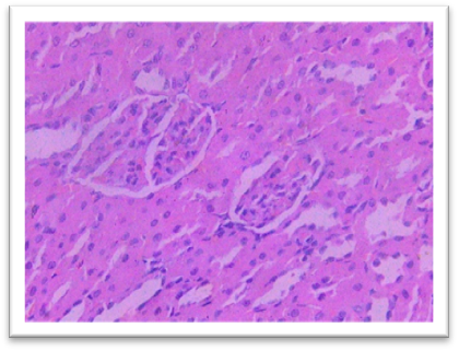
Fig. 18 Group II Kidney showing histological details comparable with normal under 400 X
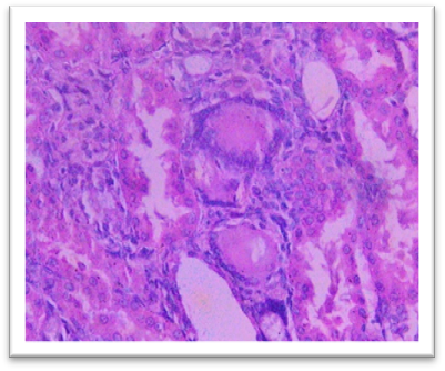
Fig. 19 Group III Kidney exhibits the crystals, congestion and haemorrhage. interstitial infiltration of mononuclear cells and fibrosis. Dilatation of the tubules, focal oedema, and focal interstitial nephritis under H & E 400X.
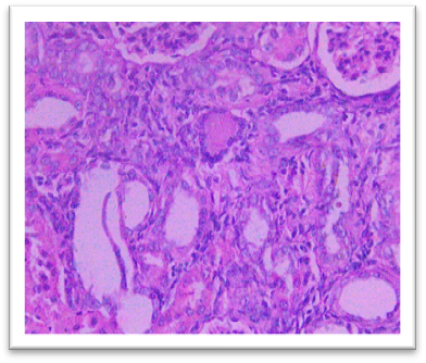
Fig. 20 Group IV Kidney shows moderate focal mononuclear cell infiltration with moderate fibrosis, and 3- 4 crystals under H & E 400 X
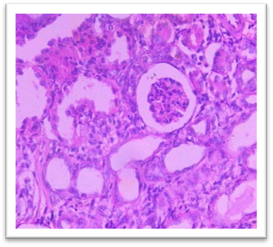
Fig. 21 Group V Kidney exhibits mild and focal interstitial fibrosis, sparse mononuclear cell infiltration, mild tubular dilatation, and 1- 2 crystals under H & E 400X.
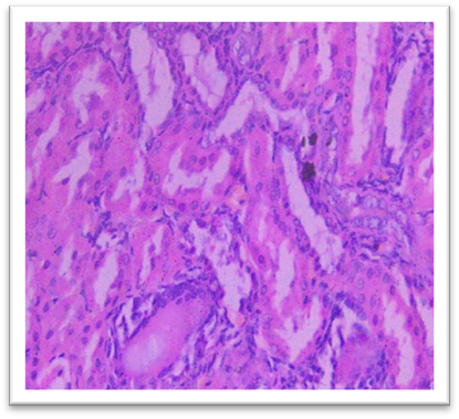
Fig. 22 Group VI Kidney shows minimal focal fibrosis alongside mononuclear cell infiltration, crystals are nearly absent under 400X.
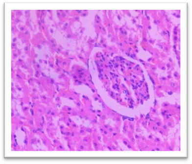
Fig. 23 Group VII Kidney shows very minimal or nearly absent lesions, including fibrosis and mononuclear cell infiltration, with crystals being absent under H & E 400X.
Histomorphometry
At the end of the study, femur bones were dissected and fixed for histological analysis. Group I (Normal) showed normal bone architecture, with well-organized chondrocytes. Group II (Lanthanum Carbonate only) maintained normal bone growth. Group III (Chronic Kidney Disease control) exhibited fibrocartilaginous changes, disorganized collagen, and impaired endochondral ossification, characteristic of Chronic Kidney Dosease. Groups IV (150 mg/kg), V (250 mg/kg), VI (500 mg/kg), and VII (750 mg/kg) demonstrated varying degrees of improvement in bone structure, with the highest dose (Group VII) showing the most significant restoration of normal bone architecture. Histomorphometry of bone: In Group III, designated as positive control for Chronic Kidney Disease, histomorphometric analysis of femur revealed significant fibrocartilaginous changes within the articular cartilage, characterized by an increased fibroblast, loss of chondrocyte organization, and disorganized collagen deposition. Furthermore, the growth plate exhibited a marked increase in the thickness of the hypertrophic zone, suggesting impaired endochondral ossification. Similar findings were observed by Saito et al. (2021) who observed that in Adenine-induced Chronic Kidney Disease, there was a reduction in bone minera density (BMD) throughout the body as well as in the femur Furthermore, micro- computed tomography (micro-CT) analysis revealed a decline in the microstructural integrity of the cortical bone, which contributed to diminished bone strength in both cortical and trabecular regions. Ferrari et al. (2014) reported that the bone microarchitecture in rats subjected to Adenine-induced renal failure exhibited significant alterations, characterized by a reduced trabecular number and an increased trabecular separation. Additionally, the presence of fibrosis was noted in the rats with Adenine-induced renal failure. Ni et al. (2018) performed a histomorphometry analysis and revealing that the bone mineral density (BMD) in the femurs of rats with Chronic Kidney Disease (CKD) was markedly lower than that observed in the control (CTL) group. Additionally, significant bone loss was evident in both cortical and trabecular bone parameters of the femurs. Lanthanum Carbonate @150mg/kg, 250mg/kg, 500mg/kg and 750 mg/kg, histomorphometric analysis revealed very minimal to nearly absent fibroblast proliferation in the articular cartilage, characterized by the absence of fibroblast- like cells and well-maintained chondrocyte organization. Additionally, the growth plate exhibited moderate proliferation of the hypertrophic zone, indicating significant improvement in endochondral ossification compared to the positive control group (Group III) and lower-dose treatment groups. These findings suggest that the highest dose of Lanthanum Carbonate (750 mg/kg) may provide the most effective therapeutic benefits in mitigating bone-related pathological changes associated with Chronic Kidney Disease Yajima et al. (2018) demonstrated that Lanthanum Carbonate enhanced the mineralization of the periosteal surface, augmented bone mass within the intracortical resorption areas, and improved mineralization on the minimodeling surface at the endocortical region. They concluded that Lanthanum Carbonate has the potential to bolster cortical stability in patients with Chronic Kidney Disease (CKD)..
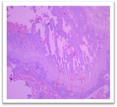
Fig. 24 Group I, Femur showing normal architectural details under H & E 400X.
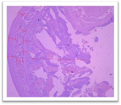
Fig 25: Group II, Femur showing histological architecture details comparable to normal under H & E 400X & 100X respectively.
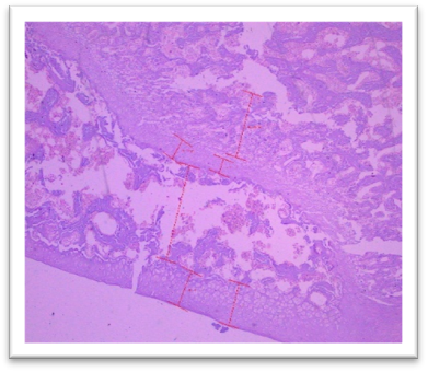
Fig. 26 Group III femur, showing fibrocartilaginous changes, fibroblasts, loss of chondrocyte organization, and disorganized collagen deposition at articular cartilage. Increased thickness of the hypertrophic zone in the growth plate

Fig. 27 Group IV Femur showing moderate fibroblastic changes at articular cartilage and moderate proliferation of hypertrophic zone under H & E 400X.
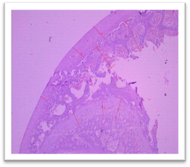
Fig. 28 Group V, Femur exhibited mild fibroblastic changes at articular cartilage and moderate proliferation of hypertrophic zone under H & E 400X.
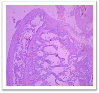
Fig. 29 Group VI showing minimal fibroblastic changes at articular cartilage and moderate proliferation of hypertrophic zone under H & E 400X.
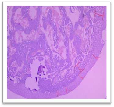
Fig. 30 Group VII Femur, showed very minimal or nearly absent lesions such as fibroblastic changes at articular cartilage and moderate proliferation of hypertrophic zone under H & E 400X.
It was concluded from the present study, that Lanthanum carbonate (Vetricare renal) have shown dose dependant effect and effective in Chronic Renal failure.
REFERENCES


S. S. Jadhav, Dr. S. M. Ghadigaonkar*, M. G. Ghadigaonkar, Evaluation of Efficacy of Lanthanum Carbonate in Rats with Chronic Kidney Disease, Int. J. of Pharm. Sci., 2025, Vol 3, Issue 6, 1712-1725. https://doi.org/10.5281/zenodo.15621357
 10.5281/zenodo.15621357