Department of Pharmacology, SHEAT College of Pharmacy, Varanasi, Uttar Pradesh
Paracetamol (PCM) is extensively used as an analgesic and also an antipyretic medication. When taken at higher doses, the drug can lead to several adverse effects, the most common being hepatotoxicity. This study offers insights into the hepatoprotective properties of abelmoschus manihot seed extract against paracetamol-induced hepatotoxicity. The hepatotoxicity induced by paracetamol was witnessed through a significant rise (P<0.05) in serum values of AST, ALT, ALP and bilirubin. Hydroalcoholic plant extracts, upon administration at doses of 100, 200, and 400 mg/kg to rats, showed protective effects against paracetamol-induced lipid peroxidation as well as against the changes in serum marker enzymes levels. Therefore, it can be concluded that the aforesaid plant extracts possess hepatoprotective activity against paracetamol-induced toxicity.
The liver is an important organ for maintaining bodily homeostasis. Protein synthesis, lipid and carbohydrate storage and metabolism, drug and toxin detoxification, bilirubin excretion, and hormone metabolism are all functions of the liver. Because liver function affects such a wide range of physiological functions, it's reasonable to assume that it affects a variety of homeostatic mechanisms, with potentially catastrophic repercussions for the individual concerned1.
The liver is highly skilled in metabolic conversion and biliary excretion, making it the principal organ in the body responsible for drug removal. It serves multiple purposes as an intricate and specialised organ. Nerves, lymphatic vessels, bile ducts, hepatic arterioles, terminal portal venules, and the acinus make up the smallest functional unit of the liver2.
The liver is crucial in metabolizing and clearing chemicals, making it vulnerable to toxicity from these substances. Some medications can damage the liver when taken in excessive doses or even within therapeutic ranges3.
EPIDEMIOLOGY
Indian population
About 10 lakh persons in India are diagnosed with liver cirrhosis each year. World Health Organisation data shows that liver illnesses rank among India's top 10 killers. Liver illness accounted for 2.44 percent of all fatalities in India in May 2014, according to the most recent data given by the World Health Organisation4.
Worldwide affected population
Roughly 30 million Americans and 29 million Europeans deal with chronic liver disease occasionally or always. Liver cirrhosis was responsible for an estimated 170,000 fatalities in Europe in 2013. There were 493,300 reported deaths in 2010 due to alcoholic liver disease (ALD), with 156,900 female and 336,400 male victims5. This disease produces liver cirrhosis. Liver cancer, according to a plethora of published studies, is the leading cause of cancer-related deaths globally in the modern era. "Worldwide, 788,000 people lost their lives to liver cancer in 2015, according to the World Health Organisation. Between 2014 and 2015, over 25,000 liver transplants were carried out worldwide, with over 5,000 of those operations taking place annually in Europe alone6.
Hepatotoxicity
One form of liver dysfunction or damage linked to the misuse of antibiotics and other medicines is hepatotoxicity. Hepatotoxins, also known as hepatotoxic agents, are substances that harm the liver7.
Mechanism of hepatotoxicity
It is impossible to see medication-related hepatotoxicity as an isolated illness. Hepatotoxicity can be caused by a variety of factors. One of these is the drug's covalent binding to cell proteins, which leads to cell death and membrane disruption. This, in turn, triggers an immune response that hinders the cell pathways that the medication uses for digestion. Another factor is the presence of abnormal bile streams caused by disruptions in subcellular actin fibres or interference with trans-port syphons, which can cause cholestasis and jaundice8. In rare cases, even with minimal cell damage, cells can undergo apoptosis through tumor-corruption factor and Fas pathways. Lastly, the drug can hinder mitochondrial function, leading to the accumulation of reactive oxygen species, lipid peroxidation, fat aggregation, and cell passing9.
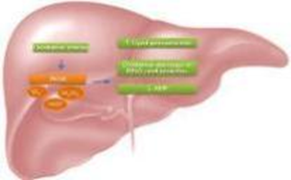
Figure 2: Mechanism of liver damage due to oxidative stress
More than 60% of the world's pharmaceuticals, health care items, and formulations made from plants have experienced extraordinary growth in recent years. The majority of pharmaceutical corporations, including global conglomerates, have been enticed by the surge in international trade in herbal medicine. In 1997, the European market was thought to have reached around $7 billion. Germany dominates the European market, accounting for half of the total at $3.5 billion, or $42.90 per capita. France ($1.8 billion), Italy ($700 million), the UK ($400 million), Spain ($300 million), and the Netherlands ($100 million) follow this market. The herbal medicine database estimates that the Asian market is worth $2.3 billion, while the Japanese market is worth 2.1 billion. But in the United States, the herbal medicine industry is booming10.
The current study aims to assess the hepatoprotective capability of chosen plants by drawing on evidence of numerous hepatoprotective chemical principles that have been previously established, as well as recommendations based on ethnomedical use and literature surveys. The study focusses on Abelmoschus manihot seeds in particular. The hepatoprotective properties of these plants were a deciding factor in their selection11.
MATERIAL AND METHODS:
Collection, identification and authentication of plant material
The seeds of Abelmoschus manihot L were gathered from the eastern region of Uttar Pradesh. The preliminary identification relied on its organoleptic and morphological traits, while the confirmation was carried out by the Botanical Survey of India in Pune.
Extraction of plant material:
The Abelmoschus manihot seeds were dried and then ground into a fine powder at room temperature. After 72 hours at around 64°C, 500 g of powdered plant material was extracted using 1.5 litres of a 70:30 mixture of ethanol and water using the Soxhlet extraction system. The filtrate was then concentrated at 40°C and lowered pressure using a rotating evaporator. At 4°C, the Abelmoschus manihot seed extract (AMSE) was then stored until it was required12. The leaves of Abelmoschus manihot demonstrated a percentage yield of 55.30%, respectively.
Drugs and chemicals
The necessary chemicals utilized during this research work have been Ethanol, Paracetamol as a hepatotoxicant and Silymarin was used as standard drugs.
Equipment
Soxhlet apparatus, Digital Balance, Desiccator, Hot Air oven, Autoanalyzer.
Phytochemical Screening
Extracts was subjected to phytochemical analysis to identify the different phytoconstituent found in the plant were put through a standard qualitative chemical examination to determine the type of phytochemical components they contained. The existence and nonexistence of various phytochemical constituents, like Alkaloids, Carbohydrates, Glycosides, Saponins, Amino acids and Proteins, Flavones and Flavonones, Tannins and Phenolics, Steroids, Fixed oils were determined using standard established techniques13.
Experimental animals
From the Animal House, Saraswati Higher Education and Technical College of Pharmacy, Gahni, Ayar, Varansi, albino rats of either sex weighing 150–200g were acquired. The animals were kept in ideal conditions, with a 12-hour light/dark cycle and room temperatures of 25 ± 1°C. They were fed a regular mouse pellet diet and given access to unlimited water, with the relative humidity being kept at 44-56%. Rats are denied food one hour before the experiment14.
Toxicity study
Rat utilized in this study underwent a fasting period of 12 hours and were randomly allocated into groups containing four and six animals each. Regarding the oral (p.o) administration, A. manihot was administered at varying doses of 200, 400, 800, 1600 and 2000 mg/kg to distinct groups of rat. The subjects were meticulously monitored for any toxicological symptoms and alterations in behavior (including piloerection, rearing, diarrhea, writhing, grooming, paw biting and licking, sedation, hyperactivity, constipation, etc.) for a duration of 2 hours following the administration of the extract''. Mortality rates were documented within a 24-hour period, and the surviving mice were further observed for an additional 14 days to identify any signs of delayed toxicity15. The LD50 was calculated employing the log dose-probit analysis method16.
Assessment of hepatoprotective activity Abelmoschus Manihot seed extract
Thirty-six (36) rats were randomly assigned to six groups (6) and treated with normal saline, varying doses of AMSE, and silymarin over a period of five days. Group A (the Control group) was administered normal saline (NS) exclusively (10 mL/kg, p.o.). Group B received Paracetamol (50 mg/kg, i.p.), while Groups C, D, E, and F were given AMSE extract at doses of 100, 200, and 400 mg/kg p.o., along with silymarin (the standard drug) at a dose of 100 mg/kg (p.o.), in addition to Paracetamol (50 mg/kg, i.p.)17.
Rats from different groups were euthanized on the sixth day of the treatment protocol after being sedated with mild ether. This procedure occurred twenty-four (24) hours after the final treatment. Blood was collected from the retro-orbital plexus and transferred into a vial devoid of anticoagulants. The serum sample was subsequently centrifuged at 5,000 rpm for a duration of 10 minutes, which was then utilized for the analysis of biochemical parameters employing standard diagnostic kits18. The liver was promptly excised and rinsed with ice-cold saline. Both the blood and liver samples were evaluated for their biochemical and antioxidant activities, in addition to histological examination.
Histopathological study
The rats were sacrificed and their livers were removed thereafter. Following a thorough blotting to remove blood and tissue fluids, this liver was then used for histological investigation. After the liver tissue had been fixated in 5% formalin for 48 hours, it was dehydrated using a series of ethyl alcohol-water solutions. After that, it was soaked in paraffin and finally xylene. Slices onto glass slides measuring 15 mm thick were made using a microtome. The liver sections were stained for three to five minutes with 10% haematoxylin, and then rinsed under running water to make the staining more visible. Subsequently, 10% eosin was applied to the sections and left to counterstain for 2 minutes19, 20. The chosen regions were shot after the slices were examined under a photomicroscope set at a magnification of 400.
Statistical analysis
Every one of the values have been expressed as Mean ±S.E.M the result consequence had been analyzed statistically via one way ANOVA followed via Dunnett’s multiple tests.
RESULTS
Phytochemical screening
The phytochemical analysis of the hydroalcoholic extract derived from the seed of Abelmoschus manihot was conducted following the methodology outlined by Khandelwal K. R. This investigation focused on the detection of specific phytochemical components, including saponins, tannins, flavonoids, cardiac glycosides, terpenoids, and alkaloids.
Table 1: Phytochemical composition of hydroalcoholic seed extracts of Abelmoschus manihot
|
Test performed |
Hydroalcoholic extract |
|
Alkaloids |
|
|
Mayer’s reagent |
+ |
|
Wagner’s reagent |
+ |
|
Saponins |
|
|
Foam test |
+ |
|
Borntrager’s test |
+ |
|
Tannins |
|
|
Lead acetate solution |
+ |
|
Ferric chloride solution |
+ |
|
Flavonoids |
|
|
Shinoda test |
+ |
|
Cardiac glycosides |
|
|
Legal test |
+ |
|
Terpenoids |
|
|
Tin and thionyl chloride test |
+ |
+: Present, - : Absent

Acute toxicity study:
The A. manihot extract, when administered via the oral route, resulted in no fatalities at the minimum dosage of 200 mg/kg and higher dose 2000 mg/kg. The estimated LD50 was determined to be >2000 mg/kg.
Table 2: Acute oral toxicity test
|
Sr. no. |
Dose |
No. of animals |
Observation |
|
1 |
200 mg/kg |
03 |
All animals survived |
|
2 |
400 mg/kg |
03 |
All animals survived |
|
3 |
800 mg/kg |
03 |
All animals survived |
|
4 |
1600 mg/kg |
03 |
All animals survived |
|
5 |
2000 mg/kg |
03 |
All animals survived |
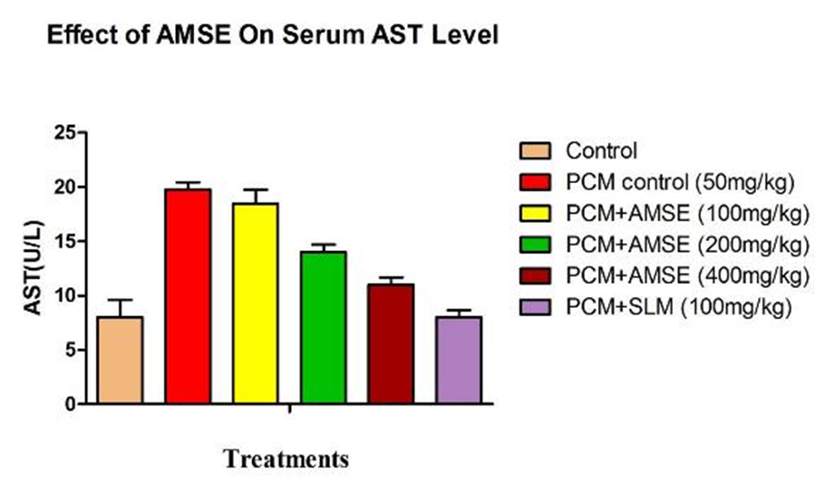
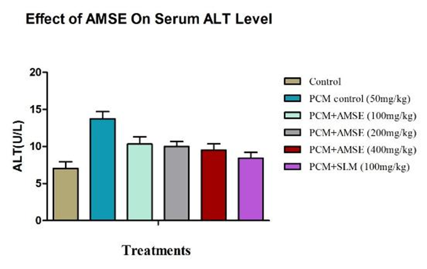
Impact of HAMP extract on Paracetamol-induced hepatotoxicity concerning serum ALT, AST, and ALP
The influence of Paracetamol, AMSE, and SLM on serum ALT, AST, ALP and Bilurubin levels is detailed in Table 1. The group receiving NS+PCM exhibited a significant increase (p<0.05) in serumALT, AST, ALP and Bilurubin when compared to the control group. Administration of varying doses of AMSE (100, 200, and 400 mg/kg) resulted in a reduction of serum ALT, AST, ALP and Bilurubin, with a notable decrease (p<0.05) observed at the AMSE 400 mg/kg dosage in comparison to the NS + Paracetamol group. Conversely, the group treated with SLM 100 mg/kg + PCM demonstrated a significant decrease (p<0.05) in serum, AST, ALP and Bilurubin levels relative to the NS + PCM treated group. (Table 3).
Table 3: Effect of Abelmoschus manihot on Paracetamol induced alterations in serum AST, ALT, ALP and Bilurubin content.
|
Groups |
AST (U/l) |
ALT (U/l) |
ALP (U/l) |
Bilurubin (U/l) |
|
Group-A |
8.00± 1.61 |
7.04±0.90 |
11.43±1.75 |
2.51±0.105 |
|
Group-B |
19.76±0.68 |
13.75±0.96 |
26.00±0.67 |
4.21±0.192 |
|
Group-C |
18.47±1.30 |
10.33±0.98 |
23.75±0.43 |
3.85±0.162 |
|
Group-D |
14.02±0.68 |
10.00±0.68 |
18.00±0.68 |
3.18±0.227 |
|
Group-E |
11.00±0.68 |
9.50±0.86 |
14.43±1.55 |
2.83±0.207 |
|
Group-F |
8.00±0.68 |
8.43±0.77 |
12.33±0.76 |
1.98±0.20 |
All the values have been expressed as Mean ±S.E.M, test employed one-way ANOVA by Dunnett’s test (n=6); significantly different from the control at *(P<0.05), ** ( P<0.01), ***( P<0.001) and ns (non-significant) when compared to the control group.


Histopathological Analysis of Liver Tissue
Normal Control:
The liver tissue from the normal control group shows a typical histological structure with well-arranged hepatic cells with clear cytoplasm and well defined nuclei. There are no sign of inflammation, necrosis and celluar damage, indicating healthy liver function.
Disease Control (PCM):
The liver tissue from the disease control group (induced with Paracetamol) exhibits significant pathological alterations. There is evidence of extensive necrosis, inflammation & congestion. Hepatocytes appear degenerated and the normal liver architecture is disrupted, indicating severe liver damage and dysfunction.
Abelmoschus manihot Seed Extract (200 mg/kg):
The liver tissue from the group treated with hydroalcoholic extract of Abelmoschus manihot L Seed at dose 200 mg/kg demonstrates noticeable hepatoprotective effects. The hepatic cells are better organized with less inflammation and cellular damage, indicating significant recovery and protection against PCM-induced liver injury.
Abelmoschus manihot Seed Extract (400 mg/kg):
The liver tissue from the group treated with hydroalcoholic extract of Abelmoschus manihot L Seed at dose 400 mg/kg shows a considerable reduction in liver damage. The hepatocytes are more organized with fewer sign of inflammation and necrosis, suggesting effective hepatoprotection.
Standard Control (Silymarin):
The liver tissue from standard control group treated with Silymarin shows a significant reduction in liver damage compared to the Disease control group.The hepatocytes are more organized with fewer sign of inflammation and necrosis, suggesting a protective effect of Silymarin against PCM-induced liver damage.
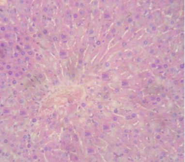
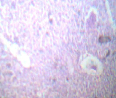
Figure 1: Normal Control Figure 2: Disease Control
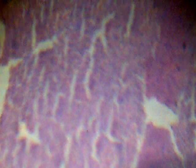

Figure 3: AMSE 200 mg/kg Figure 4: AMSE 400 mg/kg
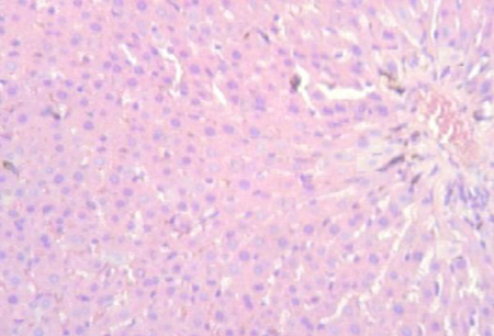
Figure 5: Standard Drug
DISCUSSION
Our study aimed to determine whether the AMSE mitigated paracetamol-induced liver injury in Wistar rats by regulating free radicals and examining changes in biochemical and histological indicators. Damage and inflammation to hepatic tissues can occur after using paracetamol, according to our analysis19. Consistent with previous studies that have shown PCM to have hepatotoxic effects, our results show that the hydroalcoholic extract of Abelmoschus manihot may protect the livers of rats against PCM-induced liver toxicity. Reversal of paracetamol-induced physiological abnormalities was also achieved by treating patients with a graded dose of hydroalcoholic extract of Abelmoschus manihot (100, 200, and 400 mg/kg) and Silymarin (100 mg/kg) in addition to paracetamol20. This indicates that the AMSE kept the membranes of the liver cells structurally intact, which stopped the hemolyzing of bilurubin, ALT, AST, and ALP. But employing paracetamol-induced hepatoxicity, Abhishek kumar et al. (2012) stated comparable results using an ethanol extract of Abelmoschus manihot seed.
A useful indicator of liver disorders, our study found that the NS + PCM treated group had higher levels of ALT, ALP, AST, and bilurubin than the control group (Achliya et al. 2004). In line with previous research, we found that groups given increasing doses of AMSE extract (100, 200, and 400 mg/kg) and Silymarin plus Paracetamol reduced ALT, ALP, AST, and bilurubin levels in a dose-dependent manner. This suggests that the liver functions were restored to a level similar to the control group21. Groups given AMSE extract or silymarin were able to undo the paracetamol-induced alterations in serum AST, ALT, ALP, and bilurubin levels. These results point to the possibility of hepatoprotective properties in the A. manihot extract.
The biochemical and histological results were similar in our investigation. Consistent with previous results by Sangeetha (2016), the group that received paracetamol showed inflammation and hepatocellular coagulative necrosis.
CONCLUSION
In this experiment it was founded that Abelmoschus manihot seed extract at dose 400mg/kg show the significant result in the paracetamol induced hepatotoxicity as the value of AST, ALT, ALP and Bilurubin, were found significant (p<0.001) compared with paracetamol treated group. The liver-protective effects of the hydro alcoholic extract of Abelmoschus manihot were demonstrated in this study. These effects were mainly brought about by the prevention of the elevation of liver biomarkers that are crucial for normal function.
Furthermore, the extract effectively reduced PCM-induced liver damage by normalizing aberrant biochemical indicators and histological changes. If we want more clinical evidence that AMSE extracts can protect the liver from paracetamol-induced liver damage, we need to do more studies.
REFERENCES

Vipul Kaul, Radhika Patel, Evaluation of Hepatoprotective Action of Abelmoschus manihot L Extracts on Experimental Animal Model, Int. J. of Pharm. Sci., 2026, Vol 4, Issue 2, 2419-2430. https://doi.org/10.5281/zenodo.18663898
 10.5281/zenodo.18663898
