
We use cookies to ensure our website works properly and to personalise your experience. Cookies policy
Shri Laxmanrao Mankar Institute of Pharmacy, Amgaon-441902
Simple and sensitive spectrofluorimetric method for phosphorus determination in food samples after microwave mineralisation has been described. The proposed method is based on the formation of a fluorescent chelate between morin and aluminium and quenching the fluorescence after addition of phosphates solution. Additionally, the method with quinine sulphate was modified and applied for food samples. The proposed procedures were compared with respect of linearity, precision and accuracy. A fluorescence spectroscopy is an accurate, sensitive reproducible method for the analysis of pharmaceutical dosage form. Fluorescence Spectroscopy is quick and sensitive tool for analysing molecular environment. The fluorimetry technique is chosen for its exceptional sensitivity, high specificity, low cost. The field of florescence Microscopy is rapidly developing, and it increase image capabilities. Many new technologies and techniques have been developed during last decade that allowed for deeper, faster and high resolution. Fluorimetry is widely established approach that is employed in wide range of applications including Industrial, Medical diagnostics, DNA sequencing. It is excellent tool for both qualitative and quantitative investigations. Fluorescence spectroscopy is also used for Determination of ruthenium, Determination of glucose, Study of marine petroleum pollutants, Determination of zinc, Determination of cadmium, determination of aluminium in alloys, etc. Fluorescence Indicators like Eosin, Quinine sulphate is also useful for analysis. Because there is a choice of wavelengths not only for the radiation produced, but also for the light that stimulates it, the process is exceedingly sensitive and specific.
Fluorometry is superior to spectrophotometry in terms of sensitivity and specificity. Fluorescence has a 10-1000-fold better sensitivity than absorbance studies. It is an analytic method for detecting and measuring fluorescence in compounds that uses ultraviolet light stimulating the compounds, causing them to emit visible light. The energy/light emitted by the substance has a linger wavelength than absorbed. This process of emitting radiation with a longer wavelength than absorbed is known as luminescence (cold light). The mechanism of phosphorescence- As phosphorescing molecules can luminesce for a much longer time than fluorochromes, there must be a difference in the way they store the excitation energy. The basis for this discrepancy is found in the two forms of excitation levels, the singlet excited state and the triplet excited state, which are based on different spin alignments.
The most popular additives in meat and fish products are nitrates, nitrites and phosphates. From the technological and health standpoints, it is essential to develop analytical methods for determination of these salts in food. Several types of method exploit the high sensitivity of fluorimetry and the fluorescence quenching with selected com pounds were described (William R. Ware 1972; Nasu & Minami 1989; Lorenz et al. 1997; Jie et al. 1999; Nainital 2003; Zhang et al. 2003). It can be noted that most of described procedures for phosphate determination based on quenching the fluorescence by phosphate ions and did not apply for real samples. The aim of the presented study was determination of phosphate ions in food samples (meat and fish products) by Spectro fluorimetry. In our work we proposed method based on the formation of a fluorescent chelate between morin and aluminium and quenching the fluorescence after addition of phosphate solution. The proposed procedure was applied for determination of phosphate ions in meat and fish samples after microwave min realisation. Additionally, we modified and ap plied method of phosphate ions determination described by William R. Ware (1972) based on precipitation of quinine. The discussed methods were validated by statistical parameters (linearity, limit of detection, DL and quantification, QL, precision and accuracy) and using certified reference materials.
FLUORESCENCE
Fluorescence is a type of luminescence caused by photons exciting a molecule, raising it to an electronic excited state. It is an optical phenomenon in which the molecular absorption of energy in the form of photons triggers the emission of fluorescent photons with a longer wavelength.
The Mechanism of Fluorescence
Fluorochromes will only fluoresce if they are illuminated with light of the corresponding wavelength. The wavelength depends on the absorption spectrum of the fluorophore and it has to be ensured that an appropriate quantity of energy is delivered to elevate the electrons to the excited state. After the electrons are excited, they can dwell in this high energy state for a very short time only. When the electrons relax to their ground state or another state with a lower energy level, energy is released as a photon.
Phosphorescence
Phosphorescence is a specific type of photoluminescence related to fluorescence. Unlike fluorescence, a phosphorescent material does not immediately re-emit the radiation it absorbs. The slower time scales of the re-emission are associated with "forbidden" energy state transitions in quantum mechanics.
The Mechanism of Phosphorescence
As phosphorescing molecules can luminesce for a much longer time than fluorochromes, there must be a difference in the way they store the excitation energy. The basis for this discrepancy is found in the two forms of excitation levels, the singlet excited state and the triplet excited state, which are based on different spin alignments.
WHAT ARE FLUOROPHORES?
The molecules showing fluorescence activity are termed as fluorophores. A fluorophore is a molecule that will absorb energy of a specific wavelength and reemit energy at a different wavelength. Fluorophores are primarily molecules with aromatic rings, such as Tyrosine, Tryptophan, and Fluorescein. Fluorophores are used as a physical marker for structural studies of macromolecules such as proteins and nucleic acids in biophysical studies. Fluorophores can be extrinsic, such as radioactive probes and dyes, or intrinsic, such as specific amino acids in protein chains. Extrinsic fluorophores are more expensive and require foreign intervention, whereas intrinsic fluorophores do not.
PRINCIPLE
The fluorescence refers to emission of electrons from the singlet ground state to the singlet excited state caused by the absorption of UV or visible radiation. The absorption of energy by incident light rays on molecules causes conformational changes, which leads to vibrational relaxation (the lowest vibrational level). If the aromatic molecule is rigid and cannot relax vibrationally to the ground state, it will reach the ground state through light emission. Fluorescence is caused by the emission of light. The electron systems of these compounds absorb the incident light first. The ground state (S0) of the system's electrons is excited to the excited energy level (S1). Furthermore, these electrons change vibrational levels in the excited state. The electrons move to the lowest vibrational energy level of the excited state by thermal energy expenditure. The state is not stable; it emits energy in the form of UV or visible radiation and returns to singlet ground state. Because these molecules can fluoresce, electrons can jump from the lowest energy level of the excited state to different vibrational energy levels of the ground state by emitting specific quantum of energy in the form of light.
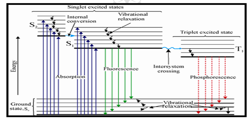
JABLONSKI DIAGRAM
Any fluorescent molecule has two characteristic spectra, the excitation spectrum and emission spectra. The excitation spectrum depicts the relative efficacy of various wavelengths of exciting light in causing fluorescence, whereas the emission spectrum depicts the relative intensity of radiation released at different wavelengths. The wavelength of the exciting radiation has no effect on the shape of the emission spectrum.
ELECTRONIC STATES
Understanding the difference between fluorescence and phosphorescence requires the knowledge of electron spin and the differences between singlet and triplet states. According to the Pauli Exclusion Principle, two electrons in an atom cannot have the same four quantum numbers {Principal (n), Azimuthal (?), Magnetic (m?), and Spin quantum number (s)}. Only two electrons can occupy each orbital where they must have opposite spin states. These opposite spin states are called spin pairing.


Spin Pairing Magnetic Field
Because of this spin pairing, most molecules do not exhibit a magnetic field and are diamagnetic. In diamagnetic molecules, electrons are not attracted or repelled by the static electric field. Free radicals are paramagnetic because they contain unpaired electrons that have magnetic moments that are attracted to the magnetic field.
Singlet State: -
When all the electron spins are paired in the molecular electronic state and the electronic energy levels do not split when the molecule is exposed to UV radiation. If there is no number of unpaired electrons, it means that (n+1) fold degeneracy (equal energy state) will be associated with the electron spin, regardless of the molecular orbital occupied. Thus, if no unpaired electrons are present (n=0), According to the formula: n+1, 0+1 = 1 spin state (singlet state).
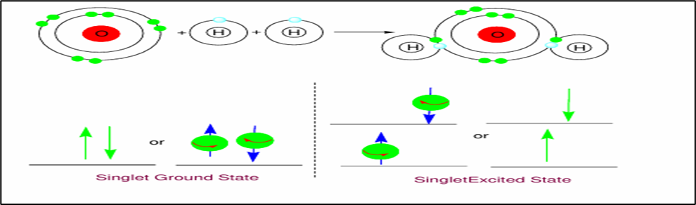
Single State (Ground & Excited)
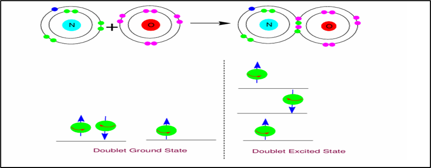
A doublet state occurs when there is an unpaired electron that gives two possible orientations when exposed to UV radiation and imparts different energy to the system.
A singlet or a triplet can form when one electron is excited to a higher energy level. In an excited singlet state, the electron is promoted in the same spin orientation as it was in the ground state (paired). In a triplet, excited stated, the electron that is promoted as the same spin orientation (parallel) to the other unpaired electron.
Doublet state (Ground & Excited)
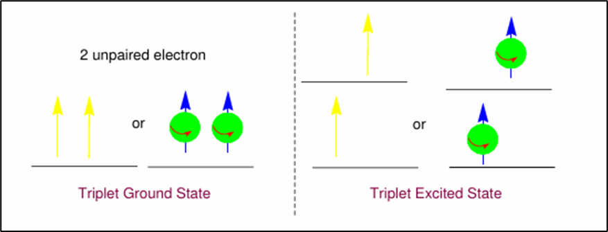
Triplet Ground State & Excited State
Singlet, doublet, and triplet is derived using the equation for multiplicity, 2S+1, Where - S is the total spin angular momentum (sum of all the electron spins). Individual spins are denoted as spin up (s = +1/2) or spin down (s = -1/2). If we were to calculate the S for the excited singlet state, the equation would be 2(+1/2 + -1/2) +1 = 2(0) +1 = 1, therefore making the centre orbital in the figure a singlet state. If the spin multiplicity for the excited triplet state was calculated, we obtain 2(+1/2 + +1/2) +1 = 2(1) +1 =3, which gives a triplet state as expected. The difference between a molecule in the ground and the excited state is that the electrons are diamagnetic in the ground state and paramagnetic in the triplet state. This difference in the spin state makes the transition from singlet to a triplet (or triplet to singlet) more improbable than the singlet-to-singlet transitions. This singlet to triplet (or reverse) transition involves a change in the electronic state. Due to that, the lifetime of the triplet state is longer the singlet state by approximately 10 seconds fold difference. The radiation that induced the transition from ground to excited triplet state has a low probability of occurring, thus their absorption bands are less intense than singlet-singlet state absorption. The excited triplet state can be populated from the excited singlet state of certain molecules which results in phosphorescence. These spin multiplicities in the ground and excited states can be used to explain the transition in photoluminescence molecules by the Jablonski diagram.
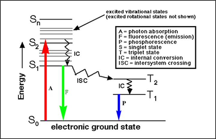
Once a molecule has absorbed energy in the form of electromagnetic radiation (longer wavelength, that is up war-pointing, red arrow, S0 ---> S1, S2, …. Sn), there are a number of routes by which it can return to ground state. If the photon emission (short wavelength that is downward-pointing, green arrow) occurs between states of the same spin state (S1 ---> S0) it is called fluorescence. If the spin state of the initial and final energy levels is different (T1 --> S0), the emission is called phosphorescence. In the diagram, this is depicted by a longer wavelength (lower energy) and therefore shorter length blue line. Since fluorescence is statistically much more likely than phosphorescence for most molecules, the lifetimes of fluorescent states are very short and phosphorescence somewhat longer.
Internal Conversion: - It is an intermolecular process by which a molecule passes to a lower energy electronic state without emission of light. Overlap of vibrational energy levels in two electronic energy levels.

External Conversion: -
External conversion is a process in which excited molecules lose their energy due to collisions with other molecules or by transfer of their energy to solvent or other unexcited molecules.

Therefore, the external conversion is influenced by temperature, solvent viscosity, as well as solvent composition.
Intersystem Crossing: -
In this process spin of an excited electron is reversed and change in multiplicity results. Most common when vibrational manifold overlap exists and when the molecule has a heavy atom substituent (e.g. Br, I).
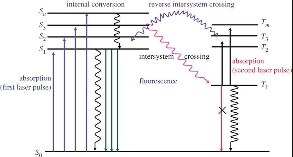
Fig: -Intersystem Crossing
Effect of Structural Nature: -
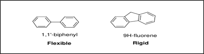
The nature of the chemical structure of a molecule in terms of flexibility and rigidity is of major influence on the fluorescence and phosphorescence signal. Molecules that have a high degree of flexibility will tend to decrease fluorescence due to higher collisional probability. However, more rigid structures have a lower probability of collisions and thus have more fluorescence potential. For example, Biphenyl has very low fluorescence quantum efficiency due to the flexible nature of the molecule while fluorine has high fluorescence quantum efficiency due to its rigidity.
Effect of Solvent Nature: -Solvents affect the luminescent behaviour of molecules. There are three common effects can be recognized –
The polarity of Solvent: - A polar solvent is preferred as the energy required for the P ---> P* is lowered.
The viscosity of Solvent: - Highly viscous solvent is preferred since collisional deactivation will be lowered at higher viscosities.
Heavy Atoms in Solvent: - If solvents contain heavy atoms, fluorescence quantum efficiency will decrease and phosphorescence will increase.
Effect of Substitution: -Substitution in the structure can also affect the fluorescence.
|
Groups increase the fluorescence intensity |
OH, Me, Et, CN, NHR, NH2, NR2, NO, NO2 |
|
Groups decrease fluorescence intensity |
COOH, CHO, COR, COOR, SH, F, Cl, Br, I |
|
Groups having no effect on fluorescence intensity |
SO3 H, NH4 +, Alkyl group |
Effect of Temperature: -
Molecule experiences larger collisional deactivation at high temperatures due to an increase in the movement and velocity of molecules. Therefore, lower temperatures are preferred for analysis.
Effect of Dissolved Oxygen: Oxygen Dissolved oxygen affects fluorescence at large scale. Molecules experience intersystem crossing due to it is paramagnetic nature.
QUENCHING: -
It refers to any process that decreases the fluorescence intensity of a sample. A variety of molecular interactions can result in quenching. Like – molecular rearrangement, Static quenching, and collisional quenching, etc.
Excited-State Reactions Quenching: -
Such reactions occur because light absorption frequently changes the electron distribution within a fluorophore, which in turn changes its chemical or physical properties. For example, a neutral solution of phenol can lose the phenolic proton in the excited state.
Molecular Rearrangement Quenching: -
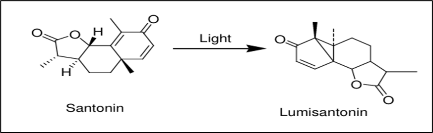
It involves the migration of a group or an atom from one centre (migration origin) to another (migration terminus) due to light and heat within the same molecule. For example, Lumi santonin a photoproduct of santonin obtained via molecular rearrangement. The C-3 carbonyl group has moved to C-2, the C-4 methyl has moved to C-1, and the C-10 carbon has been inverted.
Collisional Quenching: -
Collisional quenching occurs when the excited fluorophore experiences contact with an atom or molecule that can facilitate non radiative transitions to the ground state. Common quenchers include O2, I-, Cs+, and acrylamide. For example, quenching of quinine drug by chloride ion and quenching of tryptophan by iodide ion.
Static Quenching: -
Static quenching occurs at the ground state of the fluorescent molecule. It can be simplified by the following mechanism-
Light
(1) F + Q ———≥ F: Q ———≥ Q* + F
(2) Q*——≥ Q + Energy
Here, a complex formation occurs between the fluorescing molecule at the ground state (F) and the quencher molecule (Q) through a strong coupling. Such complex may not undergo excitation or, may be excited to a little extent reducing the fluorescence intensity of the molecule. For example, Caffeine and related xanthine and purines reduce the intensity of riboflavin by the static mechanism.
Concentration: -
Concentration quenching is a kind of self-quenching. It occurs when the concentration of the fluorescing molecule increases in a sample solution. The fluorescence intensity is reduced in a highly concentrated solution (>50 μg /ml).
Chemical Quenching: -
Chemical quenching is due to various factors like change in pH, presence of oxygen, halides, and electron-withdrawing groups, heavy metals, etc.
Change in pH: -
Aniline at pH (5-13) gives fluorescence when excited at 290 nm. But pH 13 does not show any fluorescence.
Oxygen Molecules: -
Oxygen leads to the oxidation of fluorescent substance to non-fluorescent substance and thus, causes quenching.
Halides and electron-withdrawing groups:
Halides like chloride ions, iodide ions, and electron-withdrawing groups like -NO, -COOH, -CHO groups lead to quenching.

Electron withdrawing process by nitro groups
Heavy Metals: -
The presence of heavy metals also leads to quenching because of collision and complex formation.
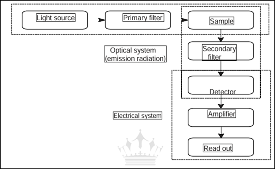
Instrumentation of Fluorimetry: -
A fluorometer or fluorimeter is a device used to measure fluorescence parameters such as intensity and wavelength of emission spectrum. These factors aid in the detection of individual molecules in a medium, as well as their quantity. Modern fluorometers are capable of detecting fluorescent molecule concentrations as low as 1 ppm.There are two basic types of fluorometers, the filter fluorometer and the spectrofluorometer. The difference between them is the way they select the wavelengths of incident light. A filter fluorometer makes use of filters, whereas a spectrofluorometer makes use of grating monochromators. Filter fluorometers are less expensive to buy/build, but they are less sensitive and have lesser resolution than spectrofluorometers.
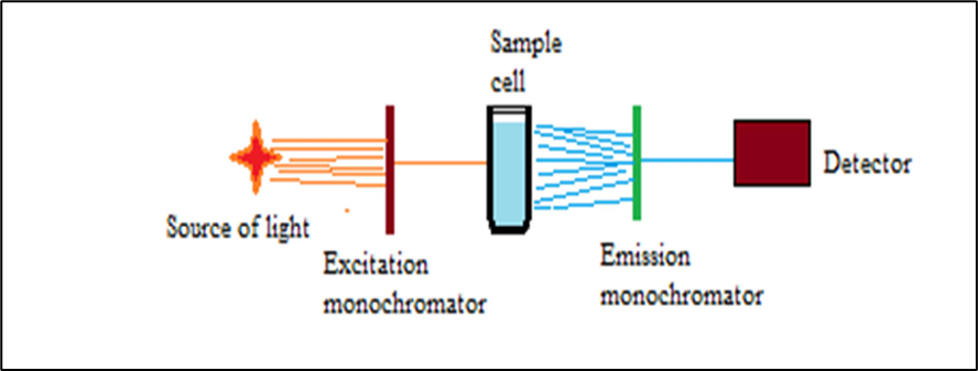
Schematic Diagram of Single-Beam Fluorimeter
FLUORIMETER
The basic components of a filter fluorometer are similar to those of a photometer. It too makes use of filters to limit the wavelengths of the excitation and emission beams.
To limit the impact of variations and drift in source intensity and detector response, filter fluorometers typically employ a single beam arrangement with source intensity control.
Spectrofluorometer mainly consists of
SOURCE OF LIGHT: -
FILTERS AND MONOCHROMATORS: -
Primary filters and secondary filters.
Excitation monochromators and Emission monochromators.
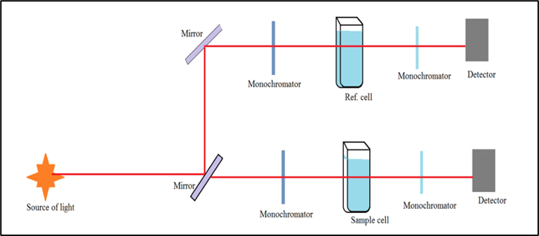
Schematic Diagram of a Typical Fluorometer
SOURCE OF LIGHT: -
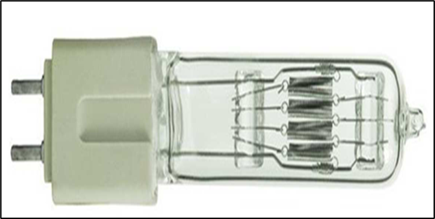
Mercury vapor Lamp: -
Mercury Vapor Lamp These lamps are ideal light sources that provide high-intensity light in the deep UV to visible regions. It consists of 2 alloys (tungsten) electrodes which are placed together in a medium containing mercury vapor and 25-50 torr of pure argon gas. These electrodes are enclosed in an elliptically shaped in a silica glass tube. It provides clear white light, high intensity with 24000 Hrs. of life.

Mercury Vapour Lamp
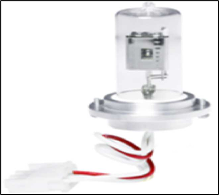
Xenon arc Lamp
It consists of two tungsten electrodes form an arc at a specific distance and xenon gas is stored (under pressure) in quartz or fused silica tube. It emits radiation with a higher intensity (500 nm) than a hydrogen discharge lamp. Wavelength: 750-1000 nm.
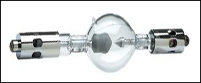
Xenon arc Lamp
Tungsten Halogen Lamp: -
It is also known as a halogen lamp. It is an incandescent light source. It is consists of a filament made up of tungsten enclosed in a quartz vessel containing an inert gas and a small quantity of Iodine or bromine (Halogen). Its 85% emitted light lies in IR and near IR region, 15 % in the visible region, and less than 1% in the UV region.
Tungsten Halogen Lamp
Deuterium and Hydrogen Lamp: -
A pair of electrodes is enclosed in a glass tube containing hydrogen or deuterium gas. When current is passed in electrodes electron discharge is occurring which exited the gas molecule which results in the emission of radiation (UV & Visible). Wavelength: 160-800 nm.Quartz window must be employed.
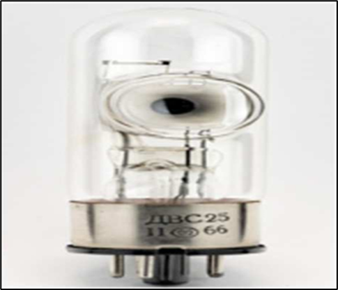
D2 Lamp Hydrogen Lamp
Filter and Monochromator: -
Filter: -
Filter is a device used to get selected wavelength. It allows the light pass through it but absorbed the light of different wavelength may partially and fully. A specific filter is used to obtain the desired wavelength for special analysis like Primary filter and Secondary filter.
Primary filter: -Absorbs visible radiation and transmit UV radiation.
Secondary filter: -Absorbs UV radiation and transmit visible radiation.
Monochromator: -
They convert polychromatic light into monochromatic light. They can isolate a specific range of wavelength or a particular wavelength of radiation from the source.
Excitation Monochromators: - Provides suitable radiation for excitation of molecules.
Emission Monochromators: - Isolate only the radiation emitted by the fluorescent
molecules.
Sample Handle or Cells or Cuvettes: -
Cuvettes are used for the handling of samples. These are rectangular or cylindrical in shape with two rough and two smooth sides, and made up of glass, quartz or fused silica.

Cuvettes
Detector: -
Detector is a device which transforms light energy into electrical signals that are observed on recorder. The characteristics of ideal detector is giving quantitative response, high sensitivity, low noise, short response time, and response quantitative to wide spectrum of radiation received.
SOME COMMONLY USED DETECTORS ARE AS FOLLOWS: -
Barrier layer cell/Photovoltaic cell: -
It is consisting of a coated silver or gold thin layer of metallic film which acts as an electrode and another metal plate acts as another electrode. Both of the layers are separated by selenium layer that act as a semiconductor. When UV radiation falls on selenium layer, an electron becomes mobile and is taken up by transparent metal layer that results a potential difference between the electrodes & causes the flow of current. When it is connected to galvanometer, a flow of current observed which is proportional to the intensity and wavelength of light falling on it.
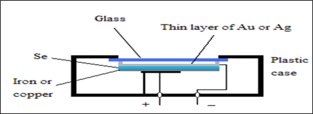
Barrier layer cell/ Photovoltaic cell
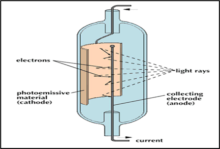
Phototubes/ Photo emissive tube: -
It is consists of an evacuated glass tube with a photocathode and a collector anode. The surface of photocathode is coated with a layer of elements like caesium, silver oxide and its mixtures. When radiant energy falls on photosensitive cathode, electrons are emitted which are attracted to anode causing flow of current. It is more sensitive than barrier layer cell.
Phototubes/ Photo emissive tube
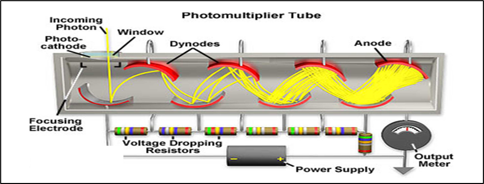
Photomultiplier tube: -
Photomultiplier tube is multiplying the photoelectrons by secondary emission of electrons. A primary photo-cathode is fixed in a vacuum tube which receives radiation from the sample. Some 08 to 10 dynodes are fixed each with increasing potential of 75-100V higher than preceding one. Near the last dynode an electron collector electrode is fixed. It is extremely sensitive to light and detect weaker or low radiation.
ADVANTAGES: -
DISADVANTAGES: -
APPLICATIONS: -
CONCLUSION
Fluorimetry is a highly sensitive analytical technique based on the principle that certain molecules emit light (fluorescence) when they absorb UV or visible radiation and return from an excited state to their ground state. The emitted light, having a longer wavelength than the absorbed light, is measured to quantify the presence and concentration of fluorescent substances. Due to its specificity, rapid response, and ability to detect low concentrations, fluorimetry is widely used in pharmaceutical analysis, biochemistry, and environmental monitoring. Its core advantage lies in the proportional relationship between fluorescence intensity and analyte concentration, enabling precise and sensitive measurements.
REFERENCES


Apurva Kapse, Rani Bhagat, Tulsidas Nimbekar, Fluorimetry: A Simple, Rapid and Sensitive Analytical Technique used in Pharmaceutical Industry, Int. J. of Pharm. Sci., 2025, Vol 3, Issue 12, 2713-2728. https://doi.org/10.5281/zenodo.17961847
 10.5281/zenodo.17961847