
We use cookies to ensure our website works properly and to personalise your experience. Cookies policy
Mount Zion College of Pharmaceutical Sciences and Research, Adoor.
Cracked heels (foot fissures) are a common dermatological condition associated with dryness and susceptibility to infection. Conventional topical treatments often show limited effectiveness due to poor skin penetration. The present study aimed to formulate and evaluate a herbal niosomal gel containing pomegranate (Punica granatum L.) peel extract for the management of foot cracks. Niosomes were prepared using the ether injection method with non-ionic surfactants and cholesterol and incorporated into a Carbopol 940-based gel. The formulation was evaluated for vesicular characteristics and physicochemical parameters, including pH, viscosity, homogeneity, spreadability, washability, and skin irritation. The study emphasizes the potential of niosomal gel systems to enhance topical delivery of herbal actives and provide controlled drug release, offering a promising alternative to conventional topical formulations for cracked heels.
Novel Drug Delivery Systems (NDDS) are advanced pharmaceutical approaches designed to enhance therapeutic efficacy, improve bioavailability, minimize adverse effects, and increase patient compliance by delivering drugs in a controlled and targeted manner. These systems utilize innovative carriers such as nanoparticles, liposomes, ethosomes, and niosomes to achieve sustained release, improved stability, and site-specific action. Among these, vesicular drug delivery systems play a crucial role by encapsulating drugs within lipid or surfactant-based vesicles, thereby enhancing skin penetration and reducing systemic side effects.
Niosomes are non-ionic surfactant-based vesicles composed of cholesterol and suitable hydration media. They are biodegradable, biocompatible, chemically stable, cost-effective, and capable of encapsulating both hydrophilic and lipophilic drugs. Due to their nanoscale size and amphiphilic structure, niosomes improve drug retention, provide sustained release, and protect active compounds from degradation. Cholesterol contributes to membrane rigidity and stability, while non-ionic surfactants enhance permeability and reduce toxicity, making niosomes suitable for topical and transdermal drug delivery.
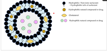
Figure no. 1: Structure of niosome
The skin, the largest organ of the human body, serves as a protective barrier against environmental, chemical, and microbial insults while maintaining homeostasis. Structurally, it consists of the epidermis, dermis, and subcutaneous tissue, each contributing to protection, sensation, immune defense, and thermoregulation. The unique structure of foot skin particularly the thick stratum corneum and absence of sebaceous glands makes it prone to dryness and cracking.
Foot cracks, also known as heel fissures, are common dermatological conditions caused by dryness, prolonged pressure, climatic factors, nutritional deficiencies, improper footwear, and systemic diseases such as diabetes and psoriasis. If untreated, fissures may deepen, causing pain, bleeding, and secondary bacterial or fungal infections. Conventional treatments include emollients, creams, and petroleum-based products; however, these often provide limited penetration and short-term relief.
Topical drug delivery offers an effective approach for managing localized skin disorders by delivering drugs directly to the site of action while avoiding first-pass metabolism and systemic side effects. Gels are preferred topical formulations due to their non-greasy nature, ease of application, good spreadability, and prolonged residence time on the skin. Incorporation of niosomes into gel bases further enhances drug permeation, stability, and controlled release.
Herbal medicines are increasingly favored due to their safety, affordability, and multifunctional therapeutic properties. Pomegranate (Punica granatum L.) peel is rich in polyphenols, flavonoids, tannins, and punicalagins, exhibiting strong antioxidant, antimicrobial, anti-inflammatory, and wound-healing activities. Incorporating pomegranate peel extract into a niosomal gel system offers a promising strategy to enhance skin penetration and therapeutic effectiveness in the treatment of cracked heels.
MATERIALS AND EXCIPIENTS
All materials and excipients used in the study were of analytical grade and were obtained from standard commercial suppliers. Tween 80, Di-sodium Hydrogen Orthophosphate Anhydrous, Sodium Dihydrogen Orthophosphate, Triethanolamine, Diethyl ether, Cholesterol, Glycerol were obtained from Isochem Laboratories Angamaly, Kochi and Tween 40, Span 60, Span 20, Carbapol 940, Curcumin, Rose water were sourced from Chemdyes Corporation Rajkot, Gujarat. Pomegranate Peel Powder was obtained from Jain Lifesciences Pvt.Ltd., Hyderabad and aloevera gel was procured from Dhathri Life Sciences Pvt. Ltd., Kayamkulam.
FORMULATION OF NIOSOMAL FOOT CRACK GEL
The formulation of niosomal foot crack gel was prepared by incorporation method. Preparation of niosome and gel is further demonstrated.
Extraction of punicalgin from pomegranate peel
Fruits were washed and peels are separated from the seeds. The peels are then dried in shade and powdered in a blender. Powdered husk was then macerated with water and methanol for 7 days with occasional stirring. After maceration, the thick puree was then filtered through muslin cloth to yield a dark brown extract. The filtrate was concentrated using rotary evaporator at 40-500oC. A semisolid thick extract is obtained.
Table no. 1 Tests for Pomegranate Peel water extract
|
Test |
Experiment |
Observation |
Inference |
|
Alkaline reagent Test |
Test solution + few drops of NaOH solution |
Intense yellow colour is obtained which turns to colourless on addition of few drops of diluted acid. |
Flavonoids are present |
|
Lead acetate test |
Test solution + lead acetate solution |
Yellow precipitation occurs |
Quercetin and other flavonoids are present |
|
Sulfuric acid test |
Test solution + sulfuric acid |
Orange to red color |
Flavanones are present |
|
Ferric chloride test |
Test solution + ferric chloride solution |
Brownish blue or green color |
Hydrolysable tannins are present |
|
Gelatin test |
Test solution + 1% gelatin solution + 10% NaCl solution |
White buff colored ppt. formed |
Tannins present |
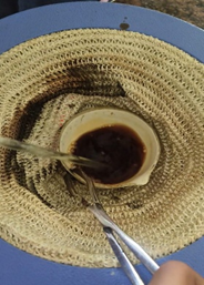
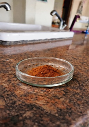
Figure no. 2: Extraction of Pomegranate peel
Method of preparation of niosome
Niosomes were prepared by ether injection method. Mixture of surfactant and cholesterol were dissolved in diethyl ether, which is then mixed with ethanol solution containing the drug. This entire solution was slowly injected at 1ml/min through a 24-guage needle into preheated buffer. This solution was stirred using a magnetic stirrer until the formation of vesicles occurred.
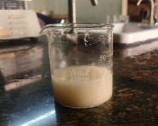
Figure no. 3: Formulation of niosome
Table no. 2 Formulation of niosome
|
Sr. No. |
Ingredients |
Weight Taken (g) |
|||
|
F1 |
F2 |
F3 |
F4 |
||
|
1 |
Span 20 |
0.1 |
- |
- |
- |
|
2 |
Span 60 |
- |
0.1 |
- |
- |
|
3 |
Tween 40 |
- |
- |
0.1 |
- |
|
4 |
Tween 80 |
- |
- |
- |
0.1 |
|
5 |
Drug |
0.1 |
0.1 |
0.1 |
0.1 |
|
6 |
Cholesterol |
0.1 |
0.1 |
0.1 |
0.1 |
Method of preparation of gel
The extract loaded niosomal gel was prepared by dispersion of carbapol 940 in distilled water and allowed to swell overnight. The swelled carbapol was stirred at 600rpm for 60 min. Add glycerine and phenoxy ethanol into the hydrated carbapol. Mix gently until uniform. Mixture was neutralized by dropwise addition of triethanolamine while stirring. Add turmeric powder and aloe vera into it. Mixing was continued until a transparent gel appeared, while the amount of base was adjusted to achieve a gel with pH 5.5.
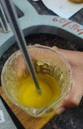
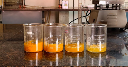
Figure no. 4 : Preparation of gel
Table no. 3 Formulation of gel
|
Ingredients |
F1 |
F2 |
F3 |
F4 |
|
Niosomal formulation |
10ml |
10ml |
10ml |
10ml |
|
Carbapol 940 |
0.21g |
0.30g |
0.19g |
0.25g |
|
Phenoxyethanol |
0.4ml |
0.4ml |
0.4ml |
0.4ml |
|
Triethanolamine |
0.18ml |
0.3ml |
0.17ml |
0.21ml |
|
Turmeric powder |
0.2mg |
0.2mg |
0.2mg |
0.2mg |
|
Aloevera gel |
1g |
1g |
1g |
1g |
|
Rose water |
3ml |
3ml |
3ml |
3ml |
|
Glycerine |
2ml |
2ml |
2ml |
2ml |
EVALUATION
Evaluation of Niosome
1. Appearance: The appearance of the drug-loaded niosomal dispersion was assessed.
2. Entrapment efficiency: Niosome entrapment efficiency was calculated using the ultracentrifugation method, which involved centrifuging the niosome dispersion for 90 minutes at 14,000 rpm. The pH 7.4 phosphate buffer was used to dilute the clear supernant from the centrifuged solution, which was then subjected to spectrophotometric analysis. The percentage entrapment efficiency (EE%) was computed using the following formula. Entrapment Efficiency is calculated as follows:
Entrapment efficiency = (Amount of drug entrapped ÷ Total amount of drug used) × 100
3. Particle Size: Determination of vesicle size can be carried out using zeta sizer instrument. This instrument contains Malvern PCS software. The sample must be diluted with distilled water before taking the result. The particle size must be required in nano range (10 - 3000nm).
4. Zeta potential: Zeta potential for niosomal formulation was performed using Zeta sizer Beckman coulter instrument. It denotes a scientific term for electro-kinetic potential in vesicle systems. This represents the potential difference between the dispersion medium and the stationary layer of fluid that is attached to the dispersed particle. Zeta potential was assessed using a flow-through cell cuvette that operates on the principle of electrophoretic light scattering (ELS). This method identifies the electrophoretic movement of charged particles in an electric field by analysing the Doppler shift of scattered light.
Evaluation of gel
1. Organoleptic evaluation: The gel formulation was tested for its color, odour and appearance.
2. pH: The amount of hydrogen ions in a solution is measured by pH, represented in moles per litre. A pH meter was used to determine the prepared gel's pH. The ideal pH range for topical preparations is 7-8. The pH meter calibration was done by using a standard buffer solution. Then weigh 0.5g of gel and dissolve in 50 ml distilled water and the pH was measured.
3. Viscosity: The sample was taken in the beaker and was allowed to rotate at 20 and 30 rpm respectively at spindle no 64. At each speed the reading was noted. Average of 3 readings was taken.
4. Homogeneity: Two transparent glass plates were taken. Spread 1g of the gel on one of the glass plates and put the second glass plate over it. Observe whether the gel spreads uniformly without lumps or particles.
5. Spreadability: It was determined by taking 0.5 gm of gel on the glass slide over the 1cm of diameter. Second slide was placed over it. Weight of 500 gm was placed on it and waited for 5 minutes. The increase in the diameter of gel was noted and average of 3 determinations were taken. It was then calculated by using following formula:
S = m.L/T
Were,
6. Washability: This test involved applying a small amount of gel to the skin, which was then rinsed off with water.
7. Irritancy: In this test patches of gel were applied on the skin and the effect to the skin on application of gel was compared with the market product.
RESULT AND DISCUSSION
The following result and discussion present the physicochemical characterization of the formulated niosome;
1. Particle size
Particle size was determined using particle size analyser. The formulation (F4) is best among rest of the formulation due to its optimal particle size (429.3nm) and uniform vesicle distribution, indicating stable niosome formation. Tween 80, a non-ionic surfactant with high HLB value, promotes the formation of flexible and stable vesicles. The obtained particle size is suitable for topical/transdermal delivery, providing better surface area and controlled drug release. A decrease in particle size enhances stability, skin penetration, and drug release, whereas an increase in particle size may lead to vesicle aggregation, reduced uniformity, and poor formulation stability. Therefore, the balanced particle size observed in formulation (F4) was represented in Figure no. 5
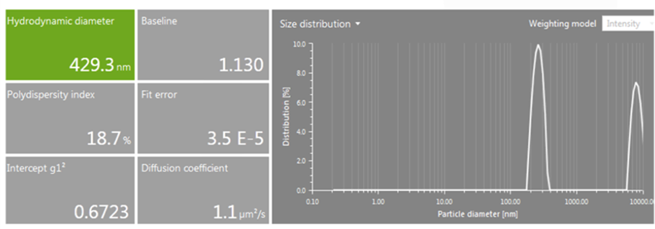
Figure no. 5: Particle Size of Niosome (F4)
Table no. 4 Particle size of different niosomal formulations
|
Formulations |
Particle size (nm) |
|
F1 |
501.9 |
|
F2 |
472.1 |
|
F3 |
458.8 |
|
F4 |
429.3 |
2. Zeta potential
Formulation (F4) prepared with Tween 80 was selected based on zeta potential because it showed an adequate negative zeta potential value (−21.5 mV), indicating sufficient electrostatic repulsion between vesicles and thus better physical stability compared to the other formulations. Zeta Potential of the formulation (F4) was represented in Figure no. 6.
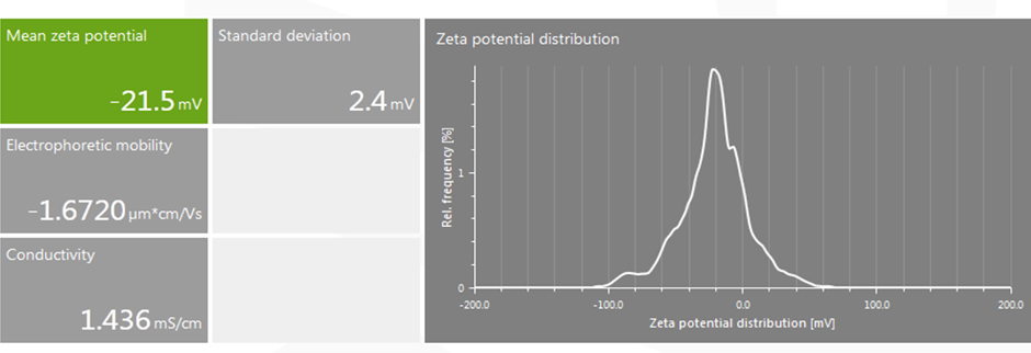
Figure no. 6 : Zeta Potential of Niosome (F4)
Table no. 5 Zeta potential of different niosomal formulations
|
Formulations |
Zeta potential (mV) |
|
F1 |
-42.1 |
|
F2 |
-35.8 |
|
F3 |
-17.6 |
|
F4 |
-21.5 |
3. Entrapment Efficiency
The niosomal formulation (F4) showed a high entrapment efficiency, indicating efficient incorporation of drug within the vesicles. High entrapment efficiency is desirable for topical delivery as it ensures sustained drug release and improved therapeutic efficacy.
Table no. 6 Entrapment efficiency of different niosomal formulations
|
Formulations |
Entrapment Efficiency (%) |
|
F1 |
67% |
|
F2 |
73% |
|
F3 |
82% |
|
F4 |
92% |
The following result and discussion present the evaluation of the developed niosomal gel;
4. pH
The pH of the formulations was determined using digital pH meter. The pH of the niosomal gel formulation (F2) is in range of 4 to 6 which is compatible to the skin. Triethanolamine is the agent that influence the variation in pH. A bar diagram Figure no.7 was plotted by taking formulation on x axis and pH value on y axis with the data's obtained from the Table no: 4
Table no. 4 pH of niosomal gel
|
Formulation |
pH |
|
F1 |
3.3 |
|
F2 |
5.50 |
|
F3 |
4.5 |
|
F4 |
3.9 |
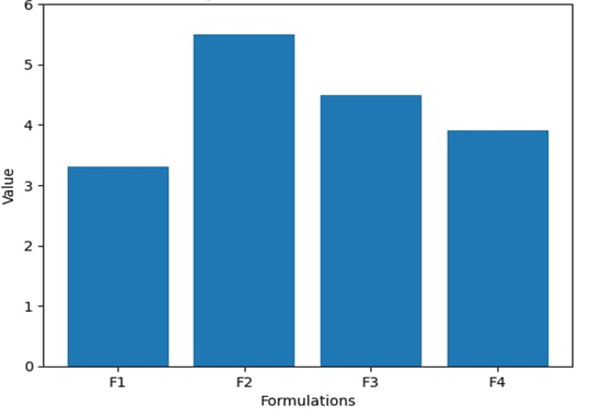
Figure no. 7 pH of different formulations
5. Viscosity
The formulation (F2) was considered better than the other formulation. it exhibited optimum viscosity ensures good spreadability, ease of application, and adequate retention at the site of application. An appropriate viscosity also contributes to better physical stability and uniform drug distribution within the gel. Viscosity of the formulation was demonstrated in Figure no.8.
Table no. 5 Viscosity of niosomal gel
|
Formulation |
Viscosity (cp) |
|
F1 |
16457 |
|
F2 |
22356 |
|
F3 |
14788 |
|
F4 |
9876 |
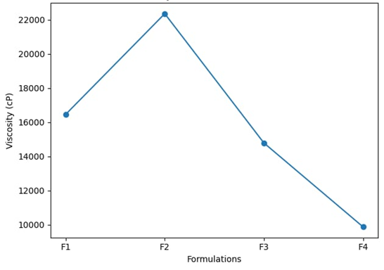
Figure no. 8 Viscosity of different formulations
6. Homogeneity
The formulation (F2) was found to have good homogeneity, others (F1,F3,F4) have satisfactory.
7. Washability
The washability test demonstrated that all niosomal gel formulations were easily washable with water, indicating good patient compliance and the absence of excessive greasiness, which is desirable for topical application.
8. Spreadability
The spreadability study revealed that all niosomal gel formulations (F1,F3,F4) exhibited satisfactory spreadability, indicating ease of application, with formulation F2 showing comparatively higher spreadability due to its optimum viscosity and uniform gel consistency.
Table no. 6 Spreadability of niosomal gel
|
Formulation |
Spreadability (g.cm/sec) |
|
F1 |
14.5 |
|
F2 |
17.6 |
|
F3 |
16.2 |
|
F4 |
15.5 |
9. Irritancy
The irritancy study showed that none of the niosomal gel formulations produced irritation at the site of application, indicating that the formulations were non-irritant and safe for topical use.
CONCLUSION
The development and assessment of an herbal niosomal gel for treating foot cracks have been finalized. Primary method of treatment is the application of topical treatment even for minor cases. Multilamellar vesicles called niosome effectively transport active substances into the deeper layers of the skin. On the basis of the study, it can be stated that the formulation (F4) is the best formulation from all the other formulation also (F4) has particle size of lowest value as compared to others i.e., Zeta potential also conducted for showing the stability of formulation. The optimized formulation was then formulated into a topical niosomal gel (F2) and various evaluating parameters like pH, viscosity, spreadability and was done. The work concluded the effectiveness of the formulation i.e. niosomal gel incorporated by particular constituent punicalgin into non-ionic surfactants and cholesterol in different ratios. Further studies can be conducted by designing the different formulation for different activities.
REFERENCES


Asna A, Anagha Ajayakumar, Ashik Nazeer, Malavika S, Mereena Johnson, Dr. Prasanth V V, Formulation and Evaluation of Niosomal Foot Crack Gel using Pomegranate Peel, Int. J. of Pharm. Sci., 2026, Vol 4, Issue 2, 1701-1710. https://doi.org/10.5281/zenodo.18610163
 10.5281/zenodo.18610163