
We use cookies to ensure our website works properly and to personalise your experience. Cookies policy
Mount Zion College of Pharmaceutical Sciences and Research, Chayalode, Adoor
Burn injuries require effective topical therapy to prevent infection and promote healing. This study developed a herbal burn cream using Saraca asoka, Cassia fistula, and chicken fat as a natural lipid base. Authenticated plant materials were cleaned, shade-dried, pulverized, and extracted, while chicken fat was rendered and purified before incorporation into a cream base. The formulation was evaluated through physical parameters and in vitro studies. Results indicated satisfactory properties and promising activity, suggesting the cream’s potential as a safe and effective treatment for burn management. Further in vivo studies are recommended to confirm its clinical efficacy and safety.
The skin is the largest organ of the human body and serves as the first line of defence against physical, chemical, and microbial damage. It plays an essential role in protection, sensation, thermoregulation, excretion, and prevention of excessive water loss. Structurally, the skin consists of three main layers: the epidermis, dermis, and hypodermis. The epidermis is the outer protective layer that prevents pathogen entry and contains melanin responsible for skin colour. The dermis lies beneath the epidermis and is composed of connective tissue, blood vessels, nerves, sweat glands, and hair follicles, providing strength, elasticity, and sensory function. The hypodermis layer consists mainly of adipose tissue and connective tissue, offering insulation, cushioning, and energy storage. The skin maintains hydration through natural oils and lipid barriers that help retain moisture and keep the skin soft, flexible, and healthy. It also contains thermoreceptors that make it sensitive to heat and cold, enabling the body to respond to temperature changes (1). Excessive exposure to heat can damage skin cells, leading to burns. Burns are injuries to the skin and underlying tissues caused by thermal agents, chemicals, electricity, radiation, or friction. Based on severity, burns are classified into first-degree burns, which affect only the epidermis and cause redness and pain; second-degree burns, which involve the epidermis and part of the dermis and result in blistering and severe pain; and third-degree burns, which destroy all layers of the skin and may extend to deeper tissues, often appearing white or charred with loss of sensation due to nerve damage. The aetiology of burns includes exposure to flames, hot liquids, acids and alkalis, electrical current, radiation, and mechanical friction. Pharmacognosy plays a significant role in burn treatment by providing natural therapeutic agents derived from medicinal plants that aid in wound healing, reduce inflammation, prevent microbial infection, and promote tissue regeneration. Medicinal plants such as Aloe vera, Curcuma longa, Azadirachta indica, Centella asiatica, and Calendula officinalis are widely used in burn management due to their bioactive constituents including polysaccharides, curcumin, nimbin, flavonoids, and triterpenoids, which exhibit anti-inflammatory, antimicrobial, antioxidant, and wound-healing properties. In addition to medicinal plants, traditional home remedies such as aloe vera gel, honey, coconut oil, and turmeric paste are commonly used for minor burns due to their soothing and protective effects (2). Furthermore, several herbal and commercial burn care products, including gels, creams, ointments, and medicated dressings containing plant extracts, are available and are extensively used to enhance healing and minimize complications associated with burn injuries.
DRUG PROFILE
1.Saraca asoka and Cassia fistula
Leaves of Saraca asoka (Leguminosae) were collected from Palode. They possess anti-inflammatory, anti-hemorrhagic, antidiabetic, CNS depressant, anti-helminthic, and cardioprotective activities. These pharmacological effects are mainly attributed to the presence of bioactive phytoconstituents such as flavonoids (quercetin, kaempferol), tannins, catechins, phenolic compounds, saponins, and glycosides in Saraca asoka.
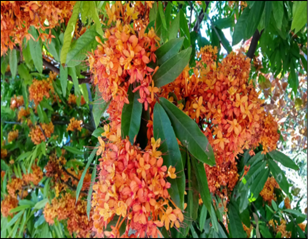
Fig 1: Saraca asoka
2. Cassia fistula
Leaves of Cassia fistula (Fabaceae) were collected from Palode. Leaves contain anthraquinones (rhein, emodin), flavonoids, tannins, phenols, and glycosides. Collectively, these constituents contribute to the observed activities through antioxidant, anti-inflammatory, hypoglycemic, neurodepressant, anthelmintic, and cardioprotective mechanism (3,4).
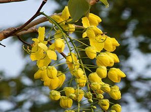
Fig 2: Cassia fistula
3.Chicken fat
Chicken fat obtained from Gallus gallus domesticus (family Phasianidae) was collected from a local slaughter shop. It is reported to exhibit beneficial effects in burn wound healing due to its emollient, anti-inflammatory, and tissue-regenerative properties, which are mainly attributed to the presence of bioactive constituents such as essential fatty acids (oleic acid, linoleic acid), triglycerides, phospholipids, and fat-soluble vitamins that promote skin barrier repair, reduce inflammation, and enhance epithelial regeneration (5).
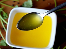
Fig 3: Chicken fat
4.Beeswax
Beeswax, obtained commercially through online sources, is a natural product secreted by honeybees (Apis species). It has been widely used in topical formulations for burn wound healing due to its protective, emollient, anti-inflammatory, and antimicrobial properties. The burn-healing activity of beeswax is mainly attributed to its chemical constituents such as long-chain fatty acid esters, free fatty acids, hydrocarbons, alcohols, flavonoids, and vitamin A. These constituents help form a protective barrier over the wound, retain moisture, reduce inflammation, prevent microbial infection, and promote skin regeneration and epithelialization (6).
Fig 4: Beeswax
5.White soft paraffin
White soft paraffin is a semi-solid purified hydrocarbon mixture made from petroleum that can be purchased commercially online. Because of its superior occlusive, emollient, and skin-protective qualities, it is frequently used as a topical base in the treatment of burn wounds. Its saturated hydrocarbons, which create a protective layer over the burned area, lower trans epidermal water loss, keep the skin hydrated, soften the tissue, and encourage epithelial regeneration, are the main reason for its positive effects on burn healing. These properties support the body's natural healing process and guard against secondary infections (7).
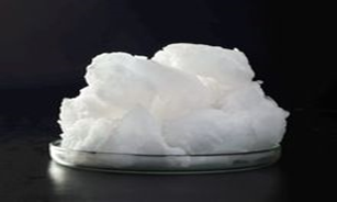
Fig 5: White soft paraffin
6.Coconut oil
Coconut oil, which is available commercially through online sources, is a natural edible oil derived from the kernel of Cocos nucifera. It is widely used in topical applications for burn wound healing due to its moisturizing, anti-inflammatory, antimicrobial, and antioxidant properties. The burn-healing activity of coconut oil is primarily due to its high concentration of medium-chain fatty acids, particularly lauric acid, as well as capric acid, caprylic acid, oleic acid, tocopherols, and polyphenolic compounds. These constituents help to maintain skin hydration, reduce inflammation, inhibit microbial growth, enhance collagen synthesis, and promote epithelialization, thus accelerating the burn wound healing process (8).
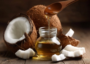
Fig 6: Coconut oil
7.Vitamin E
Tocopherols and tocotrienols are the two main fat-soluble antioxidant compounds that make up vitamin E, which can be purchased commercially online. Because of its potent antioxidant, anti-inflammatory, and skin-protective qualities, it is crucial to the healing of burn wounds. The most biologically active component of vitamin E, α-tocopherol, is mainly responsible for its beneficial effects on burn healing. It protects cell membranes from oxidative damage, lowers inflammation, improves skin hydration, encourages epithelial regeneration, and supports collagen formation, all of which speed up the healing process (9).
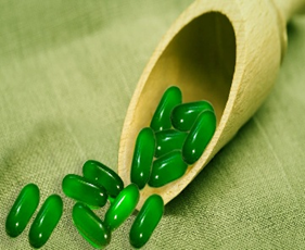
Fig 7: Vitamin E
8. Caprylyl glycol
Caprylyl glycol is a multipurpose synthetic diol that is frequently used as a preservative booster in topical formulations. Because of its humectant, emollient, and antimicrobial qualities, it shows promise in the treatment of burn injuries. Its chemical structure, especially the caprylyl (C8) alkyl chain and hydroxyl groups, which disrupt microbial cell membranes, improve skin hydration, support barrier function, and create an environment conducive to wound healing and epithelial regeneration, is largely responsible for its biological and preservative properties (10).
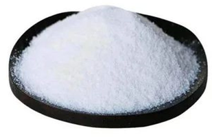
Fig 8: Caprylyl glycol
MATERIALS AND METHODS
Collection and processing of ingredients
Saraca asoka, cassia fistula, rendered chicken fat, beeswax, white soft paraffin, coconut oil, vitamin E, are the ingredients used in the formulation of burn cream. Collection and harvesting of asoka and cassia leaf were done on September at 8 am. Then dry the leaf in shade for 2 weeks. Do not expose it to sunlight. Then crush the leaf into fine powder using a grinder and sieve it. Collect the chicken from slaughterhouse, then separate chicken fat from chicken. Put the chicken fat into a pan. Add 2-3 tablespoons of water (prevents burning at the start). Heat on low to medium-low. Stir occasionally as the fat melts out. After 15-25 minutes, the skin pieces will shrink and turn golden. Pour through a fine sieve into a jar. Press gently to extract liquid fat. Let cool, then refrigerate or freeze. Rest of ingredients were bought from market.
Table 1: Formulation of burn cream
|
Sl no: |
Ingredients |
F1 |
F2 |
F3 |
F4 |
|
1. |
Saraca asoka leaf |
5.125 |
5.125 |
5.125 |
5.125 |
|
2. |
Cassia fistula leaf |
5.125 |
5.125 |
5.125 |
5.125 |
|
3. |
Rendered chicken fat |
20.5 |
20.5 |
20.5 |
20.5 |
|
4. |
Beeswax |
6.125 |
2.125 |
5.125 |
8.125 |
|
5. |
White soft paraffin |
45.125 |
49.125 |
46.125 |
43.125 |
|
6. |
Coconut oil |
15.375 |
15.375 |
15.375 |
15.375 |
|
7. |
Vitamin E |
6.15 |
6.15 |
6.15 |
6.15 |
|
8. |
Caprylyl glycol |
1.025 |
1.025 |
1.025 |
1.025 |
1.Melting the base
Beeswax and white soft paraffin should be melted together in a China dish at about 70C using a clean water bath.
Stir in the coconut oil and chicken fat until well combined.
2.The cooling stage
Let the mixture cool to around 40-45C.
If necessary, dissolve the methyl paraben in a little amount of heated oil.
3. Incorporating actives
Add the Asoka, Cassia and vitamin E extracts.
Continue stirring until evenly distributed.
4. Last step
While still semi-fluid, pour into sterile ointment jars.
Let it settle at room temperature.
Evaluation
I. Physical studies
1.Physical appearance
The colour was determined by visual examination, the odour was checked by smelling, and the nature was examined visually (11).
2.Spreadibility
The back of one's hand was scrubbed with a tiny amount of cream to make it simpler. It was noticed how the material spreads on the skin (12).
3.Determination of pH
A digital pH meter was used to determine the pH of a freshly produced emulsion at room temperature. According to the findings, the pH level of the formulation is closer to skin pH, allowing it to be safely applied on the skin (13).
4.Washability
A tiny quantity of cream was applied to the back of each hand, which was then washed clean with warm water (14).
5.Viscosity
Place the digital viscometer (IR•195BK11411) on a level surface and perform auto-zero calibration before attaching the spindle. Attach the appropriate spindle by lifting the coupling nut to reduce internal strain. Fill a beaker with the sample, avoiding air bubbles, and lower the spindle into the liquid up to the marked level. Set the spindle number and rotational speed (20 RPM) to obtain a torque between 10–100%, then start the motor. Record the stabilized viscosity reading (after ~30 seconds), along with temperature, spindle type, and RPM. After testing, switch off the instrument and clean the spindle and beaker (15).
6. Melting point
Finely powder and dry the sample for 24 hours over silica gel, then pack 4–6 mm of powder into a capillary tube. Attach the tube to a thermometer, immerse it in a heated bath starting about 10 °C below the expected melting point, and heat at ~10 °C per minute. Record the temperature at which melting begins and the temperature at which the sample completely liquefies to determine the melting range (16).
7.Acid value
Accurately weigh a 250 ml conical flask and then weigh it again after adding about 10 g of sample. Prepare a solvent mixture of 25 ml ether, 25 ml of 96% ethanol, and 1 ml phenolphthalein solution, neutralizing, if necessary, with 0.1 m KOH. Add this mixture to the flask and shake to dissolve free fatty acids; heating is usually unnecessary. After cooling to room temperature, titrate with 0.1 m KOH until a pink colour persists for 15 seconds, avoiding the addition of water, as excess KOH can hydrolyse glycerides (17).
Invitro studies
1.Antibacterial activity
Mueller–Hinton agar (15–20 mL) was poured into sterile Petri plates and allowed to solidify. The test organism was uniformly spread using a sterile cotton swab. Four wells (8 mm diameter, 20 mm apart) were made in each plate using a sterile cork borer. Test samples (50 and 100 µL from a 10 mg/mL stock) were added to wells T1 and T2. DMSO and gentamycin (40 µL from a 4 mg/mL stock) served as negative and positive controls, respectively. Plates were incubated aerobically at 36 ± 1 °C for 24 h. Zones of inhibition were measured in millimetres after incubation.
2. Anti-inflammatory studies
Cyclooxygenase (COX) assay (in vitro)
RAW 264.7 cells obtained from NCCS, Pune, were cultured to 70% confluence and stimulated with lipopolysaccharide (LPS, 1 µL of 1 µg/mL). The LPS-activated cells were treated with different concentrations of the test sample and incubated for 24 h. After incubation, cell lysates were collected and used for COX anti-inflammatory analysis.
Lipoxygenase (LOX) assay (in vitro)
RAW 264.7 cells from NCCS, Pune, were cultured to 60% confluence and activated with LPS (1 µL of 1 µg/mL). The activated cells were treated with various concentrations of the test sample and diclofenac sodium as the standard drug, then incubated for 24 h. Cell lysates were collected after incubation.5-LOX activity was measured using the method of Axelrod et al. (1981). The reaction mixture contained Tris-HCl buffer (pH 7.4), cell lysate, and sodium linoleate. LOX activity was determined by measuring the change in absorbance at 234 nm.
3. Scratch wound healing assay
Preliminary procedure
Cells (0.3 × 10? cells/well) were seeded in 6-well plates and incubated at 37 °C with 5% CO? for 24 h. Test samples were sterilized using a 0.2 µm syringe filter and added to wells with ~80% confluent cells at concentrations of 25, 50, and 100 µg/mL. Untreated wells served as controls.
Scratch assay procedure
A straight scratch was made on the cell monolayer using a sterile 200 µL pipette tip. Cells were washed once with culture medium to remove debris and fresh medium was added. Scratches of similar width were maintained in all wells. Reference marks were made near the scratch to capture the same field during imaging. Initial images were taken using a phase-contrast microscope. Plates were incubated at 37 °C, and images were captured at 0, 12, 24, and 36 h until the scratch was closed. Final images were taken by aligning the same reference points.Cell migration analysisImages were analysed using ImageJ software. The percentage of wound closure was calculated by comparing the wound area at different time points with that at 0 h. Increased wound closure indicated enhanced cell migration and wound healing activity.
RESULT AND DISCUSSION
Flavonoids and anthraquinones, which have potent anti-inflammatory, antioxidant, and antibacterial qualities, are the main chemical components of cassia leaves that are advantageous for making burn cream. Due to its high level of healthy proteins and fatty acids, chicken fat—which is mostly utilized in conventional treatments and researched in animal models—offers prospective benefits in burn healing ointments. It increases cellular activity, promotes tissue regeneration, has anti-inflammatory properties, and is hydrating and protecting.
Table 2: Organoleptic characteristics of burn cream
|
Sl no. |
Test |
Result |
|
1. |
Colour |
Dark green |
|
2. |
Odour |
Characteristic |
|
3. |
Nature |
Semi-solid |
|
4. |
Spreadability |
Good |
|
5. |
Ph |
6.4 |
|
6. |
Washability |
Washable |
|
7. |
Viscosity |
48895 cps |
|
8. |
Melting point |
60.15? |
|
9. |
Acid value |
5.81 |
Invitro studies
1.Anti-bacterial activity
Antibacterial activity testing for burn creams is critical because the destruction of the skin's barrier function in a burn injury raises the risk of infection, which is the leading cause of death in severely burned patients. Testing confirms that the cream is effective at reducing bacterial load and preventing a local infection from progressing to life-threatening systemic sepsis.
Table 3: Anti-bacterial activity
|
Name of Microorganism |
Zone of inhibition (mm) |
|||
|
Standard Gentamycin (160 µg ) |
Negative control |
T1 (500µg) |
T2 (1000µg) |
|
|
Staphylococcus aureus |
-ve |
-ve |
-ve |
-ve |
Interpretation of Results
-ve: Refers to no antibacterial activity for the given test compound (No/zero mm zone of inhibition around the well the test sample is added) against the particular microbial strain used in this study.
2.Scratch wound healing assay
Silver nanoparticles (AgNPs) at 50 and 100 µg/mL helped wounds close faster than the control. Healing increased with higher concentration, and almost complete closure was seen at 100 µg/mL within 48 hours. The healing time was reduced compared to the control. AgNPs also showed good antibacterial activity. Overall, AgNPs improve wound healing and prevent infection.
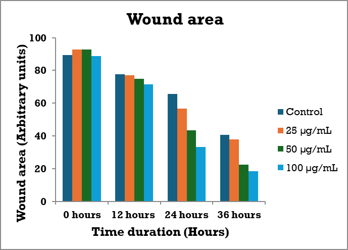
Fig 9: Time duration vs Wound area
The graph shows that the test compound reduces cell migration in a time- and dose-dependent manner, with stronger inhibition at 100 µg/mL than at 50 µg/mL over 36 hours, indicating effective suppression of cell movement.
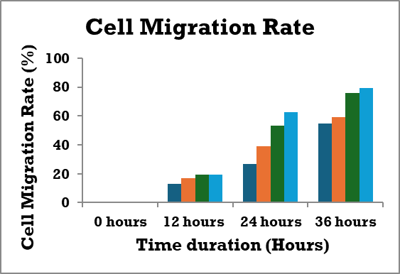
Fig 10: Time duration vs Cell migration rate
3.Anti-inflammatory studies
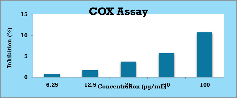
COX assay
The graph shows a dose-dependent increase in COX inhibition by diclofenac, from very low inhibition at 6.25 µg/mL to almost complete inhibition at 100 µg/mL. The linear dose–response curve (R² = 0.973) confirms good assay reliability, and diclofenac serves as a standard for validating the assay and estimating IC?? values.
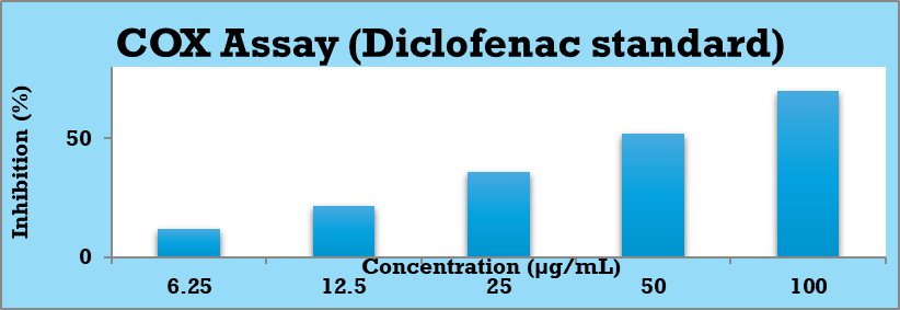
Fig 11. COX assay (Diclofenac standard)
The graph shows a linear, dose-dependent increase in COX inhibition by diclofenac, confirming assay reliability and allowing accurate estimation of IC?? values.
LOX assay
The graph shows that diclofenac inhibits LOX activity in a dose-dependent manner, with about 50% inhibition around 25 µg/mL, confirming its effectiveness as a standard anti-inflammatory inhibitor.
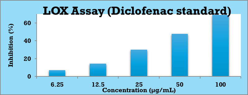
Fig 13: LOX assay (Diclofenac standard)
The graph shows that the test compound inhibits LOX activity in a dose-dependent manner, with strong inhibition at higher concentrations and moderate inhibitory potency.
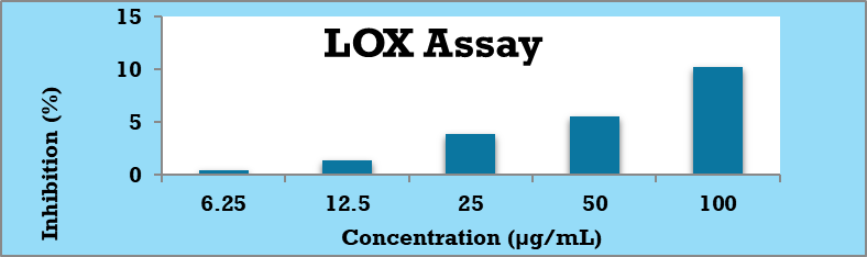
Fig 14: LOX assay of cream
ACKNOWLEDGEMENT
We acknowledged to teaching and non-teaching staff and friends to Mount Zion College of Pharmaceutical Sciences and Research, Chayalode, Adoor
REFERENCES


1. Shpichka, A., Butnaru, D., Bezrukov, E. A., Sukhanov, R. B., Atala, A., Burdukovskii, V., Zhang, Y., & Timashev, P. (2019). Skin tissue regeneration for burn injury. Stem Cell Research & Therapy, 10(1), 94. https://doi.org/10.1186/s13287-019-1203-3 2. 12. Zago, L. R., Prado, K., Benedito, V. L., & Pereira, M. M. (2021). The use of babosa (Aloe vera) in treating burns: a literature review. Brazilian Journal of Biology, 83, e249209. https://doi.org/10.1590/1519-6984.249209 3. Basri F, Sharma HP, Firdaus S, Jain P, Ranjan A. A review of ethnomedicinal plant-Vitex negundo Linn. Int. J. Adv. Res. 2014 Jan;2(3):882-94. 4. Kirtikar KR, Basu BD. Indian Medicinal plants. Vol III, Dehradun. International Book Distributor. 1987:1336-39.5. 5. Oleic acid modulation of the immune response in wound healing: a new approach for skin repair C R Cardoso et al. Immunobiology. (2011). 6. Kurek-Górecka, A., Górecki, M., Rzepecka-Stojko, A., Balwierz, R., & Stojko, J. (2020). Bee products in dermatology and skin care. Molecules (Basel, Switzerland), 25(3), 556. https://doi.org/10.3390/molecules25030556 7. Nong, Y., Maloh, J., Natarelli, N., Gunt, H. B., Tristani, E., & Sivamani, R. K. (2023). A review of the use of beeswax in skincare. Journal of Cosmetic Dermatology, 22(8), 2166–2173. https://doi.org/10.1111/jocd.15718 8. Wijaya, I. (2012). The Healing Effect of Coconut oil Used Topically on Burn Wound White Rat (Rattus novergicus) Induced by Sulfuric Acid. Thesis. 9. Traber, D. L., Hawkins, H. K., Enkhbaatar, P., Cox, R. A., Schmalstieg, F. C., Zwischenberger, J. B., & Traber, L. D. (2007). The role of the bronchial circulation in the acute lung injury resulting from burn and smoke inhalation. Pulmonary Pharmacology & Therapeutics, 20(2), 163–166. https://doi.org/10.1016/j.pupt.2005.12.006 10. Nielson, C. B., Duethman, N. C., Howard, J. M., Moncure, M., & Wood, J. G. (2017). Burns: Pathophysiology of systemic complications and current management. Journal of Burn Care & Research: Official Publication of the American Burn Association, 38(1), e469–e481. https://doi.org/10.1097/BCR.0000000000000355 11. John Wiley & Sons A/S, 2025. John Wiley & Sons Ltd. published the book. 12. Vamsi S, Satish C, Nagaveni K, Jyothi MJ, Latha P. Formulation and evaluation of polyherbal wound healing ointment. International Journal of Pharma Research & Review. 2014 Apr;3(4):66-73. 13. Jadhav MD, Ubale MP, Kadam SV, Ehtesham AM. Formulation and Evaluation of Herbal Skin Cream for Wound Healing Activity. International Research Journal of Pharmacy and Medical Sciences (IRJPMS). 2023;6(4):8-12. 14. Pal, A., Soni, M., & Patidar, K. (2014). ?Formulation and evaluation of poly herbal cream. International Journal Pharmaceutical and Biological Archives, 5, 67–71. 15. Nwose EU, Richards RS. Whole blood viscosity issue VIII: comparison of extrapolation method with diagnostic digital viscometer. North American journal of medical sciences. 2011 Jul;3(7):333. 16. J.S. QADRY, S.S. QADRY Organic Pharmaceutical and Medicinal Chemistry Fundamental and Aliphatic Compounds ,4th edition, Volume 1. (n.d.). 17. A.H. Beckett, J.B. Stenlake Practical Pharmaceutical Chemistry, part 1, Fourth edition, 146-147.
 10.5281/zenodo.18620295