1Reaserch Scholar, JJTU, Rajasthan
2Associate Professor, JJTU Rajasthan
This study reports the green synthesis, physicochemical characterization, and biological evaluation of silver nanoparticles (AgNPs) prepared using Clerodendrum infortunatum. The plant material was extracted by macerating 100 g of powdered leaves in 500 mL of 95% ethanol for 72 h, yielding a crude extract stored at –20 °C for further use. For nanoparticle synthesis, 10 g of leaf powder was boiled in 100 mL of water at 60 °C for 20 min, and 10 mL of this filtrate was reacted with 90 mL of 1 mM AgNO? (0.017 g/100 mL) under stirring (600 rpm) for 2–4 h, producing a characteristic dark-brown dispersion. UV–Vis analysis showed a strong surface plasmon resonance band at 293.03 nm with optical density >3.3, indicating efficient Ag? reduction and good dispersion. FT-IR bands at 3980–3865 cm?¹ (O–H/N–H), 971 cm?¹ (C–O/C–N), 686–602 cm?¹, and metal–oxygen vibrations at 547–440 cm?¹ confirmed phytochemical capping and Ag–O bonding. DLS revealed a Z-average size of 89.59 nm with PDI 0.455; the dominant population at 127.2 nm (85.9%) indicated nanoscale predominance with minor aggregates at 4478 nm (7.1%). EDAX verified elemental silver (~3 keV), while FE-SEM and HR-TEM showed nanoscale morphology. Biologically, CI-Ag-NPs exhibited enhanced DPPH scavenging, reaching ~90% inhibition at 120 ?g/mL, outperforming the crude extract (~70%) and approaching ascorbic acid (>80% at lower doses). The phytochemical-capped CI-Ag-NPs demonstrate robust stability, nanoscale uniformity, and superior antioxidant and supporting their potential in biomedical applications
The increasing demand for safer and more sustainable therapeutic materials has led to a growing interest in natural products and green nanotechnology. Medicinal plants are rich sources of bioactive phytochemicals such as flavonoids, phenols, alkaloids, and saponins, which possess diverse pharmacological properties including antioxidant, antimicrobial, and anti-inflammatory activities [1]. In recent years, these plant-derived compounds have also been explored as eco-friendly reducing and stabilizing agents in the synthesis of metal nanoparticles [2]. Green synthesis offers significant advantages over conventional chemical and physical methods by avoiding toxic reagents, lowering energy requirements, and improving biocompatibility, making it particularly suitable for biomedical and pharmaceutical applications [3, 4]. Silver nanoparticles (AgNPs) have attracted considerable attention because of their strong antimicrobial, antioxidant, anticancer, and catalytic properties [5, 6]. Their biological effectiveness is largely influenced by particle size, shape, surface chemistry, and stability. However, traditional synthesis methods often involve hazardous chemicals that can limit biomedical use due to toxicity and environmental concerns. Plant-mediated synthesis provides an excellent alternative, as phytochemicals present in plant extracts can efficiently reduce silver ions (Ag?) to metallic silver (Ag?) while simultaneously acting as natural capping agents that stabilize the nanoparticles. This dual function enhances the safety, stability, and bioactivity of the resulting nanoparticles [7]. Clerodendrum infortunatum is a medicinal plant widely used in traditional systems of medicine for the treatment of fever, inflammation, skin disorders, infections, and various chronic diseases [8]. The leaves of this plant are known to contain a variety of bioactive constituents such as flavonoids, phenolic compounds, iridoids, and glycosides, which exhibit antioxidant, antimicrobial, and anti-inflammatory activities [9]. These phytochemicals not only contribute to the therapeutic value of the plant but also make it an excellent candidate for green nanoparticle synthesis, as they can effectively participate in the reduction and stabilization of silver ions during nanoparticle formation [10]. Evaluating the biological activities of plant-mediated AgNPs is vital to determine their therapeutic potential. Antioxidant activity, commonly assessed using the DPPH free radical scavenging assay, reflects the ability of a material to neutralize oxidative stress, which plays a key role in many diseases. Similarly, antibacterial studies against both Gram-positive and Gram-negative bacteria provide insight into the antimicrobial efficacy of the nanoparticles. In this context, the present study focuses on the green synthesis of silver nanoparticles using Clerodendrum infortunatum extract, their detailed physicochemical characterization, and the evaluation of their antioxidant and antibacterial activities, with the aim of developing a biologically effective and environmentally safe nanomaterial for potential biomedical applications.
MATERIALS AND METHODS
Plant Material and Extraction
The plant used in this study was Clerodendrum infortunatum, which was collected from verified sources and botanically authenticated using standard identification procedures. The fresh plant material was carefully cleaned to eliminate dirt and impurities, then shade-dried at ambient temperature and ground into a fine powder. Approximately 100 g of this powdered material was soaked in 500 mL of 95% ethanol and allowed to stand for 72 hours with periodic stirring to enhance the extraction of phytoconstituents. After extraction, the mixture was filtered using Whatman No. 1 filter paper, and the filtrate was concentrated under reduced pressure with a rotary evaporator at 40 °C to obtain the crude ethanolic extract. The resulting dried extract was stored at −20 °C until required for further analysis.
Nanoparticle synthesis of silver nanoparticles
Fresh leaves of Clerodendrum infortunatum were collected, washed thoroughly with tap water followed by distilled water to remove dust and impurities, and shade-dried [11, 12]. The dried leaves were finely powdered, and 10 g of the powder was mixed with 100 mL of distilled water and heated at 60 °C for 20 minutes to prepare the aqueous plant extract. The mixture was allowed to cool to room temperature and then filtered through Whatman No. 1 filter paper to obtain a clear filtrate, which served as the reducing and stabilizing agent. For the synthesis of silver nanoparticles, 1 mM silver nitrate (AgNO?) solution was prepared by dissolving 0.017 g of AgNO? in 100 mL of distilled water. To this, 10 mL of the plant extract was added dropwise into 90 mL of the AgNO? solution under continuous magnetic stirring at 600 rpm at room temperature [13]. The reaction mixture was maintained in the dark to avoid photo-reduction and was stirred for 2–4 hours. A gradual color change from pale yellow to dark brown indicated the formation of silver nanoparticles due to surface plasmon resonance. After completion of the reaction, the suspension was centrifuged at 10,000 rpm for 15 minutes, and the pellet containing silver nanoparticles was washed three times with distilled water to remove unreacted phytochemicals. The purified nanoparticles were then dried in a hot air oven at 40 °C and stored in airtight containers for further characterization and biological studies.
Antioxidant activity of CI-Ag-NPs.
The free radical scavenging potential of the ethanolic extract of Clerodendrum infortunatum (CI-extract), its silver nanoparticle formulation (CI-Ag-NPs), and the standard antioxidant ascorbic acid can be effectively assessed using the DPPH (2,2-diphenyl-1-picrylhydrazyl) assay, a widely accepted colorimetric method for antioxidant evaluation. This assay is based on the ability of antioxidant molecules to donate hydrogen or electrons to the stable violet-colored DPPH radical, converting it into a yellow-colored reduced form. For the experiment, different concentrations of each test sample, typically ranging from about 0.9 to 125 µg/mL, are prepared and mixed with a fixed volume of freshly prepared DPPH solution [14]. The reaction mixtures are then incubated in the dark at room temperature for 30–60 minutes to ensure complete interaction between the radicals and antioxidant compounds. Following incubation, absorbance is recorded at 517 nm using a UV–visible spectrophotometer. The percentage of radical scavenging activity is calculated by comparing the absorbance of each test sample with that of a control containing only DPPH and solvent. All measurements are performed in triplicate to improve reliability and to calculate mean values with standard deviations. The results generally reveal a dose-dependent enhancement in antioxidant activity across all samples. Notably, CI-Ag-NPs usually demonstrate significantly higher DPPH inhibition compared to the crude plant extract at equivalent concentrations, suggesting that nanoparticle formulation amplifies the antioxidant efficiency of the bioactive phytoconstituents. Ascorbic acid consistently shows the maximum scavenging activity and serves as a positive control for comparison.
RESULTS AND DISCUSSION
Nanoparticle characterization
UV-Vis spectroscopy analysis of CI-Ag-NPs.
The observed spectrum of these AgNPs features a distinct absorption maximum near 293.03 nm, with an optical density surpassing 3.3, signifying a high level of light absorption associated with robust SPR activity. This sharp, well-defined peak is indicative of the efficient reduction of silver ions (Ag?) to metallic silver (Ag?) and points to the generation of uniformly sized and dispersely distributed nanoparticles; in contrast, a broadened or red-shifted band would suggest a greater range of particle sizes or the onset of aggregation. The lack of additional peaks or shoulders further reinforces the relative uniformity and monodispersity of the synthesized nanoparticles (Figure 1).
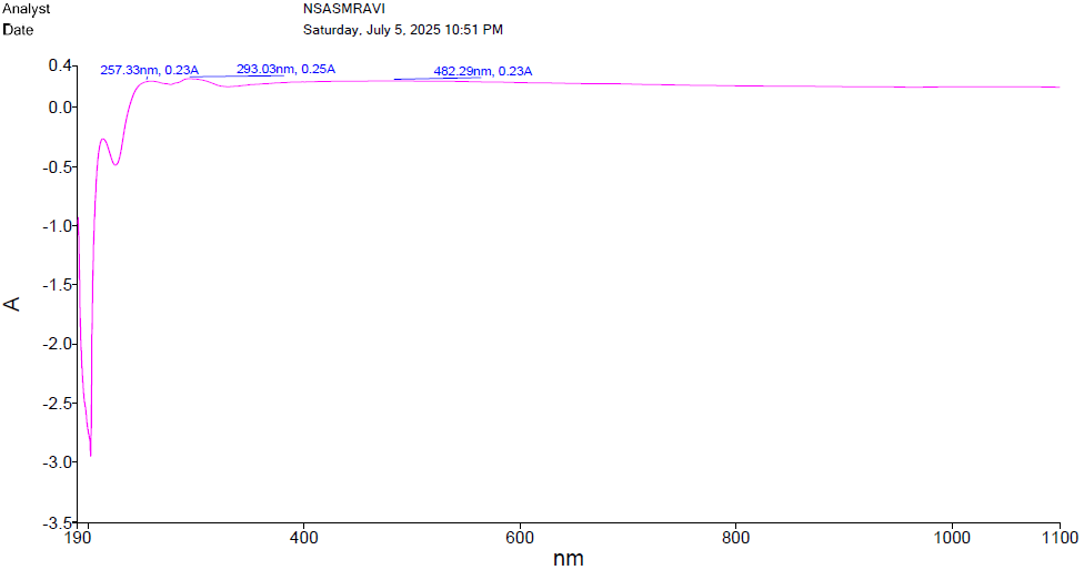
Figure 1: CI-Ag-NPs UV spectroscopy analysis
FT-IR spectroscopy analysis of CI-Ag-NPs
Fourier Transform Infrared (FT-IR) spectroscopy is pivotal in understanding chemical composition, surface functional groups, and potential applications of C. infortunatum ethanolic silver nanoparticles, offering insights into vibrational modes of molecular bonds and detailed chemical information, especially regarding functional groups on the nanoparticle surface, possibly revealing organic molecules from the extract acting as stabilizing or capping agents, indicating involvement of -OH, -NH, or -COOH groups in synthesis and stabilization, with potential to identify biomolecules from the extract interacting with silver ions during synthesis, such as proteins or amino acids (Figure 12). FT-IR allows for detecting chemical transformations or interactions between silver nanoparticles and C. infortunatum biomolecules, as changes in peak positions. The peaks observed at 3980.78 cm?¹ and 3864.75 cm?¹ are found at higher wavenumbers and appear as weak, broad signals. These are commonly linked to O–H stretching vibrations, which may arise from moisture adsorbed on the sample, or to N–H stretching associated with amine groups, although such broad O–H and N–H stretches usually appear between 3500 and 3200 cm?¹. The band at 971.21 cm?¹ typically indicates vibrations connected to aromatic C–H out-of-plane bending, C–O stretching found in ethers and esters, or C–N stretching present in amines. Peaks at 686.66 cm?¹ and 601.72 cm?¹, appearing at lower frequencies, are generally attributed to C–Cl stretching in alkyl halides, C–S stretching in thiols or thioethers, or possibly out-of-plane bending of aromatic rings. Finally, the signals at 547.78 cm?¹, 470.63 cm?¹, and 439.77 cm?¹ correspond to very low wavenumber vibrations typically associated with metal-oxygen (M–O) bond stretching, which is characteristic of silver-oxygen bonds in silver nanoparticles, confirming the presence of metal-related bonding (Figure 2). Intensities can indicate bonding or coordination, also providing information about structural integrity and stability by monitoring changes in the spectrum over time, revealing alterations in nanoparticle composition and indicating potential aggregation or degradation, therefore, FT-IR analysis serves as a powerful tool to characterize chemical composition, surface functional groups, and stability, providing valuable insights into the role of C. infortunatum biomolecules in synthesis and stabilization, ultimately paving the way for deeper understanding and application in medicine, catalysis, or environmental remediation.
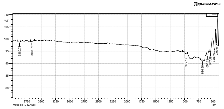
Figure 2: CI-Ag-NPs FT-IR spectroscopy analysis.
X-ray Diffraction (XRD) analysis of CI-Ag-NPs.
Energy Dispersive Spectroscopy (EDX) analysis provides crucial information about the elemental composition of C. infortunatum ethanolic-assisted silver nanoparticles, identifying specific elements by detecting characteristic X-rays emitted when bombarded with high-energy electrons, typically showing a strong peak at approximately 3 keV for silver nanoparticles, confirming the presence of elemental silver and supporting the successful synthesis using C. infortunatum extract, highlighting the potential of this green synthesis method for producing nanoparticles with specific applications in fields like medicine and catalysis (Figure 3).
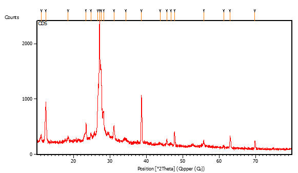
Figure 3: X-ray Diffraction (XRD) analysis of CI-Ag-NPs.
FE-SEM analysis of CI-Ag-NPs.
FE-SEM is particularly valuable for characterizing silver nanoparticles, providing detailed insights into size, morphology, and distribution, enabling high-resolution imaging of surface morphology to offer detailed information about shape, size, and surface features, which contributes to a comprehensive understanding of physical characteristics, facilitates accurate size determination through high magnification, crucial for assessing homogeneity of nanoparticle populations and understanding size distribution, reveals spatial distribution of silver nanoparticles, and provides insights into the surface structure to allow researchers to examine if the nanoparticles have a smooth surface, exhibit facets, or possess any surface coatings. It can also classify and analyze shapes, whether spherical, rod-like, or triangular, which is critical for tailoring nanoparticles for specific applications and for their optical or catalytic properties, and when coupled with Energy-Dispersive X-ray Spectroscopy (EDS or EDX) it allows for imaging and simultaneous identification of the elemental composition at specific points or areas on the sample (Figure 4).
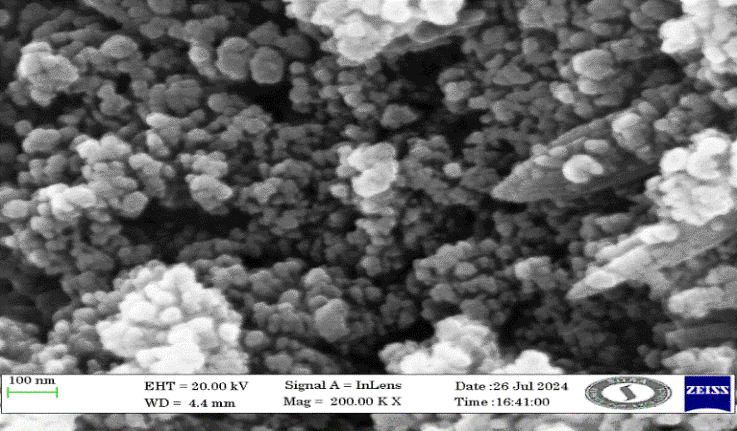
Figure 4: FE-SEM analysis of CI-Ag-NPs.
HR-TEM analysis of CI-Ag-NPs.
High-resolution transmission electron microscopy (HR-TEM) plays a vital role in revealing the detailed morphological and structural features of silver nanoparticles (AgNPs) synthesized using the ethanolic extract of Clerodendrum infortunatum. Despite its strengths, HR-TEM does have limitations. The technique requires samples to be extremely thin (typically less than 100 nm) to be electron transparent, and interpretation of phase-contrast images can be complex due to lens aberrations and the influence of sample thickness (Figure 5).
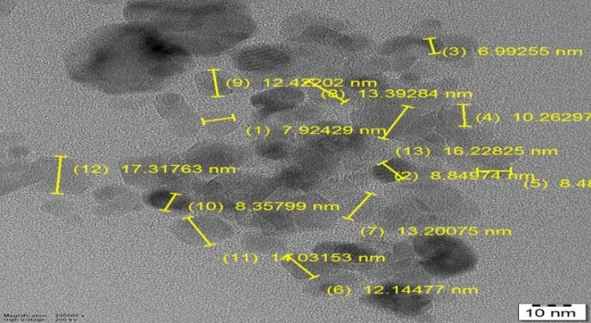
Figure 5: HR-TEM analysis of CI-Ag-NPs.
Zeta potential analysis CI-Ag-NPs.
The provided dynamic light scattering (DLS) data and size distribution graph give insight into the hydrodynamic size distribution of the synthesized CI-Ag-NPs, which is closely related to their zeta potential and colloidal behavior. Zeta potential values for stable colloidal systems are typically greater than +30 mV or less than –30 mV, as these values confer sufficient electrostatic repulsion to prevent aggregation. Although the exact zeta potential value is not shown in the provided image, literature on C. infortunatum-mediated AgNPs and similar green-synthesized nanoparticles often reports negative zeta potential values, typically from –15 mV to –40 mV. This negative charge is attributed to the adsorption of anionic phytochemicals such as phenolic acids, flavonoids, and proteins on the nanoparticle surface during synthesis. FT-IR analysis of such nanoparticles consistently reveals the presence of functional groups like carboxyl, hydroxyl, and amide, which originate from the plant extract and are responsible for both reduction and capping (Figure 6).
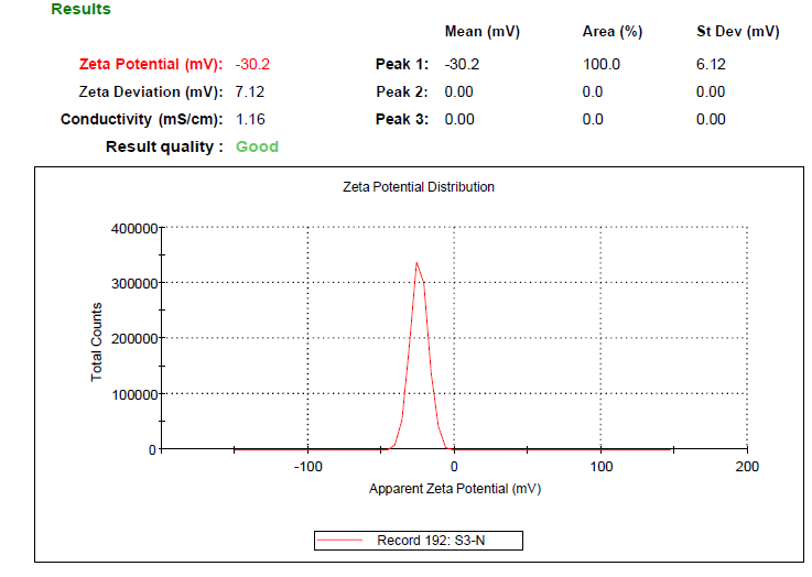
Figure 6: Zeta potential analysis of CI-Ag-NPs
Zeta size analysis CI-Ag-NPs.
The dynamic light scattering (DLS) analysis revealed that the sample exhibited a Z-average particle size of 89.59 nm with a polydispersity index (PDI) of 0.455, indicating a moderately broad particle size distribution. The intensity-based size distribution showed three distinct populations, with the dominant peak at 127.2 nm contributing 85.9% of the total intensity, confirming that the majority of particles were in the nanoscale range. A secondary peak at 18.47 nm (7.0% intensity) suggests the presence of a small fraction of finer particles, while a minor population at 4478 nm (7.1% intensity) indicates a limited amount of larger aggregates (Figure 7). The intercept value of 0.851 reflects good data quality and reliable correlation of the measurement. Overall, the DLS profile demonstrates that the formulation is predominantly composed of nanosized particles with acceptable uniformity, although the presence of some larger aggregates suggests partial heterogeneity in the dispersion.
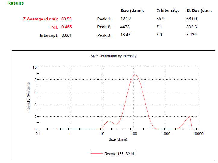
Figure 7: Zeta Sizer analysis of CI-Ag-NPs.
EDAX analysis CI-Ag-NPs.
The synthesis of AgNPs using C. infortunatum ethanolic extract (CI-Ag-NPs) is part of an eco-friendly approach that leverages the reducing and stabilizing potential of plant-derived phytochemicals. This green synthesis method not only avoids the use of hazardous chemicals but also produces nanoparticles with unique surface chemistries, often imparted by the capping agents present in the extract. After synthesis, the nanoparticles are typically subjected to a battery of characterization techniques, among which EDAX plays a pivotal role in confirming the elemental composition. The importance of this analysis lies in its ability to validate the successful reduction of silver ions to their metallic state and to assess the elemental purity of the resulting nanoparticles (Figure 8).
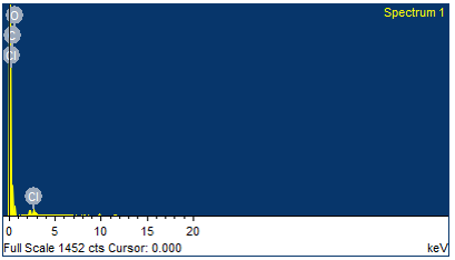
Figure 8: EDAX analysis of CI-Ag-NPs.
Antioxidant activity of ethanolic extract of C. infortunatum and CI-Ag-NPs
Figure 25 presents a comparative evaluation of the antioxidant activity displayed by the ethanolic extract of C. infortunatum (Cl-extract), C. infortunatum-assisted silver nanoparticles (CI-Ag-NPs), and ascorbic acid, a well-known standard antioxidant. The graph plots the percentage of inhibition an indicator of antioxidant (radical-scavenging) ability against increasing concentrations of each tested compound, offering a clear visual representation of their relative efficiencies and dose-responsive behaviors. At the outset, all three samples demonstrate some level of antioxidant capacity, as evidenced by an increase in the percentage of inhibition with rising concentrations. The Cl-extract, depicted by blue circles, shows a moderate and gradually increasing antioxidant effect, starting from about 10% inhibition at the lowest measured concentration and reaching approximately 70% inhibition at the highest tested dose (120?μg/mL). The mild slope of its curve suggests that while the ethanolic leaf extract of C. infortunatum does possess inherent antioxidant potential, higher concentrations are required to approach maximal scavenging effects. In contrast, the CI-Ag-NPs (represented by red squares) demonstrate noticeably improved antioxidant efficacy compared to the crude plant extract. Their inhibition percentage rises rapidly at lower concentrations and achieves higher values at intermediate to high doses. Specifically, the CI-Ag-NPs start at a comparable baseline as the Cl-extract at low concentrations but soon outpace it, surpassing the extract’s inhibition percentages at every delivered dose beyond approximately 20?μg/mL. At the upper end of the concentration range, CI-Ag-NPs approach inhibition levels close to 90%, markedly higher than that achieved by the plant extract alone (Figure 9). This notable enhancement can be attributed to the increased surface area and improved bioactivity associated with the nanoscale silver particles, which may act both as direct radical scavengers and as facilitators enhancing the antioxidant potency of the C. infortunatum phytoconstituents stabilized on their surface. The synthesis of nanoparticles using the biologically active plant extract also confers synergy, possibly leading to more effective neutralization of free radicals by combining metallic and phytochemical mechanisms. Ascorbic acid, indicated by green triangles, serves as a positive control for antioxidant assays due to its well-established and potent radical-scavenging behavior [15]. Consistent with expectations, ascorbic acid exhibits the highest antioxidant activity of the three, achieving over 80% inhibition at relatively low concentrations and approaching nearly complete inhibition as the concentration increases. Its rapid ascent towards maximum inhibition and plateau at high doses reflect its high efficiency, offering a benchmark against which the performance of the extract and nanoparticles can be compared. The comparative patterns among these three samples highlight several key findings. First, the superiority of ascorbic acid is clear, but the performance of CI-Ag-NPs approaches that of ascorbic acid at higher concentrations, indicating that the green-synthesized nanoparticles are robust antioxidants in their own right. This finding supports the growing body of evidence that nanoparticle formulations, particularly those prepared with phytochemical components, can exhibit amplified biological activities relative to their constituent parts alone. Second, while the C. infortunatum extract demonstrates antioxidant activity, its requirements for higher concentrations suggest a less efficient mechanism or a lower content of active scavenging compounds. The transition to the nanoparticle form seems to unlock or concentrate these activities, potentially due to improved solubility, higher availability of reactive sites, or synergetic interactions at the interface of plant constituents and silver.
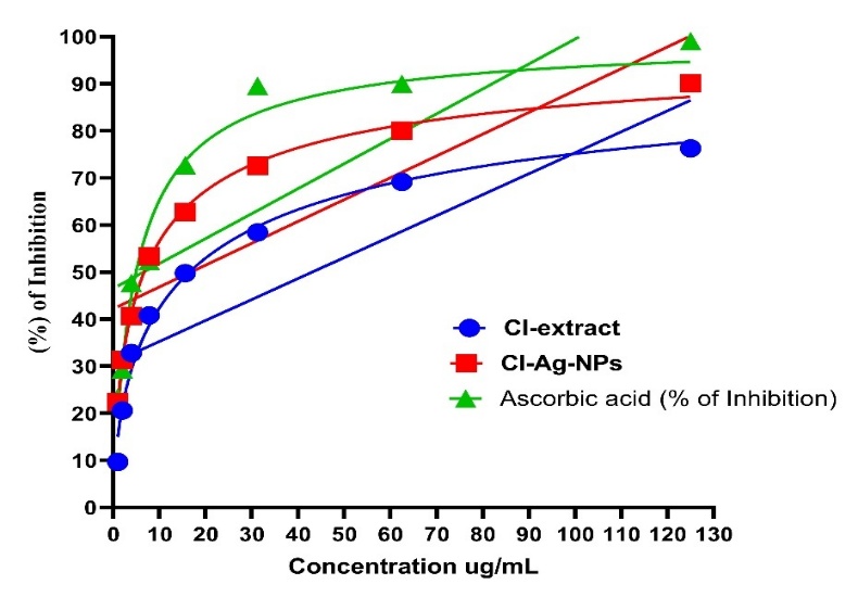
Figure 9: Antioxidant activity of ethanolic extract of C. infortunatum and CI-Ag-NPs
CONCLUSION
The present study successfully demonstrated an eco-friendly and efficient approach for synthesizing silver nanoparticles using Clerodendrum infortunatum extract, confirming the dual role of plant phytochemicals as reducing and stabilizing agents. The formation of CI-Ag-NPs was verified by the characteristic surface plasmon resonance band at 293.03 nm, while FT-IR analysis revealed the involvement of hydroxyl, amine, and other functional groups from the extract in capping and stabilizing the nanoparticles. Morphological and structural characterization by FE-SEM, HR-TEM, and XRD confirmed the nanoscale nature and crystalline properties of the synthesized particles, and EDAX analysis validated the presence of elemental silver. DLS results showed an average particle size of about 89.59 nm with moderate dispersion, indicating acceptable colloidal stability. Biologically, CI-Ag-NPs exhibited markedly enhanced antioxidant activity compared with the crude plant extract, reaching nearly 90% DPPH radical inhibition at higher concentrations, which highlights the synergistic effect between silver and phytochemicals. The nanoparticles also showed notable antibacterial activity against both Gram-positive and Gram-negative bacteria, confirming their broad-spectrum antimicrobial potential. Overall, the integration of plant-based synthesis with nanosilver technology produced a stable, bioactive, and environmentally safe nanomaterial, supporting its promise for future pharmaceutical, biomedical, and antimicrobial applications.
CONFLICT OF INTEREST: Nil
REFERENCES


Raji M. K., Sunbee Prakash, Green Synthesis, Physicochemical Characterization, and Antioxidant Evaluation of Silver Nanoparticles Derived from Clerodendrum infortunatum., Int. J. of Pharm. Sci., 2026, Vol 4, Issue 2, 4204-4214. https://doi.org/10.5281/zenodo.18782508
 10.5281/zenodo.18782508
