

We use cookies to ensure our website works properly and to personalise your experience. Cookies policy
Centre for Pharmaceutical Sciences, UCESTH, Jawaharlal Nehru Technological University, Hyderabad, Kukatpally, Telangana, India 500085
The invasive fungal infections continue to be a significant source of morbidity and mortality, especially in immunocompromised patients. Although some antifungal drugs are focused on a broad spectrum of activity, their poor aqueous solubility, erratic oral bioavailability, food-dependent absorption, and possible systemic toxicity limits clinical utility. Nanoemulsion has become a delivery system in which the above limitations may be surmounted through a promising strategy of increasing drug solubilization, and intestinal permeability, and favouring lymphatic circulation. Recent developments point to the possibilities of CD44-specific nanoemulsions in antifungal targeted to the site of action .Hyaluronic acid (HA) is a natural agonist of the CD44 receptors that are overexpressed on inflamed, infected and activated immune cells, facilitates receptor mediated uptake and enhanced tissue localization. HA-coated nanoemulsions not only increase mucoadhesion and intestinal retention but also enable localized delivery, therefore minimizing off-target toxicity and enhancing therapeutic efficiency. Parameters of critical formulation-such as oil phase selection, surfactant systems, cosurfactants, droplet size optimization (<200 nm), modulation of zeta potential and excipient safety limits shows exemplary functions on stability, augmentation of permeability, and long term safety. Regulatory considerations such as Inactive ingredients Database (IID) limits and chronic exposure safety tests by FDA are required and well assimilated when designing a formulation. This review comprehensively discusses formulation strategies, CD44-mediated targeting mechanisms, physicochemical optimization, safety profiling, regulatory perspectives, and translational challenges of HA-functionalized nanoemulsions for oral delivery of Antifungal drugs. The article further outlines future directions toward precision antifungal therapy through receptor-targeted nanocarriers.
Invasive fungal infections (IFIs) constitute a major and more and more recognized health concern at the global levels. Researchers invariably report about life-threatening fungal infections particularly those caused by Candida, Aspergillus, and Cryptococcus remain a serious health hazard on human health in the world. These organisms negatively affect individuals with weakened immunity due to conditions such as cancer, organ transplantation or critical illness. Although antifungal drugs exist, there are some shortcomings like its ability to combat these infections is hindered by resistance, toxicity and limited ability to penetrate into infected tissues[1].
Shortcomings of the existing Azole antifungal agents wherein the most commonly used agents include voriconazole that works by inhibiting one of the enzymes of the cell membrane synthesis used by fungi. Although they are wide-ranging, they experience fluctuating blood levels, interactions with drugs and toxicity, particularly with chronic treatment. The possibility of improving delivery of azole via nanotechnology has been put forward to eliminate side effects by enhancing solubility and controlled release.[2]
Nanoemulsions are tiny droplets of oil suspended in water which are capable of accommodating drugs that are poorly soluble in water, in a form that make it easier to absorb from the body. By encapsulating antifungal agents within the internal oil phase, nanoemulsions can effectively bypass the dissolution barrier that limits conventional solid dosage forms. Moreover, the contact of lipid droplets with the bile salts and digestive enzymes can bypass hepatic first-pass metabolism and increase the extent of lymphatic transport and improving systemic exposure.
Review on nanoparticle platforms (such as nanoemulsions and other systems) highlight that difficulties such as low bioavailability, tissue penetration issues and drug resistance can be overcome by these formulations. Nano-based approaches can increase uptake into infected tissues and can sustain release of drugs at places of infection [3].
Although enhancing solubility and pharmacokinetics is a must, it is necessary to deliver the drug to the target area such as inflamed and infected tissues is also another therapeutic frontier. Inflammatory responses stimulated by fungal infections are characterized by the increase in adhesion molecules, cytokines and cell surface receptors within the affected tissues.one such receptor, CD44, is a transmembrane glycoprotein abundantly expressed on epithelial cells and macrophages, and activated immune cells. Although a large portion of the literature on CD44 is on cancer, a common theme is that CD44 is a receptor which is increased in inflamed tissues and interacts well with hyaluronic acid (HA) a naturally occurring sugar molecule in the body[4]. This interaction has been utilized by researchers to obtain HA-coated nanoparticles which are absorbed more efficiently into cells which overexpress CD44 and enhance targeted delivery.
Nanocarriers functionalized with hyaluronic acid have been documented to be used extensively in oncology and CD44 mediated models of targeted delivery using inflammatory disease. These systems are exploitative of the natural interaction of HA and CD44 to facilitate receptor-mediated endocytosis, and thus, improve uptake of drugs in the CD44-enriched tissues[5]. Although its outcomes have been promising in other fields of therapy, the use of CD44-targeted nanocarriers in antifungal treatment is still very limited. Most current antifungal nanoformulations have their core centered on passive improvement of solubility rather than active targeting of infected tissues. Given the inflammatory microenvironment characteristic of fungal infections and the documented overexpression of CD44 in such settings, integrating CD44-targeting strategies with nanoemulsion platforms represents a rational and innovative approach.
This review aims to critically examine current advances in nanoemulsion-based antifungal delivery, evaluate the rationale for CD44-mediated targeting, and identify existing gaps that must be addressed to translate these strategies into clinically viable therapy.
Table 1 : Comparision of Conventional Nanoemulsions VS CD44 Targeted Nanoemulsions
|
Parameter |
Nanoemulsions |
Cd44 Targeted Nanoemulsions ( HA Functionalized) |
|
Basic Composition |
Oil + Surfactant + Co-Surfactant + Aqueous phase |
Same core nanoemulsion system + surface functionalization with hyaluronic acid(HA) |
|
Mechanism of drug delivery |
Passive diffusion & enhanced permeation due to nanoscale size |
Active receptor mediated targeting via CD44-HA interaction + passive diffusion |
|
Targeting strategy |
Non-specific (Passive accumulation) |
Active targeting towards CD44 overexpressing inflamed or infected tissues |
|
Selectivity to Diseased tissue |
Limited selectivity |
Enhanced selectivity in infected tissues with high CD44 expression |
|
Bioavailability improvement |
Improved compared to conventional dosage forms |
Further enhanced due to improved tissue localization & cellular uptake |
|
Systemic toxicity |
Reduced compared to conventional formulations but still systemic exposure |
Potentially lower systemic toxicity due to site specific accumulation |
|
Application in Antifungal therapy |
Used to improve solubility of azole antifungal drugs |
Emerging stratergy, limited but promising research in inflammatory fungal infections |
2. Pathophysiology of Fungal Infections
2.1 Mechanisms of Fungal Pathogenesis
Pathophysiology of fungal infections is divided into various categories that include: Aspergillus and Candida.
Pathogenesis: The mechanisms of pathogenesis are not well understood but it is believed that the process is complex and multi-faceted, involving enzymatic and non-enzymatic processes.
The development of the fungal infections follows a series of events that is well-coordinated to enable the normally harmless or environmental fungi to develop invasive pathogenicity, particularly in those people with a weakened immune defenses. This usually entails the initial colonization, attack of the host tissues, avoidance of immune responses, as well as, in most instances, biofilm formation. All of these adaptive strategies establish the severity and persistence of infection[6].
A. Tissue Invasion
Attachment to host surfaces is the initial stage of the pathogenesis of the fungi. Most of the pathogenic fungi express surface proteins, which are specific to them and allow them to target host components of the extracellular matrix like collagen, laminin and fibronectin. This binding process is fundamental in colonization and it comes before deeper invasion.
In Candida albicans, which is one of the most extensively studied fungal pathogens, the progress of infection occurs enormously associated with its morphological transformation capacity. Under favourable conditions,the organism shifts from a round yeast form to elongated filamentous hyphae.This transition improves its ability to enter epithelial barriers. The enzymes secreted by the fungus during the invasion, which include proteases, phospholipases and lipases, rupture the host cell membranes, structural proteins, and leads to damage of tissues and systemic spread.
On the same note, inhalation of Aspergillus fumigatus spores may extend to the lower respiratory system.These spores are normally eliminated by immunocompetent individuals. However,in immunocompromised hosts, they germinate into filamentous hyphae that invade lung tissue.A hallmark of invasive aspergillosis is angioinvasion, where hyphae infiltrate blood vessels, potentially causing thrombosis, hemorrhage and tissue necrosis.
B. Immune Evasion
Pathogenic fungi avoid or manipulate the host immune defences in order to cause an infection. One relevant strategy involves in masking structural cell wall components like as β-glucans, which would otherwise get recognized by host pattern recognition receptors such as Dectin-1. By concealing these molecules, fungi suppress early immune response and slow down inflammatory responses.
Another example of immune evasion is encapsulated fungi. The carbohydrate covering of Cryptococcus neoformans has a key part in its virulence. The capsule disrupts phagocytosis through macrophages and suppressing inflammatory signaling which enables the survival of the organism and spread especially into the central nervous system.
Candida albicans species also exhibit immune adaptation by surviving within macrophages and subsequently escaping by the way of filaments. This intracellular persistence adds to invasion of bloodstream and systemic candidiasis.
C. Biofilm Formation
The other significant determinant of fungal virulence is biofilm formation particularly in Candida infection related to medical devices. A biofilm is an organized group of fungal cells imbedded within a self-generated extracellular matrix that is made of polysaccharides, proteins, lipids, and extracellular DNA.[7]
These biofilms are common on the indwelling devices including catheters, prosthetic valves and implants. Under this structured environment, fungal cells take on altered metabolic conditions that make them resistant to antifungal agents. The surrounding matrix restricts penetration of drugs whereas biofilm-associated cells usually enhance efflux pumps and stress-response mechanisms that enhance resistance to Antifungal drugs, such as azoles and others.
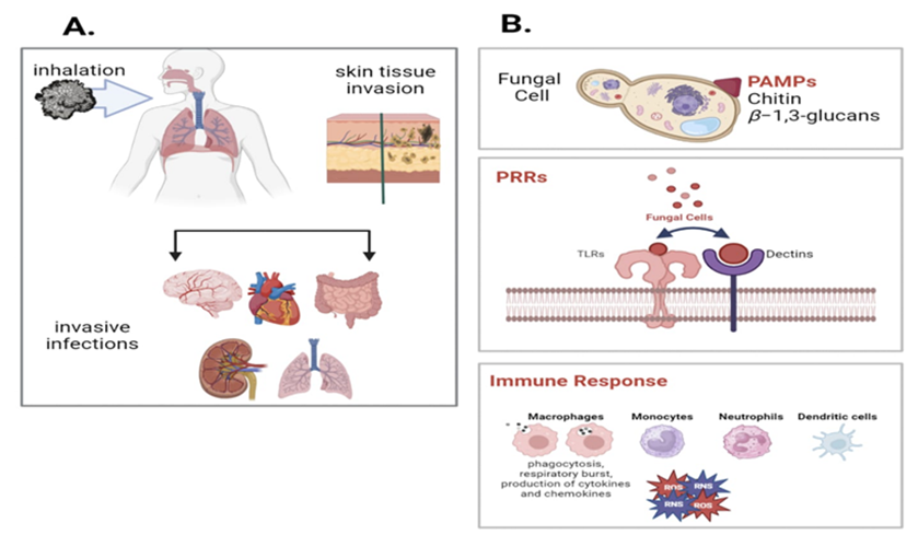
Figure 1: Schematic illustration of Tissue invasion and Immune evasion
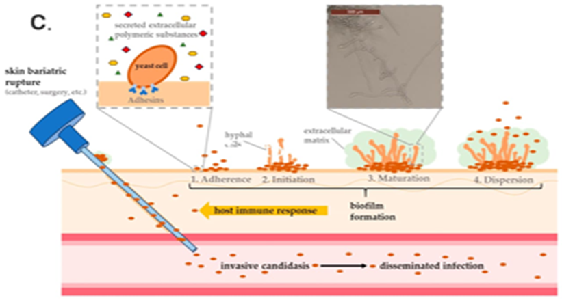
Figure 2: Schematic illustration of Biofilm formation
3. Hyaluronic Acid-Coated Nanoemulsions for CD44 targeting in Antifungal therapy
3.1 Hyaluronic Acid: Structural characteristics & molecular weight dependent behaviour
Structural Characteristics of Hyaluronic acid
Hyaluronic acid (HA) is a naturally occurring polysaccharide that forms the basic constituent of the connective, epithelial and neural tissue extracellular matrix. Unlike many other glycosaminoglycans, HA is not sulfated and it is not attached to a core protein. Instead, it exists as a free, linear polymer made up of repeating disaccharide units of the D-glucuronic acid and N-acetyl-D-glucosamine.The sugars are also interchangeably connected by β-(1→3) and β-(1→4) glycosidic bonds, and link themselves together into long chains which may be vary widely in size. This linear polymer can reach extremely high molecular weights of between a few kilodaltons up to several million daltons according to its biological source and processing.[8]
Unlike sulfated glycosaminoglycans such as heparin or chondritin sulfate, HA is synthesized at the plasma membrane and extruded directly into the extracellular space without covalent attachment to a core protein. Its single repeating structure, and lack of sulfation make it less immunogenic, which enhances its great biocompatibility and biodegradability. Enzyme degradation takes place through hyaluronidases, producing smaller fragments, which are capable of having specific biological effects.
From a drug delivery perspective, several structural features make HA highly attractive:
These structural attributes form the basis for HA’s dual role as both a biological ligand and a functional polymer in nanocarrier systems.
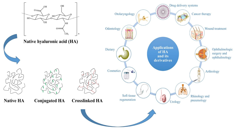
Figure 3 : Structure & Applications of hyaluronic acid
Molecular Weight-Dependent Biological Behavior
The variability of the molecular weight is one among the typical characteristics of HA. Depending on tissue origin and physiological conditions, HA chains could be a few kilodaltons up to several million daltons. This size difference is biologically significant.HA of high molecular weight is considered to be normally related to anti-inflammatory effects, structural support and hydration. In contrast, smaller-molecular-weight fragments which are frequently produced in case of tissue damage or enzyme activity are capable of taking part in inflammatory communication and immune regulation.[9]
Table 2 : Molecular weight dependent behaviour of Hyaluronic acid
|
HA Category |
Molecular weight range |
Structural/ Physicochemical features |
Biological effects |
Immunological behaviour |
Relevance in drug delivery |
|
Oligosaccharide |
<10 kDa |
Very short chain fragments; Highly diffusive; low viscosity
|
Acts as signaling molecules; rapidly cleared |
Strongly pro-inflammatory; activates TLR2/ TLR4; induces cytokine release |
Not suitable for surfaces coating, may destabilize formulation & trigger inflammation |
|
Low molecular weight |
10-100kDa |
Reduced chain length; moderate hydration; low viscosity |
Promotes angiogenesis & immune cell recruitment |
Pro-inflammatory; functions as damaged associated molecular pattern (DAMP) |
Limited use; risk of inflammatory response in infected tissues |
|
Medium molecular weight |
100-500 kDa |
Balanced flexibility & hydration; moderate viscosity |
Supports tissue repair; moderate receptor binding |
Mild immunomodulatory activity |
Preferred for nanoemulsion coating; provides efficient CD44 binding with manageable viscosity |
|
High molecular weight |
500-1000 kDa |
Long polymer chains; high water retention; increased viscosity |
Anti-inflammatory; maintains extracellular matrix integrity |
Suppresses immune activation; stabilizes tissues |
Good for targeting & prolonged retention; may increase formulation viscosity |
|
Very high molecular weight |
>1000 kDa |
Extremely long chains; highly viscous; gel forming |
Strong tissue protective & barrier effects |
Potent anti-inflammatory; inhibits excessive immune signalling |
May cause processing challenges; limited practically in nanoemulsions due to high viscosity. |
3.2 CD44 Structure and Biological Role
CD44 is a type I transmembrane glycoprotein and plays the role of a major regulator of cell adhesion, migration, immunodulators, and tissue remodeling. It is widely expressed in numerous cell types, and consists of epithelial and endothelial cells, fibroblasts, macrophages, neutrophils and lymphocytes.
The structure of CD44 consists of three major parts: an extracellular ligand domain responsible for ligand recognition, a single transmembrane segment and a cytoplasmic tail which links to intracellular signaling apparatus and the cytoskeleton. Hyaluronic acid (HA) is the best among its ligands described and most physiologically relevant binding partner.[14]
CD44 Standard and Variant Isoforms
The gene of the CD44 is highly subject to alternative splicing to produce numerous variants with distinct biological properties. The standard isoform (CD44s) contains the conserved exons encoding the HA-binding extracellular domain, transmembrane region, and cytoplasmic tail. In contrast, variant isoforms (collectively termed CD44v) arise through insertion of additional variant exons into the extracellular portion of the receptor.
These structural variations impact on the interaction of ligands and downstream signaling ability. Variant isoforms are often related to inflammatory, tissue remodelling, and pathological processes. Through communication with cytoskeletal associated proteins and signaling molecules, CD44 isoforms have the power to modulate pathways like MAPK and PI3K/Akt hence affecting cell survival, proliferation and migration.
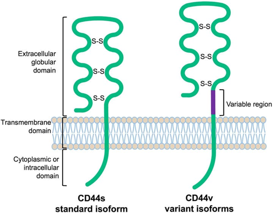
Figure 4: Schematic protein structure of the CD44 molecule.
CD44 as a molecular target in Fungal Infections
During fungal infections particularly the invasive forms, the host has a strong inflammatory response marked by cytokine release and immune cell recruitment.These proinflammatory agents stimulate a rise in the expression of CD44 on epithelial and immune cells in sites of infection. Studies of pulmonary inflammation has demonstrated that the alveolar epithelial cells and the macrophages increase CD44 in response to inflammatory stimuli. This upregulation has been found, similarly, in mucosal and systemic infections in which tissue damage and immune activation occurs.
CD44 contributes to the host defense as it helps in the adhesion and migration of leukocytes to inflamed tissues. Its cytoplasmic tail interacts with cytoskeletal components and improving the phagocytic activity. Interaction of CD44 and HA has the potential to stimulate intracellular signaling receptors controlling the production of cytokines, amplification of inflammation and tissue repair.
Therefore, CD44 is not only an adhesive molecule, but also a dynamic regulator of signalling in the course of infection.
HA-CD44 interaction and Receptor-Mediated Internalization.
Hyaluronan-binding domain is a defined domain of CD44 located in its extracellular part and which interacts with HA using a combination of hydrogen bonding and electrostatic forces. Binding of HA can stimulate receptor clustering on the cell surface. Such clustering facilitates rearrangement of cytoskeletons that may induce endocytosis via receptors.[11]
The internalization mechanism is specifically relevant to drug delivery applications. When nanoemulsions or nanoparticles are coated with HA, they can exploit CD44 recognition to facilitate targeted cellular uptake. Instead of relying soley on passive diffusion, HA-decorated systems may enter the controlled endocytic route of CD44-rich cells to enhances the intracellular drug accumulation.
Some fungal pathogens such as Candida species are able to survive within macrophages, thus delivery of drugs into the cell a significant therapeutic factor. Since the expression of CD44 is elevated in inflamed and infected tissues,HA-coated nanoemulsions can be potentially accumulate in these receptor-rich conditions. Targeting CD44 increases localization to pathological sites and facilitates immune or epithelial cell uptake via receptors.
Such targeted strategy has two possible benefits in antifungal therapy:
The biological properties of CD44 its expression in inflammation, ability to mediate endocytosis, and immune regulation role- make it a rational molecular target of HA-functionalized nanocarrier systems in the management of infectious diseases.
In targeted delivery systems such as hyaluronic acid coated nanoemulsions, HA on the surface of the nanocarrier has a specific binding to receptor of CD44 at sites of infection. The resultant receptor-mediated uptake is able to increase the localization of drugs in diseased tissues and decrease healthy cell exposure. This approach therefore enhances the effectiveness of treatment and reduces the systemic toxicity.
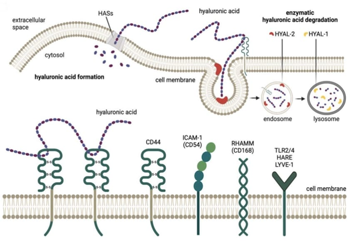
Figure 5 : Hypothetical communication between hyaluronic acid and its receptors.
4. Formulation Development of HA-Coated Nanoemulsions & Safety considerations[17].
4.1 Components of HA-Coated Nanoemulsion
(A) Drug selection
Poorly water soluble antifungal drugs are best candidates because:
(B) Oil Phase Selection
It should have a high solubilization capacity of the antifungal drug in oil phase and it should maintain acceptable safety profiles. The choice of oil is directed by solubility screening studies.
Table 3 : Safety considerations of Oils
|
Excipient
|
Type
|
Regulatory / IID Status |
Safety & Toxicology |
Typical Use Level in Oral NE |
|
Capmul MCM (glyceryl monocaprylate/ monocaprate) |
Medium-chain mono/ di-glyceride
|
Not always individually listed in IID; medium-chain glycerides are used in approved oral products |
Generally recognized as safe (GRAS-related category); USP/NF compliant; well tolerated; high doses may cause mild GI irritation |
10–30% of formulation |
|
Capryol 90 (propylene glycol monocaprylate) |
Medium-chain fatty acid ester
|
Listed in IID for oral capsules and solutions (varies by product) |
Widely used lipid solubilizer; low acute toxicity; excessive intake may cause mild laxative effect |
5–20% |
|
Miglyol 812 (medium chain triglycerides, MCT oil) |
MCT oil (C8–C10 triglycerides) |
Present in IID for oral capsules and solutions |
GRAS; metabolized via β-oxidation; high tolerability; excessive intake may cause diarrhea |
5–40% |
|
Oleic Acid
|
Long-chain fatty acid
|
IID listed for oral formulations |
Naturally occurring fatty acid; generally safe; may cause mucosal irritation at high concentrations |
1–15% |
|
Soybean Oil
|
Long-chain triglyceride
|
IID listed; widely used in oral and parenteral emulsions |
GRAS; extensively used in lipid emulsions; allergen consideration |
5–40% |
(C) Surfactant and Co-Surfactant Selection
Surfactants with appropriate hydrophilic-lipophilic balance (HLB) values are selected to ensure stable nano-sized droplets. Safety and regulatory acceptability must be considered.
Table 4 : Safety considerations of surfactants & co-surfactants
|
Excipient |
Type |
IID/ Regulatory status |
Safety & Toxicology |
Typical Use Level in Oral NE |
|
Tween 80 (Polysorbate 80)
|
Non-ionic surfactant
|
IID listed for tablets, capsules, suspensions (limits vary by dosage form)
|
GRAS; WHO ADI ~25 mg/kg/day (general guidance); rare hypersensitivity reactions; well tolerated at low–moderate doses |
1–15% |
|
Tween 20 (Polysorbate 20)
|
Non-ionic surfactant
|
IID listed for oral products
|
Similar safety to Tween 80; lower hydrophobicity; mild GI irritation at high doses |
1–10% |
|
Cremophor EL (Polyoxyl 35 castor oil)
|
Non-ionic surfactant
|
IID listed for oral capsules/solutions
|
Effective solubilizer; may cause hypersensitivity at high doses; generally safe orally |
5–20% |
|
Labrasol (Caprylocaproyl polyoxyl-8 glycerides) |
Non-ionic surfactant
|
Present in approved oral lipid formulations
|
Widely used in SEDDS; enhances permeability; mild GI effects at high doses |
10–30% |
|
Transcutol HP (Diethylene glycol monoethyl ether)
|
Cosurfactant / Solvent
|
Listed in IID for topical; oral use supported in marketed products though not always separately listed |
Extensive toxicology data; non-genotoxic; high doses in animals linked to renal effects; widely used in oral lipid systems |
5–30% |
|
Propylene Glycol
|
Solvent / Cosurfactant
|
IID listed for oral dosage forms
|
FDA approved; high doses may cause hyperosmolality; safe within recommended limits |
Up to 40% in liquids (dose dependent) |
|
PEG 400
|
Cosolvent
|
IID listed for oral capsules/solutions
|
Widely used; safe; high doses may cause osmotic diarrhea |
5–40% |
4.2 Pseudo-Ternary Phase Diagram Construction
The construction of a pseudo-ternary phase diagram is a foundational step in the rational development of nanoemulsion and self-emulsifying systems. It is characterized as pseudo because the surfactant and co-surfactant blend (Smix) is considered as a single entity and hence makes a four-component system simpler by dividing into three compositional axes of oil, Smix, and water. This is a graphical method that can be used to conduct a systematic approach of phase behaviour mapping as well as determination of areas where spontaneous nanoemulsification takes place.
The recent studies on lipid-based formulations point out that pseudo-ternary phase diagrams reduce empirical trial-and-error experimentation through clearly identifying concentration ranges which yield transparent, isotropic, and thermodynamically stable systems [19] .In antifungal nanoemulsion development, this strategy is particularly valuable since there should be enough oil to dissolve highly lipophilic drugs, but the level of surfactants should not be above the safe regulatory ranges.
The process is usually initiated by solubility screening of the drug using desired oils, surfactants and co-surfactants. Surfactant and co-surfactant are then mixed in known proportions (e.g., 1: 1, 2: 1, or 3: 1) to form Smix after the selection of appropriate components. Based on this, oil and Smix are mixed in different proportions ratios, usually 9:1 to 1: 9. Under gentle agitation, water is titrated gradually and a visual assessment of each system is done in terms of clarity, homogeneity, turbidity or phase separation.
Compositions giving clear and low-viscosity systems are plotted on triangular coordinates to trace the nanoemulsion area. The larger the nanoemulsion areas, the higher the emulsification efficiency and flexibility in formulation design. Phase diagram analysis according to Pouton and Porter is still among the reliable methods of maximizing lipid-based drug delivery system, especially SEDDS and nanoemulsions platforms.
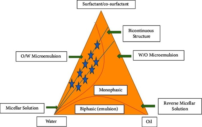
Figure 6: Schematic figure of Pseudo-Ternary phase diagram.
4.3 Methods of Nanoemulsion Preparation
Nanoemulsions can be produced through high-energy or low-energy approaches, and the choice depends on formulation requirements, scalability, and desired droplet characteristics.
High-energy methods make use of mechanical forces to rupture coarse emulsions into nanometric droplets. Intense shear and cavitation forces are exerted on the emulsion through the force of high-pressure homogenization via a narrow valve of high pressure. similarly, ultrasonication generates acoustic cavitation bubbles that collapse violently and breaking droplets down to nanoscale dimentions. These approaches are especially effective in the case when a small droplet size distribution and a precise control below 150-200 nm are required and is often desirable to enhance oral absorption of lipophilic antifungal agents [19].
Low-energy methods, on the other hand, use the physicochemical nature of surfactants and take advantage of their inherent characteristics where interfacial tension varies instead of extrinsic mechanical energy. Spontaneous emulsification takes place when an oil-surfactant mixture is introduced into the aqueous phase and causes rapid droplet formation driven by interfacial turbulence. Phase inversion techniques make use of temperature or compositional variations to induce the formation of nanoemulsions[20].
Recent formulation literature suggests that low-energy systems are appealing to oral antifungal delivery due to simplicity, cost-effectiveness, and ease of scale-up, whereas high-energy systems may offer superior control over droplet size distribution and physical stability.
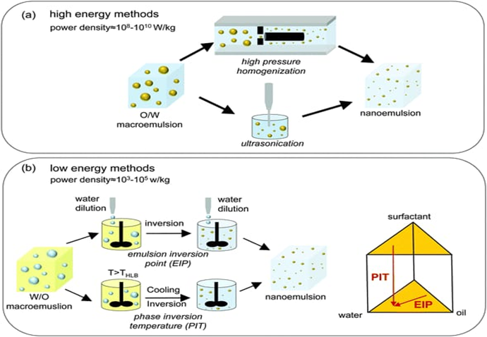
Figure 7 : Schematic representation of high energy and low energy methods for preparing O/W nanoemulsions
4.4 Hyaluronic Acid Coating Process
Surface modification with hyaluronic acid (HA) represents a strategic approach to facilitate CD44 mediated targeting. CD44 is a high-affinity transmembrane glycoprotein receptor with high affinity to HA, often expressed in inflamed or damaged epithelial tissues, such as those so caused by infection. Emerging evidence in inflammatory lung disease suggests that CD44 participates in immune cell recruitment and epithelial repair processes, supporting its relevance as a targeting ligand.
The adsorption-based methods are widely used to obtain HA coating of nanoemulsions. Because HA has negatively charged carboxyl groups, effective coating is dependent on the nature of surfaces of the pre-formed nanoemulsion. In case droplets have a slight positive or near-neutral charge, electrostatic interactions and hydrogen bonding facilitate HA deposition on the interface.
In practical terms, HA is dissolved in distilled water at low concentrations (typically 0.05–0.2% w/v). The optimized nanoemulsion is incorporated gradually with controlled stirring in order to enable homogenous adsorption.Successful surface functionalization has been confirmed by a slight droplet size increase (usually 20-50 nm) and a shift in zeta potential toward more negative values, which is indicative of HA chains on the surface.
The targeting capabilities of HA are also greatly dependent on the molecular weight. Medium-molecular-weight HA (approximately 50-300 kDa) is commonly used due to adequate CD44 binding affinity, while maintaining acceptable viscosity and colloidal stability.Excessively high molecular weight of HA may increase viscosity and aggregation, whereas very low molecular weight HA may compromise efficiency of recognition of the receptor.
By promoting CD44-mediated endocytosis, HA coating enhances intracellular accumulation of the antifungal payload, potentially improving therapeutic efficacy compared to non-targeted systems.
4.5 Characteristics of Hyaluronic Acid-Coated Nanoemulsion
The resulting HA-coated nanoemulsions exhibits physicochemical and biological properties that collectively determine its therapeutic performance.
A characteristic aspect is a droplet size in the nanoscale i.e., less than 200-250 nm. Such small droplets increase surface area, enhance dissolution of poorly soluble antifungal drugs and enhance membrane transport. A poly dispersity index of less than 0.3 indicates uniform droplets distribution and formulation homogeneity, which leads to reproducible pharmacokinetics and stability [21]
After HA coating, the zeta potential is reduced to be more negative because of exposed carboxyl groups. This negative surface charge increases the electrostatic repulsion between droplets and decreases the chance of aggregation. Additionally, the hydrated HA layer also provides steric stabilization, which further contributes to colloidal stabilization during storage.
Optimized lipid nanoemulsions typically demonstrate high drug loading and entrapment efficiency because the oil phase effectively solubilizes lipophilic antifungal agents. Importantly, the HA layer acts as a targeting shell without significantly altering drug encapsulation within the lipid core.
Another desirable quality is controlled drug release behavior. The lipid matrix is serves as a reservoir, enabling gradual diffusion of antifungal drug. Sustained release may help to maintain therapeutic concentrations at infected sites while potentially lowering systemic exposure.
Biologically, the most distinctive feature is CD44-specific interaction. HA on the droplet surface binds to CD44 receptors expressed on inflamed or infected cells which results in receptor-mediated internalization. This mechanism is able to increase intracellular drug delivery and antifungal action relative to non-targeted nanoemulsions.
Finally, the use of pharmaceutically accepted oils, non-ionic surfactants, and a naturally occurring biopolymer such as HA contributes to favorable biocompatibility and safety, supporting the translational potential of this targeted nanoemulsion system.
CONCLUSION
Hyaluronic acid–coated nanoemulsions represent an advanced and mechanistically driven approach for CD44-targeted antifungal therapy, addressing key limitations associated with conventional antifungal formulations such as poor aqueous solubility, low bioavailability, and systemic toxicity. The lipid-based nanoemulsion core enhances solubilization of hydrophobic antifungal drugs and improves absorption due to its nanoscale droplet size and large interfacial surface area. Optimized systems typically exhibit uniform particle distribution, high drug entrapment efficiency, and good thermodynamic stability, ensuring reproducible therapeutic performance.
Surface functionalization with hyaluronic acid introduces active targeting capability through specific interaction with CD44 receptors, which are often upregulated in inflamed and infected tissues. This HA–CD44 binding promotes receptor-mediated endocytosis, facilitating enhanced intracellular drug accumulation at the site of fungal infection. As a result, targeted nanoemulsions may achieve improved antifungal efficacy while reducing off-target exposure and minimizing systemic adverse effects. The negative surface charge imparted by HA further contributes to colloidal stability and biocompatibility.
Despite these advantages, certain challenges must be addressed to enable successful clinical translation. These include maintaining HA stability against enzymatic degradation, ensuring consistent receptor targeting across patient populations, optimizing large-scale manufacturing processes, and establishing long-term safety profiles. Regulatory considerations and excipient safety compliance are also essential for therapeutic approval.
Overall, hyaluronic acid–coated nanoemulsions integrate solubility enhancement, controlled drug release, and receptor-specific targeting into a single multifunctional platform. With continued research focused on formulation optimization, in vivo validation, and scalable production, this targeted nanocarrier system holds strong potential to significantly improve the effectiveness and safety of antifungal therapy in clinical settings.
REFERENCES



Rakshitha Burra, Dr. K. Anie Vijetha, Dr. M. Sunitha Reddy, Hyaluronic Acid Coated Nanoemulsions for CD44 Mediated Targeted Antifungal Therapy : Mechanistic Insights, Formulation Strategies & Considerations, Int. J. of Pharm. Sci., 2026, Vol 4, Issue 3, 763-778. https://doi.org/10.5281/zenodo.18918737
 10.5281/zenodo.18918737