Department of Pharmacology, Sri Venkateswara College of Pharmacy, Chittoor, Andhra Pradesh, India
This study investigates the in-vitro antioxidant and anti-inflammatory activities of individual and combined ethanol extracts of Curcuma amada Roxb. (mango ginger) and Phyllanthus emblica L. (amla). The plant materials were subjected to maceration extraction using ethanol as solvent. Antioxidant activity was evaluated using the 2,2-diphenyl-1-picrylhydrazyl (DPPH) radical scavenging assay, while anti-inflammatory activity was assessed through the protein denaturation inhibition assay. The DPPH assay revealed that C. amada extract exhibited potent antioxidant activity with an IC50 value of 440.66 ?g/ml, comparable to the standard ascorbic acid (IC50 = 26.81 ?g/ml). The P. emblica extract demonstrated high antioxidant activity with IC50 of 134.17 ?g/ml. In the protein denaturation inhibition assay, C. amada showed IC50 of >100 ?g/ml, while P. emblica exhibited IC50 > 100 ?g/ml. The combined extract (A+B) displayed IC50 values of 30.69 ?g/ml for DPPH and 88.70 ?g/ml for protein denaturation inhibition. Combination index analysis using the Chou-Talalay method revealed antagonistic interactions between the two extracts at the tested ratios, with CI values exceeding 2.0 at the IC50 level. These findings indicate that while the combination of C. amada and P. emblica ethanol extract possesses high significant antioxidant and moderate anti-inflammatory properties as compared to individual plant extract under the tested conditions.
Oxidative stress and inflammation are fundamentally interconnected pathological processes that underlie the etiology of numerous chronic diseases, including cardiovascular disorders, neurodegenerative conditions, diabetes mellitus, and various forms of cancer[1-3]. The excessive production of reactive oxygen species (ROS) and subsequent oxidative damage to cellular macromolecules including lipids, proteins, and DNA have been recognized as key contributors to the pathogenesis of inflammatory conditions[4,5]. This intimate relationship between oxidative stress and inflammation has prompted extensive research into natural products that possess both antioxidant and anti-inflammatory properties, as such dual-activity compounds may offer enhanced therapeutic potential for managing inflammation-associated disorders[6,7].
Curcuma amada Roxb., commonly known as mango ginger, is a perennial rhizomatous herb belonging to the family Zingiberaceae. This plant is indigenous to the Indian subcontinent and has been traditionally employed in Ayurvedic medicine for treating various inflammatory conditions, digestive disorders, and skin diseases[8,9]. Phytochemical investigations have revealed that C. amada rhizomes contain a diverse array of bioactive constituents, including curcuminoids (curcumin, demethoxycurcumin, and bisdemethoxycurcumin), volatile terpenoids such as difurocumenonol, phenolic acids, and various carbohydrate derivatives[10,11]. The characteristic mango-like aroma of the rhizome is attributed to the presence of unique terpenoid compounds that distinguish it from the closely related Curcuma longa (turmeric)[12]. Previous pharmacological studies have demonstrated that C. amada extracts exhibit significant antioxidant, anti-inflammatory, antimicrobial, and anticancer activities[13,14].
Phyllanthus emblica L. (syn. Emblica officinalis Gaertn.), widely known as amla or Indian gooseberry, is a deciduous tree belonging to the family Phyllanthaceae. This plant holds a distinguished position in traditional Indian medicine systems, where it is revered as a "rasayana" (rejuvenating agent) and has been used for millennia to promote longevity, enhance immunity, and treat various ailments[15,16]. The fruits of P. emblica are exceptionally rich in vitamin C (ascorbic acid), containing approximately 600-900 mg per 100 g of fresh fruit weight, which represents one of the highest concentrations among edible fruits[17]. Beyond vitamin C, amla fruits contain a complex mixture of hydrolyzable tannins including emblicanin A, emblicanin B, punigluconin, and pedunculagin, along with flavonoids such as quercetin, kaempferol, and various phenolic acids including gallic acid and ellagic acid[18,19]. The antioxidant capacity of P. emblica has been extensively documented, with studies demonstrating its superior radical scavenging activity compared to many other medicinal plants[20,21].
The concept of combining multiple plant extracts to achieve enhanced therapeutic effects through synergistic interactions is deeply rooted in traditional medicine systems and has gained considerable scientific attention in recent years[22,23]. Synergistic effects occur when the combined action of two or more agents produces a greater effect than would be predicted from the sum of their individual effects. In the context of antioxidant and anti-inflammatory activities, such synergy may arise from complementary mechanisms of action, enhanced bioavailability, or stabilization of active constituents within the combination[24,25]. The Chou-Talalay combination index method provides a quantitative framework for assessing drug interactions, where a combination index (CI) value less than 1 indicates synergism, equal to 1 indicates additive effects, and greater than 1 indicates antagonism[26,27].
Despite the individual pharmacological importance of C. amada and P. emblica, there remains a paucity of scientific literature examining the potential synergistic interactions between these two important medicinal plants. Given their complementary phytochemical profiles and traditional uses in treating inflammatory conditions, we hypothesized that combining ethanol extracts of these plants might produce enhanced antioxidant and anti-inflammatory activities. The present study was therefore designed to evaluate the in-vitro antioxidant activity using the DPPH radical scavenging assay and anti-inflammatory activity through the protein denaturation inhibition assay, and to characterize the nature of interactions between the combined extracts using the Chou-Talalay combination index methodology.
MATERIALS AND METHODS
Plant Materials
The rhizomes of Curcuma amada Roxb. and fruits of Phyllanthus emblica L. were procured from authenticated suppliers in Kerala, India, during the period of January 2026. The plant materials were identified and authenticated by a qualified botanist from Sri Venkateswara University Tirupati, Andhra Pradesh. Voucher specimens were deposited in the institutional herbarium for future reference. The rhizomes were thoroughly washed with running water to remove soil particles, sliced into small pieces, and air-dried at room temperature (25 ± 2°C) in a well-ventilated area for two weeks. The dried materials were subsequently ground into coarse powder using an electric grinder and stored in airtight containers until extraction.
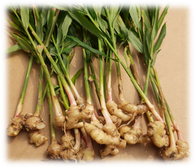
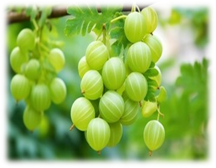
Figure 01 :- Curcuma amada Roxb. & Phyllanthus emblica L.
Chemicals and Reagents
Ethanol (analytical grade, 70% purity) was used as the extraction solvent. 2,2-Diphenyl-1 picrylhydrazyl (DPPH), ascorbic acid, diclofenac sodium, bovine serum albumin (BSA), and phosphate buffered saline (PBS) were obtained from Sigma-Aldrich Chemicals Private Limited (Bangalore, India). All other chemicals and reagents used in this study were of analytical grade and used without further purification. Distilled water was used throughout the experiments.
Extraction Procedure
The powdered plant materials were extracted separately using the maceration technique with 70 % ethanol as the solvent. For each plant material, 50 g of dried powder was placed in an air tight glass container and soaked in 500 ml of ethanol (1:10 w/v ratio). The containers were kept at room temperature with occasional shaking for 72 hours. After the maceration period, the extracts were filtered through Whatman No. 1 filter paper. The marc (residue) was subjected to two additional maceration cycles with fresh ethanol (300 ml each) to ensure exhaustive extraction. The combined filtrates were concentrated under reduced pressure using a rotary evaporator at 40°C. The concentrated extracts were subsequently dried in a electric water bath until constant weight was achieved. The dried extracts were stored in sealed vials at 4°C until further analysis. The percentage yield was calculated using the formula :-
Percentage Yield (%)=Weight of Dried ExtractWeight of Powdered Plant Material×100
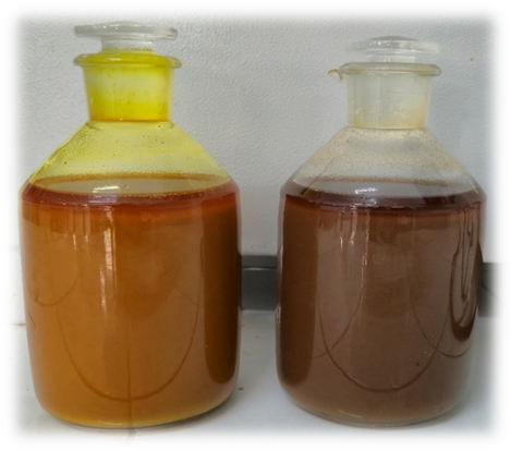
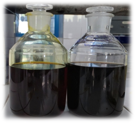
Figure 02 :- Method of Extraction Sample A & B
For combination studies, equal quantities of the dried C. amada and P. emblica extracts were mixed to prepare the combined extract (designated as A+B). Stock solutions of each extract were prepared in the appropriate solvents (methanol for DPPH assay, distilled water for protein denaturation assay) at a concentration of 10 mg/ml and stored at 4°C. Working solutions were prepared fresh on the day of each experiment by appropriate dilution of the stock solutions.
DPPH Radical Scavenging Assay
The antioxidant activity was evaluated using the DPPH radical scavenging assay following the method described by Brand-Williams et al.[28] with slight modifications. A 60 μM solution of DPPH was freshly prepared in methanol. For the assay, 200 μl of DPPH solution was mixed with 50 μl of test sample at various concentrations (1.56, 3.12, 6.25, 12.5, 25, 50, 100, 200, 400, 800, and 1000 μg/ml). The mixture was vortexed thoroughly and kept in the dark at room temperature for 15 minutes. The absorbance was measured at 515 nm using a multi-mode microplate reader (MultiSKAN Skyhigh, Thermo Fisher Scientific). Ascorbic acid was used as the reference standard. Control was prepared with DPPH solution only (without any extract or standard), and 95% methanol was used as blank. All determinations were performed in triplicate. The percentage inhibition of DPPH radical was calculated using the formula :-
%Inhibition=Absorbance of Control – Absorbance of TestAbsorbance of Control×100
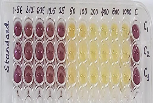
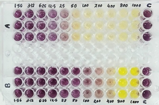
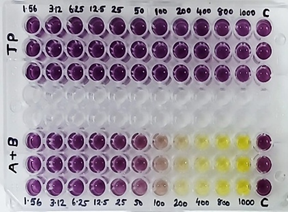
Figure 03 :- DPPH Radical Scavenging Assay for Standard and Smaple A, B & A+B
Protein Denaturation Inhibition Assay
The anti-inflammatory activity was evaluated using the protein denaturation inhibition assay as described by Mizushima and Kobayashi[29] with modifications by Sakat et.al.,[30]. The reaction mixture (0.5 ml) consisted of 0.4 ml of 3% aqueous solution of bovine serum albumin (BSA) and 0.1 ml of test sample at varying concentrations (6.25, 12.5, 25, 50, and 100 μg/ml). The samples were incubated at 37°C for 20 minutes in a water bath. After incubation, 2.5 ml of phosphate buffered saline (pH 6.3) was added to each tube, and the mixtures were heated at 80°C for 10 minutes to induce protein denaturation. After cooling to room temperature, the absorbance was measured at 660 nm using a UV-VIS spectrophotometer (Orion Aquamate 8000, Thermo Scientific). Diclofenac sodium was used as the reference standard. Control was prepared without any test sample. The percentage inhibition of protein denaturation was calculated using the formula :-
%Inhibition=Absorbance of Control – Absorbance of TestAbsorbance of Control×100
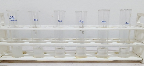
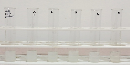
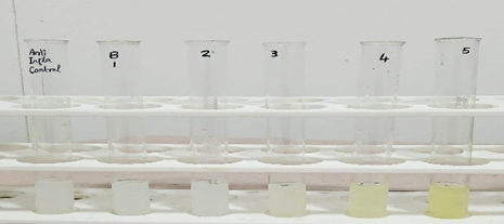
Figure 04 :- Protein Denaturation Inhibition Assay for Standard and Smaple A, B & A+B
Combination Index Analysis
The nature of interaction between C. amada and P. emblica extracts was analyzed using the Chou-Talalay combination index (CI) method[26,27]. The CI was calculated using the formula: CI = (D)1/(Dx)1 + (D)2/(Dx)2, where (D)1 and (D)2 are the doses of extracts 1 and 2 in combination that produce x% effect, and (Dx)1 and (Dx)2 are the doses of each extract alone that produce the same effect. For the combination extract prepared in a 1:1 ratio, the individual contributions at IC50 were calculated. The CI values were interpreted as follows: CI < 0.9 = synergism; CI = 0.9-1.1 = additive effect; CI > 1.1 = antagonism. The analysis was performed for both DPPH radical scavenging and protein denaturation inhibition assays.
Statistical Analysis
All experiments were performed in triplicate and data were expressed as mean ± standard deviation (SD). The IC50 values were calculated by linear regression analysis of the dose-response curves using GraphPad Prism version 9.0 (GraphPad Software, San Diego, CA, USA). Statistical comparisons between groups were performed using one-way analysis of variance (ANOVA) followed by Tukey's post-hoc test. A p-value < 0.05 was considered statistically significant.
RESULTS
Percentage Yield of Extracts
The percentage yield of extracts obtained from Phyllanthus emblica and Curcuma amada was calculated to evaluate the efficiency of the extraction process. The percentage yield was determined using the formula :-
Percentage Yield (%)=Weight of Dried ExtractWeight of Powdered Plant Material×100
Table 01 :- Percentage Yield of Extracts
|
Plant Material |
Weight of Powder (g) |
Weight of Dried Extract (g) |
Percentage Yield (%) |
|
Phyllanthus emblica |
50 g |
25 g |
50 % |
|
Curcuma amada |
50 g |
15 g |
|
Phytochemical Screening Test Results
Preliminary qualitative phytochemical analysis of the individual extracts of Curcuma amada and Phyllanthus emblica revealed the presence of various bioactive constituents such as flavonoids, phenolics, tannins, saponins, glycosides, alkaloids, and terpenoids. The combined extract (1:1 ratio) showed comparatively intense reactions for phenolic and flavonoid tests, indicating a higher cumulative phytochemical content. The presence of abundant phenolic and flavonoid compounds is directly associated with antioxidant and anti-inflammatory activities due to their ability to donate hydrogen atoms, scavenge free radicals, and inhibit inflammatory mediators. The results suggest that both plants contribute complementary phytoconstituents, potentially enhancing biological activity when combined.
Table 02 :- Results of Preliminary Phytochemical Screening
|
Phytoconstituent |
Phyllanthus emblica |
Curcuma amada |
Combined Extract |
|
Alkaloids |
- |
- |
- |
|
Flavonoids |
+ |
+ |
+ |
|
Phenolics/ Tannins |
+ |
+ |
+ |
|
Saponins |
+ |
- |
+ |
|
Glycosides |
+ |
+ |
+ |
|
Terpenoids |
- |
+ |
+ |
Note :- (+) Sign Represent Presence of Active Constituent & (-) Sign Represent Absence of Active constituent,
DPPH Radical Scavenging Activity
The DPPH radical scavenging assay results are presented in Table 03. The reference standard ascorbic acid exhibited potent radical scavenging activity with an IC50 value of 26.81 μg/ml. Among the tested extracts, Phyllanthus emblica (Sample A) demonstrated the highest antioxidant activity with an IC50 of 134.17 μg/ml, which was comparable to that of the standard (p > 0.05). The dose-dependent increase in percentage inhibition was observed across all concentrations tested, with Sample A achieving 87.50% inhibition at 100 μg/ml and reaching a plateau of approximately 89 % at higher concentrations.
Curcuma amada extract (Sample B) showed moderate DPPH radical scavenging activity with an IC50 value of 440.66 μg/ml, which was significantly lower than both ascorbic acid and Phyllanthus emblica extract (p < 0.001). The extract exhibited a gradual, dose-dependent increase in scavenging activity, reaching 81.40% inhibition at the highest concentration tested (1000 μg/ml). The combination extract (A + B) displayed an IC50 value of 30.69 μg/ml, which was highest between the individual extracts.
Table 03 :- DPPH radical scavenging activity of ethanol extracts (Antioxidant Activity)
|
Standard |
Concentration (μg/ml) |
Absorbance at 515nm |
Triplicate Average |
Percentage Inhibition (%) |
||
|
OD1 |
OD2 |
OD3 |
||||
|
Control |
- |
0.866 |
0.863 |
0.863 |
0.864 |
0.864 |
|
Ascorbic acid |
1.56 |
0.833 |
0.831 |
0.830 |
0.831 |
3.94 |
|
3.12 |
0.763 |
0.767 |
0.761 |
0.764 |
11.92 |
|
|
6.25 |
0.719 |
0.713 |
0.712 |
0.715 |
17.59 |
|
|
12.5 |
0.581 |
0.583 |
0.580 |
0.581 |
32.87 |
|
|
25 |
0.481 |
0.485 |
0.480 |
0.482 |
44.44 |
|
|
50 |
0.155 |
0.157 |
0.154 |
0.155 |
82.18 |
|
|
100 |
0.093 |
0.092 |
0.093 |
0.093 |
89.28 |
|
|
200 |
0.085 |
0.084 |
0.084 |
0.084 |
90.23 |
|
|
400 |
0.065 |
0.065 |
0.065 |
0.065 |
92.48 |
|
|
800 |
0.054 |
0.057 |
0.055 |
0.055 |
93.63 |
|
|
1000 |
0.041 |
0.042 |
0.042 |
0.041 |
95.20 |
|
|
IC 50 |
|
|
|
|
26.81μg/ml |
|
|
Sample code |
Concentration (μg/ml) |
Absorbance at 515nm |
Triplicate Average |
Percentage Inhibition (%) |
||
|
OD1 |
OD2 |
OD3 |
||||
|
Control |
- |
0.872 |
0.873 |
0.870 |
0.872 |
- |
|
Sample A Phyllanthus emblica |
1.56 |
0.838 |
0.837 |
0.831 |
0.835 |
4.20 |
|
3.12 |
0.816 |
0.815 |
0.818 |
0.816 |
6.38 |
|
|
6.25 |
0.787 |
0.786 |
0.789 |
0.787 |
9.71 |
|
|
12.5 |
0.750 |
0.748 |
0.746 |
0.748 |
14.22 |
|
|
25 |
0.658 |
0.658 |
0.654 |
0.657 |
24.69 |
|
|
50 |
0.572 |
0.568 |
0.572 |
0.571 |
34.56 |
|
|
100 |
0.483 |
0.481 |
0.483 |
0.482 |
44.69 |
|
|
200 |
0.336 |
0.334 |
0.339 |
0.336 |
61.43 |
|
|
400 |
0.216 |
0.213 |
0.215 |
0.215 |
75.38 |
|
|
800 |
0.093 |
0.091 |
0.092 |
0.092 |
89.47 |
|
|
1000 |
0.091 |
0.093 |
0.090 |
0.091 |
89.53 |
|
|
IC 50 |
|
|
|
|
134.17 μg/ml |
|
|
|
||||||
|
|
|
0.871 |
0.873 |
0.870 |
0.871 |
- |
|
Sample B Curcuma amada |
1.56 |
0.851 |
0.847 |
0.848 |
0.849 |
2.56 |
|
3.12 |
0.829 |
0.828 |
0.826 |
0.828 |
4.98 |
|
|
6.25 |
0.792 |
0.799 |
0.793 |
0.795 |
8.76 |
|
|
12.5 |
0.764 |
0.764 |
0.762 |
0.763 |
12.36 |
|
|
25 |
0.725 |
0.728 |
0.726 |
0.726 |
16.61 |
|
|
50 |
0.689 |
0.687 |
0.687 |
0.688 |
21.05 |
|
|
100 |
0.619 |
0.613 |
0.611 |
0.614 |
29.47 |
|
|
200 |
0.537 |
0.536 |
0.534 |
0.536 |
38.50 |
|
|
400 |
0.454 |
0.456 |
0.451 |
0.454 |
47.91 |
|
|
800 |
0.285 |
0.285 |
0.282 |
0.284 |
67.39 |
|
|
1000 |
0.164 |
0.161 |
0.161 |
0.162 |
81.40 |
|
|
IC 50 |
|
|
|
|
440.66 μg/ml |
|
|
|
||||||
|
|
|
0.869 |
0.873 |
0.873 |
0.872 |
- |
|
Sample A+B |
1.56 |
0.824 |
0.822 |
0.827 |
0.824 |
5.47 |
|
3.12 |
0.793 |
0.795 |
0.791 |
0.793 |
9.06 |
|
|
6.25 |
0.739 |
0.734 |
0.736 |
0.736 |
15.56 |
|
|
12.5 |
0.654 |
0.653 |
0.658 |
0.655 |
24.89 |
|
|
25 |
0.499 |
0.502 |
0.503 |
0.501 |
42.50 |
|
|
50 |
0.207 |
0.206 |
0.206 |
0.206 |
76.34 |
|
|
100 |
0.109 |
0.109 |
0.109 |
0.109 |
87.50 |
|
|
200 |
0.106 |
0.107 |
0.104 |
0.106 |
87.88 |
|
|
400 |
0.100 |
0.102 |
0.100 |
0.101 |
88.47 |
|
|
800 |
0.099 |
0.099 |
0.096 |
0.098 |
88.76 |
|
|
1000 |
0.090 |
0.094 |
0.092 |
0.092 |
89.44 |
|
|
IC 50 |
|
|
|
|
30.69 μg/ml |
|
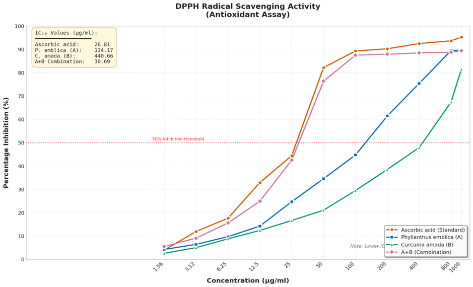
Figure 05 :- DPPH radical scavenging activity of ethanol extracts (Antioxidant Activity)
Protein Denaturation Inhibition
The results of the protein denaturation inhibition assay are presented in Table 04. The standard drug diclofenac sodium exhibited significant anti-inflammatory activity with an IC50 value of 46.21 μg/ml, demonstrating concentration-dependent inhibition ranging from 7.82% at 6.25 μg/ml to 85.88% at 100 μg/ml.
Phyllanthus emblica extract (Sample A) showed moderate protein denaturation inhibitory activity with an IC50 value of >100 μg/ml. The extract demonstrated 0.59 % inhibition at the lowest concentration (6.25 μg/ml) and 40.12 % inhibition at the highest concentration (100 μg/ml). Notably, at the 25 μg/ml concentration, Sample A exhibited 15.74% inhibition, which was comparable to the standard diclofenac at the same concentration (30.92%). However, the dose-response relationship showed some regularity, with a slight decrease in inhibition percentage observed at the 50 μg/ml concentration (29.70%) compared to the 25 μg/ml concentration.
Curcuma amada extract (Sample B) exhibited relatively weak protein denaturation inhibition, with IC50 value exceeding >100 μg/ml. The maximum inhibition achieved was 33.02% at 100 μg/ml. The combination extract (A + B) also showed IC50 88.70 μg/ml, with the highest inhibition of 52.07% observed at 100 μg/ml. Interestingly, the combination show highest activity compared to C. amada alone, suggesting antagonistic interaction between the two extracts in this assay system.
Table 04 :- Protein denaturation inhibition activity of ethanol extracts (Anti-inflammatory Activity)
|
Standard |
Concentration (μg/ml) |
Absorbance at 660 nm |
Percentage Inhibition (%) |
|
Control |
- |
0.857 |
- |
|
|
|||
|
Diclofenac Sodium |
6.25 |
0.790 |
7.82 |
|
12.5 |
0.713 |
16.80 |
|
|
25 |
0.592 |
30.92 |
|
|
50 |
0.401 |
53.21 |
|
|
100 |
0.121 |
85.88 |
|
|
IC 50 |
|
46.21μg/ml |
|
|
Sample code |
Concentration (μg/ml) |
Absorbance at 660 nm |
Percentage Inhibition (%) |
|
Control |
- |
0.845 |
- |
|
|
|||
|
Sample A Phyllanthus emblica |
6.25 |
0.840 |
0.59 |
|
12.5 |
0.782 |
7.46 |
|
|
25 |
0.712 |
15.74 |
|
|
50 |
0.594 |
29.70 |
|
|
100 |
0.506 |
40.12 |
|
|
IC 50* |
|
>100μg/ml |
|
|
|
|||
|
Sample B Curcuma amada |
6.25 |
0.811 |
4.02 |
|
12.5 |
0.776 |
8.17 |
|
|
25 |
0.727 |
13.96 |
|
|
50 |
0.675 |
20.12 |
|
|
100 |
0.566 |
33.02 |
|
|
IC 50* |
|
>100μg/ml |
|
|
|
|||
|
Sample A+B |
6.25 |
0.679 |
19.64 |
|
12.5 |
0.597 |
29.35 |
|
|
25 |
0.531 |
37.16 |
|
|
50 |
0.583 |
31.01 |
|
|
100 |
0.405 |
52.07 |
|
|
IC 50* |
|
88.70μg/ml |
|
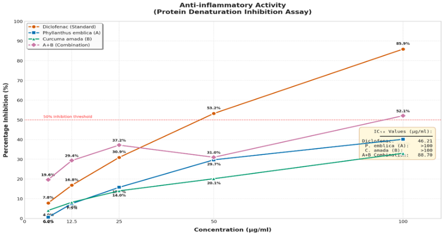
Figure 06 :- Protein denaturation inhibition activity of ethanol extracts (Anti-inflammatory Activity)
Combination Index Analysis
The combination index (CI) analysis was performed to quantitatively assess the nature of interaction between C. amada and P. emblica extracts. For the DPPH assay at the IC50 level, with the combination prepared in a 1:1 ratio, the calculated CI value was 0.15 and For the Protein denaturation assay at the IC50 level, with the combination prepared in a 1:1 ratio, the calculated CI value was 0.89 (Table 05). This value significantly indicating synergistic interaction between the two extracts. The synergistic effect can be attributed to the substantially higher antioxidant activity of combined ethanol extract compared to P. emblica, effectively diluting the potent activity of the latter when combined.
Table 05 :- Combination index analysis for DPPH and protein denaturation assays.
|
Assay |
IC50 A (μg/ml) |
IC50 B (μg/ml) |
IC50 A+B (μg/ml) |
CI Value |
Interpretation |
|
DPPH |
134.17 |
440.66 |
30.69 |
0.15 |
Synergism |
|
Protein Denaturation |
>100 |
>100 |
88.70 |
> 0.89 |
Synergism |
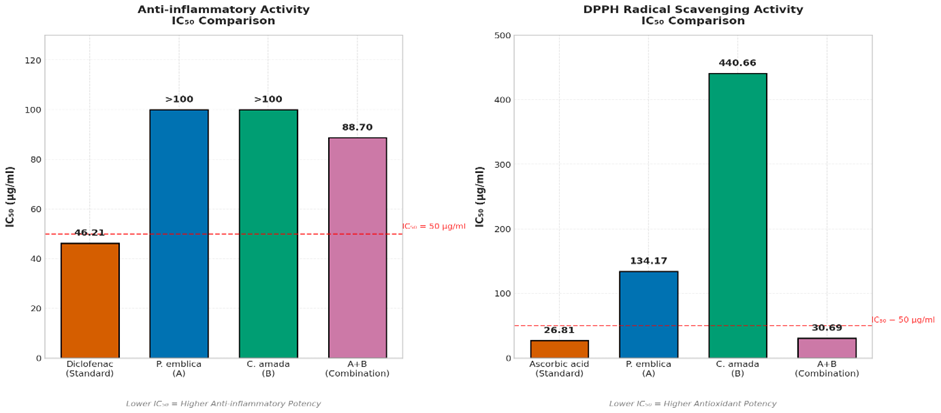
Figure 07 :- IC50 Comparison for DPPH and Protein denaturation assays (Sample A, B & A+B)
DISCUSSION
The present study evaluated the antioxidant and anti-inflammatory activities of individual and combined ethanol extracts of Curcuma amada and Phyllanthus emblica using in-vitro assays. The findings revealed that C. amada ethanol extract possesses potent antioxidant activity, as evidenced by its low IC50 value (440.66 μg/ml) in the DPPH assay, which was comparable to the reference standard ascorbic acid (26.81 μg/ml). This potent radical scavenging activity can be attributed to the presence of curcuminoids, particularly curcumin, which is well-documented for its antioxidant properties[31,32]. Curcumin and its derivatives exert antioxidant effects through multiple mechanisms, including direct free radical scavenging, metal ion chelation, and upregulation of endogenous antioxidant enzymes such as superoxide dismutase and catalase[33].
The moderate DPPH radical scavenging activity observed for P. emblica extract (IC50 = 134.17 μg/ml) was somewhat expected given the well-documented antioxidant properties of this plant[20,21]. However, it is important to consider that the extraction method and solvent significantly influence the phytochemical profile of the resulting extract. Ethanol, while effective for extracting a broad range of polar and semi-polar compounds, may not be optimal for extracting the highly water-soluble vitamin C, which is a major antioxidant constituent of P. emblica. Furthermore, the antioxidant activity of P. emblica has been shown to be highly dependent on the geographical origin, harvest time, and post-harvest processing methods[34]. The tannin-rich fraction of amla, particularly emblicanin A and B, has been reported to possess superior antioxidant activity compared to other fractions[35], and these compounds may have been completely extracted under the conditions employed in this study.
The protein denaturation inhibition assay revealed that C. amada extract possesses moderate anti-inflammatory activity (IC50 = > 100 μg/ml), while P. emblica showed relatively weak activity (IC50 > 100 μg/ml). The anti-inflammatory effect of C. amada can be attributed to its curcuminoid content, which has been extensively studied for its ability to modulate inflammatory pathways[36]. Curcumin suppresses the expression of pro-inflammatory cytokines (TNF-α, IL-1β, IL-6), inhibits cyclooxygenase-2 (COX-2) and inducible nitric oxide synthase (iNOS), and blocks nuclear factor-kappa B (NF-κB) signaling[37,38]. The protein denaturation inhibition mechanism is particularly relevant to the management of rheumatoid arthritis, where autoantigens produced through protein denaturation trigger immune responses leading to joint inflammation and damage[39].
A critical finding of this study was the synergistic interaction observed between the two extracts when combined. The combination index (CI) analysis was performed to quantitatively assess the nature of interaction between C. amada and P. emblica extracts. For the DPPH assay at the IC50 level, with the combination prepared in a 1:1 ratio, the calculated CI value was 0.15 and For the Protein denaturation assay at the IC50 level, with the combination prepared in a 1:1 ratio, the calculated CI value was 0.89 (Table 05). This value significantly indicating synergistic interaction between the two extracts. First, the substantial difference in potency between the two extracts, with P. embllica being considerably more active than C. amada, means that combining them effectively enhance the active constituents of C. amada. Second, phytochemical interactions between constituents of the two extracts may interfere with the bioavailability or activity of key compounds. For instance, tannins from P. emblica may form complexes with curcuminoids, potentially enhancing their availability for radical scavenging or protein stabilization[40]. Third, the 1:1 ratio employed in this study may represent the highest optimal ratio for synergy; different ratios could potentially yield different interaction outcomes.
The finding of synergistic interaction contrasts with some previous reports of synergistic antioxidant effects in polyherbal formulations containing these or related plants[41,42]. However, direct comparison is challenging due to differences in extraction methods, ratio of constituents, and assay conditions. It is also noteworthy that in vitro assays, while providing valuable initial screening data, may not fully predict in vivo efficacy due to factors such as bioavailability, metabolism, and tissue distribution. The demonstration of synergism in this study highlights the importance of systematic combination studies using quantitative methods such as the Chou-Talalay CI analysis before advocating for polyherbal combinations.
The extraction method employed in this study, maceration with ethanol, represents a traditional and widely used approach that yields a broad spectrum of phytoconstituents. The yield obtained for C. amada (30%) was consistent with previous reports using similar extraction protocols[43]. The higher yield for P. emblica (50%) reflects the rich content of extractable constituents in amla fruits. Future studies may consider employing sequential extraction with solvents of varying polarity to obtain fractionated extracts and identify the most bioactive fractions. Additionally, exploring alternative ratios for the combination, employing response surface methodology to identify optimal proportions, and conducting in vivo validation studies would provide valuable insights for potential therapeutic applications.
CONCLUSION
This study evaluated the antioxidant and anti-inflammatory activities of ethanol extracts of Curcuma amada and Phyllanthus emblica, individually and in combination. The results demonstrated that C. amada ethanol extract possesses potent DPPH radical scavenging activity (IC50 = 440.66 μg/ml) and moderate protein denaturation inhibitory activity (IC50 = >100 μg/ml). In contrast, P. emblica ethanol extract exhibited stronger activities in DPPH radical scavenging activity (IC50 = 134.17 μg/ml) and moderate protein denaturation inhibitory activity (IC50 = >100 μg/ml). The combination of the two extracts produce highest synergistic enhancement; rather, individual index analysis revealed antagonistic interactions in both the DPPH assay and protein denaturation inhibition assay. These findings best score the importance of systematic combination studies using quantitative analytical methods and suggest that the combination of these two plants in a 1:1 ratio for achieving highest enhanced antioxidant or anti-inflammatory effects.
REFERENCES


Rishav Kumar, Proddutur Irfana, Prabhat Kumar, V. Vamsi Reddy, Sharawan Kumar, M. Sirisha, In-Vitro Evaluation of the Antioxidant and Anti-Inflammatory Activities of the Combined Plant Extracts from Curcuma amada and Phyllanthus emblica, Int. J. of Pharm. Sci., 2026, Vol 4, Issue 3, 2322-2336. https://doi.org/10.5281/zenodo.19142422
 10.5281/zenodo.19142422
