SET’s College of Pharmacy, S R Nagar, Dharwad
Endophytes are microorganisms primarily bacteria and fungi that reside within the internal tissues of plants without causing apparent harm. These symbiotic organisms play a crucial role in plant growth promotion, stress tolerance, and defence against pathogens through the production of bioactive metabolites. The present study focuses on the isolation and characterization of endophytic microbes from selected plant species. Surface sterilization techniques were employed to eliminate epiphytic organisms, followed by culturing the internal tissues on selective media. Morphological, biochemical, and molecular methods, including 16S rRNA and ITS sequencing, were used to identify and characterize the isolated endophytes. Preliminary screening for antimicrobial activity and plant growth-promoting traits such as IAA production, phosphate solubilization, and siderophore production was also conducted. The findings underscore the potential of endophytes as a valuable source of novel bioactive compounds and their possible application in sustainable agriculture and biotechnology.
Endophytic fungi are a diverse group of microorganisms that dwell asymptomatically within plant tissues, playing critical ecological roles in plant health and ecosystem functioning. They contribute to nutrient cycling, plant growth promotion, and enhanced tolerance against biotic and abiotic stresses by modulating plant metabolic pathways and producing bioactive compounds. Historically, the discovery of endophytic fungi dates back over a century, initially observed as latent fungal inhabitants, but advances in molecular biology and cultivation techniques have significantly deepened our understanding of their taxonomy, diversity, and functional potential. Over recent decades, research on endophytic fungi has expanded across various disciplines, including agriculture, pharmaceuticals, and environmental sciences, due to their capacity to synthesize novel secondary metabolites with antimicrobial, anticancer, and antioxidant properties. The growing interest underscores the importance of studying endophytic fungi not only to harness their biotechnological applications but also to understand their role in maintaining plant resilience and ecological balance in diverse habitats1.
History:
The history of endophytic fungi research dates back to the early 19th century when German botanist Johann Heinrich Friedrich Link first described these fungi in 1809, initially considering them as plant parasitic organisms. Subsequently, the French scientist Béchamp coined the term "microzymas" to describe similar microbial entities inside plants. For much of the 19th and early 20th centuries, plants were thought to thrive best under sterile conditions, and the idea of internal microbial symbionts was overlooked until Victor Galippe's discovery in 1887 of bacteria naturally inhabiting plant tissues challenged this belief. A major milestone in the study of endophytic fungi was the seminal discovery in the 1990s of the anticancer drug paclitaxel (taxol) produced by the endophytic fungus Taxomyces andreanae isolated from the Pacific yew (Taxus brevifolia). This discovery revolutionized natural product research and pharmacology by revealing the immense secondary metabolite potential harboured by endophytes, stimulating a surge of interest in exploring endophytic fungi as prolific sources of novel bioactive compounds. The increasing recognition of secondary metabolites' pharmaceutical and agricultural significance has propelled endophyte research into a multidisciplinary frontier integrating molecular biology, biochemistry, and ecology2.
Endophytes:
The term "endophyte" (endo = within, phyto = plant) was originally coined by Anton de Bary, the father of modern plant pathology, in 1866 to describe any organism growing inside plant tissues. Over time, the definition has been refined to include only those microorganisms, primarily fungi and bacteria, that inhabit plant tissues without causing disease symptoms, and often even provide benefits to their hosts. Endophytes establish symbiotic relationships inside plants, living asymptomatically while promoting host growth, enhancing tolerance to various stresses such as pathogens, drought, or salinity, and protecting against herbivores. These microorganisms reside inter- or intracellularly and interact with their host through complex biochemical signaling pathways, leading to mutualistic outcomes. The recognition that endophytes can significantly influence plant health and productivity has driven extensive research into their biology and applications in agriculture, biotechnology, and pharmaceuticals. This refined understanding underscores the importance of endophytes as key players in plant ecosystems and promising sources of bioactive compounds3.
Classification of Endophytes:
Ecological classification divides endophytic fungi into clavicipitaceous (Class 1) and non-clavicipitaceous groups (Classes 2, 3, and 4). Clavicipitaceous endophytes (mainly in grasses) tend to have narrow host ranges and colonize shoots and rhizomes, often forming mutualistic relationships that enhance host fitness. Non-clavicipitaceous endophytes show broad host ranges and diverse colonization patterns, including root, stem, leaf, and seed tissues. Class 2 endophytes colonize multiple tissues and confer stress tolerance; Class 3 are mainly above-ground endophytes with diverse lifestyles ranging from mutualists to latent pathogens; Class 4 particularly include fungi that are horizontally transmitted and often opportunistic.
Host specificity varies, with some fungi exhibiting high specificity (e.g., Epichloë species in grasses), while others are generalists colonizing many plant species. Morphological identification relies on colony and spore morphology, but advances in DNA sequencing especially ITS rDNA regions have greatly enhanced accurate classification and phylogenetic placement. This classification framework reflects the complex evolutionary relationships and ecological versatility of endophytic fungi4.
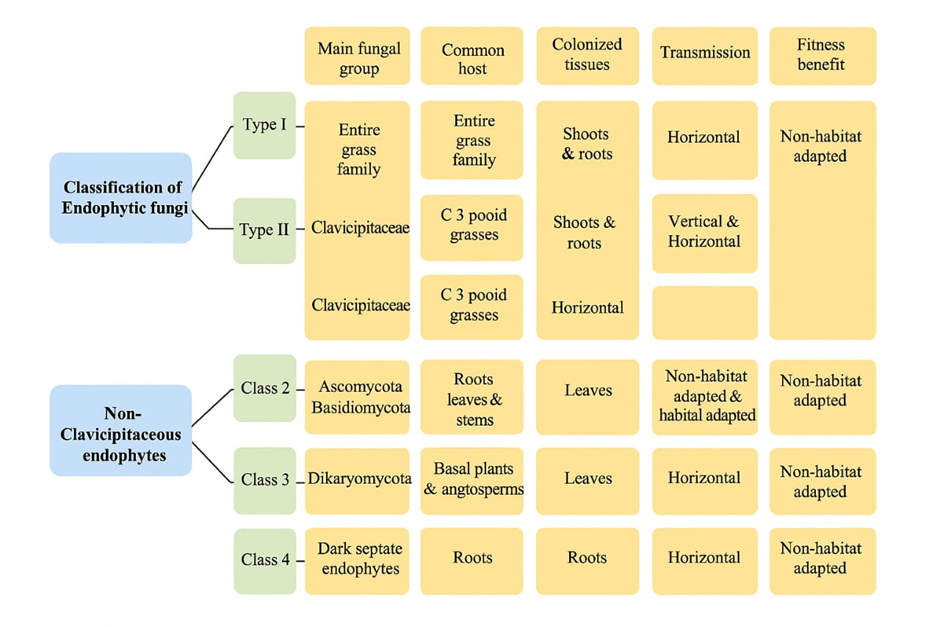
Classification of Endophytic fungi based on life history and host interaction
Endophytic Life Cycle and Interactions:
Endophytic fungi are integral members of the plant microbiome, living within plant tissues without causing visible disease symptoms. Their interactions with host plants span a continuum from mutualistic to parasitic relationships. Typically, endophytes avoid triggering strong plant immune defences, allowing a balanced coexistence with the host. This delicate interaction involves complex molecular signalling and modulation of the plant’s immune system to facilitate colonization without harm. Intriguingly, some fungi recognized as pathogens in one plant species may exist harmlessly as endophytes in others. For example, Ramularia collo-cygni is a known pathogen in barley but lives symptomless as an endophyte in other cereal hosts, illustrating the fluid boundary between pathogenic and endophytic lifestyles.
Pathogens with an Endophytic Phase:
Some pathogens may also have an endophytic phase, living quietly within a plant before causing disease when conditions favor. This dual lifestyle highlights the fluid boundary between pathogenic and endophytic roles. Thus, true endophytes are distinguished by their inability to cause disease in their host, while pathogens may transiently adopt an endophytic lifestyle5.
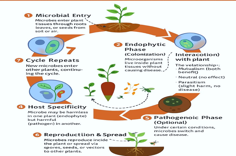
Endophytic Life Cycle and Interactions.
Endophytic fungi can be effectively visualized within plant tissues using a range of microscopic techniques that reveal their colonization patterns and interactions with the host. Light microscopy, often combined with staining methods like aniline blue, lactophenol trypan blue, or rose Bengal, highlights fungal hyphae and structures within the intercellular spaces of leaves, stems, and roots. Fluorescence microscopy, particularly confocal laser scanning microscopy with fluorescence-tagged fungi or dyes, allows three-dimensional visualization of fungal mycelia inside tissues, providing detailed insights into spatial distribution. Electron microscopy techniques, including transmission electron microscopy (TEM) and scanning electron microscopy (SEM), offer ultrastructural views, elucidating the fungal penetration mechanisms and intimate contact with host cells.
Microscopic evidence confirms that endophytes form true symbiotic relationships by inhabiting internal plant tissues and contributing to plant health through various roles such as stress tolerance, nutrient acquisition, and pathogen defence. Visualization of fungal structures inside plant tissues supports these functional associations and highlights the complexity of plant-endophyte interactions. Continuing research using advanced microscopic and molecular tools is essential to further understand endophyte diversity, ecological roles, and their potential in improving plant productivity and resilience under environmental stresses.
Regarding roles, endophytes contribute to mitigating abiotic stresses like drought and temperature extremes, alleviate biotic stresses caused by pathogens and pests, and enhance nutrient uptake, collectively promoting better plant growth and adaptation. These multifaceted functions vary depending on the host plant, environmental conditions, and developmental stages, underscoring the dynamic nature of endophytic symbiosis6.
Endophytes promoting plant adaptation to abiotic stress:
Endophytes improve water uptake and retention by promoting root growth and facilitating efficient water use, helping plants survive under water deficit conditions.
They solubilize and mobilize essential nutrients like phosphorus and nitrogen, enhancing nutrient availability and uptake, which strengthens plant growth and resilience.
Through the production of antioxidants, heat shock proteins (HSPs), and osmolytes, endophytes help plants mitigate oxidative damage and maintain cellular stability during heat or cold stress.
Endophytic microbes influence hormonal signaling to mediate stress tolerance in medicinal plants. The study highlighted that endophytes produce a range of phytohormones, including gibberellins (GAs), cytokinins, abscisic acid (ABA), jasmonic acid (JA), and salicylic acid (SA), which play crucial roles in plant growth, development, and stress responses. These hormones help plants adapt to abiotic stresses by modulating various physiological processes such as stomatal regulation, antioxidant production, and osmotic adjustment.
Some endophytes can enhance tolerance to heavy metals and alkaline soils by altering soil chemistry and supporting plant detoxification mechanisms and additionally, the endophytes enhanced the expression of metal stress-related genes in the host plants, leading to improved growth and reduced metal toxicity7.
Biocontrol potential of endophytes in plants:
Endophytes such as Penicillium citrinum stimulate plants to produce higher levels of defense-related enzymes like peroxidase (PO) and polyphenol oxidase (PPO), which help resist pathogenic fungi such as Fusarium oxysporum.
Endophytic fungi like Trichoderma hamatum activate systemic immunity by promoting the production of defense enzymes, salicylic acid, and pathogenesis-related proteins (PRPs), providing protection against diseases like downy mildew.
Endophytes synthesize secondary metabolites, enzymes, and volatile organic compounds that directly inhibit or antagonize pathogens, reducing infection and damage.
Endophytes can trigger plants to express genes involved in pathogen resistance, strengthening the plant's own defense mechanisms.
Certain endophytic fungi such as Daldinia eschscholtzii exhibit strong antagonistic activity against pathogens like Colletotrichum acutatum, effectively reducing disease severity8.
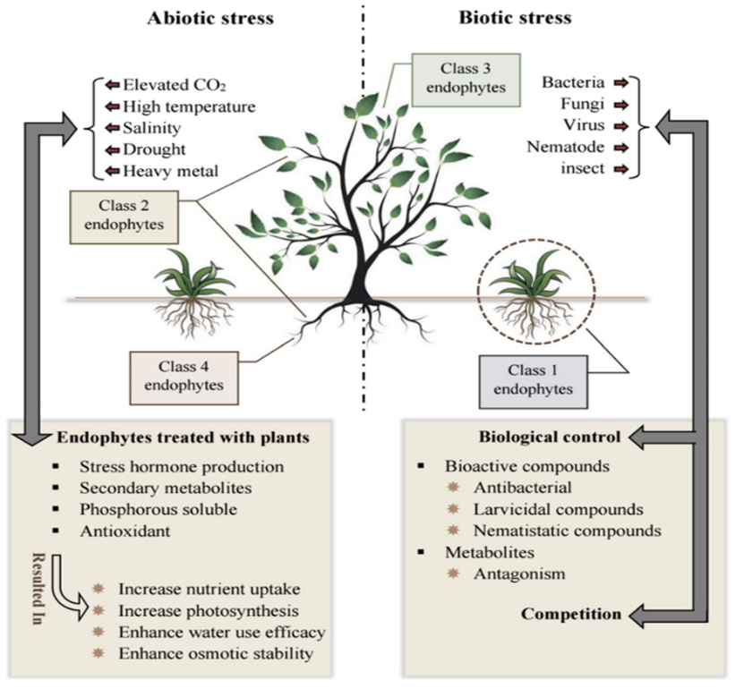
Schematic model of endophyte-plant symbiosis
Endophyte isolation techniques and identification methods:
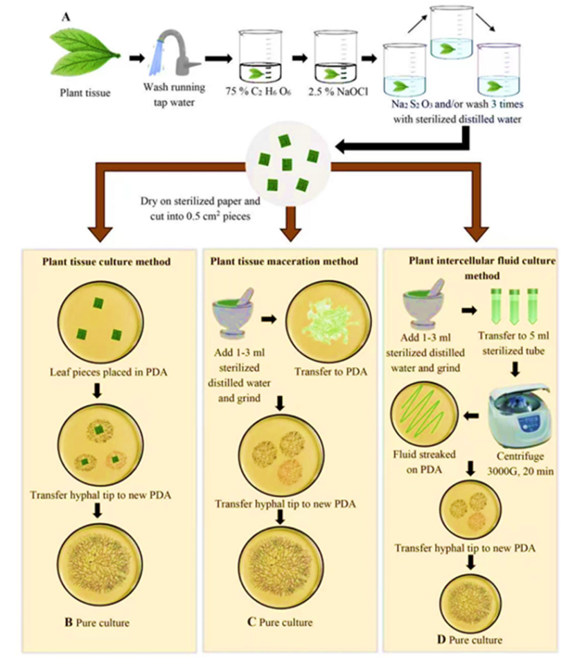
Culture-dependent isolation methods. A) plant tissue surface sterilization. B) Plant tissue culture method. C) Plant tissue maceration method. D) Plant intercellular fluid culture method.
Identification techniques:
Traditional methods:
Morphological and Cultural Characterization of Endophytic Fungi:
After initial isolation of fungi from surface-sterilized plant tissues on nutrient media, individual fungal colonies are purified by transferring hyphal tips or spores onto fresh agar plates to obtain pure cultures. This step is essential to separate mixed cultures and ensure that each isolate represents a single fungal species.
Identification of purified endophytic fungi traditionally relies on morphological characteristics observed in culture. This includes colony morphology (color, texture, growth rate), microscopic examination of fungal structures such as hyphae, conidia, and spores, as well as sporulation patterns. Because many isolates may not produce spores under standard conditions, inducing sporulation through modifying culture media or environmental conditions (e.g., adding sterilized host tissue or altering light and temperature) is often necessary to aid identification.
Despite these efforts, morphological identification can be limited by sterile isolates that lack distinctive features. Therefore, traditional identification is sometimes complemented by molecular techniques to accurately classify fungi at species or genus level10.
Modern approaches:
Molecular characterization of endophytic microorganisms:
The development of molecular biology brings a new per spective to endophyte diversity studies. Application of molecular techniques, such as DNA fingerprinting and sequencing methods, has the potential to overcome the obstacles in traditional cultivation-dependent methods.
Molecular characterization of sterile fungal isolates:
In traditional cultivation-dependent methods, fungal isolates are identified based on morphological features, primarily reproductive structures. However, a significant portion of isolates do not sporulate in culture and remain sterile, making morphological identification impossible. These non-sporulating isolates are often grouped into morphotypes based on colony characteristics such as color, texture, and growth rate, but these groupings do not reflect true species relationships.
Molecular techniques, particularly DNA sequence analysis of regions like the ITS (Internal Transcribed Spacer), are essential for accurately identifying these sterile mycelia. By analyzing genetic sequences, researchers can assign non-sporulating isolates to appropriate taxonomic groups at the genus, family, or order level. Molecular identification reveals that morphologically similar sterile isolates can belong to distantly related taxa, highlighting the limitations of morphology-based classification. Overall, molecular methods significantly enhance our understanding of fungal diversity, especially for sterile isolates that traditional techniques fail to identify11.
Molecular Identification of Endophytic Fungal Communities:
Endophytic fungal communities comprise diverse fungal groups from major phyla such as Ascomycota, Basidiomycota, and Zygomycota, making identification based solely on morphology challenging and time-consuming. To overcome these limitations, DNA sequencing, especially of the ITS region, combined with morphological data, has become a standard approach for studying endophyte diversity.
Molecular techniques allow researchers to detect a wide variety of fungal taxa across different plant tissues and ecosystems, often revealing greater species richness than morphological methods alone. Genes like 18S, 28S, and ITS are commonly used for taxonomic resolution at various levels, from broad classification to species identification. Molecular data have also uncovered novel taxa and clarified relationships within complex fungal groups. Overall, these molecular tools accelerate and enhance the understanding of fungal biodiversity in natural ecosystems, independent of the fungi’s sporulation ability in culture12.
Molecular Detection of Endophytic Fungi Within Plant Tissues:
Traditional isolation techniques for endophytic fungi often miss many species because some fungi grow slowly or do not grow on artificial media, being outcompeted by faster-growing fungi. To overcome these limitations, molecular techniques have become widely used to detect and analyze endophytic fungi directly from plant tissues.
The typical molecular detection workflow involves:
These molecular approaches can detect a broader diversity of fungi, including those that do not sporulate or grow well in culture. Studies have found significant differences between fungal communities detected by molecular methods and those isolated by traditional culturing, highlighting the complementary nature of molecular detection.
However, molecular methods also have limitations such as:
Additional techniques like Simple Sequence Repeat (SSR) markers offer robust and reproducible detection, helping to further characterize fungal diversity13.
High-Throughput Sequencing in Endophyte Studies:
High-throughput sequencing (HTS), especially next-generation sequencing (NGS) technologies like pyrosequencing and Illumina sequencing, have revolutionized the study of fungal endophyte diversity. Compared to traditional Sanger sequencing, HTS offers:
HTS has been successfully applied to diverse environments to explore fungal communities, revealing hidden diversity missed by culture-based methods. However, challenges include:
Ongoing advances in sequencing technology and bioinformatics are rapidly enhancing our ability to characterize endophytic fungi in ecological and functional contexts.
DNA Barcoding Systems in Fungal Endophyte Studies:
DNA barcoding uses short, standardized gene regions to identify species, relying on high inter-specific variation and low intra-specific variation. While the animal kingdom widely uses the CO1 mitochondrial gene, this marker is unsuitable for fungi due to the presence of introns.
For fungi, the Internal Transcribed Spacer (ITS) region of rDNA is the primary DNA barcode marker due to:
ITS has been broadly used in fungal species identification, including endophytic fungi, though it faces challenges:
In 2011, a fungal barcoding workshop and subsequent international consensus formally recommended ITS as the official DNA barcode for fungi, making it the standard marker in fungal ecology, diversity, and endophyte studies14-15.
CURRENT CHALLENGES
In endophytic fungi research include incomplete understanding of the complex interactions between endophytes and their host plants at molecular and ecological levels. The regulation of secondary metabolite biosynthesis remains poorly elucidated, hindering the optimization and consistent production of bioactive compounds. Culturing many endophytes in laboratory conditions is difficult due to their specialized nutritional and environmental requirements, leaving a significant portion of biodiversity unexplored. Scaling up metabolite production for commercial applications faces obstacles such as low yields, variability in metabolite profiles due to environmental factors, and high costs associated with fermentation and purification processes. Additionally, regulatory hurdles require extensive safety and efficacy data before endophyte-derived products can enter the market, adding to the time and complexity of commercialization. Environmental concerns also arise regarding sustainability and potential ecological impacts of manipulating endophytic populations.
FUTURE PERSPECTIVES
The future research emphasizes integrating multi-omics technologies including genomics, transcriptomics, proteomics, and metabolomics to comprehensively profile endophyte-host interactions and biosynthetic pathways. Advances in synthetic biology and metabolic engineering hold promise to enhance metabolite yields and create novel compounds. Ecological and systems biology approaches can provide holistic insights into endophyte roles in plant health and stress resilience, facilitating the development of robust bioinoculants and sustainable agricultural practices. Moreover, leveraging nanotechnology and innovative formulation strategies may improve delivery and stability of endophyte products. Expanded bioprospecting efforts targeting diverse and extreme environments are likely to uncover new endophytic taxa and metabolites beneficial for pharmaceuticals, agriculture, and environmental remediation.
CONCLUSION
The endophytic fungi represent an invaluable resource for novel bioactive compounds and plant growth-promoting agents. Realizing their full potential requires coordinated interdisciplinary research focusing on overcoming current limitations in isolation, cultivation, metabolite production, and regulatory approval. Continued innovation promises to unlock sustainable solutions addressing food security, environmental challenges, and human health.
ACKNOWLEDGEMENT
I would like to express my sincere gratitude to SET’s College of Pharmacy, Dharwad for providing the necessary infrastructure and academic support throughout the course. I am deeply thankful to my guide her expertise and mentorship have been instrumental in shaping the direction and quality of this work. I also extend my appreciation to the faculty and staff for offering excellent facilities and fostering a stimulating academic environment. Finally, I am immensely grateful to my family, friends and my mother for their constant encouragement and patience, which kept me motivated during the most challenging phases of this journey.
REFERENCE


Beeralinga G, Smita D. Madagundi, Prasanna Vasantrao Habbu, Insights into Endophytic Fungi: Historical Perspectives, Isolation Strategies, Technological Advances and Future Directions, Int. J. of Pharm. Sci., 2025, Vol 3, Issue 11, 4445-4456. https://doi.org/10.5281/zenodo.17735983
 10.5281/zenodo.17735983
