
We use cookies to ensure our website works properly and to personalise your experience. Cookies policy
JKK Munirajah Institute of Health Sciences College of Pharmacy, T. N. Palayam, Erode, Tamil Nadu, India 638506
The Electroconvulsiometer and Actophotometer are key experimental instruments in neuropharmacological research, widely employed for screening and evaluating the effects of central nervous system (CNS)–active agents. The Electroconvulsiometer functions by inducing controlled maximal electroshock seizures (MES) in rodents, providing a reliable and reproducible model for assessing the anticonvulsant efficacy of drugs, particularly those targeting generalized tonic–clonic seizures. The actophotometer assesses locomotor activity through infrared beam breaks produced by animal movement. This technique is valuable for evaluating the stimulant or depressant actions of CNS drugs, offering a non-invasive and quantitative method of behavioral analysis. Together, these instruments serve as indispensable tools in preclinical pharmacology, contributing to the discovery, characterization, and optimization of therapeutic compounds by facilitating objective assessment of their anticonvulsant, sedative, stimulant, and neuroprotective properties.
Electroconvulsiometer1-10:
The Electroconvulsiometer is a specialized scientific instrument used in neuropharmacological research to study seizure activity and evaluate the anticonvulsant potential of pharmaceutical compounds. This instrument delivers controlled electrical stimuli to laboratory animals, typically mice or rats, to induce convulsions, thereby mimicking epileptic conditions in humans. The induced seizures allow researchers to assess the protective efficacy of antiepileptic drugs by observing the alteration or suppression of convulsive phases such as tonic and clonic seizures. The Maximal Electroshock Seizure (MES) model, facilitated through the Electroconvulsiometer, is one of the most widely accepted and reproducible models for screening antiepileptic agents, especially those effective against generalized tonic-clonic seizures.
The Electroconvulsiometer remains an indispensable tool in early-phase drug discovery and in understanding the neurobiological basis of epilepsy. Its relevance is further underscored by the high translational value of the MES model, as several clinically used antiepileptic drugs such as phenytoin and carbamazepine were originally identified using this method. The Electroconvulsiometer continues to play a central role in pharmacological screening due to its reliability, simplicity, and cost-effectiveness. The Electroconvulsiometer operates on the principle of electrically induced seizures in experimental animals to evaluate the anticonvulsant potential of drugs. When a precisely controlled alternating current (AC) of defined intensity, frequency, and duration is passed through the brain via corneal or ear-clip electrodes, it causes widespread depolarization of neuronal membranes and synchronous firing of neurons. This results in a generalized tonic–clonic seizure pattern known as the Maximal Electroshock Seizure (MES). The MES consists of several phases, the most characteristic being Tonic Hind-Limb Extension (THLE), where the hind limbs extend at an angle of ≥180° to the body axis. Anticonvulsant drugs that are effective against generalized tonic–clonic epilepsy are able to prevent or shorten the THLE phase, and this forms the basis for their screening using the Electroconvulsiometer.
Major Components of Electroconvulsiometer:
The major components of an Electroconvulsiometer are as follows:
1. Stimulus Generator is the primary unit of the Electroconvulsiometer. It is responsible for producing an electrical stimulus with controlled parameters such as: Current intensity (commonly 50–150 mA), Frequency (typically 50–60 Hz), and duration (0.2 to 0.5 seconds). These parameters can be finely tuned to ensure consistency across experiments. The generator typically uses square wave pulses, as they are more effective in eliciting maximal electroshock seizures (MES).
2. Electrodes are used to deliver the electrical stimulus to the animal. They are generally made from stainless steel or other biocompatible metals and are of two main types: Ear clip electrodes – attached to the pinnae of the animal. Corneal electrodes applied to the cornea after moistening with saline to reduce resistance. The appropriate electrode is selected based on the species and responsiveness of the subject.
3. Current/Voltage Regulator is primarily used as component ensures that the voltage and current delivered are within safe and precise limits. It allows researchers to adjust the output depending on the experimental requirements. Safety cut-offs are often built-in to prevent damage to animal tissues.
4. Timer and Control Switches may be a digital or analogue timer and is crucial for maintaining uniform stimulation periods across tests. Control switches allow for starting, stopping, or resetting the device.
5. Display and Indicators provides real-time feedback on key parameters such as current, voltage, frequency, and stimulus duration. This ensures accurate monitoring during the procedure.
6. Animal Restraint Device is not part of the electrical circuitry, animal restraint systems (e.g., small cages or holders) are essential for: Preventing excessive movement, ensuring accurate electrode placement, Enhancing animal safety and experimental reproducibility.
Standard Operating Procedures:
Animals Used:
Commonly used for Maximal Electroshock Seizure (MES) testing. Adult rats weighing 150–250 g are preferred, Stimulation parameters: 150 mA, 50–60 Hz, 0.2s duration. Electrodes: corneal or ear-clip electrodes. It is primarily used to induce tonic hind-limb extension for evaluating anticonvulsant drugs.
Typical age: 6–8 weeks, weight 20–30 g, MES protocol: 50 mA, 60 Hz, 0.2 s duration. Electrodes: corneal type, with saline or tetracaine applied to improve contact. Purpose: Screening of potential anticonvulsant agents by observing seizure prevention or suppression.
Applications:
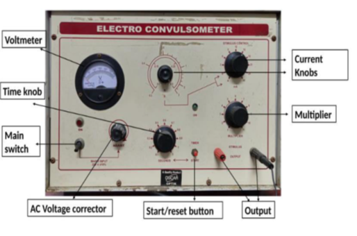
Fig.1 Diagramatic representation of Electroconvulsiometer
Actophotometer11-16
Actophotometer is an instrument used to measure locomotor (activity) behaviour in animals. It is commonly used in CNS drug screening to assess stimulant or depressant effects. The instrument detects animal movement using infrared light beams and photocells. Each time the animal crosses a beam, it interrupts the light, generating a count. The total count over a fixed period reflects the level of motor activity. Increased activity suggests CNS stimulation (e.g., with caffeine), while reduced activity suggests CNS depression (e.g., with diazepam). It is useful in testing neuroactive drugs, sedatives, anxiolytics, and stimulants.
The instrument can be digital or manual, with circular or square enclosures. Both rats and mice can be tested using different models. It provides a non-invasive and reliable method to quantify spontaneous movement. It helps to quantify drug induced changes in animal motor behaviour objectively. It is widely used in preclinical pharmacology and behavioral neuroscience research. The typical duration for activity recording is 5 to 10 minutes per animal. Animals are tested individually to avoid interference from others. The device must be placed in a quiet and undisturbed environment during testing. Both horizontal and vertical activity can be measured in advanced models. Due to its simplicity, reproducibility, and sensitivity, the actophotometer is widely used in neuropharmacology, toxicology, and behavioural research for rapid screening of CNS-active drugs, dose–response studies, and comparative activity assessments.
The actophotometer works on the principle that when an animal moves inside a closed chamber, it interrupts a beam of light (photoelectric cell), and each interruption is recorded as a count. The instrument contains a series of light beams and corresponding photoelectric sensors arranged in the activity cage. When the animal (usually a rat or mouse) crosses the light beam, the beam is broken, and the photoelectric cell converts this interruption into an electrical signal, which is counted and displayed digitally or mechanically. The total number of beam interruptions in a fixed period reflects the locomotor activity of the animal. Basic Components of Actophotometer: An actophotometer is an electronic device used to measure the spontaneous motor activity of small animals, typically in pharmacological experiments. The movement of the animal interrupts light beams inside the device, which is then recorded as activity counts. This method is widely used to assess the effect of various drugs on the central nervous system (CNS), especially stimulants and depressants
Essential components that make up an actophotometer and their respective functions:
1. Enclosure or Activity Cage Structure: The primary part of the actophotometer is the enclosed arena or cage, usually square or circular in shape, designed to restrict the animal’s movement to a specific area. Material: Typically made of transparent acrylic plastic or metal, it prevents the animal from escaping while allowing clear observation. Function: It serves as the space where the animal is placed during the experiment. The movements of the animal inside this chamber are the basis for data recording.
2. Infrared (IR) Light Source and Sensors IR Light Beams: Multiple infrared beams are placed across the floor of the enclosure. These beams are invisible to the animal and do not interfere with its natural movement. Photoelectric Sensors: Each beam is aligned with a photo sensor on the opposite side of the enclosure. When the animal moves and breaks a beam, the interruption is detected by the sensor. Function: The system detects the number of beam breaks, which reflects the locomotor activity of the animal. The more active the animal, the more beams are broken.
3. Digital Display or Counter Display Unit: A digital or analogue display unit is connected to the beam-sensor system. It shows the number of beam interruptions over a specified period. Types: Modern actophotometer come with digital LCD displays or computer interfaces for accurate data logging and analysis. Function: This component provides a real-time readout of the animal’s activity. The readings help researchers compare activity levels before and after drug administration.
4. Timer Built-in Clock or Timer: A timing mechanism is often integrated with the display unit. Function: It allows the experimenter to set the duration of the activity measurement, usually ranging from a few minutes to an hour. Importance: Consistent time intervals are crucial for comparing control and test results.
5. Electrical Power Supply Power Source: Most actophotometer operate using AC mains power or DC battery systems, depending on the model. Function: Powers the infrared lights, sensors, and display systems. Safety Features: Many systems have fuses or surge protectors to ensure safe operation during experiments.
6. Control Panel Buttons/Switches: The control panel allows the user to turn on/off the device, reset counters, and set timing functions. Advanced Models: Some include data storage, USB ports, or software interfaces for direct computer connectivity. Function: Enables customization of experimental settings and simplifies operation.
7. Calibration and Sensitivity Adjustment Knob Use: Some actophotometer offer the option to calibrate the sensitivity of the IR sensors. Function: Adjusts the threshold at which beam interruptions are recorded, helping to fine tune the system based on the size and type of animal.
Standard Operating Procedure:
Precautions:
Handle animals gently and calmly to avoid stress, ensure infrared sensors are unobstructed during the test, Keep the testing area quiet and distraction-free, use freshly prepared drug solutions, do not place more than one animal at a time.
Animals used:
In experimental pharmacology, the actophotometer is a valuable instrument used to measure the spontaneous locomotor activity of animals. To study such effects, small laboratory animals are used. The selection of the animal depends on factors such as size, handling convenience, availability, cost, and relevance to human physiology.
Most commonly used species in actophotometer studies. Advantages: Small size, easy to handle, require minimal space and food, highly sensitive to CNS-active drugs, Inbred strains provide genetic consistency.
Used when larger sample size or more tissue is needed for follow-up biochemical studies. Advantages: Larger body size allows easier administration of drugs and sampling; Behavioral responses are more stable compared to mice. Some complex tasks or longer-duration activity measurements are more reliable in rats.
Applications of Actophotometer:
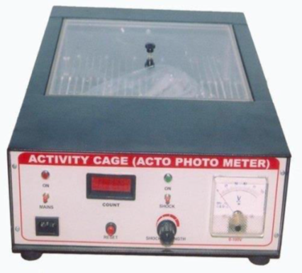
Fig.2 Diagramatic representation of Actophotometer
CONCLUSION:
The Electroconvulsiometer and actophotometer remain cornerstone instruments in pharmacological and behavioural research due to their reliability, sensitivity, and translational relevance. The Electroconvulsiometer, through the MES model, provides a standardized approach for identifying and validating anticonvulsant agents, while the actophotometer enables precise measurement of drug-induced alterations in locomotor activity. Their combined application not only enhances the understanding of drug mechanisms but also accelerates the development of novel CNS-active therapeutics. By offering reproducible and cost-effective methods, these instruments continue to play a crucial role in bridging experimental findings with clinical potential in the field of neuropharmacology.
REFERENCES

M. Nithya, G. M. Sivakumar, G. Kiruthika, A. Kokila, A. Mohanasundaram, S. Prasanna, N. Rithikadevi, V. Selvaraja, S. R. Shibi, T. Thirisha, Instrumental Approaches in Neuropharmacological Screening: A Review of Insights from Electroconvulsiometer and Actophotometer Studies, Int. J. of Pharm. Sci., 2025, Vol 3, Issue 8, 2670-2677. https://doi.org/10.5281/zenodo.16940508
 10.5281/zenodo.16940508