Department of Pharmaceutics, Rasiklal M. Dhariwal Institute of Pharmaceutical Education and Research, Chinchwad, Pune-411019.
This review explores the development and implementation of a non-destructive, in-line, dynamic pharmaceutical inspection system utilizing carbon nanotube (CNT) film-based photo-thermoelectric (PTE) imagers operating across the ultrabroadband sub-terahertz (sub-THz) to infrared (IR) spectrum. Traditional inspection techniques in pharmaceutical manufacturing often suffer from limited penetration depth, destructive sampling, and reduced detection accuracy. The presented ultrabroadband optical monitoring approach addresses these challenges by enabling label-free, non-contact identification of impurities, coating irregularities, and compositional variations within pharmaceutical pills. The system employs sub-THz, long-wave infrared (LWIR), and short-wave infrared (SWIR) radiation bands, combined with CNT PTE imagers that offer high sensitivity, mechanical flexibility, and room-temperature operation. Through transmissive and reflective imaging configurations, the system captures detailed optical signatures of pharmaceutical agents, identifying both internal and surface defects in real time. Experimental results demonstrate effective differentiation of various drug compositions and concealed impurities such as metal, glass, and plastic inclusions. The integration of automated optical stages and high-speed data acquisition enables scalable, continuous monitoring suitable for industrial environments. This review highlights the significance of ultrabroadband CNT-based PTE imaging as a promising solution for advancing pharmaceutical quality assurance, combining high accuracy, rapid inspection, and non-invasive operation
The pharmaceutical industry depends heavily on rigorous quality assurance procedures to ensure
that every product reaching the market is safe, effective, and consistent in performance [1]. The processes involved in production and distribution pharmaceutical products are complex and often prone to defects such as cracks, contamination, corrosion, and spotting on pills or tablets. These defects can compromise product quality and safety, leading to serious implications for both manufacturers and patients [2]. To find these challenges, the use of non-destructive testing (NDT) methods has become increasingly essential in latest pharmaceutical production systems. Non-destructive inspection ensures the structural integrity and chemical uniformity of products without altering or damaging them, thereby enabling continuous monitoring during manufacturing [3]. Conventional testing techniques such as magnetic, penetrative, photo-monitoring, and visual inspection using visible light have long been used in pharmaceutical quality control. These methods, have many limitations. They often rely on external reagents, are limited to surface-level examination, and may fail to detect defects or impurities embedded within the internal structure of dosage form [4]. Moreover, destructive sampling leads to material loss, increases costs, and cannot be applied to every batch in an industrial position. Consequently, the demand for advanced, non-contact, label-free, and accurate testing systems has grown significantly, particularly those proficient to monitoring a product in-line and in particular time during processing.The concept of non-destructive inspection in materials science can be traced back to 1895, with the discovery of X-rays, marking the beginning of non-invasive testing techniques [1]. Over time, various optical imaging technologies based on longer-wavelength electromagnetic radiation, such as ultraviolet (UV), visible (Vis), terahertz (THz), and infrared (IR) light, have been developed [3]. These longer wavelengths enable transparent detection of non-metallic and opaque materials such as polymers, glass, ceramics, semiconductors, and liquids that are widely used in pharmaceutical formulations and packaging. By exploiting their unique absorption and reflection properties, these optical methods can acquire detailed information about the drug composition and structure of a pharmaceutical product without physical contact [2].Recent advances in broadband photo-imaging technologies have made it possible to collect optical data from multiple wavelength bands using a single compact configuration, eliminating the need to switch between different image sensors [3]. This innovation has accelerated the development of photo-imaging systems skillful to acquiring comprehensive spectral data for each inspected sample. In addition, the intrinsic absorption features or fingerprint spectra that appear in specific spectral regions provide unique identifiers for various materials. These characteristics enrich the capability of non-destructive inspection, allowing not only the detection of physical defects but also the identification of chemical components and impurities. Compared with conventional single-point or scanning methods, photo-imaging offers higher throughput and efficiency, enabling faster and more accurate evaluation of pharmaceutical products.Pharmaceutical manufacturing involves multiple stages and diverse materials such as active pharmaceutical ingredients (APIs), excipients, and coating agents. Each of these components must be monitored carefully during production to ensure uniformity and consistency. The primary goal of non-destructive inspection is to identify the presence and distribution of these materials and detect any impurity or defect that may disturb the quality of the final product. By integrating non-destructive testing into in-line production systems, manufacturers can continuously monitor product quality without breaking the production flow. This approach minimizes the risk of defective products entering the market and improves overall process efficiency. An advanced approach to non-destructive inspection involves the use of ultrabroadband, multi-wavelength sub-terahertz to infrared (sub-THz–IR) monitoring systems. Such systems allow comprehensive evaluation of pharmaceutical materials across a wide spectral range, providing both structural and compositional information. The sub-THz–IR inspection setup typically integrates several spectroscopic techniques, including sub-THz and Fourier Transform Infrared (FTIR) spectroscopy. These techniques are particularly prime in the early stages of material evaluation, as they allow precise identification of raw material composition and detection of impurities at the molecular level.A major technological advancement in this field is initiation of the carbon nanotube (CNT) thin-film photo-thermoelectric (PTE) imagers. These imagers are highly flexible and can conform to various shapes and surfaces, allowing omnidirectional detection without blind spots. The CNT PTE imagers are designed to function efficiently across ultrabroadband regions, extending from the sub-THz to IR range, thereby providing enhanced spectral sensitivity as compared to conventional sensors [4]. The operating principle of these devices is based on the photo-thermoelectric effect, where absorbed light induces localized heating, which is then converted into an electric signal. This mechanism enables precise photo-detection without the need for external electrical bias, reducing noise and enhancing measurement stability. The integration of CNT film-based PTE imagers into non-destructive in-line inspection systems offers several distinct advantages. Their mechanical flexibility allows them to be freely attached to conveyor-based systems or curved pill surfaces, enabling real-time monitoring during manufacturing. These imagers preserve the integrity of the inspected pharmaceutical product because the process is entirely contactless and reagent-free. Furthermore, CNT-based sensors exhibit remarkable environmental stability, maintaining performance even under varying humidity, lighting, and temperature conditions. This durability makes them very acceptable for deployment in real industrial settings where conditions may fluctuate.In addition to CNT imagers, thermal detectors such as bolometers and Golay cells can also play a significant role in broadband radiation detection. These devices operate as thermal sensors that measure heat generated when electromagnetic radiation is absorbed by a material. Unlike many detectors that require cooling to extremely low temperatures, bolometers and Golay cells can operate effectively at room temperature, making them convenient and energy-efficient. Their detection range extends from sub-terahertz to infrared frequencies, enabling them to capture a wide range of radiation signals. Owing to their sensitivity and broad detection capabilities, these detectors have been applied successfully in diverse fields such as astronomy, security screening, and environmental monitoring. In pharmaceutical inspection, they can complement CNT-based imagers by providing thermal detection capabilities that enhance the accuracy and reliability of broadband monitoring systems.The combination of ultrabroadband sub-THz–IR radiation sources, advanced spectroscopic methods, CNT PTE imagers, and thermal detectors constitutes a powerful toolset for non-destructive pharmaceutical testing. This integrated system allows detailed mapping of material compositions, impurity detection, and real-time quality assessment without disrupting production. It overpassing the void space between laboratory-scale precision and industrial-scale efficiency, providing both microscopic detail and macroscopic throughput. Furthermore, these non-destructive, broadband monitoring systems are adaptable and scalable, offering the potential for widespread adoption across pharmaceutical manufacturing facilities. the development of non-destructive analysis in the pharmaceutical industry has transitioned from basic optical inspection to sophisticated ultrabroadband photo-imaging methods. The integration of carbon nanotube film-based photo-thermoelectric imagers, supported by thermal detection technologies like bolometers and Golay cells, represents a major step forward in achieving dynamic, in-line, and real-time inspection of pharmaceutical products. This proposal not only improve the accuracy and speed of defect detection but also ensures product integrity and safety without compromising the manufacturing process. The development and implementation of such systems mark an important advancement toward a new generation of intelligent, automated, and sustainable quality control in pharmaceutical production.
2. INSTRUMENTATION:
This is simple form demonstration of non-destructive in-line dynamic inspection system for pharma agent like pills with freely attachable functional CNT imager sheets and the associated ultrabroadband sub-THz–IR band photo-monitoring (Fig. 1a). the above simple demonstrated system develops in experimental setup containing with sub-THz-IR imagers and multiple photo sources which have longer wavelength extracted in single wavelength in advance (Fig. 1b). CNT detailed model which chosen as working electrode for sensing devices to their extra ordinary low detecting limit (Fig. 1c). process modeling, design master plan, and fundamental performances this all working of the PTE (Fig. 1d).
2.1 sub-THz:
Terahertz (THz) radiation, often referred to as sub-terahertz when operating below the THz range, represents a site of the electromagnetic spectrum that bridges the void space between microwaves and infrared (IR) radiation, typically frequency range from 0.1 to 10 THz. This range is totally invisible to the human eye but possesses unique physical properties that make it highly valuable for non-destructive testing and imaging applications[5]. Due to its non-ionizing characteristics and aptitude to penetrate non-metallic and dielectric materials, terahertz radiation has emerged as a useful in diverse fields such as biomedical imaging, materials science, security screening, and pharmaceutical quality control [6]. When terahertz radiation is directed toward a sample, such as a pharmaceutical tablet or any other solid material, the electromagnetic waves can travel through the sample and partially reflect back from internal interfaces. These reflections occur at boundaries where there is a change in the refractive index—such as between different material layers or between the coating and the core of a tablet [7]. Each material has a specific or different refractive index, which analyze how much it reduces the propagation of the terahertz waves. As the waves penetrate deeper, reflections from
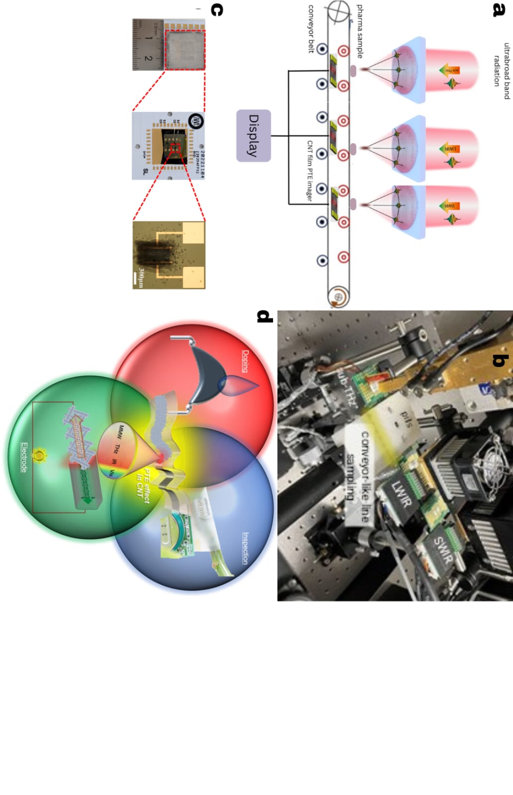
Fig 1: a) understandable diagram of non-destructive in-line dynamic ultrabroadband sub-THz-IR pharma monitoring system. b) Photograph of the experimental setup. Wavelength: 909 µm for sub-THz, 6.13 µm for LWIR, and 4.33 µm for SWIR. c) CNT film PTE imager. d) PTE effect in CNT.
internal structures take slightly longer to return compared to those from the surface because the path length increases. By precisely measuring these small differences in reflection time, known as time-of-flight measurements, it becomes possible to reconstruct the internal microstructure of the sample without physically sectioning it [8]. This capability makes sub-THz imaging an exceptionally useful and completely non-invasive tool for studying internal features of pharmaceutical dosage forms. One of the most important use of terahertz radiation in the pharmaceutical industry is the measurement of coating thickness in tablets and other solid dosage forms. The coating of a tablet serves multiple purposes, containing the active pharmaceutical ingredient (API), controlling the release rate, and improving the tablet’smechanical strength and appearance[9]. Any deflection in coating uniformity can directly affect drug performance. Terahertz time-domain spectroscopy (THz-TDS) offers a rapid and non-destructive method for assessing coating thickness and uniformity[5]. By analyzing the reflected terahertz pulses, scientists can differentiate between reflections originating from the outer tablet surface and those from the interface between the interior part and coating. The time delay between these two reflections which is correlated to layering size of the coating. Because this method does not require cutting or damaging the sample, it enables real-time, repeatable quality monitoring in manufacturing environments[6]. Advanced automated terahertz tablet scanners have been developed to extend this principle across entire tablet surfaces. These scanners systematically collect data over multiple positions to create spatial maps of coating thickness and uniformity[7]. The resulting images provide detailed insights into coating consistency and detect potential manufacturing defects such as overcoating, undercoating, or coating delamination[8]. This high-resolution mapping capability allows pharmaceutical manufacturers to improve process control and ensure that each batch of tablets meets strict regulatory and performance standards[9]. Moreover, terahertz measurements are contactless and reagent-free, decreasing contamination threat and preserving the chemical and physical integrity of the samples. The scientific basis of terahertz spectroscopy lies in its sensitivity to molecular and intermolecular interactions. Terahertz radiation interacts primarily with low-frequency vibrational and rotational modes of molecules rather than electronic transitions, which occur at higher frequencies. These vibrational and rotational motions are influenced by weak forces like van der Waal forces, dipole-dipole interaction and hydrogen dipole interaction. Consequently, terahertz spectroscopy can provide valuable information about the collective behavior of molecular assemblies, crystal structures, polymorphism, and conversion of phase. This makes it an important analytical tool for characterizing pharmaceutical materials, particularly in understanding the solid-state properties of active ingredients and excipients. Another main advantage of terahertz radiation is its relatively low photon energy, which ensures non-destructive interaction with materials. dissimilar ionizing radiation such as X-rays, terahertz waves do not alter molecular structures or induce radiation damage. This property is particularly favorable in pharmaceutical testing, where controlling the chemical stability of the product is essential. Furthermore, the penetration depth of terahertz radiation through non-metallic materials allows for the inspection of multi-layered systems, such as coated tablets, polymer films, and blister packaging. Its ability to detect internal defects, such as voids, cracks, or inclusions, enhances overall quality assurance manufacturing process is running.Terahertz absorption spectroscopy also provides insight into temperature-dependent molecular dynamics. The absorption intensity of terahertz radiation changes with dielectric losses, which can be partitioned into distinct temperature regions correlating with variations in molecular relaxation processes. This relationship enables researchers to study material transitions such as crystallization, amorphization, and dehydration in pharmaceuticals, which directly affect solubility and bioavailability. These measurements can be conducted without altering the sample, making terahertz spectroscopy a preferred choice for stability and preparation studies. Sub-terahertz radiation occupies a unique spectral niche with growing technological relevance. Its ability to probe intermolecular interactions while maintaining non-ionizing safety makes it suitable for sensitive biological and pharmaceutical samples. Continued improvements in terahertz sources and detectors—such as photoconductive antennas, quantum cascade lasers, and bolometric sensors—are expanding the applicability of terahertz techniques. Moreover, advancements in data processing and imaging system are enhancing the decision and accuracy of terahertz imaging systems, allowing for real-time monitoring and automated defect analyzing in industrial environment. From a broader perspective, the utility of sub-THz radiation extends beyond pharmaceuticals. Its capacity to penetrate non-conductive materials, distinguish molecular signatures, and operate safely under atmospheric conditions has led to its adoption in fields ranging from security screening and environmental sensing to high-speed wireless communication and biomedical diagnostics. However, challenges remain in improving signal-to-noise ratios, mitigating atmospheric absorption (especially by water vapor), and reducing equipment costs. Addressing these limitations through advancements in component design and system integration will be critical for achieving widespread industrial implementation.
2.2 IR:
Infrared (IR) radiation is one of the parts of electromagnetic energy that lies between visible light
and microwaves in the electromagnetic spectrum, typically spanning wavelengths from 0.7 to 1000 micrometers (µm) [10]. It has a lesser frequency than visible light and is widely used in spectroscopy, thermal imaging, environmental monitoring, and communication technologies [11]. Infrared radiation is primarily emitted as thermal energy, following Planck’s law, which states that any object with a temperature above zero emits electromagnetic radiation [12]. The wavelength and potency of this emission are directly related to the object’s temperature, with most room-temperature objects radiating predominantly in the infrared region. Infrared sources generate radiation in two primary ways: by heating a material until it emits thermal infrared energy or by inducing electronic or molecular transitions that produce IR photons. The emitted radiation interacts with materials in characteristic ways—being absorbed, reflected, or transmitted depending on molecular composition and structure. These interactions form the foundation of infrared spectroscopy, a technique that provides valuable insights into molecular arrangement [13].Fourier Transform Infrared (FTIR) is one of the most widely applied forms of IR spectroscopy. Rather than scanning individual wavelengths sequentially, FTIR uses an interferometer to collect all wavelengths simultaneously. The interferogram produced by the interferometer is mathematically transformed using a Fourier algorithm into an interpretable infrared spectrum [14]. This method has several advantages over traditional dispersive spectroscopy, including greater sensitivity, faster data collection, higher resolution, and improved signal-to-noise ratio. Consequently, FTIR has become a standard analytical tool for both qualitative and quantitative assessment of molecular systems [10].Infrared Absorption (IRA) spectroscopy focuses on analyzing molecular vibrations that occur within the infrared region. When IR radiation interacts with a molecule, specific frequencies are absorbed, corresponding to vibrational motions of chemical bonds such as stretching, bending, twisting, or scissoring [11]. The precise frequencies at which absorption occurs depend on atomic masses, bond strengths, and the surrounding chemical environment. Intermolecular interactions, including hydrogen bonding, dipole–dipole interactions, and van der Waals forces, can also cause subtle shifts in absorption frequencies and alter spectral intensity [12]. These unique vibrational patterns form a distinct “fingerprint” spectrum for each molecule, allowing detailed identification and structural analysis [13].Infrared spectroscopy plays a critical role in analytical chemistry and is especially valuable in pharmaceutical, biological, and materials science research. It provides both qualitative and quantitative information about chemical bonds, functional groups, and molecular structures. In pharmaceutical analysis, IR spectroscopy is widely used to identify active pharmaceutical ingredients (APIs), excipients, and contaminants, and to monitor polymorphism, crystallinity, and coating uniformity in solid dosage forms [14]. Its non-destructive nature and minimal sample preparation requirements make it ideal for in-line or at-line quality control during drug manufacturing [10].Infrared imaging techniques further enhance this capability by enabling spatial mapping of chemical components within tablets or capsules, thereby improving understanding of formulation homogeneity [11]. The effectiveness of infrared spectroscopy has been significantly enhanced by the integration of chemometrics, artificial intelligence (AI), and modern computational tools [12]. Chemometric analysis applies mathematical and statistical methods to interpret complex spectral data, enabling deconvolution of overlapping peaks and extraction of meaningful information from multicomponent samples [13]. Recent advancements in AI and machine learning algorithms have further automated spectral interpretation, pattern recognition, and predictive modeling, leading to more accurate and faster analysis [14].Despite its many advantages, infrared spectroscopy also faces challenges such as overlapping spectral bands, baseline variations, and environmental interferences from humidity or temperature fluctuations. Some probe-based systems may also experience reduced accuracy due to light scattering and surface irregularities [10]. However, continued improvements in detector sensitivity, optical design, and signal processing have greatly mitigated these issues [11].Infrared spectroscopy has also found applications beyond chemistry and pharmaceuticals. It is extensively used in biological systems for bacterial identification, tissue characterization, and protein structure analysis [12]. The ability of IR spectroscopy to analyze biological materials rapidly and non-invasively makes it a powerful technique for both clinical and industrial applications [13]. Infrared spectroscopy remains an indispensable analytical technique due to its ability to probe molecular vibrations and provide precise chemical and structural information. The development of advanced FTIR instruments, combined with chemometric and AI-based data interpretation, has expanded its capabilities, accuracy, and industrial relevance [14]. As technology continues to evolve, infrared spectroscopy will remain a cornerstone of non-destructive analysis, offering critical insights into the composition, quality, and stability of pharmaceutical products and other complex materials [10].
2.2.1 LWIR:
Long-Wave Infrared Radiation (LWIR) represents a specific region within the infrared (IR) spectrum, typically covering wavelengths between 8 and 14 micrometers (µm)[15]. This range is particularly significant because it corresponds closely to the thermal radiation naturally emitted by objects at everyday temperatures, including humans, animals, and common materials. All bodies with a temperature above absolute zero (-273.15°C) emit electromagnetic radiation as a result of their internal thermal energy, a phenomenon described by Planck’s law[16]. At standard environmental temperatures, the majority of this emission falls within the LWIR range, making it an essential domain for thermal sensing and imaging applications. LWIR is often referred to as thermal infrared radiation because it conveys information about the heat energy radiated by objects[17]. Unlike visible light, which is primarily reflected or emitted by external sources such as the sun or artificial lighting, LWIR is produced by the intrinsic thermal emission of materials themselves. This characteristic allows LWIR detection systems to visualize temperature variations independent of external illumination, enabling the observation of heat signatures invisible to the human eye. Thermal imaging devices, such as infrared cameras, detect LWIR radiation and convert it into visual representations known as thermograms. These thermograms display spatial temperature distributions, where warmer regions appear brighter and cooler regions appear darker[18]. This ability to “see” heat has made LWIR imaging a critical tool in both scientific and industrial applications.The fundamental operating principle of LWIR imaging is based on detecting differences in emitted infrared intensity, which are directly proportional to the temperature variations across an object’s surface. In practical applications, this property enables detailed thermal profiling and non-contact temperature measurement. LWIR systems are used extensively in fields such as environmental monitoring, medical diagnostics, industrial inspection, and defense[19]. For instance, in medicine, LWIR imaging assists in monitoring skin temperature variations to identify circulatory disorders, inflammation, or localized infections. In environmental science, it is used to study heat patterns in natural habitats, detect volcanic activity, and analyze thermal emissions from geological formations[20]. In industrial settings, LWIR imaging supports preventive maintenance by identifying overheating in electrical or mechanical systems before critical failures occur. A key advantage of LWIR radiation lies in its ability to penetrate certain atmospheric conditions better than visible light. It can effectively transmit through light fog, dust, or smoke, providing imaging capability in low-visibility environments[21]. This property makes LWIR particularly valuable in security surveillance, night vision, and search-and-rescue operations. However, despite these advantages, LWIR radiation is strongly absorbed by atmospheric water vapor, which can restrict its range and affect measurement accuracy in humid or wet environments. To mitigate this limitation, LWIR systems often employ specialized optics and detectors designed to optimize sensitivity and reduce atmospheric interference[22].LWIR detection technology has evolved significantly with advancements in infrared sensors and materials. Modern LWIR cameras use detectors made from mercury cadmium telluride (MCT) or microbolometer arrays that operate at or near room temperature as semiconductor[23]. Microbolometers, in particular, have enabled the development of compact, uncooled infrared cameras, reducing cost and complexity while maintaining high spatial and thermal resolution. These detectors measure changes in resistance caused by absorbed infrared energy, converting thermal variations into electrical signals that are then processed into visual images[24]. The ongoing miniaturization and improvement of LWIR sensor technology continue to expand its accessibility across scientific and industrial domains. The relevance of LWIR extends beyond thermal visualization; it also provides fundamental insights into the thermophysical properties of materials. By analyzing emitted LWIR spectra, researchers can extract information about emissivity, surface composition, and temperature-dependent behavior[25]. This capability is particularly valuable in material science, remote sensing, and pharmaceutical process monitoring, where surface temperature uniformity often indicates underlying chemical or physical changes.
2.2.2 SWIR:
Short-Wave Infrared Radiation (SWIR) refers to the portion of the infrared spectrum withwavelengths ranging approximately from 1 to 3 micrometers (µm)[26]. This spectral region presents between the visible and long-wave infrared (LWIR) bands, bridging the transition between emitted and reflected infrared energy. Unlike LWIR, which primarily captures thermal radiation emitted by objects, SWIR is dominated by reflected light—similar to the visible light behavior making it a powerful tool for imaging applications that rely on reflectance, absorption, and transmission characteristics of materials. Since SWIR wavelengths are invisible to the human eye, specialized sensors and cameras are required to estimate and visualize this radiation. SWIR imaging systems detect photons that are either reflected or scattered from an object’s surface after illumination by an external light source, such as sunlight or artificial lamps. Because of this dependence on reflected light, SWIR images exhibit high contrast and fine detail even under low density light or night time[27]. This property enables the development of night-vision systems that can capture clear images by detecting reflected SWIR signals rather than emitted thermal radiation. As a result, SWIR imaging serves as a valuable complement to visible and LWIR systems, particularly in conditions where visible imaging is limited by poor illumination or environmental interference.A key advantage of SWIR radiation lies in its ability to penetrate atmospheric obscurants such as haze, fog, smoke, and dust more effectively than visible light. This capability enables clear imaging in challenging or degraded visual environments, making SWIR an essential tool in surveillance, defense, remote sensing, and industrial inspection applications. In addition, SWIR light can also penetrate certain materials that are opaque to visible wavelengths, such as plastics, coatings, and semiconductor substrates, allowing for subsurface imaging and detection of defects. This see through ability is particularly helpful in quality control processes, enabling the identification of internal defects, contaminants, or structural inconsistencies in non-metallic materials without physical contact or sample preparation[28].Technological advancements in the infrared industry have led to significant improvements in SWIR imaging performance. Over the years, the development of advanced hardware and software has focused on enhancing signal-to-noise ratio (SNR), minimizing noise equivalent temperature difference (NETD), and increasing image resolution and detector sensitivity. Modern SWIR cameras utilize high-resolution sensors, optimized pixel pitch, and low-noise electronics to achieve precise imaging under varying environmental conditions. Indium Gallium Arsenide (InGaAs) detectors are among the most widely used technologies for SWIR imaging because of high quantum proficiency, wide dynamic range, and stability at or near room temperature. These detector changing incoming photons into electrical signals with excellent sensitivity, enabling accurate detection of reflected SWIR radiation. In analytical and industrial applications, SWIR spectroscopy plays a crucial role in material characterization and process monitoring. The absorption rays in the SWIR region correspond primarily to overtones and combinations of fundamental molecular vibrations, particularly those associated with hydrogen-containing functional groups such as O–H, N–H, and C–H[29]. As a result, SWIR spectroscopy provides valuable information about molecular composition, moisture content, and chemical bonding in inorganic and organic chemicals. In pharmaceutical manufacturing, for instance, SWIR-based hyperspectral imaging systems are used for non-destructive analysis of tablets and capsules, allowing the detection of coating uniformity, active pharmaceutical ingredient distribution, and contamination of foreign particle. This approach combines high spatial and spectral resolution, making it highly effective for real-time quality control and defect detection.SWIR imaging also finds significant use in other scientific and industrial fields. In agriculture and environmental monitoring, it aids in assessing plant health and soil moisture by exploiting differences in reflectance patterns caused by water absorption. In semiconductor and electronics industries, SWIR systems facilitate inspection of silicon wafers and integrated circuits, as silicon is transparent to SWIR wavelengths but opaque to visible light. Furthermore, SWIR’s ability to provide detailed imaging under variable lighting conditions has led to its adoption in autonomous vehicles, optical sorting systems, and precision manufacturing processes.
2.3 Carbon nanotube films (CNT):
Carbon nanotubes (CNTs) are cylindrical very fine structures made of carbon atoms structure like hexagonal lattice, resembling rolled graphene sheets. Hang on the number of concentric layers, they are mainly two class multiple walled carbon nanotube (MWCNT) or single-walled carbon nanotube(SWCNT)[30]. Because of their nanometer-scale diameters and micrometer-scale lengths, CNTs are considered pseudo-one-dimensional materials. They are allotropes of carbon, belonging to the same family as diamond, graphite, and graphene, but with distinctive physical and chemical properties that make them exceptionally useful in electronics, biosensing, and nanotechnology[31]. CNTs possess remarkable electrical, and thermal characteristics. Their tensile strength is too greater than that of steel, while their electrical conductivity can exceed that of copper by up to two orders of magnitude[32]. They also exhibit very high thermal conductivity and excellent chemical stability in both aqueous and non-aqueous environments. The fusing of these properties, along with their large surface area, enables CNTs to adsorb or interact with various molecules efficiently, making them ideal for integration into advanced sensing platforms, particularly in biosensors and electronic systems.Biosensors are analytical tools that trace biological or chemical analytes by converting biochemical interactions into measurable electrical signals. CNT-based biosensors leverage the unique structural and conductive properties of nanotubes to enhance sensitivity, selectivity, and response time[33]. Their nanoscale dimensions allow close contact with biological molecules, leading to improved transduction efficiency and faster signal generation. Depending on the detection principle, CNT biosensors are generally classified into chemical and physical types. Chemical biosensors rely on the direct interaction in the analyte and functionalized CNT surface, generating electrochemical and optical feedback. In contrast, physical biosensors detect changes in measurable parameters such as resistance, capacitance, or thermal conductivity display on the analyte. In both cases, CNTs act as the active transducer, translating molecular recognition into quantifiable electrical outputs.
CNT thin films form the strong base of many biosensing devices. These films consist of randomly stacked nanotubes forming a conductive network that ensures efficient electron and heat transport. The flexibility and robustness of such networks make them most suitable for integration into wearable or deformable sensing systems. Typically, CNT thin-film channels are supported by polyurethane (PU) substrates of approximately 8 µm thickness to provide mechanical flexibility and environmental protection. Readout electrodes are fabricated from a rubber-like conductive composite mixing of silver (Ag) nanoparticles add with binder resins, ensuring stretchability and reliable electrical contact even during bending or deformation. A figured epoxy resin layer is often added to the rear surface as a partial stiffener to prevent strain concentration. The hybrid configuration of soft and rigid layers allows the device to maintain stable photo-thermoelectric (PTE) performance under mechanical stress, such as bending or wrapping on curved surfaces.The integration of CNTs into biosensors also benefits from surface functionalization, which tailors the nanotube surface for improved biocompatibility and molecular recognition. Two primary functionalization strategies are employed: non-covalent and covalent modification. Non-covalent functionalization involves attaching biomolecules via weak interactions—such as π–π stacking or van der Waals forces without altering the CNT’s intrinsic structure[34]. This approach preserves electrical and mechanical properties but offers limited stability. Conversely, covalent functionalization introduces strong chemical bonds between functional groups and CNT surfaces, significantly improving solubility, dispersion, and compatibility with biological environments. However, this method can slightly modify the nanotube’s structure and conductivity. Enzyme-CNT electrodes represent a key application of such functionalization. In these systems, enzymes are immobilized on CNT surfaces to catalyze biochemical reactions, producing electrical signals that correspond to analyte concentration[35]. These amperometric CNT-based biosensors measure the current generated during the enzymatic reaction, enabling rapid and highly sensitive detection of biological molecules such as glucose, cholesterol, and toxins. The large surface area of CNTs provides adequate active sites for enzyme immobilization, while their high conductivity ensures efficient electron transfer, resulting in developed signal strength and time of response.Despite their advantages, CNTs face challenges related to dispersion and hydrophobicity. Because of strong van der Waals forces, CNTs tend to aggregate in both aqueous and organic media, reducing their effective surface area and hindering device uniformity. Their hydrophobic nature also limits their compatibility with biological and water based network. To overcome these issues, surface modification techniques—such as they introduce hydrophilic functional groups like –COOH or –OH—are commonly used. These treatments improve solubility, promote uniform dispersion, and enhance interaction with aqueous environments. Achieving stable and uniform dispersion is crucial for consistent film formation and reproducible biosensor performance. CNT films exhibit outstanding electrical and optical properties, contributing to their versatility in sensing applications. Their high electron mobility facilitates rapid charge transport, allowing real-time detection of chemical and biological events. Additionally, CNTs exhibit photoluminescence in the near-infrared (NIR) region, which can be utilized for optical biosensing and bioimaging. NIR light penetrates biological tissues more effectively than visible light, minimizing background interference and enabling non-invasive monitoring. These unique optoelectronic characteristics extend the utility of CNT-based sensors beyond electrochemical detection to include optical and photo-thermoelectric systems.CNT films made from single-walled CNT solutions, such as ZEO-NANO SG101 (0.5%) and EC-DH (0.2%), typically form random, non-aligned structures that deliver stable and isotropic electrical performance. The interconnected network within these films provides high mechanical flexibility and uniform conductivity, making them suitable for use in flexible and wearable biosensors. Moreover, their ability to maintain compatible performance under bending or deformation makes them promising candidates for non-destructive, in-line pharmaceutical inspection systems.The versatility of CNT-based biosensors has driven their application across many scientific and industrial levels. In medical diagnostics, they are employed to detect biomarkers and monitor metabolic indicators, providing rapid and accurate disease detection. In environmental monitoring, CNT sensors can identify pollutants, heavy metals, and toxic gases at trace concentrations. In the pharmaceutical industry, CNT films are utilized in quality testing, enabling non-destructive inspection of tablets, powders, and coating uniformity. Their flexibility and adaptability make them suitable for real-time monitoring of manufacturing processes, ensuring consistent quality control. Although CNT technology offers immense potential, challenges remain in large-scale synthesis, uniformity, and biocompatibility. Efforts are underway to refine production methods and develop hybrid composites combining CNTs with polymers, metals, or two-dimensional materials such as graphene to enhance performance and stability. Future advancements in nanofabrication, dispersion engineering, and functionalization chemistry will further expand the scope of CNT-based devices.
2.4 Photo thermoelectric (PTE) sensor:
Photo-sensing and photo-imaging measurements are powerful non-destructive techniques used to analyze the optical properties of materials under light exposure[36]. Different materials interact with light in distinct ways, exhibiting wavelength-dependent behaviors such as reflection, absorption, and transmission. By studying these optical responses across multiple spectral ranges, it becomes possible to identify materials, evaluate their structure, and monitor changes in their formation without causing any physical damage[37]. When a particle is light exposure(photo-irradiation), it either reflects, transmits, or absorbs part of the incident radiation. Measuring these interactions at various wavelengths provides valuable information about a material’s intrinsic properties. This principle forms the basis of hyperspectral imaging, an advanced method that records spectral information for every pixel in specific images. By combining optical data from multiple bands from visible to infrared regions-hyperspectral systems generate detailed spectral “fingerprints” that distinguish materials based on their optical characteristics[38]. Such imaging approaches are widely used in pharmaceuticals, materials science, and biomedical diagnostics for rapid, non-invasive assessment of samples. Photo-imaging methods are preferred over conventional testing because they are non-contact, rapid, and highly informative. They allow the inspection of delicate or valuable samples without requiring chemical reagents or physical modification. For instance, in pharmaceutical manufacturing, these methods enable real-time monitoring of pill coatings and surface consistency, checking consistent product quality.Among modern optical detection mechanisms, the photo-thermoelectric (PTE) effect has appeared as a promising technique for self-powered light sensing. The PTE effect combines two fundamental processes: photo-thermal conversion, in which absorbed light raises the local temperature of a material, and thermoelectric conversion, where the resulting temperature difference generates an electrical voltage[39]. Thus, when light irradiates a PTE material, it produces a measurable electrical signal directly related to the light intensity. A major advantage of PTE-based devices is that they operate without external bias voltage, resulting in low thermal noise and high signal-to-noise ratio (SNR). This self-powered feature enhances detection sensitivity and stability, making PTE sensors suitable for low-light and broadband applications. In a typical device, the photo-detection interface is positioned at the edge of the conductive channel to localize heating and maximize the thermal gradient. This focused heat distribution allows efficient conversion of absorbed light into an electrical signal, improving spatial resolution and accuracy.Designing an efficient PTE sensor requires optimizing the interplay between photonic, thermal, and electronic properties of the material. Matters like carbon nanotubes (CNTs), graphene, and other nanostructured conductors are ideal for PTE devices because they combine broad optical absorption, high thermal conductivity, and superior charge mobility[40]. By integrating these materials with precise structural control, modern PTE-based photo-sensing systems achieve high sensitivity, quick response, and dependable performance for real-time, non-destructive imaging and analysis.
2.5 PTE effect in channel electrode junction:
Carbon nanotube (CNT)-film-based photo-thermoelectric (PTE) sensors represent an advanced class of optoelectronic devices that combine the broadband optical absorption characteristics of CNTs with efficient thermoelectric conversion[41]. These flexible sensor sheets are uniquely suited for broadband photo-detection and non-destructive inspection applications, especially in environments requiring conformable, shape-adaptive, and omni-directional sensing. The intrinsic mechanical flexibility, thermal stability, and optoelectronic tunability of CNT films enable the realization of photo-sensing systems competent of operating reliably under physical deformation, elevated temperatures, and complex surface geometries.The fundamental working process of CNT-film-based PTE sensors relies on the photo-thermoelectric effect, which couples light absorption with thermoelectric voltage generation. When light irradiates the CNT film, part of the optical energy is converted into localized heat through photo-thermal absorption. This temperature difference (ΔT) across the CNT channel generates an electrical voltage (ΔV) via the thermoelectric effect, governed by the Seebeck coefficient of the materials. The relationship can be expressed as:
ΔV=(SCNT−Scom) × ΔT
where ΔV represents the photo-thermoelectric voltage response, SCNT is the Seebeck coefficient of the CNT film, Scom is the Seebeck coefficient of the composite component at the junction, and ΔT is the photo-induced thermal gradient. The resulting DC voltage serves as the PTE response, enabling light detection without external electrical bias[42].The self-powered nature of PTE sensors eliminates the need for external voltage, significantly reducing thermal noise and increasing the signal-to-noise ratio (SNR). This makes CNT-film-based PTE devices exceptionally sensitive, stable, and energy-efficient for broadband photo-detection across visible, infrared, and terahertz wavelengths. CNT-film-based PTE sensors can be configured using P-type and N-type materials to form PN-junction structures, improving the effective Seebeck coefficient and overall voltage output. These junctions facilitate efficient charge separation and increases the device’s thermoelectric conversion efficiency. The combination of intrinsically broadband absorption and tunable Seebeck polarity allows CNT-based PTE sensors to detect a wide range of optical wavelengths while maintaining high responsivity[43].The design flexibility of CNT-film-based devices also supports shape-deformable and shape-conformable architectures, enabling omni-directional and blind-spot-free three-dimensional (3D) monitoring. Flexible image sensor sheets can be wrapped around complex geometries or curved surfaces, eliminating the need for bulky mechanical rotation mechanisms typically required in conventional imaging systems. This approach enables continuous 3D surface mapping and defect detection on solid or composite materials. Mechanical robustness is a critical requirement for conformal and wearable sensor applications. CNT-film-based PTE sensor sheets demonstrate excellent mechanical durability, maintaining stable performance even under substantial deformation. Experimental results show that sensitivity degradation remains below 20%, as indicated by the relative increase in noise-equivalent power (NEP), under multiaxial repetitive strain ranging from 70% to 280%. The devices consistently maintain a minimum NEP value of approximately 5 pW·Hz?¹?², confirming high photo-thermoelectric sensitivity and stable operation during folding, stretching, or wrapping.Thermal reliability is equally important for field deployment. High-temperature annealing tests demonstrate that CNT-film-based PTE sensors sustain stable optical and electrical performance even at temperatures up to 200 °C, with minimal variation in NEP values. This thermal endurance indicates that the PTE mechanism continues to function effectively under elevated background temperatures. During thermal evaluation, the ambient temperature increases gradually, yet photo-irradiation continues to induce localized heating at the PN junction. These localized temperature gradients drive thermoelectric voltage generation, confirming the sustained responsiveness of the device under thermal stress. An additional advantage of CNT-film-based PTE sensor sheets lies in their ability to switch between reflective and transmission monitoring modes. This dual-mode functionality allows flexible adaptation to different types of objects—reflective mode is preferred for opaque, solid materials, while transmission mode is more effective for semi-transparent or hollow structures [44]. The capability to alternate between these modes enables comprehensive non-destructive inspection (NDI) of three-dimensional objects.
Fig 2- A carbon nanotube film-based photo-thermoelectric imager for non-destructive, ultrabroadband, multi-wavelength optical inspection of defective pharmaceutical pills. a) identification of material compositions. b) fundamental property of inspected sample. c) Transmissive line-profiling which obtained from the PTE image. d) estimation technique same but concealed foreign substance. e) fundamental property of each foreign substance. f) transmissive line-profiling which obtained from PTE image of foreign materials.The development of both reflective-type and transmission-type PTE modules broadens the application range of CNT-based sensors in real-world industrial settings. Integration with 3D printing technologies enables customizable module fabrication, allowing precise geometric adaptation to target objects. The supporting resin framework provides mechanical stability, ensuring firm attachment of the CNT-film sensor sheet and preserving device integrity during deformation or handling. This combination of material flexibility and structural adaptability positions CNT-film-based PTE sensors as a cornerstone for next-generation, conformal NDI platforms. The CNT-film-based PTE sensor sheet’s ability to maintain consistent performance under mechanical and thermal stress demonstrates its potential in wearable electronics, flexible imaging, and industrial inspection. Its ultrathin and lightweight design allows integration into curved and dynamic surfaces, enabling continuous monitoring of temperature and light intensity distributions. The broadband optical sensitivity of CNT films supports multispectral imaging, making them particularly useful for analyzing composite or layered materials in the pharmaceutical, aerospace, and materials manufacturing sectors. Furthermore, by aggregating multispectral optical data, these sensors can reveal subtle structural variations and compositional differences that conventional single-band detectors might overlook. Such capabilities are especially beneficial in pharmaceutical quality control, where differentiating between coating layers or detecting impurities requires highly sensitive, broadband, and non-contact monitoring tools.
3. Pharma monitoring demonstration:
Based on the functional CNT film PTE device and the ultrabroadband system configuration, the following demonstrations highlight the key advantages of sub-THz to infrared (IR) irradiation for non-destructive inspection of pharmaceutical agent pills [45].Fig 2 contains mainly as follow: The pharmaceutical pills tested include sedative, antipyretic, and antiplatelet agents. Using one-axis-scan transmissive imaging in the short-wave infrared (SWIR) and long-wave infrared (LWIR) ranges, the CNT film PTE device successfully differentiates the material compositions of these visually indistinguishable pills. Among the samples, the sedative agent consistently exhibits higher transmittance to external photo-irradiation at two distinct wavelengths, indicating a unique spectral response compared to the antipyretic and antiplatelet agents. Selective transmission behavior is observed among the tested agents: the antipyretic responds preferentially to SWIR irradiation, whereas the antiplatelet agent shows enhanced transmission in the LWIR range [46]. Using only the LWIRimage is insufficient to determine whether the central sample is a reflective metal object or a photo-absorbent material. The non-destructive ultrabroadband inspection system using CNT film PTE imagers demonstrated in this work is also capable ofimage is insufficient to determine whetherthe central sample is a reflective metal object or aphoto-absorbent material.
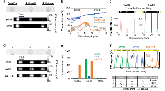
Fig 2- A carbon nanotube film-based photo-thermoelectric imager for non-destructive, ultrabroadband, multi-wavelength optical inspection of defective pharmaceutical pills. a) identification of material compositions. b) fundamental property of inspected sample. c) Transmissive line-profiling which obtained from the PTE image. d) Identification technique same but concealed foreign substance. e) fundamental property of each foreign substance. f) transmissive line-profiling which obtained from PTE image of foreign materials.
The non-destructive ultrabroadband inspection system using CNT film PTE imagers demonstrated in this work is also capable of detecting hazardous foreign substances (impurities) hidden within pharmaceutical agents, in addition to identifying the material composition of the pills themselves. In fig 2(b) This fundamental optical information—specific spectral transmittance characteristics—serves as the key identifier for each inspected pharmaceutical sample. The proposed approach enables non-destructive ultrabroadband monitoring by first earning reference spectra for various pharmaceutical agents using the CNT film PTE imager. These reference spectra allow extraction of wavelengths with characteristic transmittance distributions unique to each agent. Leveraging the CNT film PTE imager’s high sensitivity across an ultrabroadband range, the system can then detect and differentiate agents based on their spectral responses. The non-destructive inspection process is implemented using compact photo-sources tuned to these characteristic wavelengths, and abnormalities are identified by classifying the response signal intensities relative to the reference conditions. This spectroscopy-driven framework facilitates accurate, label-free detection and classification of pharmaceutical agents and defects[47]. In fig 2(c) The line profiling illustrates how the transmissive response signal intensity varies across a selected line, based on data from an individual device pixel in the acquired PTE images. In fig 2(d) The obtained results successfully identify impurities made of plastic, glass, and metal concealed within the pills. This is achieved through one-axis-scan transmissive imaging using sub-THz, LWIR, and SWIR wavelengths with the device—despite these impurities being visually indistinguishable. In the transmissive photo-thermoelectric (PTE) image of Pill-i, a distinct vertical slit is clearly visible when the sample is illuminated with two different wavelengths: long-wave infrared (LWIR) and short-wave infrared (SWIR). This vertical slit corresponds to a localized reduction in the transmitted photo-irradiation intensity through the pill. The correspondence between the response signals detected by the CNT film PTE pixels and the monochrome color scale of the image allows us to precisely identify this region as a darker area. These darker vertical slits, as shown in Figure 2d, indicate the presence of impurities concealed within the pill structure. This detecting demonstrates that the application of two-wavelength photo-irradiation combining LWIR and SWIR bands can effectively reveal impurities that are otherwise difficult to detect visually. Furthermore, the impurity within Pill-ii is also successfully visualized through transmissive imaging using only the LWIR band. These results specially denoting the capability of the CNT film PTE imager to perform sensitive, non-destructive inspection of pharmaceutical pills, enabling detection of hidden impurities by exploiting the spectral differences in transmission at multiple wavelengths. The combined use of multiple wavelengths also successfully visualizes the impurity in Pill-iii. However, from a different perspective, SWIR irradiation alone is insufficient to clearly distinguish the impurities between Pill-i and Pill-iii. Similarly, transmissive imaging using only LWIR tends to confuse the impurities present in each pill, making differentiation difficult. These observations highlight that the visibility of impurities varies depending on the specific photo-irradiation wavelength used. This variation accurately reflects the broad differences in transmittance values of different materials based on their composition and the wavelength of the sub-THz to infrared (IR) radiation applied. Glass selectively exhibits lower transmittance under LWIR irradiation, while metal shows no transmission at the three examined wavelengths due to its inherently reflective nature. Using these spectral characteristics, the device and system presented in this work can non-destructively identify the materials of impurities concealed within pharmaceutical pills. Specifically, impurity i is identified as plastic, impurity ii as glass, and impurity iii as metal. It provides lower transmittance values under external LWIR and SWIR irradiation compared to those observed at the employed sub-THz wavelength.
4. Fundamental experimental setup for pharma monitoring:
Building upon the described device materials and configurations, this study presents a non-destructive, in-line, dynamic ultrabroadband monitoring system that operates across the sub-terahertz (sub-THz) to infrared (IR) spectral radius. The system integrates carbon nanotube (CNT) film-based photo-thermoelectric (PTE) imagers, enabling real-time, contactless inspection of pharmaceutical pills during production. This approach represents a major advancement toward continuous and automated quality control in industrial pharmaceutical manufacturing[48]. The CNT film PTE imager combines several advantageous properties—broadband optical absorption, high thermoelectric conversion efficiency, and remarkable mechanical flexibility. Its thin, lightweight, and conformable design allows easy integration into existing manufacturing lines without bulky modifications. Furthermore, high-yield fabrication and stable photoelectric performance ensure reliable operation under dynamic industrial conditions[49]. These characteristics make CNT-film PTE devices ideal for implementing real-time, non-destructive inspection directly on production sites.To validate system performance, standard parameters for pill fabrication were established: 7 mm diameter, 1 mm thickness, and 5% active pharmaceutical agent concentration. The specified formulation and preparation process are provided in the material and procedure section. These standardized dimensions ensure consistent evaluation of optical properties and reproducibility of experimental results across different irradiation wavelengths. The developed in-line system employs CNT film PTE imagers capable of detecting broadband radiation spanning sub-THz to IR frequencies. Pills are transported through the optical path using a motorized belt system that allows continuous and dynamic scanning. Tunable external photo-sources provide illumination in the sub-THz, short-wave infrared (SWIR) and long-wave infrared (LWIR) bands. This tunability allows precise adjustment of the estimated wavelengths to compare the optical characteristics of specific pharmaceutical formulations, coatings, or excipients[50]. Unlike conventional spectrometers, which are limited to fixed spectral bands, this system’s flexibility enables adaptive inspection tailored to diverse materials and manufacturing conditions. Optical transmittance mapping experiments were conducted using a representative sedative pill. The results show transmittance values exceeding 50% across the sub-THz–IR range, demonstrating that ultrabroadband photo-irradiation effectively supports non-destructive pharmaceutical inspection. The obtained transmittance patterns reveal distinct wavelength-dependent variations corresponding to the pill’s composition and internal microstructure. These findings confirm that sub-THz–IR spectral data can provide crucial information for identifying defects, impurities, and compositional inconsistencies without physical alteration of the sample.The CNT film PTE imager’s ultrabroadband sensitivity allows simultaneous or sequential acquisition of multispectral data without switching sensors. This proficiency enables the construction of unique optical “fingerprints” for each pill type, facilitating rapid, label-free material differentiation. The resulting spectral maps provide an advanced platform for implementing machine learning algorithms that classify samples, monitor quality parameters, and detect anomalies in real time.Two-dimensional (2D) photo-imaging experiments using sub-THz, LWIR, and SWIR irradiation further validated the system’s accuracy and speed. Even when operated using a single-pixel CNT film PTE detector, the device produced images with spatial resolution comparable to those from conventional 2D scanners but with significantly reduced acquisition time. This high-throughput capability makes the system suitable for continuous pharmaceutical production environments where speed and precision are essential. The device maintained stable sensitivity and low noise during continue dynamic scanning, confirming its robustness under continuous operational stress. Unlike simplified laboratory-scale tests, actual pharmaceutical tablets vary in size, shape, and internal composition. The CNT film PTE imager’s flexibility and broadband responsiveness enable accurate inspection of these complex real-world targets without causing damage or chemical alteration. Its compact and easily attachable form allows integration into manufacturing lines for on-site, real-time monitoring of critical parameters such as coating uniformity, ingredient distribution, and impurity detection[51]. This feature is particularly advantageous for continuous production systems requiring in-line inspection without process interruption.
5. Pharma monitor in-line multiwavelength process
This study presents a non-destructive, in-line, and dynamic system for identifying material composition and detecting defects in pharmaceutical pills using an ultrabroadband, multi-wavelength sub-terahertz (sub-THz) to infrared (IR) monitoring setup based on carbon nanotube film (CNT) photo-thermoelectric imagers(PTE). The system enables real-time, label-free inspection of pills during production, advancing continuous quality control and safety in pharmaceutical manufacturing[52]. In the developed system, pharmaceutical pills move continuously along a linear alignment track oriented perpendicular to the optical paths of the illumination and detection components. As each pill passes through the inspection zone, it is illuminated by multiple wavelengths ranging from sub-THz to IR. CNT film PTE imagers placed along the optical path capture transmissive images in real time. This dynamic, in-line arrangement allows continuous inspection without interrupting production flow, providing rapid, non-contact analysis of composition and structure.All optical components compact photo-sources, focusing optics, and flexible CNT film sensors are aligned on a single optical axis to ensure precise illumination and uniform detection. The ultrabroadband configuration combines sub-THz, long-wave infrared (LWIR), and short-wave infrared (SWIR) illumination, capturing complementary information about each pill’s structure and material composition. Sub-THz radiation penetrates deeply to reveal internal structures, while SWIR and LWIR provide surface and compositional details. Together, these spectral bands generate a comprehensive optical signature for every sample[53]. The CNT film PTE imagers convert absorbed optical energy into electrical voltage through the photo-thermoelectric effect, which directly correlates with the pill’s transmittance properties. These signals are processed to create high-resolution transmissive images that indicate the internal uniformity, density, and presence of hidden impurities. The system connects the real-time broadband PTE response data obtained during scanning with pre-recorded reference transmittance spectra for each pharmaceutical agent. These reference spectra act as threshold benchmarks for specific materials. The measured PTE signal intensities are continuously compared with these reference values to enable automated material classification and detection of deviations.This framework allows accurate, real-time identification of pill composition and detection of impurities or defects without manual intervention. Any significant deviation from the stored spectral threshold immediately indicates a potential irregularity. Such automation establishes a foundation for Process Analytical Technology (PAT) and Industry 4.0-based smart manufacturing, where real-time feedback enhances production accuracy and reduces waste[54]. To optimize the performance of the PTE imagers, two types of CNT film channels were fabricated with different surfactant concentrations, significantly influencing electrical resistance and detection stability.
These differences directly affected noise characteristics and photo-response behavior. Channel 1 demonstrated increased noise when signal readout speeds exceeded 4 milliseconds (ms) per measurement, which degraded transient photo-signal precisely. The elevated resistance amplified Johnson–Nyquist noise, the thermal noise proportional to the square root of resistance, reducing the signal-to-noise ratio (SNR) under fast-scanning conditions. Channel 2, with lower resistance, exhibited stable and consistent transient responses even at higher readout speeds, maintaining signal clarity and fast response under dynamic operation. The reduced thermal noise and improved electrical conductivity of Channel 2 support faster, more reliable signal readout, confirming that optimized surfactant concentration and film fabrication play a critical role in improving PTE sensor fidelity and temporal resolution for high-speed monitoring applications. Signal acquisition speed in the system was controlled by the (NPLC) Number of Power Line Cycles, which defines the integration time per measurement. Three theoretical readout speeds were evaluated:
• NPLC = 1 → 20 ms readout speed
• NPLC = 0.2 → 4 ms readout speed
• NPLC = 0.02 → 0.4 ms readout speed
These configurations represent trade-offs between accuracy and speed longer integration times yield lower noise but slower imaging, while shorter integration improves speed at the expense of noise. For baseline testing, optical measurements were first conducted in a single-pixel readout mode at 20 ms (NPLC = 1), ensuring stable, low-noise characterization of the PTE response. Spatial scanning experiments were then performed at a fixed resolution of 100 µm per step, with scan-step durations of 0.70 s, 0.50 s, and 0.45 s corresponding to the respective readout speeds. The end results analyze that the system maintains signal accuracy and high spatial resolution even under the fastest scanning condition (0.45 s), demonstrating its capability for rapid, high-throughput imaging.Higher-intensity SWIR irradiation produced stronger photo-thermal responses than the sub-THz band, enabling faster and more efficient photo-detection at increased scanning speeds. Under SWIR illumination, the system achieved reliable imaging at 0.45-second scan steps while maintaining adequate signal intensity for accurate mapping. Conversely, sub-THz irradiation, though effective for deeper structural imaging, required longer integration to achieve comparable sensitivity due to lower optical power density. This difference highlights the importance of spectral power balance when configuring the system for specific inspection goals surface evaluation benefits from IR, while internal analysis favors sub-THz illumination. The results verify that the CNT film PTE-based ultrabroadband sub-THz–IR system functions effectively for in-line, dynamic, and non-destructive pharmaceutical inspection. The system integrates multi-wavelength photo-imaging, real-time spectral comparison, and adaptive scanning to identify materials and detect defects during continuous production.By correlating the PTE response with reference transmittance data, the system performs automated classification of pharmaceutical agents and impurity detection with high precision. The experimental evaluation of CNT film channels demonstrates that lowering electrical resistance through optimized fabrication significantly enhances sensitivity and speed, essential for real-time industrial operation.
This flexible, compact, and broadband platform marks a significant step toward next-generation optical inspection technologies in pharmaceutical manufacturing. Its compatibility with continuous production lines, real-time feedback control, and non-destructive sensing capabilities align with the principles of better manufacturing. Future establishment may involve the integration of multi-pixel PTE arrays, machine learning-based classification, and adaptive wavelength selection, enabling even faster, more accurate, and autonomous quality assurance systems for large-scale pharmaceutical production.
6. System automation process:
This study demonstrates a non-destructive, in-line, and dynamic monitoring approach for pharmaceutical products using carbon nanotube (CNT) film-based photo-thermoelectric imagers(PTE). These imagers operate effectively under broadband irradiation spanning from the sub-terahertz (sub-THz) to the infrared (IR) spectrum, enabling real-time analysis of pharmaceutical samples such as tablets, pills and capsules[55]. The system primarily utilizes a transmissive optical configuration, in which the incident radiation passes through the pharmaceutical sample, allowing the internal structure to be analyzed without physically altering or damaging the product. The use of sub-THz to IR irradiation enables the analysis of internal property based on differences in absorption and scattering behavior among materials inside the pill, including active pharmaceutical ingredients (APIs), excipients, and other structural components[56]. The CNT film PTE imagers are particularly well-suited for such applications due to their broadband sensitivity, operation at room temperature, and compatibility with scalable, flexible substrates[57]. These properties make them ideal for continuous and real-time monitoring in pharmaceutical production environments that demand high stability and precision.
|
Parameter |
Value / Description |
|
Scan length (current) |
500 mm |
|
Pill diameter |
7 mm |
|
Pill interval |
10 mm |
|
Pills per scan (500 mm) |
Up to 30 |
|
Scan length (extended stage) |
800 mm |
|
Pills per scan (800 mm) |
Up to 47 |
|
Imaging speed |
3 mm/s |
|
Max pills/hour (theoretical) |
635 |
|
Data acquisition |
Photo response signals from CNT film PTE imagers |
While transmissive imaging provides valuable information about internal composition and uniformity, it has certain limitations, particularly in detecting surface-level or shallowly embedded contaminants such as metallic impurities[58]. Metallic inclusions generally exhibit low transmittance and high reflectivity in the sub-THz–IR range, which can make them difficult to identify through transmission imaging alone. To solve this limitation, the integration of hybrid imaging modes combining transmissive and reflective scanning has been proposed. In this configuration, the same broadband sub-THz–IR radiation is used for both transmission and reflection. In the reflective mode, the radiation does not pass through the sample but instead captures the light reflected from the surface or near-surface regions of the tablet. This mode enhances sensitivity to high-atomic-number elements such as metals, which typically reflect or absorb strongly in the sub-THz–IR spectrum. Reflective imaging therefore provides complementary surface and near-surface information, making it possible to detect metallic or dense impurities that may be hidden under coatings or within upper layers of the pill.By combining both transmissive and reflective imaging techniques, the system provides a more comprehensive and non-destructive inspection method that allows both internal structure analysis and surface contamination detection. This dual-mode configuration strengthens product quality assurance by enabling simultaneous verification of the pill’s internal composition and external purity. Such integration not only enhances inspection accuracy but also aligns with modern pharmaceutical quality control practices that demand real-time, automated, and non-invasive analysis under continuous manufacturing conditions. The proposed hybrid imaging approach supports industry calibrate like the Good Manufacturing Practices (GMP) and Process Analytical Technology (PAT), which emphasize in-line monitoring and data-driven quality control during pharmaceutical production.Automation plays a crucial role in achieving seamless operation of the system. The setup integrates a precision optical stepping motor stage with a synchronized data acquisition module that records the photo response signals generated by the CNT film PTE imagers. The coordination between mechanical scanning and electronic readout is managed using custom LabVIEW software, ensuring precise timing and automated data collection. During operation, the optical stage moves the pharmaceutical pills through the inspection zone while the detectors continuously capture photo-thermoelectric signals. The system currently operates with a maximum scanning range of 500 millimeters, which accommodates up to 30 pills per in-line scanning cycle. Each pill, with a diameter of 7 millimeters, is spaced 10 millimeters apart along the scanning axis. Within this configuration, the system acquires spatially resolved optical data that are reconstructed into detailed images showing the pills’ internal structures and potential defects.The scalability of this system makes it particularly suitable for industrial applications. Commercially available optical stepping stages with longer scanning ranges, such as 800 millimeters, can be integrated easily, allowing up to 47 pills to be inspected in a single scanning cycle while maintaining the same pill size and spacing. Based on the maximum imaging speed of 3 millimeters per second demonstrated in this study, the system can theoretically inspect up to 635 pills per hour in a continuous, automated manner. This high-throughput capacity represents a significant advancement toward large-scale, real-time, non-destructive inspection in pharmaceutical manufacturing. Through further optimization of line speed, pill spacing, and optical resolution, even higher throughputs could be achieved without compromising accuracy.Overall, the CNT film PTE-based ultrabroadband sub-THz–IR monitoring system represents a flexible, scalable, and highly sensitive platform for non-destructive inspection of pharmaceutical products. Its ability to combine transmissive and reflective imaging modes allows both internal and surface-level defect detection, ensuring comprehensive product analysis. The integration of automation through synchronized motion control and signal acquisition further enables reliable, high-speed operation suitable for real-time industrial environments. With its potential for scalability, improved throughput, and compatibility with continuous manufacturing lines, this system provides a strong foundation for next-generation pharmaceutical quality assurance. Future improvements, including multi-pixel PTE detector arrays, faster scanning mechanisms, and machine learning-based spectral classification, could make this technology a fully autonomous solution for large-scale, intelligent pharmaceutical inspection and quality control.
CONCLUSION:
The integration of CNT film-based photo-thermoelectric (PTE) sensors within ultrabroadband sub-THz to IR optical monitoring systems represents a major advancement in non-destructive pharmaceutical inspection. By leveraging the multi-wavelength capabilities of sub-THz, LWIR, and SWIR radiation, the system effectively detects defects, impurities, and compositional inconsistencies in pharmaceutical agents without damaging or modifying them. The CNT-based PTE imagers exhibit high photo-sensitivity, mechanical flexibility, and thermal stability, enabling consistent performance under dynamic, in-line inspection conditions. Furthermore, the system’s automation through synchronized optical scanning and real-time data processing allows for high-throughput monitoring of up to 635 pills per hour, offering practical scalability for industrial production lines. The combination of transmissive and reflective imaging enhances the system’s ability to identify both internal and surface-level pollutants, improving overall drug safety and quality. In conclusion, this approach watches the feasibility of implementing broadband CNT-PTE sensor technology as an advanced, reliable, and sustainable solution for continuous, non-destructive pharmaceutical quality control and manufacturing automation.
REFERENCES


Raj Suryawanshi*, Grishma Patil, Rekha Solanki, Ritesh Terdale, Akshata Terdale, Introduction of Multiwavelength Pills Detection In-Line Process, Int. J. of Pharm. Sci., 2026, Vol 4, Issue 2, 1080-1105. https://doi.org/10.5281/zenodo.18513784
 10.5281/zenodo.18513784
