
We use cookies to ensure our website works properly and to personalise your experience. Cookies policy
1M Pharm Scholar, Department of Pharmaceutical Analysis, Laureate Institute of Pharmacy, Kathog, Jawalamukhi, Kangra, Himanchal Pradesh, India
2Professor, Department of Pharmaceutical Chemistry, Laureate Institute of Pharmacy, Kathog, Jawalamukhi, Kangra, Himanchal Pradesh, India
3Associate Professor, Department of Pharmaceutical Analysis, Laureate Institute of Pharmacy, Kathog, Jawalamukhi, Kangra, Himanchal Pradesh, India
4Assistant Professor, Department of Pharmaceutics, Laureate Institute of Pharmacy, Kathog, Jawalamukhi, Kangra, Himanchal Pradesh, India
5Professor, Department of Pharmaceutical Analysis, Laureate Institute of Pharmacy, Kathog, Jawalamukhi, Kangra, Himanchal Pradesh, India
6Assistant Professor, Department of Pharmaceutical Chemistry, Laureate Institute of Pharmacy, Kathog, Jawalamukhi, Kangra, Himanchal Pradesh, India
Background: Leishmaniasis is a neglected tropical disease caused by protozoan parasites of the genus Leishmania, transmitted through the bite of infected sandflies. It presents with diverse clinical forms, including cutaneous, mucocutaneous, and visceral leishmaniasis, each with unique manifestations and treatment challenges. Objective: This review aims to provide a comprehensive overview of leishmaniasis, emphasizing its etiology, clinical diversity, diagnostic approaches, current treatment strategies, emerging therapies, prevention, and global control efforts. Methods: A critical analysis of recent literature and global health reports was performed to synthesize current knowledge on leishmaniasis’ epidemiology, clinical presentation, diagnostics, and therapeutic options. Results: Despite progress in understanding and managing leishmaniasis, challenges such as drug resistance, diagnostic limitations, toxicity, and accessibility remain significant. Advances in nanotechnology, immunotherapy, and vaccine development show promise. Integrated vector and reservoir control, along with community engagement, are essential for effective prevention. Conclusion: Addressing leishmaniasis requires multidisciplinary research, innovative diagnostics and treatments, and sustained global commitment to reduce disease burden and achieve elimination goals.
Leishmaniasis is a complex vector-borne disease caused by protozoan parasites of the genus Leishmania, transmitted to humans through the bite of infected female phlebotomine sandflies. The disease encompasses a spectrum of clinical manifestations, including cutaneous leishmaniasis (CL), mucocutaneous leishmaniasis (MCL), and visceral leishmaniasis (VL), also known as kala-azar. First described in the early 20th century by William Leishman and Charles Donovan, leishmaniasis has since emerged as one of the most important parasitic diseases after malaria, due to its wide geographic distribution and clinical severity (Alvar et al., 2012). Leishmaniasis is classified by the World Health Organization (WHO) as one of the neglected tropical diseases (NTDs), a group of infections that predominantly affect impoverished populations in tropical and subtropical regions. Despite affecting millions globally, leishmaniasis receives disproportionately limited attention and funding in comparison to its disease burden. The “neglect” is reflected in poor healthcare access, limited drug development, and inadequate vector control programs in endemic regions (WHO, 2023). Globally, an estimated 700,000 to 1 million new cases of leishmaniasis occur annually, with over 90 countries affected. The disease is endemic in parts of Asia, East Africa, Latin America, and the Mediterranean. According to recent WHO reports, over 1 billion people are at risk, and visceral leishmaniasis alone is responsible for 20,000 to 30,000 deaths annually (WHO, 2023). The highest burden of visceral leishmaniasis is reported from India, Bangladesh, Sudan, South Sudan, Ethiopia, and Brazil, while cutaneous leishmaniasis is most prevalent in countries such as Afghanistan, Syria, and Algeria (Burza, Croft, & Boelaert, 2018). The socioeconomic impact of leishmaniasis is profound. It primarily affects poor communities with limited access to healthcare, clean water, and sanitation. The chronic nature of cutaneous lesions and disfiguring mucosal involvement often results in stigmatization and mental health challenges. In endemic regions, the disease also hampers labor productivity and economic development, exacerbating the cycle of poverty (Reithinger et al., 2007). Given the diverse manifestations of leishmaniasis and the mounting challenges associated with its treatment—such as drug resistance, toxicity, and limited therapeutic options—there is an urgent need to reassess the clinical complexity of the disease and explore innovative therapeutic and preventive strategies. This review aims to provide a comprehensive analysis of the epidemiological landscape, clinical diversity, diagnostic methods, current treatment regimens, and emerging therapeutic avenues, with a focus on addressing the global neglect of this debilitating disease.
2. Etiology and Transmission
Leishmaniasis is caused by obligate intracellular protozoan parasites belonging to the genus Leishmania, a member of the family Trypanosomatidae. More than 20 species of Leishmania are known to cause disease in humans, with specific species associated with distinct clinical syndromes. For example, Leishmania donovani, L. infantum (also known as L. chagasi in Latin America), and L. tropica are commonly linked to visceral and cutaneous forms of the disease, while L. braziliensis and L. panamensis are primarily associated with mucocutaneous leishmaniasis (Akhoundi et al., 2016).
2.1. Causative Agents: Leishmania Species
The taxonomy of Leishmania is complex, with species distributed among two major subgenera: Leishmania (Leishmania) and Leishmania (Viannia). The subgenus Leishmania is primarily found in the Old World (Asia, Africa, Europe), whereas Viannia species are mostly found in the New World (Central and South America). Each species exhibits specific geographical distributions, vector affinities, and host preferences (WHO, 2023).
2.2. Vectors: Phlebotomine Sandflies
Transmission of Leishmania parasites occurs through the bite of infected female phlebotomine sandflies. In the Old World, vectors belong mainly to the genus Phlebotomus, whereas in the New World, Lutzomyia species are the principal vectors (Ready, 2013). These nocturnal insects thrive in warm, humid environments and breed in organic-rich soils, cracks, and crevices near human dwellings. During blood feeding, sandflies inject the infective promastigote stage of Leishmania into the host’s skin.
2.3. Reservoirs: Humans, Dogs, Rodents, and Other Mammals
Leishmaniasis exists in both anthroponotic and zoonotic forms. In anthroponotic transmission, humans are the primary reservoir (e.g., L. donovani in the Indian subcontinent), whereas in zoonotic transmission, various wild and domestic animals serve as reservoirs. Dogs are the most prominent domestic reservoir, particularly for L. infantum, contributing significantly to the maintenance and spread of zoonotic visceral leishmaniasis. Rodents, sloths, and other mammals have been identified as important sylvatic reservoirs for cutaneous forms of the disease (Dantas-Torres, 2007).
2.4. Life Cycle of Leishmania Parasite
The life cycle of Leishmania involves two major stages: the extracellular promastigote stage in the sandfly vector and the intracellular amastigote stage in the mammalian host. When an infected sandfly bites a human or animal, it injects promastigotes into the skin. These promastigotes are phagocytosed by macrophages and transform into amastigotes within phagolysosomes. Amastigotes multiply and infect other macrophages, spreading the infection throughout the body. When another sandfly ingests infected macrophages during blood feeding, the amastigotes transform back into promastigotes in the sandfly midgut, completing the cycle (Kamhawi, 2006). Understanding the ecology of vectors and reservoirs, along with the parasite’s complex life cycle, is essential for designing effective control strategies aimed at interrupting transmission and minimizing outbreaks.
3. Classification and Clinical Manifestations
Leishmaniasis presents with a broad spectrum of clinical syndromes, primarily determined by the Leishmania species involved, the host immune response, and the site of infection. It is typically classified into three major clinical forms: cutaneous leishmaniasis (CL), mucocutaneous leishmaniasis (MCL), and visceral leishmaniasis (VL) or kala-azar. Atypical presentations such as diffuse cutaneous leishmaniasis (DCL) and post-kala-azar dermal leishmaniasis (PKDL) further complicate its diagnosis and management.
3.1. Cutaneous Leishmaniasis (CL)
Cutaneous leishmaniasis is the most common form of the disease, characterized by the development of localized skin lesions, typically at the site of sandfly bites. The lesions initially appear as papules or nodules, which may ulcerate over time and form painless but disfiguring sores. The incubation period ranges from weeks to months (Reithinger et al., 2007). There is a notable distinction between Old World CL—caused by L. major, L. tropica, and L. aethiopica, primarily found in parts of Asia, Africa, and the Mediterranean—and New World CL, caused predominantly by L. mexicana and species of the Viannia subgenus (e.g., L. braziliensis, L. panamensis) in Central and South America (Alvar et al., 2012). In some cases, CL may progress to diffuse cutaneous leishmaniasis (DCL), a rare and severe form associated with immunosuppression or poor cell-mediated immunity. DCL presents with widespread non-ulcerative nodules resembling lepromatous leprosy and is refractory to conventional treatments. Additionally, New World species such as L. braziliensis can metastasize to mucosal tissues, progressing to mucocutaneous leishmaniasis if not treated early (Gupta et al., 2020).
Figure 1: Cutaneous Leishmaniasis condition
3.2. Mucocutaneous Leishmaniasis (MCL)
Mucocutaneous leishmaniasis is a destructive form of the disease characterized by parasitic invasion of the mucous membranes of the nose, mouth, pharynx, and occasionally the larynx. The condition usually develops months to years after the healing of a primary cutaneous lesion caused by L. braziliensis or L. panamensis (de Vries et al., 2015). MCL leads to severe inflammation and erosion of mucosal tissues, often resulting in facial disfigurement, breathing difficulty, and social stigmatization. The disease is endemic mainly in South and Central America, with significant prevalence in countries like Brazil, Peru, and Bolivia. Diagnosis and treatment are challenging due to its insidious onset and potential for relapse (Burza et al., 2018).
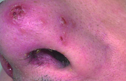
Figure 2: Mucocutaneous Leishmaniasis condition
3.3. Visceral Leishmaniasis (VL) or Kala-azar
Visceral leishmaniasis is the most severe and life-threatening form of the disease. It is caused predominantly by L. donovani (in Asia and East Africa) and L. infantum (in the Mediterranean and Latin America). The parasite disseminates through the bloodstream to infect vital organs including the spleen, liver, lymph nodes, and bone marrow (Singh et al., 2016). The clinical presentation includes prolonged fever, hepatosplenomegaly, pancytopenia, weight loss, and hypergammaglobulinemia. If left untreated, VL has a high fatality rate. Diagnosis is difficult in early stages due to non-specific symptoms and may require invasive procedures such as bone marrow or splenic aspirate microscopy, although rapid diagnostic tests like the rK39 immunochromatographic test are increasingly used (WHO, 2023). An important sequela of VL is Post-Kala-azar Dermal Leishmaniasis (PKDL), characterized by hypopigmented macules, papules, or nodules appearing on the skin after apparent cure of visceral disease. PKDL is especially common in Sudan and India and acts as a parasite reservoir, contributing to disease transmission (Zijlstra et al., 2003).
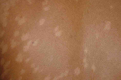
Figure 3: Visceral Leishmaniasis (VL) or Kala-azar condition
4. Diagnostic Approaches
Accurate and timely diagnosis of leishmaniasis is critical for effective treatment and control. However, the diversity in clinical manifestations, overlapping symptoms with other endemic diseases, and varying accessibility to diagnostic tools present substantial challenges, especially in resource-limited settings.
4.1. Clinical Diagnosis
Clinical suspicion of leishmaniasis arises from characteristic signs such as chronic skin ulcers (cutaneous leishmaniasis), mucosal disfigurement (mucocutaneous leishmaniasis), or prolonged fever with hepatosplenomegaly (visceral leishmaniasis), particularly in patients from endemic regions. However, these features are not pathognomonic and can mimic other infectious or inflammatory diseases, making clinical diagnosis alone insufficient (Reithinger et al., 2007).
4.2. Laboratory Tests
Microscopy
Direct microscopic examination of Giemsa-stained smears from skin lesions (for CL and MCL), bone marrow, splenic, or lymph node aspirates (for VL) remains a cornerstone of diagnosis. It enables identification of amastigote forms (Leishman-Donovan bodies). Although highly specific, sensitivity varies from 50–70% and depends heavily on sample quality and operator expertise (Singh et al., 2016).
Serological Tests
Serological assays such as the direct agglutination test (DAT) and rK39-based immunochromatographic tests are widely used for VL diagnosis. The rK39 test is particularly valuable due to its simplicity and high sensitivity (>90%) in South Asia, although its performance is less reliable in East Africa (Boelaert et al., 2014). Serology is less useful for CL and MCL due to the limited systemic immune response.
Molecular Diagnostics
Polymerase Chain Reaction (PCR)-based assays targeting kinetoplast DNA or ribosomal RNA are highly sensitive and specific, capable of detecting low parasite loads. PCR is valuable for species identification and confirming diagnosis in atypical or relapse cases. However, its application is limited in endemic areas due to the need for infrastructure and technical expertise (Cruz et al., 2013).
4.3. Point-of-Care Diagnostics in Endemic Areas
Rapid diagnostic tests (RDTs) like the rK39 strip test offer significant advantages in remote settings—being fast, user-friendly, and cost-effective. These tests enable prompt field-level screening and have become integral to kala-azar control programs in South Asia (WHO, 2023). Nevertheless, regional variability in test performance and cross-reactivity with other diseases remain limitations.
4.4. Limitations and Needs for Improved Diagnostics
Current diagnostic tools for leishmaniasis have several limitations:
There is an urgent need for non-invasive, highly sensitive, affordable, and field-adaptable diagnostic tools. Innovations like loop-mediated isothermal amplification (LAMP) and CRISPR-based diagnostics show promise but require further validation (Adams et al., 2021). An ideal diagnostic strategy would integrate clinical, serological, and molecular tools in a tiered approach tailored to regional needs.
5. Treatment Strategies and Challenges
The treatment of leishmaniasis is complex due to the diversity of its clinical forms, variability in drug response by region and parasite species, toxicity profiles, and increasing drug resistance. The development of effective and accessible therapies is essential to reduce disease burden and improve outcomes in endemic populations.
5.1. Current Treatment Options
Several antileishmanial drugs are currently in use, with selection depending on the type of leishmaniasis, species involved, patient status, and regional guidelines.
Table 1. Overview of Current Antileishmanial Drugs
|
Drug |
Mechanism of Action |
Form of Leishmaniasis |
Route |
Key Features |
|
Sodium stibogluconate |
Inhibits parasite metabolism |
CL, MCL, VL |
IV/IM |
First-line in many countries; resistance is rising |
|
Amphotericin B (Liposomal) |
Binds to ergosterol-like sterols in parasite membrane |
VL (esp. HIV co-infection) |
IV |
High efficacy, low relapse rate, high cost |
|
Miltefosine |
Disrupts membrane and mitochondrial function |
CL, VL |
Oral |
First oral drug; teratogenic, GI side effects |
|
Paromomycin |
Inhibits protein synthesis |
VL (used in India) |
IM |
Used in combination; less toxic, low cost |
|
Combination Therapy |
Multi-drug targeting different mechanisms |
CL, VL |
Varies |
Reduces duration, resistance risk, improves efficacy |
(Source: Sundar & Singh, 2018; WHO, 2023)
5.2. Treatment Limitations and Challenges
Despite available treatments, several challenges limit the success of current therapeutic strategies.
5.2.1. Drug Resistance and Relapse
Resistance to pentavalent antimonials is a major concern, particularly in regions like Bihar, India, where cure rates have declined significantly (Singh et al., 2012). Miltefosine resistance is also emerging due to widespread monotherapy use and long half-life.
5.2.2. Toxicity and Side Effects
Many antileishmanial drugs are associated with severe adverse effects. Amphotericin B (conventional) is nephrotoxic, while miltefosine can cause gastrointestinal disturbances and hepatotoxicity. Parenteral drugs can also cause injection site reactions and systemic effects (Croft et al., 2006).
5.2.3. High Treatment Cost
Liposomal amphotericin B, although safer and effective, is prohibitively expensive in many endemic regions. This restricts its widespread use despite being a preferred option (WHO, 2023).
5.2.4. Lengthy Treatment Duration
Traditional regimens often require prolonged hospitalization or repeated injections over weeks. This leads to poor patient compliance and increased risk of relapse or incomplete treatment (Olliaro et al., 2005).
5.2.5. Regional Variation in Drug Efficacy
Drug efficacy varies with Leishmania species and geographical location. For example, miltefosine shows high efficacy in India but reduced success rates in South America and East Africa due to parasite variability (Ponte-Sucre et al., 2017).
5.2.6. Access Issues in Endemic and Rural Settings
Health infrastructure in endemic areas is often inadequate. Lack of access to diagnostic and therapeutic facilities delays treatment initiation and contributes to morbidity and transmission (Alvar et al., 2006).
Table 2. Summary of Treatment Challenges in Leishmaniasis
|
Challenge |
Impact |
Potential Solution |
|
Drug resistance |
Reduced efficacy, relapse |
Use of combination therapy, drug rotation |
|
Toxicity |
Treatment discontinuation |
Safer formulations (e.g., liposomal amphotericin B) |
|
High cost |
Limited access to effective drugs |
Subsidized pricing, generic production |
|
Long treatment duration |
Poor compliance |
Development of short-course regimens |
|
Regional drug efficacy variation |
Inconsistent treatment success |
Region-specific protocols, molecular diagnostics |
|
Access in rural areas |
Delayed care, continued transmission |
Decentralized care, mobile clinics, point-of-care tools |
6. Emerging Therapeutic Approaches
Due to the limitations of conventional treatments—such as toxicity, resistance, and high cost—research is shifting toward innovative therapies for leishmaniasis. These include novel drug candidates, advanced drug delivery systems, immunotherapies, vaccines, and natural remedies. These emerging approaches aim to improve efficacy, reduce side effects, and target resistant strains.
6.1. New Drug Candidates and Clinical Trials
Several novel compounds are currently in preclinical and clinical development to address resistance and improve pharmacokinetic properties.
6.2. Nanotechnology and Targeted Drug Delivery
Nanotechnology offers precise drug delivery, improved bioavailability, and targeted accumulation at infected macrophages:
6.3. Immunotherapy and Host-Directed Therapy
Immunomodulatory strategies aim to boost host responses to enhance parasite clearance and prevent relapse:
6.4. Vaccines Under Development
Despite decades of research, no human vaccine is commercially available, but several candidates are in the pipeline:
6.5. Role of Traditional and Herbal Medicines
Ethnomedicine remains an underutilized area in anti-leishmanial therapy. Several plant-derived compounds exhibit antiparasitic activity:
7. Prevention and Control Strategies
Preventing and controlling leishmaniasis is a multifaceted challenge that requires a comprehensive and integrated approach. Strategies span vector control, reservoir management, public education, surveillance, and community-based interventions.
7.1 Vector Control
Controlling the sandfly population is the cornerstone of leishmaniasis prevention. Effective methods include:
Table 3. Common Vector Control Strategies and Their Effectiveness
|
Strategy |
Description |
Effectiveness |
Challenges |
|
Indoor Residual Spraying (IRS) |
Spraying homes with long-lasting insecticide |
High |
Insecticide resistance, coverage gaps |
|
Insecticide-treated nets (ITNs) |
Nets treated with pyrethroids |
Moderate |
Compliance, durability |
|
Environmental management |
Clearing vegetation, improving waste disposal |
Moderate |
Requires sustained community effort |
Insecticide resistance is a growing concern, especially in endemic areas in India, Brazil, and Sudan (Killick-Kendrick, 1999; WHO, 2022).
7.2 Reservoir Control
Animal reservoirs, particularly domestic dogs, play a significant role in transmission, especially for zoonotic species like Leishmania infantum.
Table 4. Reservoir Control Interventions
|
Intervention |
Target Animal |
Outcome |
Limitations |
|
Canine vaccination |
Dogs |
Reduced infection and transmission |
Limited availability and uptake |
|
Dog culling |
Dogs |
Temporary decline in cases |
Ethical concerns, low long-term impact |
|
Insecticide collars |
Dogs |
Significant reduction in sandfly bites |
Cost and need for regular replacement |
7.3 Community Awareness and Health Education
Health education and community participation are crucial in sustaining prevention efforts:
Studies have shown that communities informed about leishmaniasis are more likely to adopt preventive behaviors (Alvar et al., 2012).
7.4 Surveillance and Outbreak Management
Effective surveillance ensures timely identification of cases and outbreaks:
WHO's Global Leishmaniasis Surveillance system collects standardized data from over 90 countries, yet underreporting remains a challenge (WHO, 2022).
7.5 Integrated Disease Management Approaches
Integrated approaches combine multiple strategies for more effective and sustainable control:
8. Global Initiatives and Policy Frameworks
The global response to leishmaniasis as a neglected tropical disease (NTD) has gained momentum through coordinated strategies, frameworks, and policy initiatives led by international and national organizations. These efforts aim to reduce disease burden, improve access to diagnostics and treatment, and prioritize research for innovative interventions.
8.1 WHO Roadmap for NTDs
The World Health Organization (WHO) launched its "Ending the neglect to attain the Sustainable Development Goals: A roadmap for neglected tropical diseases 2021–2030", which prioritizes leishmaniasis control and elimination efforts. The roadmap emphasizes integrated approaches, country ownership, and innovation in diagnostics and treatment.
Key targets for leishmaniasis by 2030 include:
8.2 Sustainable Development Goals (SDGs) and Leishmaniasis
Leishmaniasis control is embedded within SDG 3: Good Health and Well-being, specifically Target 3.3, which aims to end the epidemics of AIDS, tuberculosis, malaria, and neglected tropical diseases by 2030.
The disease also intersects with:
Meeting SDG targets requires sustained political commitment, financial resources, and innovation in public health strategies (United Nations, 2015).
8.3 National and International Programs
Several global and regional programs support leishmaniasis elimination:
8.4 Funding and Research Priorities
Despite advancements, leishmaniasis remains underfunded. WHO and partners have highlighted priority areas including:
Table 5. Key Global Actors in Leishmaniasis Policy and Research
|
Organization / Program |
Role / Contribution |
Focus Area |
|
WHO |
Policy roadmap, global technical guidance |
Control & elimination targets |
|
DNDi |
Drug R&D, clinical trials, public-private partnerships |
Oral and affordable therapies |
|
G-FINDER |
Research funding tracker and gap analysis |
Advocacy and policy-making |
|
TDR (WHO/UNICEF/UNDP/World Bank) |
Capacity building, research funding |
Diagnostics and implementation research |
|
National Elimination Programs |
Country-level implementation and surveillance |
Diagnosis, treatment, and vector control |
9. Future Perspectives and Research Needs
Despite decades of research and some therapeutic advancements, leishmaniasis remains a formidable public health challenge. The future of effective control and elimination depends on addressing current gaps and innovating across scientific, clinical, and policy domains.
9.1 Gaps in Knowledge and Unmet Clinical Needs
Persistent issues such as limited understanding of host-pathogen interactions, immune evasion mechanisms, and species-specific responses to treatment present critical barriers to progress. Additionally, there's a lack of pediatric formulations, non-invasive diagnostics, and effective vaccines (Sundar & Singh, 2018).
9.2 Multidisciplinary Research Approaches
Integrating immunology, molecular biology, pharmacology, and epidemiology is essential to designing more effective interventions. Collaboration between public health experts, entomologists, molecular scientists, and data analysts can enhance prediction models, drug efficacy testing, and community-based implementation strategies (de Vries et al., 2015).
9.3 Role of Genomics, Proteomics, and Bioinformatics
High-throughput technologies like next-generation sequencing, proteomic mapping, and transcriptomics are shedding light on parasite biology, drug targets, and resistance genes. Bioinformatics-driven in silico modeling is also accelerating vaccine design and drug repurposing (Alvar et al., 2021).
9.4 Public–Private Partnerships for Drug and Vaccine Development
Organizations such as DNDi, Wellcome Trust, and Bill & Melinda Gates Foundation play vital roles in funding R&D for NTDs. Collaborative frameworks between academia, pharmaceutical companies, and governments are necessary to sustain pipelines for new antileishmanial agents and safe, effective vaccines (Ponte-Sucre et al., 2017).
CONCLUSION
Leishmaniasis remains a neglected tropical disease with diverse clinical manifestations, affecting millions in impoverished and rural settings. The burden is compounded by diagnostic limitations, drug resistance, treatment toxicity, and weak healthcare infrastructure. There is an urgent need for global attention, sustained investments, and interdisciplinary research. Only through a united effort involving governments, international agencies, academia, and the private sector can we move closer to elimination goals and equitable access to care for all affected populations.
REFERENCES

Gitesh Kumar, Parvez Khan, Dr. Aditi Kaushik, Arti Devi, Pratibha Choudhary, Ruchika Sharma, Dr. Amardeep Ankalgi, Dhiraj Kumar*, Dimple Kumari, Leishmaniasis: A Neglected Tropical Disease with Diverse Manifestations and Treatment Challenges, Int. J. of Pharm. Sci., 2025, Vol 3, Issue 5, 4866-4880. https://doi.org/10.5281/zenodo.15548213
 10.5281/zenodo.15548213