
We use cookies to ensure our website works properly and to personalise your experience. Cookies policy
RJS College of Pharmacy, Kokamthan, Kopargaon.
Microneedle technology has surfaced as a promising method for transdermal drug delivery, aimed at addressing the shortcomings of oral and hypodermic methods such as low bioavailability, first-pass metabolism, discomfort, and risks associated with needles. By creating miniature channels through the stratum corneum, microneedles facilitate painless and minimally invasive administration of various therapeutics, including vaccines, peptides, proteins, biomolecules, and cosmeceuticals. Their distinctive designs—solid, coated, hollow, and dissolving or biodegradable—allow for both rapid and sustained drug release.[12] Progress in microfabrication techniques and biomaterials has enhanced the mechanical strength of microneedles, their penetration efficiency, and their controlled release capabilities, enabling applications that range from small-molecule drugs to larger macromolecules. This review aims to underscore recent developments in microneedle delivery systems, focusing on fabrication methods, material advancements, therapeutic uses, and safety aspects. Research indicates that microneedles significantly improve skin penetration, extend drug release duration, enhance stability, and boost patient compliance when compared to traditional methods. Additionally, microneedles eliminate sharp waste and mitigate needle-related anxiety, making them an appropriate choice for self-administration. Nonetheless, challenges persist, including the delivery of large biomolecules, ensuring consistent penetration through the skin, addressing large-scale production complexities, and fulfilling regulatory standards. Overall, microneedles serve as an innovative, safe, and user-friendly platform with considerable potential for future clinical applications.[3]
In recent years, transdermal drug delivery systems (TDDS) have received a lot of attention Many medications have been developed that can be injected into the bloodstream straight via the skin. In 1979, the Food and Drug Administration (FDA) granted FDA approval for the first transdermal patch. An anti-motion sickness patch was used to apply to the skin. The FDA authorized four nicotine patches between the end end of 1991 and the beginning of 1992 after the pharmaceutical firms began developing a nicotine patch to assist smokers stop smoking in the mid-1980s [1] Controlled drug release and painless administration are the key benefits of this approach. A Transdermal patch clings to the skin and delivers the medicine to the skin. When using a Transdermal Patch, there are various components that play an important part in the delivery of the medication through the skin, such as liners, adherents, and drug reservoirs. Transdermal patches are available in a variety of forms and may be applied in a variety variety of ways. To tak take advantage of its many benefits, it has become one of the most researched areas in the pharmaceutical industry the benefits and drawbacks of the transdermal patch, as well as the ways of applying, the care that must be taken when applying, the kinds and applications of transdermal patch, and new patents and market market items, have been reviewed here. Evidence of percutaneous medication absorption may be detected in blood levels of the drug and its metabolites, as well as in the patient's clinical reaction to the pharmacological treatment.[3]
History of patch according to generation
1.1 First-Generation Delivery Systems
Transdermal Clinical usage of tiny, lipophilic, low-dose medicines has continued to rise steadily over the last several years.
1.2 Second-Generation Delivery Systems
There There are are additionally therapeutic solutions that have been developed employing chemical enhancers, noncavitational ultrasound, and Jontophoresis. The capability to regulate delivery rates in real time gives further usefulness.
1.3 Third-Generation Delivery Systems :
Microneedles, thermal ablation, microderm abrasion, electroporation, and cavitational ultrasound are used to target the stratum comeum's barrier layer. Insulin, parathyroid hormone, and influenza vaccine are among the macromolecules and vaccines now being tested using microneedles and thermal ablation. When second and third generation transdermal delivery augmentation technologies are used, their influence influence on medicine will soar.There has been a rise in interest in Transdermal over the last several years due to a number of factors, including new clinical data from technology firms and pharmaceutical companies. looking for new ways to prolong patents on their[1,15]
Skin Anatomy and Transdermal Drug Delivery Systems:
The skin is a complex organ that functions as both a protective barrier and a potential drug delivery route. It is composed of three main layers: the epidermis, dermis, and hypodermis. The epidermis is the outermost layer (≈150–200 µm). Its top portion, the stratum corneum (10–20 µm), is formed by dead keratinized cells embedded in lipid bilayers. This thin but highly resistant layer is the primary barrier to transdermal drug absorption, allowing only small, lipophilic molecules (<500 Da) to pass through. Beneath it, the viable epidermis (stratum granulosum, spinosum, and Basale) contains living cells but no blood supply. The dermis (1.5–3 mm) is composed of collagen, elastin, and connective tissue. It houses blood capillaries, lymphatic vessels, and sensory nerves, making it the key site for systemic absorption once drugs penetrate the epidermis. The hypodermis (3–100 mm) consists mainly of adipose tissue and provides cushioning, insulation, and attachment to deeper Tissues. Conventional transdermal systems struggle to cross the stratum corneum, restricting drug types. Microneedles (MNs) overcome this challenge by creating microscale channels across the outer barrier without reaching nerve-rich regions, thereby avoiding pain and bleeding. This technique enables efficient delivery of macromolecules such as peptides, proteins, and vaccines, expanding the scope of transdermal therapy.[3]
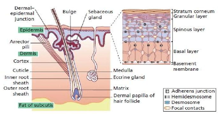
Figure: structure of skin and routs of penetration of a molecule across the stratum corneum
Microneedle: Transdermal drug delivery has long been studied as an alternative to conventional routes, but the presence of the stratum corneum acts as a major barrier to drug permeation. To overcome this limitation, microneedles (MNs) have been developed as a novel approach in pharmaceutical technology.[12] Microneedles are micron-sized projections, generally ranging from a few hundred micrometres in length, which can pierce the outer skin layers to create temporary microchannel that facilitate the passage of therapeutic agents. Microneedles can be fabricated in various forms such as solid, hollow, coated, and dissolving systems, each designed for specific applications in drug and vaccine delivery. Advanced techniques including micromolding, lithography, and 3D printing have enabled the development of precise and reproducible microneedle arrays suitable for both research and clinical use. In recent years, microneedle-based systems have gained significant attention in pharmaceutics, not only for their potential in drug administration but also for their applications in diagnostics and cosmetic treatments. Their growing role highlights the importance of continued research and development in this field, marking Microneedles as an emerging platform in modern drug delivery systems.[3]
The microneedle concept was proposed in the 1970s. The first Microneedles reported in literature were developed by Hashmi et al for intracellular delivery and gene transfection. Since the first studies of enhancement of transdermal delivery by Henry et al in 1998, there has been rapid increase in interest by the microfabrication industry to develop novel fabrication technology for fabricating Microneedles for pharmaceutical applications.[16] Micro-electromechanical systems are used for making Microneedles from a range of substrate materials including silicon, metals, polymers, titanium, glass and sugar. Microneedles may vary according to their tip shape, example volcano like micro hypodermis and snake fang design or the overall shape example pyramidal, spiked, candle like and spear shaped structures. Micro-electromechanical systems are also useful in preparing other delivery devices like micro pumps, micro valves, implantable microchips and self-regulated micro devices. The micromachining process includes three major steps, i.e., deposition of material, patterning process for incorporation of desired micro features and removing (or etching) portions of material.[17]
Ideal Characteristics of Microneedles
1. Mechanical Strength & Insertion Ability:
Must be capable of insertion deep into the skin without breaking. Require sufficient rigidity to penetrate the stratum corneum but should minimize tissue damage.
2. Size & Geometry
Sharp enough to pierce skin effectively but short enough to avoid reaching deeper nerves (painless application). Can have shapes like triangular, rounded, or arrow-shaped tips.
3. Drug Delivery Efficiency
Should allow controlled and predictable drug release at a definite rate. Ability to localize drug at the site of action, enhancing bioavailability.
4. Biocompatibility & Safety
Made from safe materials such as silicon, metals (stainless steel, nickel, palladium, cobalt), biodegradable polymers, or glass. Must not cause irritation, toxicity, or immune response.
5. Fabrication Properties
Rugged and reproducible. Manufactured with optimum size to prevent bending or breaking. Should support large-scale production with consistent quality.
6. Pain Minimization
Penetration should be painless or cause minimal discomfort, unlike hypodermic needles. Shorter length ensures less nerve stimulation.
7. Drug Compatibility
Should be capable of delivering both hydrophilic and hydrophobic drugs. Should not degrade or chemically interact with loaded drugs.
8. Performance in Patches
Advantages of Microneedles
1. Minimally invasive – Microneedles penetrate only the superficial layers of the skin, avoiding pain and tissue damage associated with conventional hypodermic needles.
2. Improved patient compliance – Since the application is almost painless and easy to use, patients are more likely to accept therapy.
3. Bypassing first-pass metabolism – Drugs delivered through microneedles avoid degradation in the gastrointestinal tract and hepatic metabolism, improving bioavailability.
4. Self-administration – Microneedle patches can be applied without clinical supervision, reducing the need for hospital visits.
5. Enhanced drug delivery – Suitable for both small molecules and biologics (proteins, peptides, vaccines) that are difficult to administer orally.
6. Reduced risk of infection – Compared to traditional injections, microneedles cause minimal bleeding and reduce cross-contamination chances.
7. Controlled release – Microneedles can be designed to provide rapid or sustained release depending on the formulation.
8. Stability – Solid or coated microneedles allow drugs, especially vaccines, to remain stable at room temperature, reducing cold chain requirements.
Disadvantages of Microneedles
1. Limited dose capacity – Only a small amount of drug can be loaded compared to oral or injectable routes.
2. Skin variability – Thickness, hydration, and elasticity of skin can affect penetration and drug absorption.
3. Manufacturing challenges – Precise fabrication of microneedles requires advanced technology, increasing cost.
4. Regulatory hurdles – Being a relatively new dosage form, long-term safety data and standardized approval guidelines are still developing.
5. Risk of needle breakage – If poorly designed, microneedles may break and remain embedded in the skin.
6. Irritation or allergy – Some patients may experience redness, itching, or allergic reactions at the application site.
7. Not suitable for all drugs – Molecules requiring very high doses or those unstable at the skin surface are difficult to deliver
|
Parameter |
Conventional dosage forms (Tablet, capsules, syrup, injection) |
Microneedle Drug Delivery System (MNs) |
|
Drug release |
Immediate, uncontrolled |
Controlled, sustained and localized |
|
Bioavailability |
Oral forms suffer from first pass metabolism injections bypass but are painful |
High bioavailability by bypassing first pass effect |
|
Patient compliance |
Oral forms easy but frequent dosing injection painful |
Painless, minimally invasive, self-administrable |
|
Target specificity |
Non-specific, systemic exposure |
Targeted delivery through skin layers |
|
Side effects |
Higher due to systemic circulation |
Reduced due to localized delivery |
|
Stability of Drug |
Simple cost, effective |
Biologics (proteins, vaccines, peptides, protected) |
|
Application |
Mass use for acute illness |
Vaccination, insulin, cancer therapy, gene/drug delivery |
Comparison of Conventional Dosage Forms vs Microneedle Drug Delivery Systems
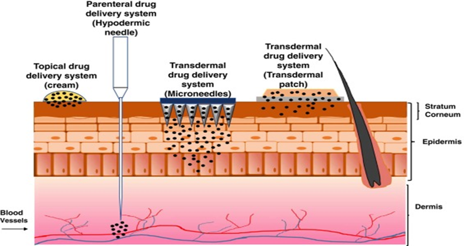
Figure: comparison of drug delivery of microneedle and hypodermic needle
Recent Approaches in the Drug Delivery System:
Microneedle (MN) technology is one of the most promising recent advances in drug delivery. Microneedles are micron-sized projections (25–1000 μm) that puncture the stratum corneum without injuring nerves or blood vessels, making the process painless and minimally invasive. they overcome the limitations of oral and injectable routes by bypassing first-pass metabolism, avoiding gastrointestinal degradation, and improving bioavailability. Microneedle also reduce needle phobia, allow self-administration, and enable sustained drug release. In vaccination, Microneedles deliver antigens into the dermis where they are processed by Langerhans cells and dendritic cells, activating T cells and B cells to trigger strong immune responses. Thus, microneedles provide a safe, effective, and patient-friendly approach for vaccines, chronic disease management, and localized therapies, representing a revolutionary step in modern drug delivery system.[12]
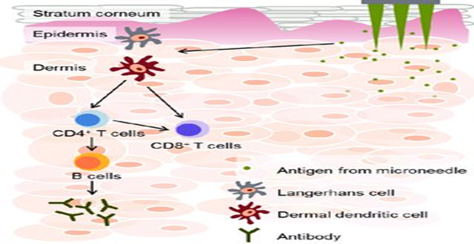
Figure 2: Working of microneedle
Type:
1. Solid Microneedle- Solid microneedles are among the earliest forms of microneedle systems, typically ranging from 50–900 µm in length, designed to penetrate the stratum corneum and create microchannels that facilitate transdermal drug delivery. They function via the “poke and patch” approach, wherein the microneedles first disrupt the skin barrier and a drug formulation, such as a patch, gel, or cream, is subsequently applied over the treated site for diffusion. Fabricated from metals, polymers, ceramics, or silicon through techniques like micro-molding, lithography, or laser cutting, solid microneedles have been explored for the administration of vaccines, insulin, anesthetics, and cosmetic treatments.[15] Their advantages include simple design, low production cost, and strong mechanical strength. However, limitations such as the requirement of a two-step process, rapid closure of microchannels, risk of infection, and possible needle breakage reduce their clinical convenience compared to coated, hollow, or dissolving microneedles.
2. Coated Microneedle: Coated microneedles are solid microneedles whose surfaces are coated with a thin layer of drug formulation. When inserted into the skin, the drug coating quickly dissolves in the interstitial fluid, releasing the active substance into the epidermis or dermis. They allow precise delivery of small drug doses, especially vaccines, peptides, and proteins, and are suitable for rapid onset of action Coated microneedles are simple, avoid the two-step “poke and patch” process, and provide better patient compliance compared to solid microneedles. However, their major limitation is the limited drug-loading capacity due to the small coating surface area.[12,15]
3. Hollow Microneedle: Hollow microneedles are tiny needles with a central lumen (hollow bore) that allow liquid drugs to be directly injected into the skin layers, similar to miniature hypodermic needles. Unlike solid or coated microneedles, they can deliver larger drug volumes, continuous infusions, or even viscous formulations. They are particularly useful for vaccines, insulin, and biologics that require precise and controlled dosing. Their advantages include the ability to bypass the stratum corneum barrier and deliver macromolecules in a single step. However, challenges such as complex fabrication, risk of needle clogging, and higher cost limit their widespread us.
4. Dissolving Microneedle: Dissolving microneedles are fabricated from biodegradable and water-soluble polymers such as hyaluronic acid, polyvinylpyrrolidone (PVP), and carboxymethylcellulose (CMC). When applied onto the skin, these microneedles pierce the stratum corneum and dissolve upon contact with interstitial fluid, thereby releasing the encapsulated drug directly into the epidermis or dermis. Since the microneedles completely dissolve, there is no generation of sharp waste, making the system safer and more patient friendly. They are especially useful for vaccines, peptides, proteins, and small-molecule drugs, offering painless delivery, improved compliance, and reduced risk of infection. However, limitations include relatively low drug loading capacity and the need for sufficient mechanical strength to penetrate the skin.[9]
5. Hydrogel-Forming Microneedles : Hydrogel-forming microneedles are prepared from cross-linked, hydrophilic polymers that swell upon absorbing interstitial fluid after insertion into the skin. Unlike dissolving microneedles, they do not release the drug by degrading but instead create aqueous microchannels, enabling drugs to diffuse from an attached reservoir or patch into deeper skin layers. These systems are advantageous as they are biocompatible, leave no polymer residue, and can provide controlled and sustained drug delivery. They have shown great potential for delivering peptides, proteins, vaccines, and long-acting therapeutics. However, challenges such as maintaining mechanical strength and optimizing swelling behavior still need to be addressed.
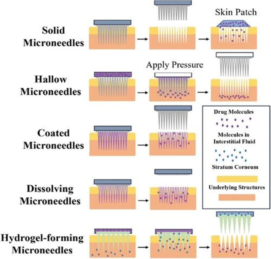
figure: Type of Microneedle
Applications of Microneedle Dosage Forms
1. Dermatological Applications : Microneedles are widely used in dermatology for the treatment of psoriasis, acne, scars, and even skin cancers. By creating microchannels in the stratum corneum, they enable localized delivery of corticosteroids, retinoids, and anticancer drugs directly to the affected skin layers. This improves drug penetration and therapeutic efficacy while reducing systemic side effects compared to conventional topical or oral therapy .
2. Vaccination and Infectious Diseases. : One of the most promising applications of microneedles is in vaccine delivery. Microneedle patches have been developed for vaccines against influenza, hepatitis B, measles, and most recently COVID-19. Since the skin is rich in antigen-presenting cells, vaccines delivered via microneedles induce a strong immune response even with smaller doses. Additionally, they are suitable for self-administration and mass immunization, particularly in resource-limited settings[4] (Prausnitz & Langer, 2008; Arya et al., 2017).
3. Endocrine and Metabolic Disorders : In diabetes management, dissolving microneedle patches have shown excellent potential for insulin and glucagon-like peptide-1 (GLP-1) agonist delivery. Unlike conventional insulin injections, microneedle provide a minimally invasive and painless alternative, allowing controlled and sustained release of the drug. This not only enhances patient comfort but also improves compliance and reduces the burden of frequent injections (Ling et al., 2021).
4. Cardiovascular Diseases : Microneedle are explored for delivering cardiovascular drugs such as propranolol, carvedilol, and heparin. They enable transdermal drug transport with controlled and sustained release, ensuring stable plasma drug concentrations. This approach reduces the limitations of oral therapy such as first-pass metabolism and gastrointestinal irritation, thus enhancing bioavailability and patient compliance ( yuqi Zhang et al., 2021).
5. Oncology (Cancer Therapy) : Microneedle are increasingly used in cancer therapy for localized delivery of chemotherapeutic agents such as doxorubicin and cisplatin. By targeting drugs directly to the tumor site, microneedle reduce systemic toxicity and adverse effects commonly associated with chemotherapy. They also allow controlled release, which improves drug retention in tumours and enhances therapeutic outcomes ( Xintong Li et al., 2023).
6. Neurological Disorders : For central nervous system disorders, microneedle offer a potential route to bypass the blood–brain barrier. Drugs for Alzheimer’s, Parkinson’s, and migraine therapy can be delivered intradermally or intranasally using microneedle, improving bioavailability and therapeutic efficacy. This method represents a minimally invasive alternative to systemic administration, which often suffers from limited brain penetration (shuna Tan et al.Int J pharm 2024).
7. Ophthalmic Drug Delivery : Microneedle are being studied for ocular diseases such as glaucoma, age-related macular degeneration, and uveitis. By directly delivering drugs into the corneal or scleral tissue, microneedle improve local bioavailability and therapeutic efficiency compared to conventional eye drops, which suffer from poor penetration and rapid clearance (Thakur et al., 2016).
8. Reproductive and Hormonal Applications : Microneedle patches have been investigated for long-acting contraceptive delivery of hormones such as levonorgestrel and etonogestrel. These patches ensure sustained drug release over weeks or months, reducing the need for daily dosing. Such applications are particularly beneficial in improving compliance and accessibility in reproductive health (Baek et al., 2017).
9. Respiratory Disorders : Recent research explores microneedle-assisted formulations for respiratory diseases like asthma and COPD. Microneedle patches can deliver bronchodilators and corticosteroids transdermally, bypassing the need for inhalers, which often require proper technique for effectiveness. This innovation could improve management of chronic respiratory conditions (Baek et al., 2017).
Drug Selection Criteria for levonorgestrol in microneedle patch:
Drug profile of Levonorgestrel for microneedle patch: ( hormonal contraceptive)
|
Parameter |
Levonorgestrel profile |
Relevance for microneedle patch |
|
Drug class |
Synthetic progesterone (second generation progestin) |
Widely used and safe contraceptive |
|
Molecular weight |
~312.4 Dalton |
Small-suitable for skin permeation via microneedle |
|
Log p( lipophilicity) |
~3.3(moderately lipophilic) |
Ensures balanced partitioning across skin layers |
|
Aqueous solubility |
Very low(<0.01mg/ml) |
Limitation; can be improved with liposomes,cyclodextrins, polymer |
|
Half- life (t1/2) |
20-30hours |
Support sustained /controlled release |
|
Therapeutic plasma level |
~200-300 pg/ml |
Low plasma requirement achievable with microneedle patches |
|
Daily dose requirement |
30-40microgram/ day |
High potency fits microneedle limited loading |
|
Current dosage forms |
Oral pills,subdermal implants, emergency contraceptive |
Microneedle patch offers non-invasive , user-friendly option |
|
Mechanism of action |
Inhibits ovulation, thickens cervical mucus, alters endometrium |
Proven contraceptive mechanism , effective at low dose |
|
Stability for microneedle patch |
Chemically stable under storage /fabrication condition |
Ensures reliable microneedle formulation |
|
Suitability for microneedle patch |
High potency, long half- life low dose, stable |
Strong candidate for sustained – release microneedle contraception |
Levonorgestrel is considered an appropriate candidate for microneedle delivery because it fulfills the main requirements of drug selection for this system. Its low molecular weight (312 Da) and moderate lipophilicity (log P ~3.3) allow it to diffuse efficiently once delivered across the skin barrier. The drug is highly potent, with contraceptive efficacy achieved at microgram doses, which fits the limited loading capacity of microneedle arrays. In addition, levonorgestrel has a long elimination half-life (20–30 hours), which supports the design of sustained and controlled release formulations capable of maintaining therapeutic plasma levels (~200–300 pg/mL) for several weeks to months. The compound is also chemically stable during fabrication and storage, and its safety and effectiveness are well established through decades of clinical contraceptive use. A limitation is its low aqueous solubility, but this can be addressed by using carriers such as liposomes, cyclodextrins, or polymer-based matrices, ensuring a consistent sustained release profile when delivered via microneedle patches.
Composition and Excipients Used in Levonorgestrel Microneedle Patch –
|
Components |
Example |
Function in microneedle formulation |
Relevance for Levonorgestrel |
|
Polymeric matrix (structural Base) |
PVP, PVA, CMC, hyaluronic acid, chitosan |
Provide mechanical strength, forms dissolving matrix, controls release rate |
Ensures penetration into skin and drug embedding; choice affects dissolution and release profile |
|
Biodegradable polymers |
PLGA,PLA |
Slow- degrading matrix for sustained release |
Suitable for long- acting contraceptive Levonorgestrel delivery |
|
Plasticizers/ mechanical modifiers |
Glycerol,PEG400, Sorbitol |
Reduce brittleness, improve flexibility and insertion |
Maintains integrity during storage and insertion |
|
Solubilizers/ Dispersion aids |
Cyclodextrins,tween80, Poloxamers, ethanol, (Process aid) |
Enhance solubility of lipophilic Levonorgestrel, promote uniform drug distribution |
Improves Levonorgestrel loading and prevents crystallization in polymer matrix |
|
Stabilizers/ Lyoprotectants |
Trehalose, mannitol, Sucrose |
Protect drug during drying, improve storage stability |
Prevents Levonorgestrel degradation during processing |
|
Permeation Enhancers (optional) |
Oleic acid , propylene glycol, short- chain fatty acids |
Enhance drug transport across skin |
May be used if deeper penetration is required must assess irritation risk |
|
Backing layer/ adhesive |
Silicone adhesive films, PSA (pressure – sensitive adhesives) |
Provides patch adhesion, prevents drug loss to backing side |
Ensures unidirectional delivery into skin |
|
Release modifiers |
Blends of fast- and slow dissolving polymers, drug loaded nanoparticles |
Tailor drug release kinetics |
Enables either rapid or controlled Levonorgestrel release |
In making Levonorgestrel microneedle patches, the choice of excipients is very important because they decide how well the patch works. The excipients must give enough strength for the microneedle to enter the skin, improve the solubility of the drug, keep the formulation stable during preparation and storage, and control how the drug is released. Fast-dissolving polymers like PVP, PVA or hyaluronic acid help in quick release, while biodegradable materials such as PLGA are useful for long-term delivery. Extra agents like Solubilizers, sugars, or backing layers further improve safety and performance. By selecting the right combination, the patch can provide reliable contraception, better patient comfort, and long-lasting action ( Rajput et al., 2022).
Method :
Preparation of Liposomes: Liposomes are nanoscale or microscale vesicles made from phospholipid bilayers that can entrap both hydrophilic and lipophilic drugs, making them attractive carriers for Levonorgestrel. Their preparation generally involves dissolving phospholipids and cholesterol, along with the drug, in a suitable organic solvent such as ethanol or chloroform. The solvent is removed under reduced pressure, forming a thin lipid film on the container surface. When this film is hydrated with an aqueous buffer, the lipids self-assemble into multilamellar vesicles. To improve uniformity and reduce vesicle size, techniques such as sonication or extrusion through polycarbonate membranes are applied, producing small unilamellar vesicles. In some cases, solvent injection is used, where a lipid solution in ethanol is injected into an aqueous buffer to trigger vesicle formation. Another approach is reverse-phase evaporation, which creates large unilamellar vesicles with higher entrapment capacity. Regardless of the method, the prepared liposomes are further analyzed for particle size, zeta potential, encapsulation efficiency, and stability. For Levonorgestrel delivery, liposomes not only enhance solubility and protect the drug from degradation but also allow sustained release, which makes them highly compatible with microneedle patch systems.[15,10]
Characterization of Levonorgestrel-Loaded Liposomes
Levonorgestrel-loaded liposomes are designed to improve the delivery and sustained release of the drug. After making the liposomes, it is important to check their properties to ensure they work effectively.
Particle size is measured because it affects how well the liposomes circulate in the body; smaller liposomes are usually more stable and deliver the drug better. This is often done using dynamic light scattering. Surface charge, or zeta potential, tells us how stable the liposomes are; liposomes with higher negative or positive charges do not clump together easily. The amount of drug inside the liposomes is called encapsulation efficiency, were calculated using equation 1&2 respectively [8]
Drug loading =total drug amount – free drug amount /total lipid amount ×100
Entrapment efficiency= total drug amount – free drug amount/total drug amount ×100
and it is calculated as the percentage of drug successfully loaded compared to the total used. The shape and structure of liposomes are checked using electron microscopy, which shows if they are round and smooth. Finally, drug release and stability are studied to see if Levonorgestrel is released slowly over time and if the liposomes maintain their size and structure during storage. These tests together ensure that the liposomes are safe, stable, and effective for delivering Levonorgestrel (Rajput et al., 2022).
Fabrication of Levonorgestrel Microneedle Array Patch
The fabrication of a Levonorgestrel microneedle patch starts with selecting a suitable biocompatible polymer, such as PVP, PVA, or HPMC, which can form either dissolving or solid microneedle. The drug formulation, often containing Levonorgestrel-loaded liposomes, is mixed thoroughly with the polymer to ensure the drug is evenly distributed throughout the microneedle matrix. This mixture is then carefully poured into a microneedle mold that has tiny needle-shaped cavities. To make sure the mixture fills every cavity and does not trap air bubbles, the mold is usually subjected to centrifugation or vacuum application. After the mold is filled, the mixture is dried under controlled temperature and humidity, allowing the microneedle to solidify and maintain their shape. Drying can take several hours depending on the type of polymer used. Once dried, the microneedle patch is gently removed from the mold and inspected to ensure all needles are sharp, uniform, and structurally intact. Often, a flexible backing layer is added to the patch to support handling and application. Proper fabrication is critical because it ensures that the microneedle can penetrate the skin effectively, release Levonorgestrel in a controlled manner, and provide consistent contraceptive action. Each step—from polymer selection, drug mixing, mold filling, drying, to mold removal—directly affects the performance, stability, and effectiveness of the final microneedle patch (Rajput et al., 2022
list of evaluation parameters for Levonorgestrel microneedle patch:
RESULT :
From the collected studies, it is clear that microneedle patches are strong enough to cross the skin barrier and can deliver drugs in a controlled way. They improve drug penetration, reduce pain compared to injections, and provide better patient comfort. In the case of Levonorgestrel, microneedle patches were found to release the drug slowly and maintain steady levels in the body, which is useful for long-term contraception. Overall, research shows that microneedle systems are effective for different drugs and therapeutic areas.
DISCUSSION:
Microneedle technology has gained attention as an effective alternative to conventional drug delivery methods. The main advantage is its ability to cross the stratum corneum barrier without causing pain, unlike injections. This makes the system more acceptable to patients and useful for long-term therapies. From the literature, it is clear that microneedle can deliver a wide range of drugs including hormones, vaccines, anticancer agents, and peptides. For contraceptive applications, Levonorgestrel-loaded microneedle patches show promising results. They provide sustained release, maintain steady plasma concentrations, and improve patient compliance compared with oral tablets or implants. This suggests that microneedle patches could be a reliable option for long-acting contraception. At the same time, some limitations remain. The amount of drug that can be loaded is restricted, and large-scale manufacturing requires cost-effective techniques. Long-term safety and regulatory approval are also important challenges that need further study. Despite these issues, the overall evidence shows that microneedle represent a strong future platform for painless, controlled, and patient-friendly drug delivery.
Marketed Example:
|
Brand name |
Company |
Strength |
Cost |
|
Conscious chemist Dissolving Microneedle patch® |
Conscious chemist, India |
2%salicylic acid + azelaic acid + niacinamide( Acne management) |
?449 (6 patches, widely available in India) |
|
Mighty patch micropoint for dark spots® |
Hero cosmetics, USA |
395 Microneedles with tranexamic acid vitamin c, niacinamide, licorice root |
~USD $13-15 (1500-2000 rupees, import) |
|
Acropass soothing Q® |
Acropass, South Korea |
Anti- itch microneedle patch |
~Rupees 5800 (12 patches, import) |
|
Acropass trouble cure® |
Acropass, south korea |
Hyaluronic acid+ niacinamide Microneedles ; salicylic acid swab(Acne treatment) |
Rupees 1200 (local seller) Rupees 5600 (import packs) |
|
Peace out wrinkles® |
Peace out skincare, USA |
~450 microneedle with hyaluronic acid, retinol, vitamin c , peptide |
~USD $ 28 ( Rupees 4000-4200, 6 patches imported in india) |
|
Dermaroller® microneedle patch |
Derma spark, canada |
Stainless steel/ polymer microneedle patches |
~USD $30-50 (2400-4200) |
Recent Research on Levonorgestrel Microneedle Patches
1. Effervescent PLGA Microneedle Patch
A patch prepared with biodegradable PLGA needles and an effervescent base can insert into the skin and detach within one minute. Animal experiments showed that a single patch could maintain LNG levels high enough for pregnancy prevention for almost one month. A placebo version tested among women in India, Nigeria, and the USA indicated strong user acceptance compared with oral or injectable methods.
2. Liposome-Based Microneedle System (India Contribution)
Researchers at IIT Bombay developed a patch where LNG was loaded into liposomes before being incorporated into microneedles. This method increased drug entrapment and provided a smoother release pattern. In vitro and animal studies confirmed that this system could deliver LNG consistently for about 28 days, showing higher efficiency than plain drug-loaded patches.
3. Silk Fibroin Microneedles
Another innovation involved silk fibroin, a natural protein with high stability at room temperature. When LNG was directly added to the silk microneedles, release lasted for around 100 days. When the drug was first packed into silk microparticles and then inserted into the needles, release extended beyond one year, offering the possibility of a long-acting, low-maintenance contraceptive.
4. Core–Shell Microneedle Design
To further extend duration, researchers created a multi-layer microneedle with a PLGA core (holding LNG), surrounded by stronger polymer shells. This controlled the release profile, reduced the “burst effect,” and achieved sustained release for over six months, making it a potential substitute for implants.
5. Three-Layer Microneedle Array (TIMN)
A more recent design used three polymer layers, providing strength and skin safety. About one-fourth of the drug was released on the first day, while the rest was delivered gradually over two weeks. Animal studies confirmed that this system was well tolerated without causing irritation or damage
CONCLUSION
Microneedle patches with levonorgestrel are a modern and convenient method of contraception. They are easy to use, cause little or no pain, and can release the drug steadily for a long time. Research so far is encouraging, and these patches may become a safe and effective alternative to pills or injections in the future.
REFERENCES

Sakshi Ghorpade, Aparna Sawant, Shruti Gosavi, Hrutuja Chavan, Amrita Singh., Microneedles: A Smart Approach for Advanced Drug Delivery – A Review, Int. J. of Pharm. Sci., 2025, Vol 3, Issue 12, 3058-3072. https://doi.org/10.5281/zenodo.18000194
 10.5281/zenodo.18000194