
We use cookies to ensure our website works properly and to personalise your experience. Cookies policy
St. Thomas College, Bhilai, Chhattisgarh, India
Nanomedicine brought a wise revolution in the field of medical biology which is significantly affecting the treatment of various major diseases like cancer, neurological disorders and cardiovascular diseases. The usage of different nanoparticles in the field of nanomedicine for diagnosis, monitoring, control, prevention and treatment of diseases is increasing day by day. Cancer remains a highly lethal disease and it is the second leading cause of death throughout the world. Traditional Cancer treatment and conventional immunotherapeutic approaches have inherent limitations that cannot properly distinguish the malignant cells from the healthy ones. Most importantly, both conventional chemotherapy and several immunomodulators in non-targeted cancer therapies often remove tumor cells at the expense of numerous normal cells, causing undesirable and sometimes fatal side effects. These limitations of cancer treatments have increased the interest in the field of nanomedicine to develop more effective and safer cancer therapy. The role of nanoparticles in cancer therapeutics has been to improve the overall therapeutic index by improving drug pharmacokinetics and reduce the systemic toxicities of chemotherapies by selectively targeting and delivering anticancer drugs to neoplastic tissues. Additionally, Nano-sized carriers effectively used for nanomedicine can be conjugated with specific ligand which specifically target malignant cells. Various nanoparticles like polymeric nanoparticles, dendrimers, liposomes, quantum dots, and metal nanoparticles (especially gold) are being explored and clinically approved for cancer therapy, imaging, and diagnostics. There are many nanotechnology-enabled therapeutic modulators which are currently under clinical inspection like for instance; chemotherapy, hyperthermia, radiation therapy, gene or RNA interference (RNAi) therapy and immunotherapy which can be probably become future therapeutic strategies for cancer.
Cancer is a condition where certain cells in the body begin to grow uncontrollably and can spread to other parts. According to the most recent Global Cancer Statistics, about 19.3 million new cancer cases and nearly 10 million deaths were reported globally in 2020. Factors like population growth, pollution, lifestyle changes, and increasing exposure to risk factors are expected to cause a sharp rise in cancer cases over the next 20 years. Cancer can develop in almost any part of the body. Although it is not contagious, the number of new cases continues to rise each year. Despite significant progress in science and medicine, cancer still has limited treatment options and remains one of the top causes of death worldwide, but early detection of cancer significantly increases the chances of complete recovery from the disease [1]. There are several conventional cancer therapies, such as MRI, laboratory test, pathological characteristic analysis, morphological analysis of tumour cell and tissue, surgery, chemotherapy and radiotherapy. These approaches have significantly contributed to increased survival rates in many cancer patients. However, their effectiveness is often limited when dealing with advanced or metastatic cancers. Among all of these techniques, chemotherapy is most widely used due to its simplicity and convenience in treating cancer, but it has many significant drawbacks as it can induce cytotoxicity, damaging not only cancer cells but also healthy cells [2]. It also induces multidrug resistance. Also, the drugs used in chemotherapy are nonspecific, have short life span and poor solubility, this attribute negatively affects the working potential of chemotherapy [3].
To overcome the drawbacks and damages caused by chemotherapy, nanoscience and nanotechnology have emerged as promising tools for cancer diagnosis and treatment. The term “nano” is derived from Greek word which means “dwarf” or something very small and depict one thousand millionth of a meter (10-9)[4]. Nanoparticles exhibit unique optical, magnetic, electronic, and structural properties. A diverse array of nanomaterials ranging from organic and inorganic substances to lipids, proteins, glycans, and synthetic polymers are utilized in the development of cancer treatments. Among these, nanoparticles (NPs) hold a significant position in diagnosis and therapy of cancer. Nanoparticle are small particles, having diameter of less than 100 nm and cannot be seen through naked eyes [2]. Their unique properties make them ideal candidates for medical applications through the field known as nanomedicine. Nanomedicine refers to the application of nanotechnology in healthcare for the treatment, diagnosis, monitoring, and regulation of biological systems. It involves designing and synthesizing nanoparticles or nanostructures by manipulating atoms and molecules to sizes comparable to biological molecules, enabling efficient interaction with human cells [7]. This approach offers new possibilities for early disease detection and targeted therapy. In the context of cancer, nanomedicine enables smart treatment strategies by activating the body’s natural repair mechanisms and delivering therapeutic agents directly to tumour cells [8].
The most widely used nanomedicine for drug delivery are liposomes, polymeric drugs conjugates, and polymeric micelles. These customized nanomaterials enhance the targeted delivery of chemotherapeutic agents directly to tumour sites, minimizing damage to healthy tissues, reducing multidrug resistance, and aiding in tumour diagnosis, imaging and improving therapeutic indices[9]. Unlike conventional treatment methods which primarily aim to eliminate diseased cells, nanomedicine offers a smarter, more precise approach either by killing specific cell or repair them one at a time based on biosensors [10].
Overview and Evolution of Nanomedicine:
Nanotechnology is one of the most promising scientific advancements of the 21st century. Richard Feynman was the pioneer of nanotechnology who introduced the concept of nanotechnology in 1959. The use of nanoparticles in medicine is widely referred to as nanomedicine began gaining significant attention in the early 2000s. The evolutionary progress in nanomedicine can be categorised into 3 significant stages-
First Stage (1964- 1995)
This period marked as the foundational research, beginning with the discovery of liposome in the 1964. It culminated in the FDA’s approval of the first nanotherapeutic, liposomal doxorubicin (Doxil) in 1995. This was a groundbreaking moment, showcasing the potential of nanotechnology in drug delivery.
Second Stage (1995-2007)
During this phase, nanotherapeutic underwent clinical validation and commercialization, this period established the feasibility and effectiveness of nanomedicine in the real-world application.
Third Stage (2008- Present)
The current era is characterized by highly targeted nanomedicine systems. Modern nanotherapeutics are being designed to respond to specific stimuli and these "smart" systems are capable of active targeting, meaning they can seek out and interact specifically with cancer cells or diseased tissues, minimizing harm to healthy cells. As a result, treatments are becoming more precise and effective, with fewer side effects. Many of these sophisticated formulations are now undergoing clinical trials, signalling a promising future for personalized medicine and innovative treatment strategies [4].
According to the Medical Standard Committee of ESF nanomedicine can be defined as the “the science and the technology of diagnosing, treating and preventing disease and traumatic injury, of relieving pain and of preserving and improving human health using molecular tools and molecular knowledge of the human body’’. It also outlines five main disciplines: analytical tools, nanoimaging, nanomaterials and nanodevices, novel therapeutics and drug delivery systems, and clinical, regulatory, and toxicological issues [12][13].
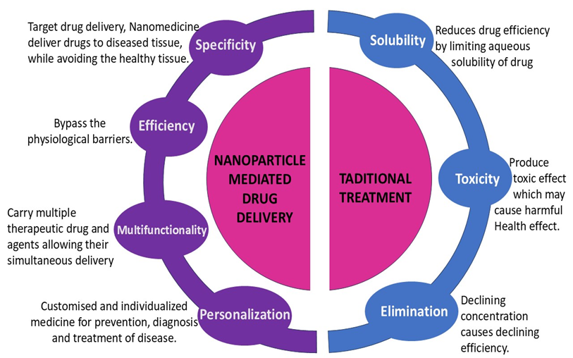
Figure 1: Comparison of Nanoparticle-Mediated Drug Delivery and Traditional Treatment
Rational Design Strategies for Nanomedicine Formulations
There are many physicochemical attributes such as size, shape, surface charge, and surface chemistry which must be considered to precisely engineer nanomedicine1973 to overcome biological barriers. These properties directly influence how nanoparticle interacts with the cells and protein.
Nanoparticle size is a crucial feature as it significantly influences the half-life, encapsulation efficiency and cellular uptakes. Size also shows how the nanoparticle will interact with the other biomolecules like lipids, peptides and DNA. For nanoparticles to effectively fight tumour, they need to overcome a few key challenges. They must circulate in the bloodstream long enough to reach the tumour, then exit the tumour’s blood vessels and get into the tumour tissue. Crucially, these nanoparticles should not cross the vessel walls in normal tissues thereby causing adverse effects. Normal blood vessels have tiny pores, typically 6 to 12nm wide. To prevent nanoparticles from escaping into healthy tissues, they should be larger than 12 nm. However, tumour blood vessels are often "leaky," with much larger pores, generally ranging from 40 to 200 nm. The main goal should be to design nanoparticles that are big enough to stay in normal vessels but small enough to slip through these larger tumour pores. Nanoparticles also interact with tumour vessel wall openings through and also get through the vessel walls with the help of hydrodynamic forces and electrostatic interactions. Therefore, the ratio of the nanoparticles size to the pore size significantly influences these interactions [35].
Nanoparticle shape significantly impacts their interactions with biological systems, influencing circulation, interstitial penetration, internalization, clearance, and cellular entry. Recent research studies prove that non-spherical particles offer many advantages, particularly in blood circulation. However, nanoparticles of rods and discs shape exhibit greater tendency to move towards vessel walls and higher wall adhesion in dynamic flow compared to spherical nanoparticles. This is attributed to biased hydrodynamic and Brownian forces. Filomicelles, a type of elongated nanoparticle, have been observed to circulate in the bloodstream for up to a week—ten times longer than spherical particles. Furthermore, ellipsoid-shaped nanoparticles demonstrate higher lateral drifting velocities than spherical ones, facilitating their movement away from the central flow region of a blood vessel.
Nanoparticle surface charge is another critical factor in drug delivery system; it influences the interaction with micro vessels and tumour. Surface charge affects solubility, biodistribution, stability, cellular uptake, and cytotoxicity. Within the bloodstream, electrostatic interactions become prominent as nanoparticles near the vessel walls. Positively charged (cationic) particles tend to attract negatively charged proteins, leading to their adsorption, immune recognition, and subsequent clearance from the body. Additionally, the negatively charged endothelial basal lamina easily absorbs cationic nanoparticles, limiting their circulation. Conversely, negatively charged (anionic) nanoparticles are repelled from the cell-free layer (CFL), which limits their ability to extravasate. For these reasons, neutral nanoparticles are generally preferred for vascular transport. Interactions between cationic particles and vessel walls can enhance extravascular transport into tumours. Experimental and theoretical studies suggest that electrostatic interactions with the negatively charged glycocalyx of vascular endothelial cells can facilitate the extravasation of cationic nanoparticles more readily than anionic or neutral nanoparticles.
In addition to electrostatic charge, other surface properties are crucial for selecting appropriate nanocarriers for delivery of drug. For example, encapsulating low-solubility drugs within nanocarriers possessing hydrophobic or amphiphilic surfaces is an effective strategy for drug delivery.
Principles of Engineering Nanomedicine Products for Cancer Therapy
Nanomedicine offers several advantages for anticancer therapeutic development. Following are the statements in favour of nanosized therapeutic development:
Many anticancer drugs have poor water solubility which hinder the development and bioavailability of early agents of anticancer drugs. Hydrophilic nanocarriers solves this problem by encapsulating these poorly soluble drugs, thus, significantly increasing their delivery and uptake. Encapsulation within nanocarriers can also enhance the chemical stability of drug thus preventing their degradation [15].
Nanocarriers safeguard anticancer drugs from enzymatic degradation and premature elimination [10]. For instance, siRNA is vulnerable to degradation by endonucleases in the plasma, while therapeutic proteins can be broken down by digestive enzymes like trypsin or pepsin in the gastrointestinal tract. By encapsulating these sensitive molecules, nanocarriers shield them from such hostile environments. Moreover, Encapsulation of the drugs allow protection and better control over the pharmacokinetics profile of the drug[10].
Nanotechnology can enhance the delivery and localization of anticancer medicines within the body. Nanomedicine enables better penetration of therapeutic agents into tumour tissues and selectively targets drugs to target cell. This targeted approach is achieved through both active and passive targeting strategies which involves surface modification of nanoparticles with ligands that recognize specific markers on cancer cells.
Nanocarriers can be engineered to release their payload in response to specific stimuli present in the tumour microenvironment. For example- Doxorubicin, a drug independent of pH, can be coupled with pH sensitive nanoparticle. This coupling increases the uptake and intracellular release of drug.
These approaches reduce tumour resistance from anticancer drugs and enhance drug effectiveness while minimizing nonspecific interactions [15].
Nanoparticles Used in Nanomedicine Targeting
Nanoparticles are coated with a wide range of organic or inorganic materials and these coating determines the physiological property of nanoparticles. Nanoparticles are considered as nanocarrier due to their property like water dispersity, biocompatibility and biodegradability. These characteristics contribute to improved solubility and half-life of the drug that increasing the pharmacokinetics of chemotherapeutic agents. They promote drug accumulation in cancer tissue via enhancing permeability and retention effect. Combining nanoparticles with anticancer drug can improve therapeutic efficiency and reducing side effects by targeting specific cancer site using ligand [16]. Many types of nanoparticles can be used for targeting nanomedicine towards cancerous cells. The different types of nanoparticles are summarized in Figure 2.
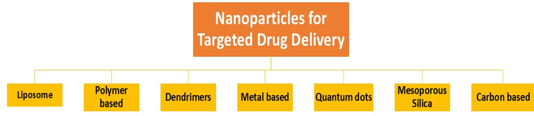
Figure 2: Different types of Nanoparticles used in target drug delivery
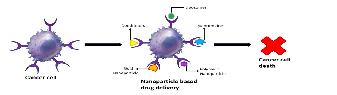
Figure 3: Nanoparticle based drug delivery
PNPs are colloidal macromolecules with size ranging from 10 to 100 nm. PNPs acts as a drug carrier in which the drug that need to be transported to the target site are encapsulated or attached to the surface of PNPs, thus forming a nano capsule. Initially nonbiodegradable polymers like polymethyl methacrylate (PMMA), Polyacrylamide, Polystyrene, Polyacrylate were used in PNPs but the problem with these materials was that they induced toxicity and chronic inflammation due to their accumulation in tissue and in addition to that they were difficult to degrade, excrete and cannot be physically removed. This problem was solved by using biodegradable polymers such as polylactic acid (PLA), poly(ε caprolactone) (PCL), poly(lactic-co glycolic acid) (PLGA), gelatine, albumin in PNPs [3].
The use of biodegradable polymers reduces the toxicity, improved drug release kinetics and increased biocompatibility and stability of the drug. For example- PNPs loaded with Cisplatin such as dexamethasone or alpha tocopheryl succinate have been employed in chemotherapy which prevents cisplatin induced ototoxicity. Therefore, the shift from non-biodegradable to biodegradable polymers has significantly enhanced the safety and efficacy of PNPs as drug delivery system. To increase the bioavailability, the PNPs are coated with polymers like polysorbate which helps it to interact with blood brain barrier endothelial cell membrane and facilitate endocytosis. An enhanced and modified version of PNPs is FNPs (Fluorescent Polymeric Nanoparticle) which is a theragnostic material, that combines diagnosis and treatment at the same time. A FNPs usually consists of fluorescent protein, biocompatible biopolymers, inorganic quantum dots and organic dyes which performs the function of tumour imaging and the treatment is provided by drugs which are loaded by π-π bond or hydrophobic interactions in fluorescence assays which eventually increases the anti-cancer efficacy of nanomedicine [17].
Table 1: Types of Polymeric Nanoparticle
|
Types Of Polymeric Nanoparticle |
Advantages |
Disadvantages |
|
|
Nanogels |
Biocompatibility and degradability Hydrophilicity Long contact time on skin surface High drug delivery efficiency |
Fast clearance Off target accumulation Difficulties in controlling both degradation and drug release |
[15] |
|
Chitosan |
Biocompatibility and degradability Mucoadhesive Easily enzymatically soluble Low toxicity |
Poor solubility at physiological pH Different chitosan-based delivery system affects the properties of chitosan nanoparticles in vivo |
|
|
Micro-emulsions
|
Thermodynamically stable Lowering the skin barrier effect Improving loading capacity and stability |
Functionalizing the dynamic oil/ water interface |
Dendrimers are well defined spherical or globular shaped structure, consisting of highly branched polymer chain, centre core and an outer layer of multivalent functional group which can interact with other molecules. The inner cavity of dendrimers is hydrophobic in nature and can encapsulate uncharged, non-polar drug molecule through hydrophobic interaction. The outer functional groups can be modified to release drug in response to specific stimuli, such as change in pH or presence of specific enzyme [17]. RGD (Arginylglycylaspartic acid) peptides or monoclonal antibodies are some targeting molecules which can be attached to the surface of dendrimers to guide the drug delivery to specific cell or tissue. Doxorubicin and Paclitaxel are hydrophobic drugs which can be covalently attached to the dendrimers for their specific targeting. Dendrimers such as poly (glutamic acid)-b-poly(phenylalanine) copolymers can self-assemble into micelles, which can then encapsulate and deliver drug [9]. Dendrimers based micelles are being investigated in clinical trials to deliver Paclitaxel for the treatment of breast cancer, lung cancer and pancreatic cancer.
Liposomes are the smallest form of nano vectors with the size of 90-150nm which are made up of lipids. The liposomes are made up lipid bilayer and contain an aqueous phase core to store drug. Liposomes are formed by self-assembling of lipid into a bilayer structure by forming hydrophobic interaction between fatty acids and head group, surrounding an aqueous environment. This unique structure of liposomes allows encapsulation of both hydrophobic and hydrophilic drug. The liposomes are used as drug delivery system because of their incredible biodegradable property of the fat globules [9] When liposomes are administrated intravenously, the body’s reticuloendothelial system (RES) readily takes them up and the liposomal drugs gets accumulated in the liver, lungs, and bone marrow as well as in the other tissues. This targeted delivery mechanism significantly improves the therapeutics effectiveness of drugs while simultaneously reducing their undesirable side effect [18]. Liposomal therapy is a well-developed technology for the delivery of chemotherapy drug; they enclose the drug/medication in the core and the fatty layer of liposome protects the drug until the liposome is delivered and adheres to the outer membrane of target cancer cell. Liposome have a considerable persistence in the blood i.e., they remain in blood for a longer duration compared to some other drug delivery system. This prolonged circulation time facilitates efficient drug delivery to target tissue. The properties of liposomes can be manipulated by using different types of lipids. Variation in fatty acid chain length, head group, melting temperature allow for the construction of liposomes with specific functionalities. By carefully selecting the lipid composition, temperature-sensitive or pH-sensitive liposomes can be engineered to encapsulate drugs and release them in response to specific environmental triggers at the target site, such as elevated temperature or acidic pH.
Magnetic nanoparticles are a type of nanoparticle that are primarily composed of pure metal like iron, cobalt, nickel, or rare earth metal or combination of metal and polymers. MNPs have a distinct characteristic that they can be controlled by an external magnetic field [20]. Magnetic nanoparticle is widely used in biomedicine and bioimaging due to their distinct magnetic properties. These particles are being explored for a wide range of applications like MRI contrast agent, magnetic thermotherapy, target drug delivery, separation, binding, and purification of biomolecules and cells. MNPs should have the ability to generate a uniform magnetic field, which helps to prevent particle aggregation and promotes consistent and uniform behaviour. Magnetic nanoparticles have superparamagnetic nanometre size and spherical shape, enhanced saturation magnetization, high magnetization rate and high chemical stability. Some MNPs are approved by the Food and Drug Administration for clinical trial but some MNPs have potential side effects which can adversely affect the vital organs like liver, brain, and skin as well as issue with gastric function and even neurological damage [21].
Semiconducting nanoparticles, more commonly known as quantum dots are small particles or nanocrystals of size between 2 to10 nm. These quantum dots can be synthesised using variety of different compound. Commonly quantum dots consist of a semiconductor core, the core consists of II-IV, IV-VI, III-V elements of the periodic table which are again surrounded by a semiconductor shell having wide bandgap (Zn). Quantum dots may be of heavy metals or Silicon based and each has its specific property [22]. Quantum dots absorb the white light and emit it at specific wavelength. The wavelength of emitted light varies from blue to near infrared depending on the different size and composition of the quantum dots. The quantum dots have an ability to concentrate in a single internal organ which make quantum dots a potential drug delivery system. The latest advancement in quantum dots combines biomolecules, peptides and antibodies on their surface which make a potential tumour targeting system and have application in cancer imaging and treatment. Quantum dots some time combines with prostate specific antigen to label cancer, while other use of quantum dots is to synthesize biomarkers which produce more stable and bright light intensity compared to traditional fluorescent immunomarker leading to more reliable and sensitive detection. Quantum dots-based probes enable multicolour imaging of cancer cells, allowing for detection of tumour. Highly sensitive probe based on quantum dots can also be used to detect ovarian cancer [23]. Quantum dots immunohistochemistry offers advantages over traditional immunohistochemistry, particularly in detecting low expression level of HER2 in breast cancer and enabling multicolour detection.
Metal nanoparticle has garnered significant attention due to their versatile properties, particularly in cancer treatment. Some of the examples of metal nanoparticle is gold, silver, iron/iron oxide, copper, nickel, zinc. Among all these gold nanoparticles exhibit superior characteristics, making them prominent in cancer research, while silver nanoparticle also shows promising results. Their potential lies in offering cost effecting alternative to the traditional cancer therapies [24]. Due to their unique physiochemical characteristic AuNPs have a wide range of biomedical application including tumour targeting. The anti-cancer effect of AuNPs can be improved by surface functionalization or coating. The gold core of gold nanoparticles is considered safe and their unique light activated properties allows for controlled release of the therapeutic cargo. These modified nanoparticles can be used diagnostic, therapeutic, bioimaging, and prognostic purposes.
Example of AuNPs in cancer treatment is biosynthesised gold nanoparticles using Brazilian red propolis, showed cytotoxic effect against T24 bladder cancer and PC3 prostate cancer cell line.[25]. Polyethylene glycol (PEG) along with the tumour targeting agents are added on the surface of the gold nanoparticle, the gold nanoparticles are used to treat advance solid tumour including sarcomas and melanomas by conjugating gold nanoparticle with tumour necrosis factor alpha. Similarly superparamagnetic iron oxide nanoparticles are being developed for drug delivery. These require the application of local hyperthermia or oscillation to release their conjugate drug. Additionally magnetic field can be used to guide these nanoparticles to their specific target area in the body [17]. The following table show the properties of other metal nanoparticle
Table 2: Types of metal nanoparticle
|
Type of metal nanoparticle |
Targeted cancer |
Targeting role |
|
|
Glyco-gold nanoparticles |
Cervical cancer (HeLa and L929 cells) |
Conjugated Rnase |
Ref. [25] |
|
Silver nanoparticle |
Pancreatic cancer (PANC-1, AsPC-1, and MIA PaCa-2 cells) |
Oxygen based (ROS) OR Nitrogen based (RNS) reactive species |
|
|
Mesoporous platinum nanoparticle |
Breast cancer (MCF-7/ADR Cells) |
Photothermal therapy/ doxorubicin |
|
|
Superparamagnetic iron oxide nanoparticle |
Breast cancer bone metastasis (MDA-MB-231/ Bone marrow cells) |
Bone targeting peptide- conjugated |
|
|
Nickle oxide nanoparticle |
Triple-negative breast cancer cells (Vero cell line and MDA-MB-231 cell) |
Folic acid decorated and pH sensitive |
Table 3: Nanoparticle used for drug delivery
|
Np Type |
Composition |
Properties/Application |
|
|
Polymeric nanoparticle |
Polymers with suspended nanosized substance /drug |
Synthetic and natural origin biodegradable and biocompatible polymers are used. |
Ref. [15] |
|
Dendrimer nanoparticle |
Polymeric, monomeric or oligomeric core shell nanostructure having a substance/ drug encapsulated in their branch or absorbed on their surface |
Biocompatible, has low polydispersity, and can be hydrophobic as well as hydrophilic |
|
|
Liposome |
Composed of Phospholipid and cholesterol. Bilayer lipid containing enteral aqueous portion. The lipids are cationic and neutral of phosphatidylcholine class and sterol |
Biodegradable, biocompatible, nonimmunogenic, amphiphilic, alternating lipid modification and drug delivery capacity |
|
|
Quantum dots |
Semiconducting nanoparticle, artificial nanostructure metalloid crystalline core usually manufactured from CdSe or CdTe surrounded by zinc sulphide outer shell |
Electron transporter, they can emit light of various colours. They have application in composites, solar cells and fluorescent biological labels, optical imaging |
|
|
Gold particle |
Gold combined with a substance/drug |
Biocompatible, within the range of 5 to 100nm, in the drug delivery system |
|
|
Niosome |
Formed by self-association of non-ionic surfactant and cholesterol in an aqueous phase |
Biodegradable, biocompatible, have non-immunogenic skeleton and therefore serve as promising drug carrier |
|
|
Carbon based nanoparticle |
Pure carbon |
Highly electrical, heat conductor, highly stability, low toxicity, biocompatibility, application in drug delivery |
|
|
Silica nanoparticle |
Composed of an amorphous network of silicon and oxygen |
Biocompatible, large surface area, easy functionalization, bioactive and commonly used in drug delivery and contrast imaging |
|
|
Magnetic nanoparticle |
They contain major component such as magnetite, Fe, Ni, and Co |
Ferromagnetic, contrasting agent in the case of MRI and certain therapy |
Targeting Mechanisms of Nanomedicine
Nanoparticles can be designed to deliver drugs selectively to specific tissues and cells in vivo. Nanoparticles need to be design to evade the immune system and prevent non-specific uptake by the cells, allowing them to circulate and reach the target site (Stealth). Once at the target site, Nanoparticles should be able to adhere to specific tissue and interact with or be taken up by the desired cell (Stickiness). Recent research focuses on designing “smart Nanoparticles”. Nanoparticles capable of transitioning from a stealth state to sticky state use dynamic switching mechanisms to enhance biodistribution and improve tumour targeting [26].
There are two mechanisms for targeting-
Passive Targeting: Passive targeting is the accumulation of nanomedicine in tumour tissue based on their specific physical and chemical properties. In passive targeting, through the EPR effect the nanomedicine gets accumulated in tumour tissue. Passive targeting of nanomedicine utilizes the size of nanoparticle and take advantages of unique characteristics of tumour vasculature and environment. As the tumour grow, they trigger angiogenesis, which is the process of blood vessels to supply oxygen and nutrients. Unlike normal blood vessels, angiogenesis vessels in the tumour have structural abnormalities, including gaps between endothelial cells measuring 600-800nm. Additionally, poor lymphatic drainage in tumour tissue results in the enhanced permeability and retention (EPR) effect. Due to these gaps’ nanoparticles carrying nanomedicine can selectively leak out from the blood vessels and accumulate in the tumour [23].
Despite of the advantages of passive targeting through the EPR effect, there are certain limitations of passive targeting, such as solid tumour exhibits elevated levels of interstitial fluid pressure, this high pressure can hinder the movement of drugs from interstitial space into the cells. The presence and accumulation of nanomedicine in solid tumours are directly affected by their concentration in the blood stream. This in turn is influenced by various pharmacokinetics factor. Therefore, to overcome this problem a deep understanding of biological process and pharmacokinetics behaviour of nanocarrier is required.
Active Targeting: In active targeting, a substance with strong ability is attached to the surface of nanocarrier. These attached ligands are specifically designed to selectively bind to the receptor that are present on the surface of the target cells. A wide range of ligand types has been employed for active targeting like carbohydrates, folic acids, amides, proteins and oligonucleotides. An important subtype of active targeting is cell targeting. The cell targeting aims to target specific cells such as cancer cells, immune cells and stem cells. In these approach nanoparticles capable of transitioning from a stealth state to sticky state. The ligand mediated recognition of the target substrate receptor is a crucial component of this strategy, ensuring selective binding to the desired cells [10].The receptor on the target cell causes intermembrane folding which leads to the internalization of nanoparticle via receptor mediated endocytosis. After internalisation, the engulfed nanoparticle is transported to nuclear endosome where further particle processing and drug release occur due to the change in the pH [27].
Stimuli Responsive System/ Targeted Release: Stimuli responsive system is a type of drug delivery system in which the drug is released in response to specific triggers. These triggers can be physical, chemical or biological. The mechanism of drug release is achieved by these triggers, which interacts with the fundamental property of the nanocarrier, such as phase, structure, or conformation. The stimuli responsive system shares the similarity with the feedback mechanism of the biological system. This implies a dynamic and controlled release mechanism. The stimuli are broadly categorized into two types: internal stimuli and external stimuli. Internal stimuli, also known as endogenous stimuli, arise from the body's intrinsic patho-physiological or patho-chemical conditions. Examples include pH, redox potential, ionic strength, and shear stress. External stimuli, or exogenous stimuli, are physical cues applied from outside the body, such as temperature, light, ultrasound, magnetic force, and electric field [11].
The increased presence of specific molecule in diseased tissue as compared to the normal tissue can be use as trigger for drug release. Enzymes that are found at higher levels are more active in diseased area and can be exploited for trigger drug release. Physical stimuli, such as local hyperthermia, is a method for externally triggering drug release in targeted therapy. Thermos responsive nanocarrier like thermoDox are design to release their payload when exposed to elevated temperature within the range of 37 to 420C. This controlled hyperthermia not only induce drug release bit also enhances vascular permeability, allowing anticancer agents to penetrate deeper into tumour tissue [8].
Nanomedicine Approved for Cancer Treatment
The first FDA-approved nanomedicine is Doxil which is a polyethylene glycol (PEG)-modified liposome encapsulating the chemotherapeutic drug, doxorubicin. After the first FDA approval of Doxil in 1995, five additional cancer nanomedicine, including Abraxane, was introduced the clinical market in the United States and there are currently over thirty nanoparticle formulations are in the clinical trial [28]. The following table shows clinically approved nanomedicine by Food and Drug Administration (FDA) and European Medicines Agency (EMA). These drugs are in phase I/III clinical trials or have been approved in some countries.
Table 4: Lipid based nanoparticle
|
Product |
Drug |
Carrier component |
Np size |
Nano formulation Advantage |
Indication |
Approval Year |
|
|
Doxil |
Doxorubin |
Liposome: HSPC, cholesterol, mPEG- DSPE (3:1:1 weight ratio) |
80-90 nm |
increase Blood circulation time, increase Tumour uptake, decrease cardio toxicity. |
Kaposi's sarcoma Ovarian cancer, multi myeloma |
FDA (1995) |
Ref. [29] [30]
|
|
Myocet |
Doxorubin |
Liposome: Phosphatidylcholine, cholesterol (55:45 molar ratio) |
150 nm |
increase Blood circulation time, increase Tumour uptake, decrease cardio toxicity. |
Metastatic breast cancer |
EMA (2000) |
|
|
Mepact |
Mifanuritide |
Liposome POPC, OOPS |
- |
increase Blood circulation time, increase Tumour uptake, decrease toxicity. |
Osteosarcoma |
EMA (2009) |
|
|
Vyxeos |
Cytarabine and Darnorubicin |
Liposome: DSPC, DSPG, Cholesterol (7:2:1) |
100 nm |
increase Blood circulation time, increase accumulation in bone marrow |
Acute myeloid leukaemia |
EMA (2018) |
Table 5: Protein drug conjugates
|
Product |
Drug |
Nano formulation advantage |
Indication |
Approval |
|
|
Oncaspar |
Pegaspargase |
increase Blood circulation time, increase Tumour uptake |
Acute lymphoblastic leukaemia |
FDA (1994) |
Ref. [29] [30]
|
|
Kadcyla |
DM1 (or Emtansine) |
Increased solubility, increase blood circulation time, decrease several toxicities |
HER2 + breast cancer |
EMA (2013) |
|
|
Ontak |
Denileukin Diftitox |
increase Blood circulation time, increase Tumour uptake Increase selectivity, decrease several toxicities |
cutaneous T cell lymphoma |
FDA (1999) |
|
|
Pazenir |
Paclitaxel |
Increase solubility, increase Blood circulation, increase Tumour uptake, decrease several toxicities |
Metastatic breast cancer, non-small lung cancer |
EMA (2019) |
Table 6: Metallic nanoparticle
|
Product |
Nanotechnology platform |
Drug |
Nano formulation advantage |
Indication |
Approval |
|
|
Nanotherm |
Nanoparticle of superparamagnetic iron oxide coated with amino silane |
F2O3 |
increase Blood circulation time, increase Tumour uptake - Heat production under stimulation with EMF |
Glioblastoma, prostate and pancreatic cancer |
EMA (2013) |
Ref. [30] |
Future Prospects of Nanomedicine
In the recent years, nanotechnology has emerged as one the modern and most high-tech methods for the treatment of cancer. The nanotechnology along with pharmacology has revolutionaries the treatment of disease especially in cancer treatment. This convergence starts a new era of investigation about the new effect of nanodrugs on patients and the mechanism in their body. Nanotechnology can help in better drug delivery and enhances the effect of drug on their target site at the same time [31].
The future outlook of nanomedicine should focus on conducting more clinical trials to validate the efficacy of nano formulation, optimizing dosage and minimizing long term side effects. Nanomedicine holds immense promise for the future of cancer treatment by offering the potential for more precise, effective and personalised therapies with reduced side effects. Selecting the appropriate nanodrug based on a specific patient’s disease indication will become crucial for tailoring therapeutic modalities and improving treatment outcome. Future efforts will increase the focus on co-loading multiple therapeutic cargoes within a single nanoparticle platform. This strategy will aim to overcome the limitation of current combination therapies by enabling the simultaneous and aggressive targeting of various nanomedicine [32]. The availability of a wide range of core materials will facilitate the development of hybrid nanoparticle composition. These advanced materials will be crucial for packaging drug with diverse structural and physiochemical properties within a single delivery system. The development of advanced nanomedicine presents both a valuable opportunity to revolutionize the treatment of cancer and significant challenge in term of research, design and clinical translation [33]. The future of nanomedicine lies in creating increasingly sophisticated and targeted drug delivery system that can precisely address pathological conditions at the cellular and molecular level, leading to more effective and safer therapies [34].
CONCLUSION
Nanomedicine represents a new era in cancer diagnosis and therapy, offering promising solutions to overcome the limitations of conventional treatments. By manipulating the unique properties of nanomaterials, nanomedicine aims to achieve early and precise cancer detection, enhance drug delivery to tumour sites, minimize off-target toxicity, and ultimately improve patient outcomes.The development of various nanoparticles, including polymeric nanoparticles, dendrimers, liposomes, quantum dots, and metal nanoparticles, has provided a versatile toolkit for addressing the complexities of cancer treatment. These nanocarriers can be engineered with specific properties for improved drug solubility, stability, protection from degradation, enhanced tumour targeting through passive and active mechanisms, and stimuli-responsive drug release. The progress in nanomedicine is evident in the increasing number of nanomedicine formulations undergoing clinical trials and the approval of several cancer nanomedicines globally. These advancements underscore the translational potential of nanotechnology in revolutionizing cancer care. Furthermore, the emergence of cancer nano vaccines highlights the role of nanomedicine in stimulating the immune system to combat cancer cells. The future of nanomedicine requires clinical trials, dosage optimization, and long-term safety assessments. Personalized therapy through the selection of appropriate nanodrugs for specific patient conditions will be crucial for improved outcomes. Nanomedicine presents immense potential for precise and effective cancer therapies, significant challenges remain in research, design, and clinical translation. Ultimately, the future aims to develop increasingly sophisticated and targeted drug delivery systems at the cellular and molecular level, leading to safer and more effective cancer treatments.
REFERENCES


Gautam Nishita, Patel Kritika, Sulagna Ghosh Barman, Shanthi V, Nanomedicine in Cancer Diagnosis and Treatment, Int. J. of Pharm. Sci., 2025, Vol 3, Issue 10, 1323-1338. https://doi.org/10.5281/zenodo.17342091
 10.5281/zenodo.17342091