View Article

  • Obesity And Gut Microbiota: A Comprehensive Review

  • 1,2,4Department of Pharmacology, Shiva Institute of Pharmacy, Chandpur, Distt. Bilaspur, Himachal Pradesh- 174004, India.
    3Department of Pharmacology, University School of Pharmaceutical Sciences, Rayat Bahra University, Mohali, Punjab-140301, India.

Abstract

Obesity is the multifactorial metabolic disease with worldwide interest, which leads to high non-communicable disease burden like type 2 diabetes, cardiovascular disease, and metabolic syndrome. Over the recent years, gut microbiota has been revealed as a master regulator of obesity pathogenesis. The imbalance of the composition of microbes in the gut or dysbiosis has been linked to higher energy harvest, lower energy expenditure, a change in lipid metabolism, and failing to control the appetite. Remarkably, a higher Firmicutes to Bacteroidetes ratio among the obese is a common behavior, which improves fermentation of the indigestible polysaccharides and leads to the production of short-chain fatty acids (SCFA) to facilitate adipogenesis. In addition, metabolites produced in the gut, including SCFAs, have context-specific effects on the metabolism of the host, and current evidence demonstrates that SCFAs exert both positive and negative effects on the rate of lipogenesis and inflammation. Neuroendocrine signaling by microbial metabolites regulating the gut-brain axis mediates the role of appetite and energy homeostasis. Moreover, Gram-negative bacterial endotoxin, lipopolysaccharides (LPS), causes metabolic endotoxemia and low-grade systemic inflammation to enhance insulin resistance and accumulation of lipids. Taken together, these mechanisms emphasise the imperative role of gut microbiota in development of obesity. Knowledge of the intricate host microbiota interactions may have therapeutic potentials, such as microbiota-specific treatments, namely probiotics, prebiotics, dietary manipulations, and fecal microbiota transplantation to treat the treatment of obesity.

Keywords

Gut Microbiota, Obesity, Dysbiosis, Short-Chain Fatty Acids, Gut-Brain Axis

Introduction

Introduction of Obesity:

Obesity: The disease is obesity, which has become one of the major problems of global health in all aspects, as far as the present times are concerned. Obesity has come to be the 5th cause of deaths all over the world. World Health Organization defines obesity as excess or abnormal fat which may negatively affect the health, and the primary cause of obesity is, imbalance of energy i.e. excess body calories taken against the calories burnt by body.[1,2] Obesity is another international public health problem of high prevalence among all age strata. It produces significant social and economic effects, as it influences the health and quality of lives of people.[3] There is evidently a raging problem of obesity as far as worldwide health is concerned; and according to estimates, obesity and overweight rate will rise to 25 per cent and 32 per cent overall respectively in 2030.[4] Obesity is a 21 st century disease and a serious nutritional problem, which comes along with heart disease, diabetes, cancer in addition to inflammation and metabolic syndrome.[5] The health status obesity is a critical contributor of metabolic syndrome which are cluster of metabolic abnormalities such as elevated blood sugar, excess body fat, excessive serum triglycerides, blood pressure and diminished volume of high-density-lipoproteins. The metabolic syndrome is allied among an exorbitant hazard of acute disease like type 2 diabetes, cardiovascular disease and stroke.[6,7] Its causation is caused by the interaction between various factors with a support on genetic and environmental factors, specifically, over-consumption of food and lifestyle. According to research conducted in 2018, it was revealed that even among kids younger than five years old, 5.9 percent people (40 million people) were classified as overweight whereas in 2016 an estimated 2 billion adults fell into this category.[8] According to some estimates, 18% of males and 21% of women in the population will become obese by the year 2025.[9,10] Another report released by WHO in 2021 shows that at least 1 billion people were already obese, with 650 million adults, 340 million adolescents, and 39 million of them being children according to data collected in 2016. The global trend of overweight and obesity has risen by 27 percent in adults and by 47 percent in children between the period 1980 and 2013.[11] This figure is still rising and WHO puts the number at it is bound to rise to about 167 million people by 2025.[12]

Classification of Obesity:

The degree of BMI rise has historically been used to define obesity. Body Mass Index (BMI) and ethnicity-specific cut-points have formed the foundation for the traditional classifications of overweight and obesity.[13] Overweight is defined as having a BMI of 25.0 to 29.9 kg/m2, whereas class 1, class 2, class 3, class 4 (Super Obesity) and class 5 (Hyper Obesity)  include BMIs of 30.0 to 34.9 kg/m2, 35.0 to 39.9 kg/m2, 40.0 to 49.9 kg/m2, 50.0 to 59.9 kg/m2, and over 60 kg/m2 and those with a BMI of 18.5 to 24.9 are considered normal weight.[14,15] In the case of Asian populations, BMI between 23.0 and 24.9 kg/m2 were weighted as overweight, 25.0 kg/m2 and above were weighted as obese and 18.5 to 22.9 kg/m2 were taken as normal.[13,16] The ease of measurement index of Body Mass Index (BMI) that is [(weight in kg)/(height in m2 ) serves as a convenient guide in the classification of the adult body as underweight, overweight or obese. BMI was invented in the 1830s by the Belgian mathematician and sociologist, who still applies this value in the assessment of obesity rates.[17]

Table 1.1 WHO Classification of Weight Status WHO (2000) [17]

Body Mass Index (BMI) (Kg/m2)

Classification

Weight Status

<16.0

Severe thinness

Underweight

16.0-16.9

Moderate thinness

Underweight

17.0-18.4

Mild thinness

Underweight

18.5-24.9

Normal range

Healthy weight

25.0-29.9

Overweight

Overweight

30.0-34.9

Obesity class I

Obese

35.0-39.9

Obesity class II

Obese

40.0-49.0

Obesity class III

Morbidly obese

50.0-59.9

Obesity class IV

(Super obesity)

Extremely obese

≥60.0

Obesity class V

(Hyper obesity)

Extremely obese

Causes of Obesity: Obesity causes are multilateral and interrelated. To ease the discussion we refer to them here as non-modifiable and modifiable factors. Non-Modifiable factors include Genetic,[18] leptin-melanocortin pathway,[19,20] Hypothalamic obesity[21] and the modifiable factors are Epigenetics,[22,23] Physical inactivity,[24] Excessive caloric intake,[25] The intrauterine environment,[26] Postnatal influences,[27] Insufficient sleep,[28] Drugs,[29] Medical conditions, Socioeconomic status,[30] Ethnicity, Psychosocial stress, Endocrine disrupting chemicals,[31] Gastrointestinal microbiome.[32,34]

Pathophysiology of Obesity:

Such long-term stability of body mass and body composition is possible with the energy intake being equal to his or her energy expenditure. Sufficient body energy supply has to go in effect. Our bodies have many short-term and long-regulations that come into action to maintain the utilization of the energy, storage of energy and the energy consumption. The hypothalamus has a number of nerve centers that have a great control on the quantity of food that we consume. The feeding centre is the lateral nucleus of the hypothalamus. The stimulation of this area or center causes hyperphagia. The hypothalamus paraventricular nucleus, dorsomedial nucleus, and the arcuate nucleus are also important in controlling the food intake levels. Lesions on paraventricular nuclei consistently cause over-consumption of food. In case of damage or any lesion to the dorsomedial nuclei, the same tends to decrease the stimuli to consume food. Usually, the food intake and the expenditure of energy are regulated by the arcuate nuclei of the hypothalamus. Many gastrointestinal humorous and the adipose product hormones are believed to act here. The gastrointestinal tract sends neural messages to the hypothalamus. These nerve impulses provide sensory data which indicates the stomach is full. Also, the glucose, amino acids, and fatty acids present in the blood send messages concerning satiation to the brain through their molecules. The feeding behavior is determined by the information within the hypothalamus of the cerebral cortex (sight, taste and smell). Hypothalamic feeding and satiety centers also possess a large number of receptors to many neurotransmitters and hormones which mostly manipulate the actions of feeding heavily. [35,36,37,38]

Overview of Human Gut Microbiota:

The gastrointestinal tract (GI) is one of the major surfaces in the human body (250–400 m2) where antigens, the host, and the environment come into touch. About 60 tons of food pass through the human GI tract at any given time throughout an average lifespan, along with the numerous microbes that the body meets.[39] The gut microbiota is the collection of all the bacteria, archaea, and eukarya that live in the GI tract. Over thousands of years, they have co-evolved with the host to form a complex mutually beneficial relationship.[40,41] The GI tract's germs are thought to number over 1014, which is more than 100 times the human genome's genomic content (microbiome) and 1014 times more bacterial cells than human cells.[40, 42] However, a revised perspective has shown that the human:bacterial cell ratio is 1:1.[43] The microorganisms and body that harbors them are sometimes referred to as superorganisms because of the large numbers of bacterial cells in the body.[44] The microbiota has considerable benefits to the host, in various physiological functions, like integrity as well as strength of the gut, shaping of intestinal epithelium,[45] energy harvesting,[46] protection of the pathogens as well as controlling host immunity.[47,48] Yet, these processes can be interfered with due to the altered microbial composition, which is called dysbiosis. As more and more advanced ways of profiling and characterising complex ecosystems emerge, a contribution of the microbiota to most intestinal and extra-intestinal diseases emerged increasingly strongly.[49,50]

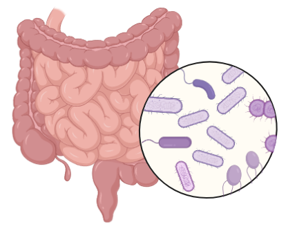
Figure 1.1 Human Gut Microbiota

Organization and makeup of the human gastrointestinal microbiota

The great bulk of data on the mature microbiota of humans was being collected by time-consuming culture-based fixations around 10 years ago.[51] The development of culture-independent techniques, such as high throughput and inexpensive sequencing methods, has greatly improved our ability to survey the extent of gut microbiota in recent years. Since the (16S) ribosomal RNA (rRNA) genome is present in all living organisms and can be easily distinguished between species thanks to its nine highly variable sections (V1 to V9), it is one of the most often targeted genes.[52,53] Earlier, the focus was on sequencing the whole 16S rRNA gene. 76 percent of the rRNA sequences obtained in an adult male fecal sample were in unique and uncharacterized species, highlighting the severe insensitivity as well as bias of culturing procedures in an early experiment using this methodology.[54] Though shorter read lengths introduce mistakes, the sequencing of 16S rRNA has advanced in recent years to concentrate on specific gene subregions in greater detail.[52,53] Because whole-genome shotgun metagenomics approaches have better resolution and sensitivity, they may also provide more accurate estimates of the diversity and composition of microbiotas. Joint data on the MetaHit and that of the Human Microbiome Project have yielded the largest picture ever of the human-associated microbial repertoire so far.[55,56] Summarized data of these studies had one hundred and twenty phyla of two thousand and seventeen species isolated in human beings out of which ninety three and half percent belonged to Proteobacteria, Firmicutes, Actinobacteria and Bacteroidetes. The identified phyla were reduced to three single species species isolated of the 12 phyla only one of which, the intestinal species, was identified as Akkermansia muciniphila and was the sole representative of the Verrucomicrobia phylum that was known.  Within man 386 of the species so far identified completely anaerobic and therefore will tend to be located in areas of mucosa like the mouth and the GI tract.[55] The gut microbiota exhibits high degrees of functional redundancy and is not as diverse in makeup as the community of microbes at certain other body sites.[57,58,59]Recently, a thorough inventory concerning the human gut microbiome's functional potential was obtained, and utilizing 248 newly sequenced and 1018 reported samples, 9879896 genes were found.[56] As a result of the study's identification of nation-specific microbial signatures, the gut microbiota's composition is influenced by environmental factors, such as nutrition and maybe host genetics. It is however important to also note that although different microbiotas are different in the sense that they vary in composition, there could be functional redundancy of some sort in different microbiotas resulting in similarity in protein or metabolic profile.[60] Such information is vital in establishing therapeutic measures to alter and manipulate the microbial community in disease.

Human gastrointestinal microbiota's development

It is suggested that the microbiota's development starts at the time of birth, but this dogma is questioned by a small body of experiments where the microbes were found in the tissues of the womb, including the placenta. [61,62] The GI tract is quickly colonized following birth and life experience like illness, antibiotic therapy or dietary alteration leads to the random experimenting.[63] The microbiota composition and the manner of delivery appear to be related as well; children born vaginally had a high lactobacilli abundance in their microbiota during the first few days, which is indicative of the high lactic acid bacteria in the vaginal environment.[64,65] On the other hand, facultative anaerobes such as Clostridium species inhabit the microbiota of newborns delivered via caesarean section, which is shortened and delayed in the colonization of Bacteroides genus.[66,67] While the fecal microbiota of 72% of newborns delivered vaginally is comparable with the one of their mothers, this percentage drops to 41% in babies born via caesarean section.[68] During early development, microbiota are typically low-diversity and only contain 2 primary phyla, namely Actinobacteria and Proteobacteria.[62,69] The microbial diversity rises and microbiota composition tends to mature with a specific adult-like microbial profile during the first year of life with unique and different temporal patterns between the different babies.[70] By about 2.5 years old, the microbiome make-up, variety and functionality of the infant microbiota can match adult microbiome functionality.[62,63] Undoubtedly, the makeup of the gastrointestinal tract's microbiota remains relatively constant in adulthood, nonetheless, it is prone to disruption by life events.[71] In the subjects older than 65 years, microbial community is changed, and there are more Bacteroidetes phylum and Clostridium cluster IV, unlike younger participants who have a more predominant cluster XIVa.[72] On the other hand, another study found that while the gut bacteria among a young population and a group of seniors (70 years) were very similar, the microbioma diversity of a cohort of seniors was much lower.[73] The enhanced prevalence of facultative anaerobes (like Escherichia coli) and changes in the pattern of butyrate makers (like a decrease in Faecalibacterium prausnitzii) were two more group-specific modifications in the centenarian microbiome.[72,73] Diversity and living arrangements, whether in the community or in long-term residential care, have been found to be egregiously correlated among elderly citizens.[74]

In general, metabolic activities of the microbiota such as the production of short-chain fatty acids (SCFAs), amylolysis are impaired in old age, whereas proteolytic activity is enhanced.[75] Since it became more evidenced that SCFAs are crucial metabolic and immune mediators, the hypothesis was generated that the fall of SCFAs provides the environment that fosters the intestinal inflammation-aging process of elderly individuals.[76]

Gut Microbiota's Pivotal Role in The Onset and Progression of Obesity:

Complex microorganism community or microbiota in the gastrointestinal tract, gut microbiota, is crucial in causing and exacerbation of obesity. Change in its makeup usually called as dysbiosis can result into new influence on various physiological systems, such as energy homeostasis, lipid metabolism, appetite control, as well as continual inflammation. All of these mechanisms lead to additional energy taking, fat deposition, deranged appetite signals, and low-grade inflammation, which are features manifestations of obesity.

1. Energy homeostasis disturbance:

1.1 Increased Digestible Energy Absorbing Mass:

The gut microbiota of obese people has an increased ability of metabolism of the food in terms of retrieving energy. This is mainly because of increased abundance of Firmicutes and raised Firmicutes/Bacteroides ratio, which is positively correlated with digestion of otherwise un-digestable polysaccharides into absorbable monosaccharides and SCFAs like acetate and butyrate.[77,78] Of particular importance is increasing energy uptake though increasing glucose transporter (Glut2) components (Firmicutes phylum) through the over-expression of fatty acid translocase (CD36).[77] Moreover, on gut bacteria enriched with obesity, the expression of enzyme, such as, a-amylase and amylomaltase, which enables fermentation of carbohydrates into energy-yielding SCFAs, is high by rate.[78] These SCFAs are taken up in the colon and may be able to furnish 5-15 percent of the daily dietary energy intake as well as 70 percent of the energy demand of colonic epithelial cells.[79] Besides, the microbial interspecies hydrogen (H2 ) has been used to boost the fermentation efficiency. Methanogenic Archaea that use H2, partner with H2-producing bacteria to alleviate thermodynamic constraints of fermentation thereby enhancing outputs of SCFA and energy recovery.[80] Such synergy between microbes occurs more often in microbiomes of obese people and further adds to the calorie uptake.

1.2 Impaired Energy Expenditure: 

Other than rising dietary consumption, another reason why obese individuals have low energy expenditure is due to gut microbiota dysbiosis. Gut microbes control the activation of bile acid that activate TGR5 and FXR receptors that control thermogenesis. Adipose tissue that is brown, TGR5 agonist enhanced the expression of PPARy coactivator-1alpha and iodothyronine- deiodinase type 2, which facilitates mitochondria production and increases the rate of energy burning through heat.[81] In the intestine, FXR stimulates FGF15/19 that alters bile acid composition and enhances white adipose tissue browning as well as BAT stimulation.[82,41] Nonetheless, in obesity, the amount of bacteria that manufacture bile acid including the structure of the bacteroides and Lactobacillus are lowered,[83] and these pathways are damaged thus lowering thermogenic capability. Short-chain fatty acids (SCFAs), are involved in energy expenditure in a complex role as well. On the one hand they inhibit release of fasting-induced adipose factor (FIAF), lowering the stimulation of AMP-activated kinase (AMPK) and thus minimizing of.[84,85,86] Conversely, SCFAs such as butyrate have the potential to induce AMPK and the increase of the mitochondrial uncoupling protein 1 (UCP1) and peroxisome proliferator-activated receptor (PGC-1alpha) in BAT to stimulate thermogenesis and fatty acid oxidation.[87] These two reasons represent a complicated context-specific control of energy expenditure that should be further studied.

2. Modulation of Lipid Synthesis and Storage:

2.1 Gut Microbiota and Hepatic Lipogenesis:

Deregulated gut microbiota alters liver lipid metabolism mainly acting via bile acids signaling. Depleted bile acids cause lower FXR body positioning in the liver, which leads to a decrease inhibition of liver receptor homolog 1 and enhance SREBP1c which is a transcription factor that is involved in de novo lipogenesis.[88] Activating intestinal FXR also triggers FGF19 that suppressed hepatic lipogenesis through FGFR4 activation and inhibition of co-activators such as PGC-1B or activation of SHP.[89] Therefore, liver stores fat in obesity via modifications in the activities of these bile acid routes. Enhanced level of serum glucose as a result of escalated expression of Glut2 also augments hepatic lipogenesis as a result of escalated SREBP1 and carbohydrate response element binding protein (ChREBP).[90] Also, acetate, another SCFA that is elevated in obesity is a precursor of fatty acid and cholesterol production, and butyrate boosts the production of lipids via the β-hydroxy-β-methylglutaryl CoA pathway.[91,92] In offspring, butyrate supplementation increased expressions of lipogenic genes and decreased lipolysis in offspring.[93] Butyrate has, however, also been shown by other studies to inhibit lipogenesis through decreasing PPAR.[90] This contradicts the twofold utility depending upon biological circumstance proposed in the study.[94]

2.2 Gut Microbiota and Lipid Storage: 

Obese people's gut flora contributes to increased fat accumulation through hormone changes and inflammation. Increased LPS concentrations of Gram-negative bacteria such as of a type of bacteria known as Veillonella, trigger the metabolic endotoxemia and the inflammatory process through the activation of the TLR4 signaling pathway.[95,96,97] LPS produces activation of macrophages and adipocytes which stimulates the release of cytokines that promote inflammation (TNF-a, IL-6, and MCP-1), which inhibit insulin signaling through phosphorylation of the insulin receptor consequently facilitating the buildup of lipids in the adipose tissue and liver.[98,99] Adipocyte precursor proliferation caused by LPS is also mediated by the CD14 and Activin A pathways.[100] Additionally, the gut microbiota can facilitate storage of fat by decreasing the expression of such anorexigenic peptides encoding genes as Gcg and Bdnf and causing leptin resistance by inducing SOCS3.[101] Decrease in the level of the “Lactobacillus paracasei” in obesity dissolves its repression to the lipoprotein lipase and permits triglycerides to be stored in adipocytes.[102]

3. Regulation of Central Appetite and Feeding Behavior:

3.1 The Microbiota-Gut-Brain Axis:

A two-way communication system: Gut-brain axis is a communication system that includes neural, hormonal and immune channels. Central nervous system can be subjected to microbial metabolites and signals, and its effect influences the appetite and feeding behavior.[103,104,105] Food intake and energy balance can be related to obesity dysbiosis through this axis.[106,107]

3.2 Microbial Control of Satiety and Mood: 

Low-intensity metabolomics, such as lactate (of Lactobacillus and Bifidobacterium), maintains the energy needs of the neurons and enhances satiety.[108] Acetate has an effect on the expression of hypothalamic neuropeptides though its entry into the citric acid cycle[109] whereas butyrate is known to stimulate vagal afferents and the hypothalamus immediately upon crossing the blood-brain barrier.[110] Further, SCFAs and bile acids regulate anorectic bowel hormones (GLP-1 and PYY), which in turn directed the brain through their receptors and results in the reduction of appetite.[111,112] The gut microbiome as well affects production of neurotransmitters e.g. serotonin and GABA. GABA enhances appetite and serotonin blocks the impulse to eat and support the balance of energy[113,114] by effect on melanocortin neurons. Microbial signals also mediate emotional and hedonic mechanisms of regulating eating behavior, as SCFA propionate inhibits brain reward reactions to high-calorie food using striatal pathways.[107]

4. Chronic Low-Grade Inflammation:

4.1 LPS-Mediated Inflammation: 

One of the characteristics of obesity is low grade inflammation which is predominantly microbial LPS induced. The gram negative bacteria like the yaquo Veillonella multiply in a body that is obese, there is excessive LPS95. This endotoxin destroys the resistance of the intestinal barrier through the TLR4 / MyD88 / IRAK4 signaling pathway in epithelial cells, and LPS translocation into the organism occurs.[115] Depleted abundance of a mucosal-integrity bacterium, the “Akkermansia muciniphila ” enhances this leaks[116] and high-fat diets increase the absorption of LPS through chylomicron transportation.[117] LPS becomes systemic and then binds to LBP and CD14 to create a complex which activates TLR4 on macrophages and adipocytes, thereby resulting in the inflammatory cytokines releasing through the process of NF-kB (TNF- six, IL-6, MCP-1).[96,97] This inflammation attracts macrophage into the adipose tissue in an ongoing inflammatory cycle.[118]

4.2 Anti-inflammatory Role of SCFAs: 

SCFAs, and particularly butyrate have been shown to be anti-inflammatory, through stimulation of IL-18, differentiation of regulatory T cells, and activation of GPR109a.[119,120] Butyrate prevents LPS-induced NF-kB activity as well as inducing PPAR-y, decreasing inflammation.[121]

CONCLUSION: 

Gut microbiota contributes significantly to the pathogenesis of obesity involving numerous interrelated mechanisms. Dysbiosis increases digestible energy and the absorption of live energy, lowers energy consumption, favors the deposition of lipids and changes in the regulation of appetite and tabulates with chronic inflammation. As much as SCFAs have been shown to be beneficial as well as harmful in certain situations, they continue to be the main connection between the metabolism of host and the microbes in the gut. This knowledge of these complex microbial-host interactions has paved a way to a new treatment of obesity with microbiota-based therapies.

REFERENCES

  1. Camacho S, Ruppel A. Is the calorie concept a real solution to the obesity epidemic?. Global health action. 2017 Jan 1;10(1):1289650.
  2. Ibrahim S, Akram Z, Noreen A, Baig MT, Sheikh S, Huma A, Jabeen A, Lodhi M, Khan SA, Hudda A. Overweight and obesity prevalence and predictors in people living in Karachi. J. Pharm. Res. Int. 2021;33:194-202.
  3. Heo MG, Choung SY. Anti-obesity effects of Spirulina maxima in high fat diet induced obese rats via the activation of AMPK pathway and SIRT1. Food & function. 2018;9(9):4906-15.
  4. Zarrati M, Aboutaleb N, Cheshmazar E, Shoormasti RS, Razmpoosh E, Nasirinezhad F. The association of obesity and serum leptin levels with complete blood count and some serum biochemical parameters in Iranian overweight and obese individuals. Medical journal of the Islamic Republic of Iran. 2019 Jul 22;33:72.
  5. Jodayree S, Patterson ZR, MacKay H, Abizaid AB, Tsopmo A. Blood and liver antioxidant capacity of mice fed high fat diet supplemented with digested oat bran proteins. Int J Food Sci Nutr Eng. 2014 Apr 23;4(1):9-14.
  6. Narayanaswami V, Dwoskin LP. Obesity: Current and potential pharmacotherapeutics and targets. Pharmacology & therapeutics. 2017 Feb 1;170:116-47.
  7. Bano N, Ahmed A, Tanveer M, Khan GM, Ansari MT. Pharmacological evaluation of Ocimum sanctum. J Bioequiv Availab. 2017;9(3):387-92.
  8. Mohanty S, Pattnaik A. Scientific evaluation of anti-obesity potential of methanolic leaves extract of Ocimum sanctum (Linn.) in monosodium glutamate-high fat diet induced obese mice. Indian J Pharmac Educ Res. 2021 Apr 1;55(2):S535-43.
  9. Fernández-Navarro T, Díaz I, Gutiérrez-Díaz I, Rodríguez-Carrio J, Suárez A, de Los Reyes-Gavilán CG, Gueimonde M, Salazar N, González S. Exploring the interactions between serum free fatty acids and fecal microbiota in obesity through a machine learning algorithm. Food Research International. 2019 Jul 1;121:533-41.
  10. Barakat B, Almeida ME. Biochemical and immunological changes in obesity. Archives of biochemistry and biophysics. 2021 Sep 15;708:108951.
  11. Avgerinos KI, Spyrou N, Mantzoros CS, Dalamaga M. Obesity and cancer risk: Emerging biological mechanisms and perspectives. Metabolism. 2019 Mar 1;92:121-35.
  12. Jin X, Qiu T, Li L, Yu R, Chen X, Li C, Proud CG, Jiang T. Pathophysiology of obesity and its associated diseases. Acta Pharmaceutica Sinica B. 2023 Jun 1;13(6):2403-24.
  13. Busebee B, Ghusn W, Cifuentes L, Acosta A. Obesity: a review of pathophysiology and classification. InMayo Clinic Proceedings 2023 Dec 1 (Vol. 98, No. 12, pp. 1842-1857). Elsevier.
  14. Li Z, Daniel S, Fujioka K, Umashanker D. Obesity among Asian American people in the United States: a review. Obesity. 2023 Feb;31(2):316-28.
  15. Ding Y, Deng Q, Yang M, Niu H, Wang Z, Xia S. Clinical classification of obesity and implications for metabolic dysfunction-associated fatty liver disease and treatment. Diabetes, Metabolic Syndrome and Obesity. 2023 Dec 31:3303-29.
  16. Safaei M, Sundararajan EA, Driss M, Boulila W, Shapi'i A. A systematic literature review on obesity: Understanding the causes & consequences of obesity and reviewing various machine learning approaches used to predict obesity. Computers in biology and medicine. 2021 Sep 1;136:104754.
  17. World Health Organization. Obesity: preventing and managing the global epidemic: report of a WHO consultation.
  18. Montague CT, Farooqi IS, Whitehead JP, Soos MA, Rau H, Wareham NJ, Sewter CP, Digby JE, Mohammed SN, Hurst JA, Cheetham# CH. Congenital leptin deficiency is associated with severe early-onset obesity in humans. Nature. 1997 Jun 26;387(6636):903-8.
  19. Loos RJ, Yeo GS. The genetics of obesity: from discovery to biology. Nature Reviews Genetics. 2022 Feb;23(2):120-33.
  20. Campos A, Port JD, Acosta A. Integrative hedonic and homeostatic food intake regulation by the central nervous system: insights from neuroimaging. Brain sciences. 2022 Mar 24;12(4):431.
  21. Dubern B, Mosbah H, Pigeyre M, Clément K, Poitou C. Rare genetic causes of obesity: diagnosis and management in clinical care. InAnnales d'endocrinologie 2022 Feb 1 (Vol. 83, No. 1, pp. 63-72). Elsevier Masson.
  22. Herrera BM, Keildson S, Lindgren CM. Genetics and epigenetics of obesity. Maturitas. 2011 May 1;69(1):41-9.
  23. Speliotes EK, Willer CJ, Berndt SI, Monda KL, Thorleifsson G, Jackson AU, Allen HL, Lindgren CM, Luan JA, Mägi R, Randall JC. Association analyses of 249,796 individuals reveal 18 new loci associated with body mass index. Nature genetics. 2010 Nov;42(11):937-48.
  24. Ludwig DS, Ebbeling CB. The carbohydrate-insulin model of obesity: beyond “calories in, calories out”. JAMA internal medicine. 2018 Aug 1;178(8):1098-103.
  25. Hall KD, Farooqi IS, Friedman JM, Klein S, Loos RJ, Mangelsdorf DJ, O’Rahilly S, Ravussin E, Redman LM, Ryan DH, Speakman JR. The energy balance model of obesity: beyond calories in, calories out. The American journal of clinical nutrition. 2022 May 1;115(5):1243-54.
  26. Young AI, Wauthier F, Donnelly P. Multiple novel gene-by-environment interactions modify the effect of FTO variants on body mass index. Nature communications. 2016 Sep 6;7(1):12724.
  27. Miller MA, Kruisbrink M, Wallace J, Ji C, Cappuccio FP. Sleep duration and incidence of obesity in infants, children, and adolescents: a systematic review and meta-analysis of prospective studies. Sleep. 2018 Apr;41(4):zsy018.
  28. Kline CE. The bidirectional relationship between exercise and sleep: implications for exercise adherence and sleep improvement. American journal of lifestyle medicine. 2014 Nov;8(6):375-9.
  29. Dayabandara M, Hanwella R, Ratnatunga S, Seneviratne S, Suraweera C, de Silva VA. Antipsychotic-associated weight gain: management strategies and impact on treatment adherence. Neuropsychiatric disease and treatment. 2017 Aug 22:2231-41.
  30. Dinsa GD, Goryakin Y, Fumagalli E, Suhrcke M. Obesity and socioeconomic status in developing countries: a systematic review. Obesity reviews. 2012 Nov;13(11):1067-79.
  31. Darbre PD. Endocrine disruptors and obesity. Current obesity reports. 2017 Mar;6:18-27.
  32. Backhed F, Ley RE, Sonnenburg JL, Peterson DA, Gordon JI. Host-bacterial mutualism in the human intestine. science. 2005 Mar 25;307(5717):1915-20.
  33. Turnbaugh PJ, Ley RE, Mahowald MA, Magrini V, Mardis ER, Gordon JI. An obesity-associated gut microbiome with increased capacity for energy harvest. nature. 2006 Dec;444(7122):1027-31.
  34. Masood B, Moorthy M. Causes of obesity: a review. Clinical Medicine. 2023 Jul 1;23(4):284-91.
  35. Hall JE. Guyton and Hall Textbook of Medical Physiology, Jordanian Edition E-Book. Elsevier Health Sciences; 2016 Nov 17.
  36. Obradovic M, Sudar-Milovanovic E, Soskic S, Essack M, Arya S, Stewart AJ, Gojobori T, Isenovic ER. Leptin and obesity: role and clinical implication. Frontiers in endocrinology. 2021 May 18;12:585887.
  37. Roger C, Lasbleiz A, Guye M, Dutour A, Gaborit B, Ranjeva JP. The role of the human hypothalamus in food intake networks: An MRI perspective. Frontiers in nutrition. 2022 Jan 3;8:760914.
  38. Das B, Khaled KL. Pathophysiology of Obesity: An Extensive Review. Indo Global Journal of Pharmaceutical Sciences. 2024 Jun 13;14:1-0.
  39. Bengmark S. Ecological control of the gastrointestinal tract. The role of probiotic flora. Gut. 1998 Jan 1;42(1):2-7.
  40. Backhed F, Ley RE, Sonnenburg JL, Peterson DA, Gordon JI. Host-bacterial mutualism in the human intestine. science. 2005 Mar 25;307(5717):1915-20.
  41. Neish AS. Microbes in gastrointestinal health and disease. Gastroenterology. 2009 Jan 1;136(1):65-80.
  42. Gill SR, Pop M, DeBoy RT, Eckburg PB, Turnbaugh PJ, Samuel BS, Gordon JI, Relman DA, Fraser-Liggett CM, Nelson KE. Metagenomic analysis of the human distal gut microbiome. science. 2006 Jun 2;312(5778):1355-9.
  43. Sender R, Fuchs S, Milo R. Revised estimates for the number of human and bacteria cells in the body. PLoS biology. 2016 Aug 19;14(8):e1002533.
  44. Luckey TD. Introduction to intestinal microecology. The American journal of clinical nutrition. 1972 Dec 1;25(12):1292-4.
  45. Natividad JM, Verdu EF. Modulation of intestinal barrier by intestinal microbiota: pathological and therapeutic implications. Pharmacological research. 2013 Mar 1;69(1):42-51.
  46. Den Besten G, Van Eunen K, Groen AK, Venema K, Reijngoud DJ, Bakker BM. The role of short-chain fatty acids in the interplay between diet, gut microbiota, and host energy metabolism. Journal of lipid research. 2013 Sep 1;54(9):2325-40.
  47. Bäumler AJ, Sperandio V. Interactions between the microbiota and pathogenic bacteria in the gut. Nature. 2016 Jul 7;535(7610):85-93.
  48. Gensollen T, Iyer SS, Kasper DL, Blumberg RS. How colonization by microbiota in early life shapes the immune system. Science. 2016 Apr 29;352(6285):539-44.
  49. Chang C, Lin H. Dysbiosis in gastrointestinal disorders. Best practice & research Clinical gastroenterology. 2016 Feb 1;30(1):3-15.
  50. Schroeder BO, Bäckhed F. Signals from the gut microbiota to distant organs in physiology and disease. Nature medicine. 2016 Oct;22(10):1079-89.
  51. Moore WE, Holdeman LV. Human fecal flora: the normal flora of 20 Japanese-Hawaiians. Applied microbiology. 1974 May;27(5):961-79.
  52. Poretsky R, Rodriguez-R LM, Luo C, Tsementzi D, Konstantinidis KT. Strengths and limitations of 16S rRNA gene amplicon sequencing in revealing temporal microbial community dynamics. PloS one. 2014 Apr 8;9(4):e93827.
  53. Mizrahi-Man O, Davenport ER, Gilad Y. Taxonomic classification of bacterial 16S rRNA genes using short sequencing reads: evaluation of effective study designs. PloS one. 2013 Jan 7;8(1):e53608.
  54. Suau A, Bonnet R, Sutren M, Godon JJ, Gibson GR, Collins MD, Dore? J. Direct analysis of genes encoding 16S rRNA from complex communities reveals many novel molecular species within the human gut. Applied and environmental microbiology. 1999 Nov 1;65(11):4799-807.
  55. Hugon P, Dufour JC, Colson P, Fournier PE, Sallah K, Raoult D. A comprehensive repertoire of prokaryotic species identified in human beings. The Lancet Infectious Diseases. 2015 Oct 1;15(10):1211-9.
  56. Li J, Jia H, Cai X, Zhong H, Feng Q, Sunagawa S, Arumugam M, Kultima JR, Prifti E, Nielsen T, Juncker AS. An integrated catalog of reference genes in the human gut microbiome. Nature biotechnology. 2014 Aug;32(8):834-41.
  57. Schluter J, Foster KR. The evolution of mutualism in gut microbiota via host epithelial selection. PLoS biology. 2012 Nov 20;10(11):e1001424.
  58. Costello EK, Lauber CL, Hamady M, Fierer N, Gordon JI, Knight R. Bacterial community variation in human body habitats across space and time. science. 2009 Dec 18;326(5960):1694-7.
  59. Pérez-Cobas AE, Gosalbes MJ, Friedrichs A, Knecht H, Artacho A, Eismann K, Otto W, Rojo D, Bargiela R, von Bergen M, Neulinger SC. Gut microbiota disturbance during antibiotic therapy: a multi-omic approach. Gut. 2013 Nov 1;62(11):1591-601.
  60. Moya A, Ferrer M. Functional redundancy-induced stability of gut microbiota subjected to disturbance. Trends in microbiology. 2016 May 1;24(5):402-13.
  61. Aagaard K, Ma J, Antony KM, Ganu R, Petrosino J, Versalovic J. The placenta harbors a unique microbiome. Science translational medicine. 2014 May 21;6(237):237ra65-.
  62. Rodríguez JM, Murphy K, Stanton C, Ross RP, Kober OI, Juge N, Avershina E, Rudi K, Narbad A, Jenmalm MC, Marchesi JR. The composition of the gut microbiota throughout life, with an emphasis on early life. Microbial ecology in health and disease. 2015 Dec 1;26(1):26050.
  63. Koenig JE, Spor A, Scalfone N, Fricker AD, Stombaugh J, Knight R, Angenent LT, Ley RE. Succession of microbial consortia in the developing infant gut microbiome. Proceedings of the National Academy of Sciences. 2011 Mar 15;108(supplement_1):4578-85.
  64. Avershina E, Storrø O, Øien T, Johnsen R, Pope P, Rudi K. Major faecal microbiota shifts in composition and diversity with age in a geographically restricted cohort of mothers and their children. FEMS microbiology ecology. 2014 Jan 1;87(1):280-90.
  65. Aagaard K, Riehle K, Ma J, Segata N, Mistretta TA, Coarfa C, Raza S, Rosenbaum S, Van den Veyver I, Milosavljevic A, Gevers D. A metagenomic approach to characterization of the vaginal microbiome signature in pregnancy. PloS one. 2012 Jun 13;7(6):e36466.
  66. Jakobsson HE, Abrahamsson TR, Jenmalm MC, Harris K, Quince C, Jernberg C, Björkstén B, Engstrand L, Andersson AF. Decreased gut microbiota diversity, delayed Bacteroidetes colonisation and reduced Th1 responses in infants delivered by caesarean section. Gut. 2014 Apr 1;63(4):559-66.
  67. Salminen S, Gibson GR, McCartney AL, Isolauri E. Influence of mode of delivery on gut microbiota composition in seven year old children. Gut. 2004 Sep 1;53(9):1388-9.
  68. Bäckhed F, Roswall J, Peng Y, Feng Q, Jia H, Kovatcheva-Datchary P, Li Y, Xia Y, Xie H, Zhong H, Khan MT. Dynamics and stabilization of the human gut microbiome during the first year of life. Cell host & microbe. 2015 May 13;17(5):690-703.
  69. Bäckhed F. Programming of host metabolism by the gut microbiota. Annals of Nutrition and Metabolism. 2011 Aug 1;58(Suppl. 2):44-52.
  70. Palmer C, Bik EM, DiGiulio DB, Relman DA, Brown PO. Development of the human infant intestinal microbiota. PLoS biology. 2007 Jul;5(7):e177.
  71. Dethlefsen L, Relman DA. Incomplete recovery and individualized responses of the human distal gut microbiota to repeated antibiotic perturbation. Proceedings of the National Academy of Sciences. 2011 Mar 15;108(supplement_1):4554-61.
  72. Claesson MJ, Cusack S, O'Sullivan O, Greene-Diniz R, de Weerd H, Flannery E, Marchesi JR, Falush D, Dinan T, Fitzgerald G, Stanton C. Composition, variability, and temporal stability of the intestinal microbiota of the elderly. Proceedings of the National Academy of Sciences. 2011 Mar 15;108(supplement_1):4586-91.
  73. Biagi E, Nylund L, Candela M, Ostan R, Bucci L, Pini E, Nikkïla J, Monti D, Satokari R, Franceschi C, Brigidi P. Through ageing, and beyond: gut microbiota and inflammatory status in seniors and centenarians. PloS one. 2010 May 17;5(5):e10667.
  74. Claesson MJ, Jeffery IB, Conde S, Power SE, O’connor EM, Cusack S, Harris HM, Coakley M, Lakshminarayanan B, O’sullivan O, Fitzgerald GF. Gut microbiota composition correlates with diet and health in the elderly. Nature. 2012 Aug 9;488(7410):178-84.
  75. Woodmansey EJ, McMurdo ME, Macfarlane GT, Macfarlane S. Comparison of compositions and metabolic activities of fecal microbiotas in young adults and in antibiotic-treated and non-antibiotic-treated elderly subjects. Applied and environmental microbiology. 2004 Oct;70(10):6113-22.
  76. Biagi E, Candela M, Turroni S, Garagnani P, Franceschi C, Brigidi P. Ageing and gut microbes: perspectives for health maintenance and longevity. Pharmacological research. 2013 Mar 1;69(1):11-20.
  77. Woting A, Pfeiffer N, Loh G, Klaus S, Blaut M. Clostridium ramosum promotes high-fat diet-induced obesity in gnotobiotic mouse models. MBio. 2014 Oct 31;5(5):10-128.
  78. Turnbaugh PJ, Ley RE, Mahowald MA, Magrini V, Mardis ER, Gordon JI. An obesity-associated gut microbiome with increased capacity for energy harvest. nature. 2006 Dec;444(7122):1027-31.
  79. Brahe LK, Astrup A, Larsen LH. Is butyrate the link between diet, intestinal microbiota and obesity?related metabolic diseases?. Obesity reviews. 2013 Dec;14(12):950-9.
  80. Zhang H, DiBaise JK, Zuccolo A, Kudrna D, Braidotti M, Yu Y, Parameswaran P, Crowell MD, Wing R, Rittmann BE, Krajmalnik-Brown R. Human gut microbiota in obesity and after gastric bypass. Proceedings of the National Academy of Sciences. 2009 Feb 17;106(7):2365-70.
  81. Broeders EP, Nascimento EB, Havekes B, Brans B, Roumans KH, Tailleux A, Schaart G, Kouach M, Charton J, Deprez B, Bouvy ND. The bile acid chenodeoxycholic acid increases human brown adipose tissue activity. Cell metabolism. 2015 Sep 1;22(3):418-26.
  82. Fang S, Suh JM, Reilly SM, Yu E, Osborn O, Lackey D, Yoshihara E, Perino A, Jacinto S, Lukasheva Y, Atkins AR. Intestinal FXR agonism promotes adipose tissue browning and reduces obesity and insulin resistance. Nature medicine. 2015 Feb;21(2):159-65.
  83. Ridlon JM, Kang DJ, Hylemon PB. Bile salt biotransformations by human intestinal bacteria. Journal of lipid research. 2006 Feb 1;47(2):241-59.
  84. Mandard S, Zandbergen F, van Straten E, Wahli W, Kuipers F, Muller M, Kersten S. The fasting-induced adipose factor/angiopoietin-like protein 4 is physically associated with lipoproteins and governs plasma lipid levels and adiposity. Journal of Biological Chemistry. 2006 Jan 13;281(2):934-44.
  85. Long YC, Zierath JR. AMP-activated protein kinase signaling in metabolic regulation. The Journal of clinical investigation. 2006 Jul 3;116(7):1776-83.
  86. Woting A, Blaut M. The intestinal microbiota in metabolic disease. Nutrients. 2016 Apr 6;8(4):202.
  87. Lin HV, Frassetto A, Kowalik Jr EJ, Nawrocki AR, Lu MM, Kosinski JR, Hubert JA, Szeto D, Yao X, Forrest G, Marsh DJ. Butyrate and propionate protect against diet-induced obesity and regulate gut hormones via free fatty acid receptor 3-independent mechanisms. PloS one. 2012 Apr 10;7(4):e35240.
  88. Trauner M, Claudel T, Fickert P, Moustafa T, Wagner M. Bile acids as regulators of hepatic lipid and glucose metabolism. Digestive diseases. 2010 May 7;28(1):220-4.
  89. Bhatnagar S, Damron HA, Hillgartner FB. Fibroblast growth factor-19, a novel factor that inhibits hepatic fatty acid synthesis. Journal of Biological Chemistry. 2009 Apr 10;284(15):10023-33.
  90. Bäckhed F, Ding H, Wang T, Hooper LV, Koh GY, Nagy A, Semenkovich CF, Gordon JI. The gut microbiota as an environmental factor that regulates fat storage. Proceedings of the national academy of sciences. 2004 Nov 2;101(44):15718-23.
  91. Amabebe E, Robert FO, Agbalalah T, Orubu ES. Microbial dysbiosis-induced obesity: role of gut microbiota in homoeostasis of energy metabolism. British Journal of Nutrition. 2020 May;123(10):1127-37.
  92. Birt DF, Boylston T, Hendrich S, Jane JL, Hollis J, Li L, McClelland J, Moore S, Phillips GJ, Rowling M, Schalinske K. Resistant starch: promise for improving human health. Advances in nutrition. 2013 Nov 1;4(6):587-601.
  93. Huang Y, Gao S, Chen J, Albrecht E, Zhao R, Yang X. Maternal butyrate supplementation induces insulin resistance associated with enhanced intramuscular fat deposition in the offspring. Oncotarget. 2016 Dec 30;8(8):13073.
  94. Den Besten G, Bleeker A, Gerding A, van Eunen K, Havinga R, van Dijk TH, Oosterveer MH, Jonker JW, Groen AK, Reijngoud DJ, Bakker BM. Short-chain fatty acids protect against high-fat diet–induced obesity via a PPARγ-dependent switch from lipogenesis to fat oxidation. Diabetes. 2015 Jul 1;64(7):2398-408.
  95. Yun Y, Kim HN, Kim SE, Heo SG, Chang Y, Ryu S, Shin H, Kim HL. Comparative analysis of gut microbiota associated with body mass index in a large Korean cohort. BMC microbiology. 2017 Dec;17:1-9.
  96. Neal MD, Leaphart C, Levy R, Prince J, Billiar TR, Watkins S, Li J, Cetin S, Ford H, Schreiber A, Hackam DJ. Enterocyte TLR4 mediates phagocytosis and translocation of bacteria across the intestinal barrier. The Journal of Immunology. 2006 Mar 1;176(5):3070-9.
  97. Vijay-Kumar M, Aitken JD, Carvalho FA, Cullender TC, Mwangi S, Srinivasan S, Sitaraman SV, Knight R, Ley RE, Gewirtz AT. Metabolic syndrome and altered gut microbiota in mice lacking Toll-like receptor 5. Science. 2010 Apr 9;328(5975):228-31.
  98. Weisberg SP, McCann D, Desai M, Rosenbaum M, Leibel RL, Ferrante AW. Obesity is associated with macrophage accumulation in adipose tissue. The Journal of clinical investigation. 2003 Dec 15;112(12):1796-808.
  99. Hotamisligil GS, Peraldi P, Budavari A, Ellis R, White MF, Spiegelman BM. IRS-1-mediated inhibition of insulin receptor tyrosine kinase activity in TNF-α-and obesity-induced insulin resistance. Science. 1996 Feb 2;271(5249):665-70.
  100. Gomes AC, Hoffmann C, Mota JF. The human gut microbiota: Metabolism and perspective in obesity. Gut microbes. 2018 Jul 4;9(4):308-25.
  101. Schéle E, Grahnemo L, Anesten F, Hallén A, Bäckhed F, Jansson JO. The gut microbiota reduces leptin sensitivity and the expression of the obesity-suppressing neuropeptides proglucagon (Gcg) and brain-derived neurotrophic factor (Bdnf) in the central nervous system. Endocrinology. 2013 Oct 1;154(10):3643-51.
  102. Aronsson L, Huang Y, Parini P, Korach-André M, Håkansson J, Gustafsson JÅ, Pettersson S, Arulampalam V, Rafter J. Decreased fat storage by Lactobacillus paracasei is associated with increased levels of angiopoietin-like 4 protein (ANGPTL4). PloS one. 2010 Sep 30;5(9):e13087.
  103. van Son J, Koekkoek LL, La Fleur SE, Serlie MJ, Nieuwdorp M. The role of the gut microbiota in the gut–brain axis in obesity: mechanisms and future implications. International Journal of Molecular Sciences. 2021 Mar 15;22(6):2993.
  104. Vascellari S, Melis M, Cossu G, Melis M, Serra A, Palmas V, Perra D, Oppo V, Fiorini M, Cusano R, Morelli M. Genetic variants of TAS2R38 bitter taste receptor associate with distinct gut microbiota traits in Parkinson's disease: A pilot study. International Journal of Biological Macromolecules. 2020 Dec 15;165:665-74.
  105. Rao SS, Xiang X, Yan Y, Rattanakovit K, Patcharatrakul T, Parr R, Ayyala D, Sharma A. Randomised clinical trial: linaclotide vs placeboa study of bi?directional gut and brain axis. Alimentary pharmacology & therapeutics. 2020 Jun;51(12):1332-41.
  106. Wall R, Cryan JF, Ross RP, Fitzgerald GF, Dinan TG, Stanton C. Bacterial neuroactive compounds produced by psychobiotics. Microbial endocrinology: The microbiota-gut-brain axis in health and disease. 2014 Jun 9:221-39.
  107. Byrne CS, Chambers ES, Alhabeeb H, Chhina N, Morrison DJ, Preston T, Tedford C, Fitzpatrick J, Irani C, Busza A, Garcia-Perez I. Increased colonic propionate reduces anticipatory reward responses in the human striatum to high-energy foods. The American journal of clinical nutrition. 2016 Jul 1;104(1):5-14.
  108. Silberbauer CJ, Surina-Baumgartner DM, Arnold M, Langhans W. Prandial lactate infusion inhibits spontaneous feeding in rats. American Journal of Physiology-Regulatory, Integrative and Comparative Physiology. 2000 Mar 1;278(3):R646-53.
  109. Zhou J, Martin RJ, Tulley RT, Raggio AM, McCutcheon KL, Shen L, Danna SC, Tripathy S, Hegsted M, Keenan MJ. Dietary resistant starch upregulates total GLP-1 and PYY in a sustained day-long manner through fermentation in rodents. American Journal of Physiology-Endocrinology and Metabolism. 2008 Nov;295(5):E1160-6.
  110. Wong DH, Beiko RG. Transfer of energy pathway genes in microbial enhanced biological phosphorus removal communities. BMC genomics. 2015 Dec;16:1-3.
  111. Gribble FM, Reimann F. Enteroendocrine cells: chemosensors in the intestinal epithelium. Annual review of physiology. 2016 Feb 10;78(1):277-99.
  112. Richards P, Parker HE, Adriaenssens AE, Hodgson JM, Cork SC, Trapp S, Gribble FM, Reimann F. Identification and characterization of GLP-1 receptor–expressing cells using a new transgenic mouse model. Diabetes. 2014 Apr 1;63(4):1224-33.
  113. Heisler LK, Jobst EE, Sutton GM, Zhou L, Borok E, Thornton-Jones Z, Liu HY, Zigman JM, Balthasar N, Kishi T, Lee CE. Serotonin reciprocally regulates melanocortin neurons to modulate food intake. Neuron. 2006 Jul 20;51(2):239-49.
  114. Xu Y, Jones JE, Kohno D, Williams KW, Lee CE, Choi MJ, Anderson JG, Heisler LK, Zigman JM, Lowell BB, Elmquist JK. 5-HT2CRs expressed by pro-opiomelanocortin neurons regulate energy homeostasis. Neuron. 2008 Nov 26;60(4):582-9.
  115. Guo S, Nighot M, Al-Sadi R, Alhmoud T, Nighot P, Ma TY. Lipopolysaccharide regulation of intestinal tight junction permeability is mediated by TLR4 signal transduction pathway activation of FAK and MyD88. The Journal of Immunology. 2015 Nov 15;195(10):4999-5010.
  116. Derrien M, Vaughan EE, Plugge CM, de Vos WM. Akkermansia muciniphila gen. nov., sp. nov., a human intestinal mucin-degrading bacterium. International journal of systematic and evolutionary microbiology. 2004 Sep;54(5):1469-76.
  117. Torres-Fuentes C, Schellekens H, Dinan TG, Cryan JF. The microbiota–gut–brain axis in obesity. The lancet Gastroenterology & hepatology. 2017 Oct 1;2(10):747-56.
  118. Kanda H, Tateya S, Tamori Y, Kotani K, Hiasa KI, Kitazawa R, Kitazawa S, Miyachi H, Maeda S, Egashira K, Kasuga M. MCP-1 contributes to macrophage infiltration into adipose tissue, insulin resistance, and hepatic steatosis in obesity. The Journal of clinical investigation. 2006 Jun 1;116(6):1494-505.
  119. Macia L, Tan J, Vieira AT, Leach K, Stanley D, Luong S, Maruya M, Ian McKenzie C, Hijikata A, Wong C, Binge L. Metabolite-sensing receptors GPR43 and GPR109A facilitate dietary fibre-induced gut homeostasis through regulation of the inflammasome. Nature communications. 2015 Apr 1;6(1):6734.
  120. Singh N, Gurav A, Sivaprakasam S, Brady E, Padia R, Shi H, Thangaraju M, Prasad PD, Manicassamy S, Munn DH, Lee JR. Activation of Gpr109a, receptor for niacin and the commensal metabolite butyrate, suppresses colonic inflammation and carcinogenesis. Immunity. 2014 Jan 16;40(1):128-39.
  121. Mattace Raso G, Simeoli R, Russo R, Iacono A, Santoro A, Paciello O, Ferrante MC, Canani RB, Calignano A, Meli R. Effects of sodium butyrate and its synthetic amide derivative on liver inflammation and glucose tolerance in an animal model of steatosis induced by high fat diet. PloS one. 2013 Jul 5;8(7):e68626.

Reference

  1. Camacho S, Ruppel A. Is the calorie concept a real solution to the obesity epidemic?. Global health action. 2017 Jan 1;10(1):1289650.
  2. Ibrahim S, Akram Z, Noreen A, Baig MT, Sheikh S, Huma A, Jabeen A, Lodhi M, Khan SA, Hudda A. Overweight and obesity prevalence and predictors in people living in Karachi. J. Pharm. Res. Int. 2021;33:194-202.
  3. Heo MG, Choung SY. Anti-obesity effects of Spirulina maxima in high fat diet induced obese rats via the activation of AMPK pathway and SIRT1. Food & function. 2018;9(9):4906-15.
  4. Zarrati M, Aboutaleb N, Cheshmazar E, Shoormasti RS, Razmpoosh E, Nasirinezhad F. The association of obesity and serum leptin levels with complete blood count and some serum biochemical parameters in Iranian overweight and obese individuals. Medical journal of the Islamic Republic of Iran. 2019 Jul 22;33:72.
  5. Jodayree S, Patterson ZR, MacKay H, Abizaid AB, Tsopmo A. Blood and liver antioxidant capacity of mice fed high fat diet supplemented with digested oat bran proteins. Int J Food Sci Nutr Eng. 2014 Apr 23;4(1):9-14.
  6. Narayanaswami V, Dwoskin LP. Obesity: Current and potential pharmacotherapeutics and targets. Pharmacology & therapeutics. 2017 Feb 1;170:116-47.
  7. Bano N, Ahmed A, Tanveer M, Khan GM, Ansari MT. Pharmacological evaluation of Ocimum sanctum. J Bioequiv Availab. 2017;9(3):387-92.
  8. Mohanty S, Pattnaik A. Scientific evaluation of anti-obesity potential of methanolic leaves extract of Ocimum sanctum (Linn.) in monosodium glutamate-high fat diet induced obese mice. Indian J Pharmac Educ Res. 2021 Apr 1;55(2):S535-43.
  9. Fernández-Navarro T, Díaz I, Gutiérrez-Díaz I, Rodríguez-Carrio J, Suárez A, de Los Reyes-Gavilán CG, Gueimonde M, Salazar N, González S. Exploring the interactions between serum free fatty acids and fecal microbiota in obesity through a machine learning algorithm. Food Research International. 2019 Jul 1;121:533-41.
  10. Barakat B, Almeida ME. Biochemical and immunological changes in obesity. Archives of biochemistry and biophysics. 2021 Sep 15;708:108951.
  11. Avgerinos KI, Spyrou N, Mantzoros CS, Dalamaga M. Obesity and cancer risk: Emerging biological mechanisms and perspectives. Metabolism. 2019 Mar 1;92:121-35.
  12. Jin X, Qiu T, Li L, Yu R, Chen X, Li C, Proud CG, Jiang T. Pathophysiology of obesity and its associated diseases. Acta Pharmaceutica Sinica B. 2023 Jun 1;13(6):2403-24.
  13. Busebee B, Ghusn W, Cifuentes L, Acosta A. Obesity: a review of pathophysiology and classification. InMayo Clinic Proceedings 2023 Dec 1 (Vol. 98, No. 12, pp. 1842-1857). Elsevier.
  14. Li Z, Daniel S, Fujioka K, Umashanker D. Obesity among Asian American people in the United States: a review. Obesity. 2023 Feb;31(2):316-28.
  15. Ding Y, Deng Q, Yang M, Niu H, Wang Z, Xia S. Clinical classification of obesity and implications for metabolic dysfunction-associated fatty liver disease and treatment. Diabetes, Metabolic Syndrome and Obesity. 2023 Dec 31:3303-29.
  16. Safaei M, Sundararajan EA, Driss M, Boulila W, Shapi'i A. A systematic literature review on obesity: Understanding the causes & consequences of obesity and reviewing various machine learning approaches used to predict obesity. Computers in biology and medicine. 2021 Sep 1;136:104754.
  17. World Health Organization. Obesity: preventing and managing the global epidemic: report of a WHO consultation.
  18. Montague CT, Farooqi IS, Whitehead JP, Soos MA, Rau H, Wareham NJ, Sewter CP, Digby JE, Mohammed SN, Hurst JA, Cheetham# CH. Congenital leptin deficiency is associated with severe early-onset obesity in humans. Nature. 1997 Jun 26;387(6636):903-8.
  19. Loos RJ, Yeo GS. The genetics of obesity: from discovery to biology. Nature Reviews Genetics. 2022 Feb;23(2):120-33.
  20. Campos A, Port JD, Acosta A. Integrative hedonic and homeostatic food intake regulation by the central nervous system: insights from neuroimaging. Brain sciences. 2022 Mar 24;12(4):431.
  21. Dubern B, Mosbah H, Pigeyre M, Clément K, Poitou C. Rare genetic causes of obesity: diagnosis and management in clinical care. InAnnales d'endocrinologie 2022 Feb 1 (Vol. 83, No. 1, pp. 63-72). Elsevier Masson.
  22. Herrera BM, Keildson S, Lindgren CM. Genetics and epigenetics of obesity. Maturitas. 2011 May 1;69(1):41-9.
  23. Speliotes EK, Willer CJ, Berndt SI, Monda KL, Thorleifsson G, Jackson AU, Allen HL, Lindgren CM, Luan JA, Mägi R, Randall JC. Association analyses of 249,796 individuals reveal 18 new loci associated with body mass index. Nature genetics. 2010 Nov;42(11):937-48.
  24. Ludwig DS, Ebbeling CB. The carbohydrate-insulin model of obesity: beyond “calories in, calories out”. JAMA internal medicine. 2018 Aug 1;178(8):1098-103.
  25. Hall KD, Farooqi IS, Friedman JM, Klein S, Loos RJ, Mangelsdorf DJ, O’Rahilly S, Ravussin E, Redman LM, Ryan DH, Speakman JR. The energy balance model of obesity: beyond calories in, calories out. The American journal of clinical nutrition. 2022 May 1;115(5):1243-54.
  26. Young AI, Wauthier F, Donnelly P. Multiple novel gene-by-environment interactions modify the effect of FTO variants on body mass index. Nature communications. 2016 Sep 6;7(1):12724.
  27. Miller MA, Kruisbrink M, Wallace J, Ji C, Cappuccio FP. Sleep duration and incidence of obesity in infants, children, and adolescents: a systematic review and meta-analysis of prospective studies. Sleep. 2018 Apr;41(4):zsy018.
  28. Kline CE. The bidirectional relationship between exercise and sleep: implications for exercise adherence and sleep improvement. American journal of lifestyle medicine. 2014 Nov;8(6):375-9.
  29. Dayabandara M, Hanwella R, Ratnatunga S, Seneviratne S, Suraweera C, de Silva VA. Antipsychotic-associated weight gain: management strategies and impact on treatment adherence. Neuropsychiatric disease and treatment. 2017 Aug 22:2231-41.
  30. Dinsa GD, Goryakin Y, Fumagalli E, Suhrcke M. Obesity and socioeconomic status in developing countries: a systematic review. Obesity reviews. 2012 Nov;13(11):1067-79.
  31. Darbre PD. Endocrine disruptors and obesity. Current obesity reports. 2017 Mar;6:18-27.
  32. Backhed F, Ley RE, Sonnenburg JL, Peterson DA, Gordon JI. Host-bacterial mutualism in the human intestine. science. 2005 Mar 25;307(5717):1915-20.
  33. Turnbaugh PJ, Ley RE, Mahowald MA, Magrini V, Mardis ER, Gordon JI. An obesity-associated gut microbiome with increased capacity for energy harvest. nature. 2006 Dec;444(7122):1027-31.
  34. Masood B, Moorthy M. Causes of obesity: a review. Clinical Medicine. 2023 Jul 1;23(4):284-91.
  35. Hall JE. Guyton and Hall Textbook of Medical Physiology, Jordanian Edition E-Book. Elsevier Health Sciences; 2016 Nov 17.
  36. Obradovic M, Sudar-Milovanovic E, Soskic S, Essack M, Arya S, Stewart AJ, Gojobori T, Isenovic ER. Leptin and obesity: role and clinical implication. Frontiers in endocrinology. 2021 May 18;12:585887.
  37. Roger C, Lasbleiz A, Guye M, Dutour A, Gaborit B, Ranjeva JP. The role of the human hypothalamus in food intake networks: An MRI perspective. Frontiers in nutrition. 2022 Jan 3;8:760914.
  38. Das B, Khaled KL. Pathophysiology of Obesity: An Extensive Review. Indo Global Journal of Pharmaceutical Sciences. 2024 Jun 13;14:1-0.
  39. Bengmark S. Ecological control of the gastrointestinal tract. The role of probiotic flora. Gut. 1998 Jan 1;42(1):2-7.
  40. Backhed F, Ley RE, Sonnenburg JL, Peterson DA, Gordon JI. Host-bacterial mutualism in the human intestine. science. 2005 Mar 25;307(5717):1915-20.
  41. Neish AS. Microbes in gastrointestinal health and disease. Gastroenterology. 2009 Jan 1;136(1):65-80.
  42. Gill SR, Pop M, DeBoy RT, Eckburg PB, Turnbaugh PJ, Samuel BS, Gordon JI, Relman DA, Fraser-Liggett CM, Nelson KE. Metagenomic analysis of the human distal gut microbiome. science. 2006 Jun 2;312(5778):1355-9.
  43. Sender R, Fuchs S, Milo R. Revised estimates for the number of human and bacteria cells in the body. PLoS biology. 2016 Aug 19;14(8):e1002533.
  44. Luckey TD. Introduction to intestinal microecology. The American journal of clinical nutrition. 1972 Dec 1;25(12):1292-4.
  45. Natividad JM, Verdu EF. Modulation of intestinal barrier by intestinal microbiota: pathological and therapeutic implications. Pharmacological research. 2013 Mar 1;69(1):42-51.
  46. Den Besten G, Van Eunen K, Groen AK, Venema K, Reijngoud DJ, Bakker BM. The role of short-chain fatty acids in the interplay between diet, gut microbiota, and host energy metabolism. Journal of lipid research. 2013 Sep 1;54(9):2325-40.
  47. Bäumler AJ, Sperandio V. Interactions between the microbiota and pathogenic bacteria in the gut. Nature. 2016 Jul 7;535(7610):85-93.
  48. Gensollen T, Iyer SS, Kasper DL, Blumberg RS. How colonization by microbiota in early life shapes the immune system. Science. 2016 Apr 29;352(6285):539-44.
  49. Chang C, Lin H. Dysbiosis in gastrointestinal disorders. Best practice & research Clinical gastroenterology. 2016 Feb 1;30(1):3-15.
  50. Schroeder BO, Bäckhed F. Signals from the gut microbiota to distant organs in physiology and disease. Nature medicine. 2016 Oct;22(10):1079-89.
  51. Moore WE, Holdeman LV. Human fecal flora: the normal flora of 20 Japanese-Hawaiians. Applied microbiology. 1974 May;27(5):961-79.
  52. Poretsky R, Rodriguez-R LM, Luo C, Tsementzi D, Konstantinidis KT. Strengths and limitations of 16S rRNA gene amplicon sequencing in revealing temporal microbial community dynamics. PloS one. 2014 Apr 8;9(4):e93827.
  53. Mizrahi-Man O, Davenport ER, Gilad Y. Taxonomic classification of bacterial 16S rRNA genes using short sequencing reads: evaluation of effective study designs. PloS one. 2013 Jan 7;8(1):e53608.
  54. Suau A, Bonnet R, Sutren M, Godon JJ, Gibson GR, Collins MD, Dore? J. Direct analysis of genes encoding 16S rRNA from complex communities reveals many novel molecular species within the human gut. Applied and environmental microbiology. 1999 Nov 1;65(11):4799-807.
  55. Hugon P, Dufour JC, Colson P, Fournier PE, Sallah K, Raoult D. A comprehensive repertoire of prokaryotic species identified in human beings. The Lancet Infectious Diseases. 2015 Oct 1;15(10):1211-9.
  56. Li J, Jia H, Cai X, Zhong H, Feng Q, Sunagawa S, Arumugam M, Kultima JR, Prifti E, Nielsen T, Juncker AS. An integrated catalog of reference genes in the human gut microbiome. Nature biotechnology. 2014 Aug;32(8):834-41.
  57. Schluter J, Foster KR. The evolution of mutualism in gut microbiota via host epithelial selection. PLoS biology. 2012 Nov 20;10(11):e1001424.
  58. Costello EK, Lauber CL, Hamady M, Fierer N, Gordon JI, Knight R. Bacterial community variation in human body habitats across space and time. science. 2009 Dec 18;326(5960):1694-7.
  59. Pérez-Cobas AE, Gosalbes MJ, Friedrichs A, Knecht H, Artacho A, Eismann K, Otto W, Rojo D, Bargiela R, von Bergen M, Neulinger SC. Gut microbiota disturbance during antibiotic therapy: a multi-omic approach. Gut. 2013 Nov 1;62(11):1591-601.
  60. Moya A, Ferrer M. Functional redundancy-induced stability of gut microbiota subjected to disturbance. Trends in microbiology. 2016 May 1;24(5):402-13.
  61. Aagaard K, Ma J, Antony KM, Ganu R, Petrosino J, Versalovic J. The placenta harbors a unique microbiome. Science translational medicine. 2014 May 21;6(237):237ra65-.
  62. Rodríguez JM, Murphy K, Stanton C, Ross RP, Kober OI, Juge N, Avershina E, Rudi K, Narbad A, Jenmalm MC, Marchesi JR. The composition of the gut microbiota throughout life, with an emphasis on early life. Microbial ecology in health and disease. 2015 Dec 1;26(1):26050.
  63. Koenig JE, Spor A, Scalfone N, Fricker AD, Stombaugh J, Knight R, Angenent LT, Ley RE. Succession of microbial consortia in the developing infant gut microbiome. Proceedings of the National Academy of Sciences. 2011 Mar 15;108(supplement_1):4578-85.
  64. Avershina E, Storrø O, Øien T, Johnsen R, Pope P, Rudi K. Major faecal microbiota shifts in composition and diversity with age in a geographically restricted cohort of mothers and their children. FEMS microbiology ecology. 2014 Jan 1;87(1):280-90.
  65. Aagaard K, Riehle K, Ma J, Segata N, Mistretta TA, Coarfa C, Raza S, Rosenbaum S, Van den Veyver I, Milosavljevic A, Gevers D. A metagenomic approach to characterization of the vaginal microbiome signature in pregnancy. PloS one. 2012 Jun 13;7(6):e36466.
  66. Jakobsson HE, Abrahamsson TR, Jenmalm MC, Harris K, Quince C, Jernberg C, Björkstén B, Engstrand L, Andersson AF. Decreased gut microbiota diversity, delayed Bacteroidetes colonisation and reduced Th1 responses in infants delivered by caesarean section. Gut. 2014 Apr 1;63(4):559-66.
  67. Salminen S, Gibson GR, McCartney AL, Isolauri E. Influence of mode of delivery on gut microbiota composition in seven year old children. Gut. 2004 Sep 1;53(9):1388-9.
  68. Bäckhed F, Roswall J, Peng Y, Feng Q, Jia H, Kovatcheva-Datchary P, Li Y, Xia Y, Xie H, Zhong H, Khan MT. Dynamics and stabilization of the human gut microbiome during the first year of life. Cell host & microbe. 2015 May 13;17(5):690-703.
  69. Bäckhed F. Programming of host metabolism by the gut microbiota. Annals of Nutrition and Metabolism. 2011 Aug 1;58(Suppl. 2):44-52.
  70. Palmer C, Bik EM, DiGiulio DB, Relman DA, Brown PO. Development of the human infant intestinal microbiota. PLoS biology. 2007 Jul;5(7):e177.
  71. Dethlefsen L, Relman DA. Incomplete recovery and individualized responses of the human distal gut microbiota to repeated antibiotic perturbation. Proceedings of the National Academy of Sciences. 2011 Mar 15;108(supplement_1):4554-61.
  72. Claesson MJ, Cusack S, O'Sullivan O, Greene-Diniz R, de Weerd H, Flannery E, Marchesi JR, Falush D, Dinan T, Fitzgerald G, Stanton C. Composition, variability, and temporal stability of the intestinal microbiota of the elderly. Proceedings of the National Academy of Sciences. 2011 Mar 15;108(supplement_1):4586-91.
  73. Biagi E, Nylund L, Candela M, Ostan R, Bucci L, Pini E, Nikkïla J, Monti D, Satokari R, Franceschi C, Brigidi P. Through ageing, and beyond: gut microbiota and inflammatory status in seniors and centenarians. PloS one. 2010 May 17;5(5):e10667.
  74. Claesson MJ, Jeffery IB, Conde S, Power SE, O’connor EM, Cusack S, Harris HM, Coakley M, Lakshminarayanan B, O’sullivan O, Fitzgerald GF. Gut microbiota composition correlates with diet and health in the elderly. Nature. 2012 Aug 9;488(7410):178-84.
  75. Woodmansey EJ, McMurdo ME, Macfarlane GT, Macfarlane S. Comparison of compositions and metabolic activities of fecal microbiotas in young adults and in antibiotic-treated and non-antibiotic-treated elderly subjects. Applied and environmental microbiology. 2004 Oct;70(10):6113-22.
  76. Biagi E, Candela M, Turroni S, Garagnani P, Franceschi C, Brigidi P. Ageing and gut microbes: perspectives for health maintenance and longevity. Pharmacological research. 2013 Mar 1;69(1):11-20.
  77. Woting A, Pfeiffer N, Loh G, Klaus S, Blaut M. Clostridium ramosum promotes high-fat diet-induced obesity in gnotobiotic mouse models. MBio. 2014 Oct 31;5(5):10-128.
  78. Turnbaugh PJ, Ley RE, Mahowald MA, Magrini V, Mardis ER, Gordon JI. An obesity-associated gut microbiome with increased capacity for energy harvest. nature. 2006 Dec;444(7122):1027-31.
  79. Brahe LK, Astrup A, Larsen LH. Is butyrate the link between diet, intestinal microbiota and obesity?related metabolic diseases?. Obesity reviews. 2013 Dec;14(12):950-9.
  80. Zhang H, DiBaise JK, Zuccolo A, Kudrna D, Braidotti M, Yu Y, Parameswaran P, Crowell MD, Wing R, Rittmann BE, Krajmalnik-Brown R. Human gut microbiota in obesity and after gastric bypass. Proceedings of the National Academy of Sciences. 2009 Feb 17;106(7):2365-70.
  81. Broeders EP, Nascimento EB, Havekes B, Brans B, Roumans KH, Tailleux A, Schaart G, Kouach M, Charton J, Deprez B, Bouvy ND. The bile acid chenodeoxycholic acid increases human brown adipose tissue activity. Cell metabolism. 2015 Sep 1;22(3):418-26.
  82. Fang S, Suh JM, Reilly SM, Yu E, Osborn O, Lackey D, Yoshihara E, Perino A, Jacinto S, Lukasheva Y, Atkins AR. Intestinal FXR agonism promotes adipose tissue browning and reduces obesity and insulin resistance. Nature medicine. 2015 Feb;21(2):159-65.
  83. Ridlon JM, Kang DJ, Hylemon PB. Bile salt biotransformations by human intestinal bacteria. Journal of lipid research. 2006 Feb 1;47(2):241-59.
  84. Mandard S, Zandbergen F, van Straten E, Wahli W, Kuipers F, Muller M, Kersten S. The fasting-induced adipose factor/angiopoietin-like protein 4 is physically associated with lipoproteins and governs plasma lipid levels and adiposity. Journal of Biological Chemistry. 2006 Jan 13;281(2):934-44.
  85. Long YC, Zierath JR. AMP-activated protein kinase signaling in metabolic regulation. The Journal of clinical investigation. 2006 Jul 3;116(7):1776-83.
  86. Woting A, Blaut M. The intestinal microbiota in metabolic disease. Nutrients. 2016 Apr 6;8(4):202.
  87. Lin HV, Frassetto A, Kowalik Jr EJ, Nawrocki AR, Lu MM, Kosinski JR, Hubert JA, Szeto D, Yao X, Forrest G, Marsh DJ. Butyrate and propionate protect against diet-induced obesity and regulate gut hormones via free fatty acid receptor 3-independent mechanisms. PloS one. 2012 Apr 10;7(4):e35240.
  88. Trauner M, Claudel T, Fickert P, Moustafa T, Wagner M. Bile acids as regulators of hepatic lipid and glucose metabolism. Digestive diseases. 2010 May 7;28(1):220-4.
  89. Bhatnagar S, Damron HA, Hillgartner FB. Fibroblast growth factor-19, a novel factor that inhibits hepatic fatty acid synthesis. Journal of Biological Chemistry. 2009 Apr 10;284(15):10023-33.
  90. Bäckhed F, Ding H, Wang T, Hooper LV, Koh GY, Nagy A, Semenkovich CF, Gordon JI. The gut microbiota as an environmental factor that regulates fat storage. Proceedings of the national academy of sciences. 2004 Nov 2;101(44):15718-23.
  91. Amabebe E, Robert FO, Agbalalah T, Orubu ES. Microbial dysbiosis-induced obesity: role of gut microbiota in homoeostasis of energy metabolism. British Journal of Nutrition. 2020 May;123(10):1127-37.
  92. Birt DF, Boylston T, Hendrich S, Jane JL, Hollis J, Li L, McClelland J, Moore S, Phillips GJ, Rowling M, Schalinske K. Resistant starch: promise for improving human health. Advances in nutrition. 2013 Nov 1;4(6):587-601.
  93. Huang Y, Gao S, Chen J, Albrecht E, Zhao R, Yang X. Maternal butyrate supplementation induces insulin resistance associated with enhanced intramuscular fat deposition in the offspring. Oncotarget. 2016 Dec 30;8(8):13073.
  94. Den Besten G, Bleeker A, Gerding A, van Eunen K, Havinga R, van Dijk TH, Oosterveer MH, Jonker JW, Groen AK, Reijngoud DJ, Bakker BM. Short-chain fatty acids protect against high-fat diet–induced obesity via a PPARγ-dependent switch from lipogenesis to fat oxidation. Diabetes. 2015 Jul 1;64(7):2398-408.
  95. Yun Y, Kim HN, Kim SE, Heo SG, Chang Y, Ryu S, Shin H, Kim HL. Comparative analysis of gut microbiota associated with body mass index in a large Korean cohort. BMC microbiology. 2017 Dec;17:1-9.
  96. Neal MD, Leaphart C, Levy R, Prince J, Billiar TR, Watkins S, Li J, Cetin S, Ford H, Schreiber A, Hackam DJ. Enterocyte TLR4 mediates phagocytosis and translocation of bacteria across the intestinal barrier. The Journal of Immunology. 2006 Mar 1;176(5):3070-9.
  97. Vijay-Kumar M, Aitken JD, Carvalho FA, Cullender TC, Mwangi S, Srinivasan S, Sitaraman SV, Knight R, Ley RE, Gewirtz AT. Metabolic syndrome and altered gut microbiota in mice lacking Toll-like receptor 5. Science. 2010 Apr 9;328(5975):228-31.
  98. Weisberg SP, McCann D, Desai M, Rosenbaum M, Leibel RL, Ferrante AW. Obesity is associated with macrophage accumulation in adipose tissue. The Journal of clinical investigation. 2003 Dec 15;112(12):1796-808.
  99. Hotamisligil GS, Peraldi P, Budavari A, Ellis R, White MF, Spiegelman BM. IRS-1-mediated inhibition of insulin receptor tyrosine kinase activity in TNF-α-and obesity-induced insulin resistance. Science. 1996 Feb 2;271(5249):665-70.
  100. Gomes AC, Hoffmann C, Mota JF. The human gut microbiota: Metabolism and perspective in obesity. Gut microbes. 2018 Jul 4;9(4):308-25.
  101. Schéle E, Grahnemo L, Anesten F, Hallén A, Bäckhed F, Jansson JO. The gut microbiota reduces leptin sensitivity and the expression of the obesity-suppressing neuropeptides proglucagon (Gcg) and brain-derived neurotrophic factor (Bdnf) in the central nervous system. Endocrinology. 2013 Oct 1;154(10):3643-51.
  102. Aronsson L, Huang Y, Parini P, Korach-André M, Håkansson J, Gustafsson JÅ, Pettersson S, Arulampalam V, Rafter J. Decreased fat storage by Lactobacillus paracasei is associated with increased levels of angiopoietin-like 4 protein (ANGPTL4). PloS one. 2010 Sep 30;5(9):e13087.
  103. van Son J, Koekkoek LL, La Fleur SE, Serlie MJ, Nieuwdorp M. The role of the gut microbiota in the gut–brain axis in obesity: mechanisms and future implications. International Journal of Molecular Sciences. 2021 Mar 15;22(6):2993.
  104. Vascellari S, Melis M, Cossu G, Melis M, Serra A, Palmas V, Perra D, Oppo V, Fiorini M, Cusano R, Morelli M. Genetic variants of TAS2R38 bitter taste receptor associate with distinct gut microbiota traits in Parkinson's disease: A pilot study. International Journal of Biological Macromolecules. 2020 Dec 15;165:665-74.
  105. Rao SS, Xiang X, Yan Y, Rattanakovit K, Patcharatrakul T, Parr R, Ayyala D, Sharma A. Randomised clinical trial: linaclotide vs placeboa study of bi?directional gut and brain axis. Alimentary pharmacology & therapeutics. 2020 Jun;51(12):1332-41.
  106. Wall R, Cryan JF, Ross RP, Fitzgerald GF, Dinan TG, Stanton C. Bacterial neuroactive compounds produced by psychobiotics. Microbial endocrinology: The microbiota-gut-brain axis in health and disease. 2014 Jun 9:221-39.
  107. Byrne CS, Chambers ES, Alhabeeb H, Chhina N, Morrison DJ, Preston T, Tedford C, Fitzpatrick J, Irani C, Busza A, Garcia-Perez I. Increased colonic propionate reduces anticipatory reward responses in the human striatum to high-energy foods. The American journal of clinical nutrition. 2016 Jul 1;104(1):5-14.
  108. Silberbauer CJ, Surina-Baumgartner DM, Arnold M, Langhans W. Prandial lactate infusion inhibits spontaneous feeding in rats. American Journal of Physiology-Regulatory, Integrative and Comparative Physiology. 2000 Mar 1;278(3):R646-53.
  109. Zhou J, Martin RJ, Tulley RT, Raggio AM, McCutcheon KL, Shen L, Danna SC, Tripathy S, Hegsted M, Keenan MJ. Dietary resistant starch upregulates total GLP-1 and PYY in a sustained day-long manner through fermentation in rodents. American Journal of Physiology-Endocrinology and Metabolism. 2008 Nov;295(5):E1160-6.
  110. Wong DH, Beiko RG. Transfer of energy pathway genes in microbial enhanced biological phosphorus removal communities. BMC genomics. 2015 Dec;16:1-3.
  111. Gribble FM, Reimann F. Enteroendocrine cells: chemosensors in the intestinal epithelium. Annual review of physiology. 2016 Feb 10;78(1):277-99.
  112. Richards P, Parker HE, Adriaenssens AE, Hodgson JM, Cork SC, Trapp S, Gribble FM, Reimann F. Identification and characterization of GLP-1 receptor–expressing cells using a new transgenic mouse model. Diabetes. 2014 Apr 1;63(4):1224-33.
  113. Heisler LK, Jobst EE, Sutton GM, Zhou L, Borok E, Thornton-Jones Z, Liu HY, Zigman JM, Balthasar N, Kishi T, Lee CE. Serotonin reciprocally regulates melanocortin neurons to modulate food intake. Neuron. 2006 Jul 20;51(2):239-49.
  114. Xu Y, Jones JE, Kohno D, Williams KW, Lee CE, Choi MJ, Anderson JG, Heisler LK, Zigman JM, Lowell BB, Elmquist JK. 5-HT2CRs expressed by pro-opiomelanocortin neurons regulate energy homeostasis. Neuron. 2008 Nov 26;60(4):582-9.
  115. Guo S, Nighot M, Al-Sadi R, Alhmoud T, Nighot P, Ma TY. Lipopolysaccharide regulation of intestinal tight junction permeability is mediated by TLR4 signal transduction pathway activation of FAK and MyD88. The Journal of Immunology. 2015 Nov 15;195(10):4999-5010.
  116. Derrien M, Vaughan EE, Plugge CM, de Vos WM. Akkermansia muciniphila gen. nov., sp. nov., a human intestinal mucin-degrading bacterium. International journal of systematic and evolutionary microbiology. 2004 Sep;54(5):1469-76.
  117. Torres-Fuentes C, Schellekens H, Dinan TG, Cryan JF. The microbiota–gut–brain axis in obesity. The lancet Gastroenterology & hepatology. 2017 Oct 1;2(10):747-56.
  118. Kanda H, Tateya S, Tamori Y, Kotani K, Hiasa KI, Kitazawa R, Kitazawa S, Miyachi H, Maeda S, Egashira K, Kasuga M. MCP-1 contributes to macrophage infiltration into adipose tissue, insulin resistance, and hepatic steatosis in obesity. The Journal of clinical investigation. 2006 Jun 1;116(6):1494-505.
  119. Macia L, Tan J, Vieira AT, Leach K, Stanley D, Luong S, Maruya M, Ian McKenzie C, Hijikata A, Wong C, Binge L. Metabolite-sensing receptors GPR43 and GPR109A facilitate dietary fibre-induced gut homeostasis through regulation of the inflammasome. Nature communications. 2015 Apr 1;6(1):6734.
  120. Singh N, Gurav A, Sivaprakasam S, Brady E, Padia R, Shi H, Thangaraju M, Prasad PD, Manicassamy S, Munn DH, Lee JR. Activation of Gpr109a, receptor for niacin and the commensal metabolite butyrate, suppresses colonic inflammation and carcinogenesis. Immunity. 2014 Jan 16;40(1):128-39.
  121. Mattace Raso G, Simeoli R, Russo R, Iacono A, Santoro A, Paciello O, Ferrante MC, Canani RB, Calignano A, Meli R. Effects of sodium butyrate and its synthetic amide derivative on liver inflammation and glucose tolerance in an animal model of steatosis induced by high fat diet. PloS one. 2013 Jul 5;8(7):e68626.

Photo
Dr. Saurabh Sharma
Corresponding author

Department of Pharmacology, Shiva Institute of Pharmacy, Chandpur, Distt. Bilaspur, Himachal Pradesh- 174004, India

Photo
Chandan Sood
Co-author

Department of Pharmacology, Shiva Institute of Pharmacy, Chandpur, Distt. Bilaspur, Himachal Pradesh- 174004, India

Photo
Saurav Anand
Co-author

Department of Pharmacology, University School of Pharmaceutical Sciences, Rayat Bahra University, Mohali, Punjab-140301, India

Photo
Neha Gupta
Co-author

Department of Pharmacology, Shiva Institute of Pharmacy, Chandpur, Distt. Bilaspur, Himachal Pradesh- 174004, India

Chandan Sood, Dr. Saurabh Sharma*, Saurav Anand, Neha Gupta, Obesity and Gut Microbiota: A Comprehensive Review, Int. J. of Pharm. Sci., 2025, Vol 3, Issue 7, 1965-1980. https://doi.org/10.5281/zenodo.15908683

More related articles
Lifestyle Intervention: The Quintessential tool to...
J.C Balachandar, Sanjeev Kumar Yadav, Amit Kumar Agrawal, S. Irsh...
Extensive Review of Gas Chromatography in Pharmace...
Deshoju Srinu, G. Sampath Kumar Reddy, B. Jainendra Kumar, ...
A Review On Anti-Diabetic Effect Of Neem (Azadirac...
Yogita Dhuri , Vishal Biswas, Shamili Singh, Shruti Rathore, Divy...
Primary Sjogren’s Syndrome Presenting as Life-Threatening Hypokalemic Paralysi...
Rohmotullah mondal, Mohd Aheteshamuddin, Praveen kumar, Rohith A., ...
The Blueprint of Healing: Exploring the Frontiers of Gene Therapy...
Prajwal Aher, Sakshi Waikar, Gauri Salave, Pooja Gangurde, Dr. Atul Bendale, Dr. Anil Jadhav, ...
Formulation And Evaluation of Eczema Cream...
Srushti Mitkari, Shraddha Sonune, Swati Tijara, Snehal Khiradkar, Shraddha Chikte, Samiksha Jaiswal,...
Related Articles
A Pharmacognostical & Phyto Pharmacological Review of Terminalia Catappa: An Upd...
P. Veeresh Babu, K. Vamshi Meghana, M. Ramya, P. Lavanya, ...
Analytical Profiling of Eliglustat for Gaucher Disease: Method Development and V...
Abhishek Lawsare, Abhishek Lawsare, Vaishnavi Umretkar, Shyam Rangari, Milind Umekar, ...
Lifestyle Intervention: The Quintessential tool to prevent and reverse Type 2 Di...
J.C Balachandar, Sanjeev Kumar Yadav, Amit Kumar Agrawal, S. Irshad Ahamed, A. Ayeshamariam, M. Jaya...
More related articles
Lifestyle Intervention: The Quintessential tool to prevent and reverse Type 2 Di...
J.C Balachandar, Sanjeev Kumar Yadav, Amit Kumar Agrawal, S. Irshad Ahamed, A. Ayeshamariam, M. Jaya...
Extensive Review of Gas Chromatography in Pharmaceutical Applications: Emphasizi...
Deshoju Srinu, G. Sampath Kumar Reddy, B. Jainendra Kumar, ...
A Review On Anti-Diabetic Effect Of Neem (Azadirachta Indica) Leaves...
Yogita Dhuri , Vishal Biswas, Shamili Singh, Shruti Rathore, Divyani Soni, ...