
We use cookies to ensure our website works properly and to personalise your experience. Cookies policy
1 Shri Venkateshwara University, Gajraula, Uttar Pradesh, India.
2 Azad Institute of Pharmacy & Research, Lucknow, Uttar Pradesh, India.
3 City Women's College, Jankipuram, Lucknow, Uttar Pradesh, India.
4 Goel Institute of Pharmacy & Sciences, Lucknow, Uttar Pradesh, India.
The unusual, irritating contact dermatitis known as paederus dermatitis is typified by the abrupt development of erythematobullous lesions on exposed body parts. A bug from the genus Paederus is the cause of the illness. Although this beetle doesn't bite or sting, it does emit a coelomic fluid that includes the powerful vesicant chemical paederin when it is accidentally brushed against or crushed against the skin. Three medical staff members on a medical mission boat on the Amazon River developed this dermatitis, which is described in this article. The therapy and prevention of Paederus dermatitis are addressed, along with its prevalence and pathophysiology. Between 1990 and 2019, the number of incident and prevalent cases of pediatric AD worldwide rose by almost 0.7 and 5.7 million, respectively. Between 1990 and 2019, the global age-standardised prevalence and incidence fell by -0.17% (-0.19% to -0.16%) and -0.12% (-0.13% to -0.11%) per year, respectively. The earliest recorded account of paederus dermatitis was published in 1901 by Vorderman, who described a dermatitis outbreak among employees of the Anjet-Kidoel lighthouse in Jawa brought on by insects known as semoet-kalong in the area. Paederus peregrinus, which Vorderman described, is thought to be a variant of Paederus fuscipes. Pirajá da Silva documented a second outbreak in Brazil in 1912, which was brought on by Paederus columbinus. The pathogenesis, aetiology, diagnosis, treatment, risk factors, and prospects of paederus dermatitis are all covered in this review article.
Paederus dermatitis is categorized as a non-infectious irritant contact dermatitis resulting from exposure to pederin, a vesicant toxin found in the hemolymph of Paederus beetles. This dermatological condition is characterized by its specific etiology, which results not from a bite or sting, but from the unintentional compression of the beetle against human skin. The released toxin induces a delayed inflammatory response, resulting in erythema, blister formation, and, in severe instances, necrosis. Despite being self-limiting, insufficient management of the affected region may lead to complications such as secondary bacterial infections, extended hyperpigmentation, and ocular involvement if toxin-contaminated hands come into contact with the eyes. The unique linear arrangement of lesions, commonly known as “kissing lesions,” arises from the transfer of toxins to neighboring skin areas (1).
1.2 Global Distribution and Epidemiology
Paederus dermatitis is commonly observed in tropical and subtropical regions, characterized by a significant presence of Paederus beetles. Seasonal outbreaks are frequently reported in countries across Southeast Asia, Africa, and Latin America, particularly during times of elevated humidity and rainfall that facilitate beetle activity.
Recent research has demonstrated that climate change and deforestation are impacting beetle distribution, thereby elevating the likelihood of outbreaks in regions that were previously unaffected. The distribution of Paederus species into temperate regions has been documented, prompting apprehensions regarding the occurrence of cases in non-endemic areas (2).
1.3 Pathophysiology and Toxicity Mechanism
The underlying toxic agent responsible for Paederus dermatitis is pederin, a polyketide amide compound characterized by its significant cytotoxic properties. Pederin exerts its effects by inhibiting protein and DNA synthesis through the disruption of ribosomal function, which subsequently results in epidermal cell apoptosis and the activation of inflammatory responses.
Key pathophysiological mechanisms include:
It is noteworthy that pederin is not synthesized by the beetle itself; rather, it is produced by endosymbiotic Pseudomonas bacteria residing within Paederus species. This interdependent relationship has garnered significant attention in contemporary studies, particularly regarding its potential applications in the development of antimicrobial and anticancer pharmaceuticals (3).
1.4 Clinical and Public Health Significance
Although Paederus dermatitis is not life-threatening, its effects on affected populations can be significant:
Extensive occurrences have been recorded in educational institutions, residential halls, and military facilities, where artificial illumination draws Paederus beetles inside. Enhanced awareness and prevention strategies are essential for reducing the impact of this condition (4).
1.5 Research Gaps and Future Directions
Despite advancements in understanding Paederus dermatitis, several aspects require further investigation:
This review will explore the biological, clinical, and epidemiological aspects of Paederus dermatitis, along with advancements in treatment and prevention strategies.
2. Genus Paederus and Its Characteristics
2.1 Taxonomy and Classification
The genus Paederus is classified under the family Staphylinidae, which is commonly referred to as rove beetles, and is situated within the order Coleoptera. The family Staphylinidae represents one of the most extensive groups of beetles, encompassing thousands of species globally, while the genus Paederus includes over 600 recognized species. The distribution of these beetles spans tropical, subtropical, and certain temperate regions. Their ecological function is noteworthy, as they play a crucial role in pest management through the predation of various small insects (5-6).
Taxonomic Hierarchy of Paederus
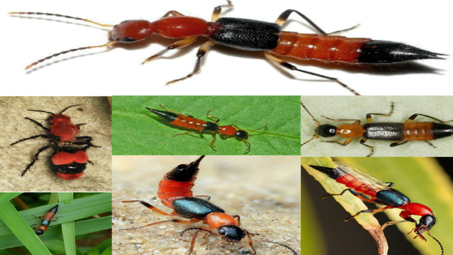
Among the various species of Paederus, some are more commonly associated with Paederus dermatitis, including Paederus fuscipes, Paederus littoralis, Paederus sabaeus, and Paederus riparius.
2.2 Morphological Features
Paederus beetles are small, measuring between 7–10 mm in length and 0.5–1 mm in width. Their slender and elongated body allows them to move quickly, making them highly mobile predators.
Key Morphological Characteristics
2.3 Behavioral and Ecological Traits
2.3.1 Habitat Preferences
These beetles flourish in warm and humid conditions and are frequently observed in agricultural fields, wetlands, riverbanks, and regions characterized by decaying organic matter. These organisms exhibit a preference for moist soil conditions and are frequently found in environments abundant in vegetation, including rice paddies, marshlands, and compost piles (7).
2.3.2 Nocturnal Activity and Attraction to Light
Paederus beetles exhibit primarily nocturnal behavior and demonstrate a significant attraction to artificial light sources. This phenomenon, referred to as positive phototaxis, frequently results in their intrusion into residential areas, medical facilities, and educational institutions, thereby heightening the likelihood of human interaction. Their affinity for artificial illumination renders urban settings especially susceptible to occurrences of Paederus dermatitis, particularly in the rainy season.
2.3.3 Role in Pest Control
Paederus beetles function as natural predators of agricultural pests, including aphids, whiteflies, and small caterpillars, contributing to ecological balance. Their presence in agricultural fields plays a significant role in integrated pest management by diminishing the dependence on chemical pesticides. Nonetheless, their advantageous function is mitigated by their medical implications upon interaction with humans (8).
2.4 Pederin: The Toxic Compound
2.4.1 Chemical Structure and Properties
Pederin (C??H??O?N) is a notable polyketide amide recognized for its cytotoxic and vesicant characteristics. It impedes the processes of DNA and protein synthesis, ultimately leading to apoptosis in epidermal cells. Structurally, it bears resemblance to specific marine toxins identified in sponges, and its biological activity has garnered attention within the pharmaceutical field.
2.4.2 Source of Pederin
In contrast to venoms generated by the organisms that utilize them, pederin is synthesized by an endosymbiotic bacterium (Pseudomonas species) residing within the beetle. The beetle does not produce the toxin independently; rather, it acquires it via its bacterial symbionts. This distinctive mechanism has prompted investigations into cytotoxins derived from bacteria, which may have significant biomedical applications.
2.4.3 Effects of Pederin on Human Skin
2.5 Role of Paederus Beetles in Public Health
While Paederus beetles do not serve as vectors for infectious diseases, their influence on dermatological health is considerable. Instances of Paederus dermatitis are commonly documented in military installations, educational institutions, and urban environments where artificial illumination draws beetles inside.
2.6 Economic and Public Health Importance
2.7 Future Research Directions
Despite extensive research, several aspects of Paederus beetles remain poorly understood:
3. Pathophysiology of Paederus Dermatitis
3.1 Introduction to Pathophysiology
Paederus dermatitis represents a form of irritant contact dermatitis that arises from exposure to pederin, a highly potent vesicant toxin present in the hemolymph of Paederus beetles. In contrast to allergic or infectious dermatitis, the inflammatory response observed in Paederus dermatitis arises from chemical irritation rather than an immune-mediated reaction. The pathophysiological process is defined by epidermal necrosis, inflammation, and a delayed onset of symptoms subsequent to toxin exposure.
3.2 Mechanism of Pederin Toxicity
3.2.1 Chemical Structure and Action of Pederin
Pederin is a polyketide amide compound recognized for its cytotoxic characteristics. It functions by irreversibly obstructing protein and DNA synthesis, resulting in cellular apoptosis and inflammatory damage. The toxin exhibits structural similarities to other polyketides that possess established antitumor properties, thereby positioning it as a significant focus within pharmacological research.
3.2.2 Effects on Cellular Structures
Upon skin exposure, pederin disrupts normal cellular function by:
The degree of tissue damage depends on concentration, exposure duration, and mechanical spread of the toxin. Unlike bacterial or viral infections, no infectious agent is involved in the progression of the disease.
3.3 Stages of Cutaneous Reaction
3.3.1 Initial Contact Phase (0–12 Hours)
3.3.2 Inflammatory Phase (12–48 Hours)
3.3.3 Vesiculation and Necrotic Phase (2–5 Days)
3.3.4 Resolution Phase (1–2 Weeks)
3.4 Factors Influencing Severity of Lesions
Several factors determine the extent of tissue damage and recovery:
3.5 Systemic and Ocular Complications
While Paederus dermatitis is primarily localized to the skin, rare systemic effects have been reported:
3.5.1 Ocular Involvement (Paederus Keratoconjunctivitis)
3.5.2 Secondary Bacterial Infections
3.5.3 Rare Systemic Toxicity
3.6 Immune Response and Healing
Unlike allergic contact dermatitis, which involves a T-cell mediated immune response, Paederus dermatitis is driven by direct chemical irritation and innate immune activation. The healing process is characterized by:
3.7 Pathophysiology Compared to Other Dermatitis Types
|
Feature |
Paederus Dermatitis |
Herpes Zoster |
Phytophotodermatitis |
|
Cause |
Pederin toxin exposure |
Varicella-zoster virus |
Plant-derived photosensitizers + UV light |
|
Onset |
Delayed (8–24 hrs) |
Prodrome (1–3 days) |
Delayed (24–72 hrs) |
|
Primary Lesion |
Linear vesicles & ulcers |
Dermatomal blisters |
Irregular erythematous patches |
|
Healing Time |
1–2 weeks |
2–4 weeks |
2–3 weeks |
|
Complications |
Secondary infections, hyperpigmentation |
Postherpetic neuralgia |
Hyperpigmentation, peeling |
3.8 Future Research Directions
While much is known about Paederus dermatitis, certain areas remain underexplored:
3.9 Conclusion
The pathophysiology of Paederus dermatitis is distinct from allergic or infectious skin conditions, as it is purely a chemical-induced inflammatory reaction. The delayed onset, vesiculation, and necrosis result from pederin-mediated inhibition of protein synthesis, leading to cellular apoptosis and inflammatory cytokine activation. While the condition is self-limiting, ocular involvement, bacterial superinfection, and severe necrotic lesions can pose significant medical concerns. Further research is needed to develop targeted treatments to neutralize pederin and prevent its harmful effects on human skin.
4. Clinical Presentation of Paederus Dermatitis
4.1 Introduction
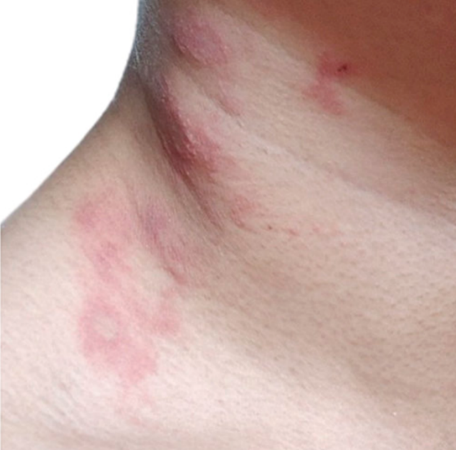
Paederus dermatitis represents a specific type of irritant contact dermatitis, which is induced by pederin, a vesicant toxin that is released upon the crushing of Paederus beetles against the skin. In contrast to allergic reactions or infectious skin diseases, Paederus dermatitis is characterized by a delayed-onset inflammatory process, which includes linear erythema, vesiculation, ulceration, and post-inflammatory hyperpigmentation. The intensity of symptoms is contingent upon the duration of exposure, the concentration of toxins, and the mechanical dispersion of the hemolymph. The condition primarily impacts areas of the body that are exposed, and the distinctive "kissing lesions" arise from the transfer of toxins to neighboring skin surfaces. In severe instances, complications may arise, including secondary bacterial infections and involvement of the ocular region.
4.2 Skin Lesions
The skin manifestations of Paederus dermatitis progress through four clinical stages, each exhibiting unique morphological characteristics.
4.2.1 Early Erythematous Stage (8–24 hours post-exposure)
4.2.2 Vesiculobullous Stage (24–48 hours post-exposure)
4.2.3 Ulcerative and Desquamation Stage (3–7 days post-exposure)
4.2.4 Resolution and Hyperpigmentation Stage (1–2 weeks post-exposure)
4.3 Symptoms of Paederus Dermatitis
The clinical symptoms depend on the severity of exposure and the patient’s skin sensitivity.
4.3.1 Localized Symptoms
4.3.2 Systemic Symptoms (Rare Cases)
Although Paederus dermatitis is primarily a localized skin reaction, some individuals may experience mild systemic effects, including:
4.3.3 Ocular Involvement (Paederus Keratoconjunctivitis)
If pederin-contaminated fingers or hands touch the eyes, a severe ocular reaction known as Paederus keratoconjunctivitis may develop:
If left untreated, corneal involvement may occur, requiring urgent ophthalmologic intervention.
4.4 Commonly Affected Body Areas
4.5 Differential Diagnosis
The unique linear and vesiculobullous lesions of Paederus dermatitis can mimic other dermatological conditions. Proper diagnosis is essential to avoid unnecessary antiviral or antibiotic treatments.
|
Condition |
Key Differences from Paederus Dermatitis |
|
Herpes Zoster |
Dermatomal distribution, grouped vesicles, pain precedes rash. |
|
Phytophotodermatitis |
Irregular hyperpigmentation, history of plant exposure + UV light. |
|
Bullous Impetigo |
Honey-colored crusts, bacterial etiology. |
|
Contact Dermatitis |
Pruritic, non-vesicular, associated with allergens/irritants. |
|
Chemical Burns |
Immediate burning, necrosis often deeper. |
4.6 Clinical Variants and Complications
4.6.1 Atypical Presentations
4.6.2 Secondary Complications
4.7 Conclusion
The clinical presentation of Paederus dermatitis follows a predictable progression, with delayed-onset erythema, vesiculation, ulceration, and eventual resolution with hyperpigmentation. The condition is self-limiting but can be painful and cosmetically distressing, particularly in cases involving facial or ocular exposure. Early recognition and supportive treatment can help minimize complications and accelerate healing.
4.1. Skin Lesions
Introduction
Paederus dermatitis is characterized by distinct cutaneous lesions resulting from contact with pederin, a vesicant toxin released when Paederus beetles are crushed against the skin. Unlike insect bites or allergic reactions, these lesions exhibit a linear or streak-like distribution, reflecting the movement of the toxin across the skin. The inflammatory reaction progresses through four distinct stages, with hallmark features including erythema, vesiculation, ulceration, and post-inflammatory hyperpigmentation. The severity of lesions depends on the concentration of pederin, duration of contact, and mechanical spread. In some cases, secondary bacterial infections or persistent pigmentation changes can prolong the healing process.
Types of Skin Lesions in Paederus Dermatitis
The lesions in Paederus dermatitis evolve through four clinically identifiable stages, each with distinct morphological characteristics.
1. Erythematous Phase (8–24 hours post-exposure)
2. Vesiculobullous Phase (24–48 hours post-exposure)
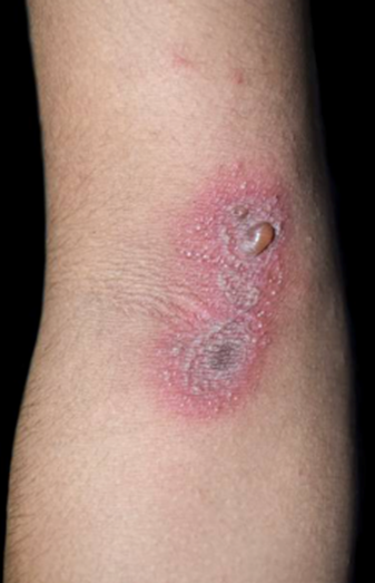
3. Ulcerative and Desquamation Phase (3–7 days post-exposure)
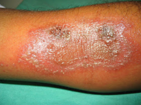
4. Resolution and Hyperpigmentation Phase (1–2 weeks post-exposure)
Morphological Variations of Skin Lesions
While the classic linear streak pattern is the most common presentation, variations exist depending on exposure circumstances, environmental conditions, and skin characteristics.
1. Kissing Lesions
2. Diffuse or Atypical Lesions
3. Necrotic or Deep Ulcerative Lesions
Most Commonly Affected Areas
Differentiating Paederus Dermatitis from Other Skin Conditions
|
Condition |
Key Differences from Paederus Dermatitis |
|
Herpes Zoster |
Unilateral, dermatomal blisters with severe pain before rash onset. |
|
Phytophotodermatitis |
Irregular hyperpigmentation with history of plant exposure + UV light. |
|
Bullous Impetigo |
Yellow-crusted vesicles, often in children, caused by bacterial infection. |
|
Chemical Burns |
Immediate burning with deep necrosis, no delayed onset. |
|
Contact Dermatitis |
Pruritic, non-vesicular rash associated with allergens or irritants. |
Complications Associated with Skin Lesions
While Paederus dermatitis is usually self-limiting, complications may arise in untreated or severe cases.
1. Secondary Bacterial Infection
2. Post-inflammatory Pigmentation Changes
3. Rare Systemic Symptoms
Conclusion
The skin lesions of Paederus dermatitis exhibit a predictable progression from erythema to vesiculation, ulceration, and hyperpigmentation. The condition is self-limiting but can be painful, cosmetically distressing, and prone to secondary complications. Understanding the lesion morphology and progression helps in early diagnosis and appropriate management to prevent prolonged symptoms and minimize scarring.
4.2. Symptoms
Introduction
The symptoms of Paederus dermatitis arise due to direct chemical irritation caused by pederin, a potent vesicant toxin released from Paederus beetles when crushed against the skin. Unlike allergic reactions or infectious skin conditions, Paederus dermatitis follows a delayed-onset inflammatory response, typically appearing 8–24 hours after exposure. Symptoms progress through distinct stages, including erythema, vesiculation, ulceration, and post-inflammatory hyperpigmentation. The severity and duration of symptoms depend on factors such as the amount of toxin exposure, mechanical spread, skin sensitivity, and environmental conditions. In some cases, complications such as secondary infections, persistent pigmentation changes, and ocular involvement may occur.
4.2.1 Localized Symptoms
The skin manifestations of Paederus dermatitis follow a predictable clinical course and primarily affect exposed body areas.
1. Burning and Tingling Sensation (8–12 hours post-exposure)
2. Erythema and Edema (12–24 hours post-exposure)
3. Vesiculation and Blister Formation (24–48 hours post-exposure)
4. Ulceration and Peeling (3–7 days post-exposure)
5. Post-inflammatory Hyperpigmentation (1–2 weeks post-exposure)
4.2.2 Systemic Symptoms (Rare Cases)
While Paederus dermatitis is primarily localized, some patients—especially those with multiple lesions or extensive skin involvement—may experience mild systemic symptoms. These include:
4.2.3 Ocular Symptoms (Paederus Keratoconjunctivitis)
If pederin-contaminated hands or towels come into contact with the eyes, severe conjunctival inflammation can develop, leading to a condition known as Paederus keratoconjunctivitis. Symptoms include:
4.2.4 Complications of Paederus Dermatitis Symptoms
While Paederus dermatitis is generally self-limiting, some cases may develop complications due to excessive toxin spread, improper wound care, or secondary infections.
1. Secondary Bacterial Infection
2. Persistent Hyperpigmentation
3. Extensive Ulceration and Scarring
4. Psychological and Cosmetic Impact
4.2.5 Differential Diagnosis of Symptoms
Since Paederus dermatitis shares similar symptoms with various dermatological conditions, distinguishing features are critical for accurate diagnosis and appropriate treatment.
|
Condition |
Key Differences from Paederus Dermatitis |
|
Herpes Zoster |
Unilateral dermatomal vesicles with severe pain before rash onset. |
|
Phytophotodermatitis |
Irregular hyperpigmentation with history of plant exposure + UV light. |
|
Bullous Impetigo |
Yellow-crusted vesicles, often in children, caused by bacterial infection. |
|
Chemical Burns |
Immediate burning with deep necrosis, no delayed onset. |
|
Contact Dermatitis |
Pruritic, non-vesicular rash associated with allergens or irritants. |
4.2.6 Prognosis and Resolution of Symptoms
Conclusion
The symptoms of Paederus dermatitis follow a predictable progression, beginning with erythema and burning, followed by vesiculation, ulceration, and post-inflammatory hyperpigmentation. While most cases resolve without complications, ocular involvement, secondary bacterial infections, and prolonged pigmentation changes may require medical intervention. Early recognition and supportive management can minimize discomfort and prevent complications.
5. Diagnosis of Paederus Dermatitis
5.1 Introduction
The diagnosis of Paederus dermatitis is primarily clinical, based on characteristic skin lesions, exposure history, and symptom progression. Unlike allergic or infectious skin conditions, Paederus dermatitis follows a predictable timeline with delayed-onset erythema, vesiculation, and ulceration, often in a linear or streaked pattern. Given its non-specific early symptoms, Paederus dermatitis is often misdiagnosed as herpes zoster, phytophotodermatitis, or chemical burns. A careful patient history, including potential exposure to Paederus beetles in endemic areas, is essential for accurate identification and prevention of unnecessary treatments.
5.2 Physical Examination
A thorough dermatological examination is the cornerstone of diagnosis. Key clinical features include:
5.2.1 Skin Lesion Characteristics
5.2.2 Commonly Affected Body Areas
5.2.3 Associated Symptoms
5.4 Diagnostic Tools and Laboratory Investigations
Although Paederus dermatitis is diagnosed clinically, additional investigations may be needed in atypical cases or suspected complications.
5.4.1 Dermoscopy
5.4.2 Skin Biopsy (Rarely Needed)
5.4.3 Microbiological Testing (Only for Secondary Infections)
5.5 Diagnostic Pitfalls and Challenges
Despite its characteristic presentation, Paederus dermatitis is often misdiagnosed due to:
5.6 Importance of Early Recognition
5.7 Conclusion
The diagnosis of Paederus dermatitis relies on clinical recognition of its characteristic skin lesions and exposure history. While laboratory tests are rarely required, differentiating it from infectious, allergic, and phototoxic skin conditions is essential for proper management. Early identification can prevent complications, unnecessary treatments, and recurrence through effective preventive measures (22-25).
5.1. Physical Examination
Introduction
The physical examination is the primary diagnostic tool for Paederus dermatitis. Since the condition results from direct chemical irritation by pederin rather than an allergic or infectious process, it exhibits distinctive clinical features. The examination should focus on skin lesion morphology, distribution, progression, and associated symptoms. A detailed patient history—including recent outdoor activity, exposure to artificial lights at night, and potential beetle encounters—can help confirm the diagnosis.
5.1.1 General Skin Inspection
A thorough dermatological assessment should be performed under adequate lighting to identify the unique lesion patterns of Paederus dermatitis.
Key Clinical Features
Lesions are often unilateral and appear in areas exposed during sleep or outdoor activities.
Lesion Evolution During Examination
|
Stage |
Physical Findings |
|
Early Phase (8–24 hrs) |
Erythematous streaks, mild swelling, possible tingling. |
|
Vesiculobullous Phase (24–48 hrs) |
Formation of vesicles, bullae, or pustules, burning sensation. |
|
Ulcerative Phase (3–7 days) |
Ruptured blisters, crusting, peeling, necrosis in severe cases. |
|
Healing Phase (1–2 weeks) |
Hyperpigmentation, mild residual erythema, rare hypopigmentation. |
5.1.2 Commonly Affected Body Areas
The physical examination should focus on exposed body sites, as Paederus beetles typically contact uncovered skin.
Most Frequent Sites of Lesions
Special attention should be given to skin folds and flexural areas, as kissing lesions may form when affected skin surfaces come into contact.
5.1.3 Palpation and Sensory Examination
A careful palpation of the lesions can help differentiate Paederus dermatitis from other dermatological conditions.
Tactile Findings
Pain and Sensation Assessment
5.1.4 Ocular Examination (Paederus Keratoconjunctivitis)
If the patient reports eye irritation or redness, an ocular examination should be performed to rule out Paederus keratoconjunctivitis.
Ophthalmologic Findings
If ocular involvement is suspected, urgent referral to an ophthalmologist is required.
5.1.5 Signs of Complications
While Paederus dermatitis is usually self-limiting, the physical examination should assess for secondary infections or atypical presentations.
Indicators of Secondary Bacterial Infection
Signs of Severe Ulceration or Necrosis
If these complications are noted, topical or systemic antibiotic therapy may be required.
5.1.6 Differentiating Paederus Dermatitis from Similar Skin Conditions
During the physical examination, the clinician should compare Paederus dermatitis with other dermatological conditions that present with vesiculobullous or ulcerative lesions.
|
Condition |
Distinctive Features Compared to Paederus Dermatitis |
|
Herpes Zoster |
Unilateral dermatomal distribution, intense prodromal pain before vesicles appear. |
|
Phytophotodermatitis |
Irregular hyperpigmented patches, history of plant exposure + UV light. |
|
Bullous Impetigo |
Honey-colored crusts, bacterial etiology, common in children. |
|
Contact Dermatitis |
Pruritic, non-vesicular rash linked to allergen exposure. |
|
Chemical Burns |
Immediate pain, deeper necrosis, history of chemical exposure. |
A careful review of lesion morphology and patient history is key to making an accurate diagnosis.
Conclusion
The physical examination in Paederus dermatitis focuses on identifying distinctive linear erythematous and vesiculobullous lesions, assessing common exposure sites, and ruling out secondary infections or atypical presentations. A detailed lesion assessment and history of beetle exposure can help confirm the diagnosis without the need for laboratory tests.
5.2. Differential Diagnosis
Introduction
The clinical presentation of Paederus dermatitis—characterized by delayed-onset erythema, vesiculation, ulceration, and hyperpigmentation—can mimic various dermatological conditions. Due to its linear lesion distribution and lack of immediate pain, it is often misdiagnosed as viral, bacterial, or chemical-induced dermatitis. An accurate differential diagnosis is crucial to prevent unnecessary antiviral, antifungal, or antibiotic treatments and to ensure appropriate symptom management.
5.2.1 Common Conditions Mimicking Paederus Dermatitis
The following dermatological conditions can resemble Paederus dermatitis but have distinctive clinical features that help differentiate them.
1. Herpes Zoster (Shingles)
|
Feature |
Herpes Zoster |
Paederus Dermatitis |
|
Cause |
Reactivation of Varicella-zoster virus (VZV) |
Chemical irritation from pederin toxin |
|
Lesion Pattern |
Dermatomal distribution, unilateral |
Linear streaks, often irregular |
|
Onset |
Prodromal pain (burning, tingling) 2–3 days before rash |
No prodrome, rash appears 8–24 hrs after exposure |
|
Primary Lesion |
Grouped vesicles on an erythematous base |
Vesicles/bullae with erythematous streaks |
|
Healing Time |
2–4 weeks |
1–2 weeks, with hyperpigmentation |
|
Complications |
Postherpetic neuralgia (PHN), secondary bacterial infection |
Hyperpigmentation, secondary bacterial infection (rare) |
Key Differentiating Factors:
2. Phytophotodermatitis
|
Feature |
Phytophotodermatitis |
Paederus Dermatitis |
|
Cause |
Plant-derived psoralens + UV exposure |
Pederin toxin from Paederus beetle |
|
Lesion Pattern |
Irregular, blotchy erythema, often with drips or streaks |
Linear streaks with kissing lesions |
|
Onset |
Delayed (24–72 hrs) after sun exposure |
Delayed (8–24 hrs) after beetle exposure |
|
Primary Lesion |
Bullae, hyperpigmentation |
Bullae, ulceration, hyperpigmentation |
|
Healing Time |
2–4 weeks, pigmentation may persist |
1–2 weeks, pigmentation may persist longer |
|
Complications |
Severe hyperpigmentation, prolonged skin sensitivity to sunlight |
Hyperpigmentation, secondary infection (rare) |
Key Differentiating Factors:
3. Bullous Impetigo
|
Feature |
Bullous Impetigo |
Paederus Dermatitis |
|
Cause |
Staphylococcus aureus infection |
Pederin toxin from Paederus beetle |
|
Lesion Pattern |
Localized or widespread, fragile bullae |
Linear vesiculobullous streaks |
|
Onset |
Rapid (within hours) |
Delayed (8–24 hrs) |
|
Primary Lesion |
Flaccid bullae, honey-colored crusts |
Tense bullae, ulceration, peeling |
|
Healing Time |
7–10 days, can spread |
1–2 weeks, localized |
|
Complications |
Bacterial superinfection, systemic illness in neonates |
Hyperpigmentation, secondary infection (rare) |
Key Differentiating Factors:
4. Allergic Contact Dermatitis (ACD)
|
Feature |
Allergic Contact Dermatitis |
Paederus Dermatitis |
|
Cause |
Allergens (e.g., nickel, fragrances, latex) |
Pederin toxin exposure |
|
Lesion Pattern |
Patchy erythema, pruritic papules or vesicles |
Linear vesicles, streaked erythema |
|
Onset |
Delayed (24–48 hrs) after allergen contact |
Delayed (8–24 hrs) after exposure |
|
Primary Lesion |
Red, swollen plaques, weepy vesicles |
Tense bullae, ulceration |
|
Healing Time |
1–3 weeks, resolves with allergen avoidance |
1–2 weeks, hyperpigmentation persists |
|
Complications |
Chronic eczematous dermatitis with repeated exposure |
Hyperpigmentation, secondary infection (rare) |
Key Differentiating Factors:
5. Chemical Burns
|
Feature |
Chemical Burns |
Paederus Dermatitis |
|
Cause |
Acidic or alkaline chemical exposure |
Pederin toxin exposure |
|
Lesion Pattern |
Sharply demarcated burns, deep necrosis |
Linear vesiculobullous streaks |
|
Onset |
Immediate pain and damage |
Delayed symptoms (8–24 hrs post-exposure) |
|
Primary Lesion |
Necrotic tissue, ulceration, eschar formation |
Vesicles, ulceration, peeling |
|
Healing Time |
Weeks to months, may cause scarring |
1–2 weeks, usually no scarring |
|
Complications |
Deep burns, permanent scarring, systemic toxicity |
Hyperpigmentation, secondary infection (rare) |
Key Differentiating Factors:
Conclusion
The differential diagnosis of Paederus dermatitis requires careful evaluation of lesion morphology, symptom onset, and patient history. The linear, vesiculobullous streaks with a delayed onset and residual hyperpigmentation distinguish it from herpes zoster, phytophotodermatitis, bullous impetigo, allergic contact dermatitis, and chemical burns. Proper differentiation helps prevent unnecessary antiviral, antibiotic, or steroid treatments, allowing for accurate patient management and effective preventive measures.
6. Treatment Options for Paederus Dermatitis
6.1 Introduction
The treatment of Paederus dermatitis focuses on symptom relief, prevention of complications, and accelerated healing. Since the condition is caused by pederin, a non-infectious chemical toxin, antibiotics and antivirals are not required unless secondary bacterial infections occur. Early recognition and proper wound care can significantly reduce pain, inflammation, and post-inflammatory hyperpigmentation. Management strategies include topical and systemic therapies, supportive care, and preventive measures to minimize the risk of recurrence.
6.2 First-Aid and Immediate Management
Early intervention can prevent severe vesiculation and ulceration. Patients with suspected exposure should:
6.2.1 Immediate Actions After Exposure
Early cleansing within one hour of exposure can significantly reduce the severity of the reaction.
6.3 Topical Therapies
6.3.1 Corticosteroids
6.3.2 Antihistamines
6.3.3 Antiseptic and Antibiotic Ointments
6.3.4 Moisturizers and Skin Protectants
6.4 Systemic Therapies
6.4.1 Non-Steroidal Anti-Inflammatory Drugs (NSAIDs)
6.4.2 Systemic Corticosteroids (Severe Cases Only)
6.4.3 Antibiotics (Only for Secondary Infections)
6.5 Ocular Involvement Treatment (Paederus Keratoconjunctivitis)
If pederin exposure affects the eyes, immediate irrigation is critical to prevent corneal damage.
Ocular Treatment Approach:
6.6 Preventive Strategies
To reduce the risk of recurrence, the following preventive measures should be implemented, especially in endemic areas.
6.6.1 Personal Protection
6.6.2 Environmental Control
6.6.3 Public Awareness and Education
6.7 Future Research Directions in Treatment
Despite effective symptomatic treatments, further research is needed to:
6.8 Conclusion
The treatment of Paederus dermatitis involves early skin decontamination, anti-inflammatory therapy, pain management, and prevention of complications. Topical corticosteroids and antihistamines are the mainstays of therapy, while systemic treatments are reserved for severe cases. Preventive strategies, including protective clothing, environmental control, and public education, are crucial in endemic regions to reduce exposure and recurrence.
6.1. Topical Therapies
Introduction
Topical treatments are the first-line approach for managing Paederus dermatitis, aiming to reduce inflammation, relieve symptoms, prevent complications, and accelerate healing. Since the condition is caused by a chemical toxin (pederin) rather than an infectious agent, antibiotics and antivirals are unnecessary unless secondary bacterial infection occurs.
The primary goals of topical therapy include:
6.1.1 Corticosteroids (First-line Treatment)
Topical corticosteroids are the most effective treatment for reducing inflammation and preventing lesion progression when applied early.
Types of Corticosteroids Used
|
Severity of Lesions |
Recommended Corticosteroid |
Strength |
Dosage |
|
Mild cases (erythema, no vesicles) |
Hydrocortisone 1% cream |
Low |
Twice daily for 3–5 days |
|
Moderate cases (vesiculation, mild ulceration) |
Betamethasone valerate 0.1% |
Medium |
Twice daily for 5–7 days |
|
Severe cases (extensive bullae, severe inflammation) |
Clobetasol propionate 0.05% |
High |
Once daily for 3–5 days, then taper |
Mechanism of Action
Application Guidelines
6.1.2 Antihistamines (For Pruritus Relief)
Topical antihistamines help reduce itching and irritation, especially in the vesiculobullous phase.
Commonly Used Topical Antihistamines
Application Guidelines
6.1.3 Antiseptic and Antibiotic Ointments (For Secondary Infection Prevention)
Although Paederus dermatitis is not primarily infectious, open vesicles and ulcerated lesions increase the risk of bacterial superinfection.
Indications for Use
Recommended Topical Antibiotics
|
Antibiotic |
Spectrum of Activity |
Indications |
Dosage |
|
Mupirocin 2% ointment |
Staphylococcus aureus, Streptococcus pyogenes |
Small areas with suspected bacterial colonization |
Apply twice daily for 5–7 days |
|
Fusidic acid cream |
Gram-positive bacteria |
Localized pustules |
Apply twice daily |
|
Silver sulfadiazine cream |
Broad-spectrum antibacterial |
Extensive ulceration |
Apply once daily |
Application Guidelines:
6.1.4 Moisturizers and Barrier Creams (For Skin Protection and Healing)
Moisturizing agents help maintain skin hydration, reduce irritation, and accelerate healing.
Recommended Skin Protectants
Application Guidelines
6.1.5 Cooling Agents and Astringents (For Symptom Relief and Vesicle Drying)
Astringents help dry out vesicles, reduce exudation, and promote crust formation.
Common Astringent Solutions
|
Agent |
Mechanism |
Application |
|
Burow’s solution (aluminum acetate 5%) |
Reduces weeping and inflammation |
Apply soaked compress for 15 min, 2–3 times daily |
|
Potassium permanganate solution (1:10,000 dilution) |
Acts as an antiseptic and drying agent |
Use diluted solution for soaks or compresses |
6.1.6 Emerging Topical Treatments (Under Research)
6.1.7 Summary of Topical Therapy Recommendations
|
Treatment Type |
Primary Function |
Commonly Used Agents |
Application Guidelines |
|
Corticosteroids |
Reduces inflammation |
Hydrocortisone, betamethasone, clobetasol |
Apply twice daily, avoid prolonged use |
|
Antihistamines |
Relieves itching |
Diphenhydramine, doxepin |
Use sparingly, avoid excessive application |
|
Antibiotics (if infected) |
Prevents/treats secondary infection |
Mupirocin, fusidic acid |
Apply twice daily, only if needed |
|
Moisturizers & Protectants |
Soothes and repairs skin |
Aloe vera, zinc oxide, calamine |
Use 2–3 times daily |
|
Astringents |
Dries vesicles, reduces swelling |
Burow’s solution, potassium permanganate |
Use as compresses |
Conclusion
Topical therapies play a central role in the management of Paederus dermatitis, helping to control inflammation, prevent infection, and promote skin healing. Corticosteroids are the most effective agents for reducing inflammation, while antihistamines and astringents provide symptom relief. Moisturizers and barrier creams aid in skin regeneration, and antibiotic ointments should be reserved for infected lesions. Early and appropriate topical treatment can minimize complications and accelerate recovery.
6.2. Systemic Therapies
Introduction
Systemic therapies for Paederus dermatitis are reserved for moderate to severe cases, where inflammation is extensive, pain is significant, or secondary infections are suspected. While topical treatments remain the first-line approach, oral medications can help control inflammation, pain, pruritus, and bacterial superinfection in more severe cases.
The primary goals of systemic therapy include:
6.2.1 Non-Steroidal Anti-Inflammatory Drugs (NSAIDs)
Indications:
Commonly Used NSAIDs
|
Drug |
Dosage |
Administration |
Precautions |
|
Ibuprofen |
400–600 mg |
Every 6–8 hours |
Avoid in patients with gastric ulcers or kidney disease |
|
Paracetamol (Acetaminophen) |
500–1000 mg |
Every 6–8 hours |
Safe in most patients, does not reduce inflammation |
|
Naproxen |
250–500 mg |
Every 12 hours |
May cause gastric irritation |
Mechanism of Action:
Application Guidelines:
6.2.2 Systemic Corticosteroids (For Severe Cases)
Indications:
Commonly Used Corticosteroids
|
Drug |
Dosage |
Duration |
Precautions |
|
Prednisolone |
20–40 mg/day |
5–7 days |
Taper gradually to avoid rebound effects |
|
Dexamethasone |
4–8 mg/day |
3–5 days |
More potent, use only for severe inflammation |
Mechanism of Action:
Application Guidelines:
6.2.3 Oral Antihistamines (For Pruritus and Inflammation Control)
Indications:
Commonly Used Oral Antihistamines
|
Drug |
Dosage |
Administration |
Precautions |
|
Loratadine |
10 mg |
Once daily |
Non-sedating, preferred for daytime use |
|
Cetirizine |
10 mg |
Once daily |
Mild sedation in some individuals |
|
Diphenhydramine |
25–50 mg |
Every 6–8 hours |
Causes drowsiness, best for nighttime relief |
|
Hydroxyzine |
10–25 mg |
Every 6–8 hours |
Stronger sedative effect, used for severe pruritus |
Mechanism of Action:
Application Guidelines:
6.2.4 Systemic Antibiotics (For Secondary Bacterial Infections)
Indications:
Commonly Used Antibiotics
|
Antibiotic |
Dosage |
Spectrum |
Indications |
|
Cephalexin |
500 mg every 6–8 hrs |
Gram-positive |
Mild to moderate infection |
|
Doxycycline |
100 mg every 12 hrs |
Broad-spectrum |
Resistant S. aureus or extensive infection |
|
Amoxicillin-Clavulanate |
875 mg every 12 hrs |
Broad-spectrum |
Moderate to severe infections |
Mechanism of Action:
Application Guidelines:
6.2.5 Systemic Therapy for Paederus Keratoconjunctivitis (Ocular Involvement)
Indications:
Recommended Systemic Treatments
6.2.6 Summary of Systemic Therapy Recommendations
|
Treatment Type |
Indications |
Commonly Used Drugs |
Dosage & Frequency |
|
NSAIDs |
Pain, mild inflammation |
Ibuprofen, naproxen |
400–600 mg every 6–8 hrs |
|
Corticosteroids |
Severe inflammation |
Prednisolone, dexamethasone |
20–40 mg/day (short course) |
|
Antihistamines |
Pruritus, inflammation |
Loratadine, cetirizine |
10 mg/day |
|
Antibiotics |
Secondary infections |
Cephalexin, doxycycline |
Based on severity |
Conclusion
Systemic therapies play a crucial role in managing moderate to severe Paederus dermatitis by controlling inflammation, pain, and secondary infections. NSAIDs and antihistamines provide symptomatic relief, while corticosteroids are reserved for severe inflammation. Oral antibiotics are only necessary if bacterial superinfection occurs. Proper systemic therapy selection ensures faster recovery, reduced complications, and better patient outcomes.
7. Preventive Measures for Paederus Dermatitis
7.1 Introduction
Preventing Paederus dermatitis primarily involves avoiding exposure to Paederus beetles and reducing human contact with pederin toxin. Since Paederus beetles do not bite or sting, dermatitis occurs only when their bodies are crushed against the skin, releasing pederin, a potent vesicant toxin. Preventive strategies focus on environmental control, personal protective measures, and public awareness to minimize beetle-human interactions.
7.2 Avoidance Strategies
Avoiding direct contact with Paederus beetles is the most effective way to prevent dermatitis.
7.2.1 Personal Protective Measures
7.2.2 Indoor Protection and Behavior Modification
7.3 Environmental Control
Controlling Paederus beetle populations in surrounding habitats can significantly reduce the risk of outbreaks.
7.3.1 Habitat Modification
7.4 Public Awareness and Education
7.4.1 Educating Communities in Endemic Areas
7.4.2 Dissemination of Preventive Information
7.5 Preventing Ocular Involvement (Paederus Keratoconjunctivitis)
Since accidental transfer of pederin to the eyes can cause severe conjunctivitis, the following precautions should be taken:
7.6 Preventive Measures During Outbreaks
7.7 Future Directions in Prevention
7.8 Conclusion
Prevention of Paederus dermatitis relies on a combination of personal protective measures, environmental control, public education, and outbreak monitoring. Avoiding direct contact with beetles, reducing artificial lighting, and maintaining proper hygiene can significantly lower the risk. Integrating community awareness and scientific research will help develop more effective, sustainable prevention strategies in the future.
7.1. Avoidance Strategies
Introduction
The most effective way to prevent Paederus dermatitis is to avoid direct contact with Paederus beetles and minimize exposure to pederin toxin. Unlike biting or stinging insects, Paederus beetles cause dermatitis only when crushed against the skin, releasing their toxic hemolymph. Implementing personal protective measures and modifying behaviors in beetle-prone areas can significantly reduce the risk of exposure.
7.1.1 Personal Protective Measures
1. Avoid Direct Contact with Beetles
2. Immediate Skin Decontamination
3. Wear Protective Clothing
4. Use Insect Repellents
7.1.2 Indoor Protection and Behavior Modification
1. Reduce Artificial Lighting at Night
2. Use Window Screens and Bed Nets
3. Close Windows and Doors at Night
4. Maintain a Clean Indoor Environment
7.1.3 Safe Handling of Beetles and Contaminated Objects
1. Proper Disposal of Beetles
2. Wash Contaminated Items
3. Educate Household Members and Workers
7.1.4 Avoidance Strategies in High-Risk Occupations
Individuals working in beetle-infested environments—such as farmers, soldiers, construction workers, and outdoor laborers—are at increased risk of exposure.
1. Farmers and Field Workers
2. Military Personnel and Outdoor Laborers
3. School and Office Workers
7.1.5 Seasonal and Climate-Based Precautions
1. Avoid Outdoor Activities During Peak Beetle Seasons
2. Monitor Beetle Activity in Endemic Regions
Conclusion
Avoiding Paederus beetles is the most effective strategy for preventing Paederus dermatitis. Personal protective measures, behavior modifications, and environmental management all play a role in reducing human-beetle interactions. By minimizing artificial lighting, wearing protective clothing, using repellents, and ensuring proper handling techniques, individuals can significantly lower their risk of exposure to pederin toxin and its dermatological effects.
7.2. Environmental Control
Introduction
Environmental control is a crucial strategy in preventing Paederus dermatitis, as it focuses on reducing Paederus beetle populations and limiting their interactions with human habitats. Since these beetles thrive in warm, humid environments and are attracted to artificial lights, modifying these factors can significantly reduce the risk of beetle infestations and human exposure.
Effective environmental control measures include:
7.2.1 Habitat Modification
Paederus beetles are commonly found in moist environments, such as wetlands, rice paddies, and areas with decaying vegetation. Reducing these breeding sites near human settlements can significantly decrease beetle populations.
1. Drainage and Moisture Control
2. Vegetation and Waste Management
7.2.2 Light Management to Reduce Beetle Attraction
Paederus beetles exhibit positive phototaxis (strong attraction to artificial lights), which increases their presence in homes, schools, and workplaces. Managing light sources is one of the most effective ways to prevent beetle invasions.
1. Reduce Artificial Lighting at Night
2. Modify Indoor Lighting Practices
7.2.3 Physical Barriers and Protective Structures
1. Install Window Screens and Door Seals
2. Use Bed Nets in High-Risk Areas
3. Protective Enclosures for Agriculture and Workplaces
7.2.4 Chemical Control Strategies
1. Targeted Insecticide Use
2. Eco-Friendly and Natural Repellents
7.2.5 Biological Control Methods
7.2.6 Environmental Surveillance and Monitoring
1. Establishing Beetle Surveillance Programs
2. Community-Based Reporting Systems
7.2.7 Large-Scale Urban and Rural Planning Considerations
1. Urban Planning to Minimize Beetle Habitats
2. Rural Management for Agricultural Workers
7.2.8 Preventive Actions During Beetle Outbreaks
Conclusion
Environmental control is a key strategy in preventing Paederus dermatitis by reducing beetle habitats, modifying human surroundings, and using targeted chemical and biological interventions. Through proper waste management, light control, physical barriers, and habitat modification, the risk of human-beetle interactions can be significantly minimized. Public health surveillance and integrated pest management further enhance long-term prevention strategies.
7.2. Environmental Control
Introduction
Environmental control is a crucial strategy for preventing Paederus dermatitis, focusing on reducing beetle populations and minimizing their presence in human habitats. Since Paederus beetles are attracted to artificial light and thrive in humid, vegetated areas, modifying these environmental factors can significantly lower human exposure.
The key environmental control measures include:
7.2.1 Habitat Modification
Paederus beetles breed and thrive in moist environments with abundant organic material. By modifying these habitats, we can disrupt their lifecycle and reduce their population density.
1. Eliminating Breeding Sites
2. Vegetation Control Around Human Dwellings
3. Urban and Rural Planning Adjustments
7.2.2 Light Management to Reduce Beetle Attraction
Paederus beetles are highly attracted to artificial lights, particularly white, blue, and ultraviolet (UV) light sources. Adjusting lighting practices can significantly reduce beetle infestations in homes, workplaces, and public areas.
1. Modify Outdoor Lighting
2. Modify Indoor Lighting Practices
7.2.3 Physical Barriers and Structural Modifications
1. Install Protective Screens and Seals
2. Use Bed Nets in High-Risk Areas
3. Protect Agricultural Storage and Workplaces
7.2.4 Chemical Control Strategies
1. Targeted Use of Insecticides
2. Eco-Friendly and Non-Toxic Approaches
7.2.5 Biological Control Methods
7.2.6 Environmental Surveillance and Monitoring
1. Establishing Beetle Surveillance Programs
2. Community-Based Reporting and Public Health Alerts
7.2.7 Large-Scale Agricultural and Urban Planning
1. Agricultural Adjustments
2. Urban Planning for Beetle-Prone Regions
7.2.8 Emergency Response Measures During Outbreaks
Conclusion
Environmental control plays a key role in reducing Paederus beetle populations and minimizing their interactions with humans. Strategies such as habitat modification, light management, structural barriers, targeted insecticide use, and biological control can significantly reduce the risk of Paederus dermatitis. Public awareness and surveillance programs further enhance long-term prevention efforts. By integrating these environmental strategies, communities can create safer living and working conditions with reduced beetle-related risks.
8. Case Studies and Research on Paederus Dermatitis
8.1 Introduction
Paederus dermatitis has been widely reported in tropical and subtropical regions, particularly in Africa, Asia, and South America, where Paederus beetles thrive. Case studies and research have significantly contributed to understanding the epidemiology, pathophysiology, clinical presentation, and management strategies of this condition. This section explores notable case studies and recent research findings on Paederus dermatitis, focusing on outbreak reports, clinical investigations, and advancements in prevention and treatment.
8.2 Case Studies of Paederus Dermatitis
8.2.1 Outbreak in a Military Camp (India, 2015)
Key Findings:
8.2.2 Paederus Dermatitis in a Boarding School (Malaysia, 2018)
Key Findings:
8.2.3 Paederus Dermatitis in an Urban Hospital (Brazil, 2020)
Key Findings:
8.3 Recent Research on Paederus Dermatitis
8.3.1 Epidemiological Studies
8.3.2 Advances in Treatment
8.3.3 Environmental and Behavioral Research
8.4 Implications for Future Prevention and Management
Based on case studies and research findings, the following key takeaways have emerged:
1. Improved Public Awareness and Education
2. Enhanced Environmental Control
3. Development of New Treatment Approaches
4. Strengthened Surveillance Systems
8.5 Conclusion
Case studies and research have provided critical insights into the epidemiology, risk factors, and treatment strategies for Paederus dermatitis. Military camps, schools, hospitals, and agricultural communities remain high-risk settings, but effective prevention measures—such as reducing artificial lighting, using protective barriers, and maintaining sanitation—can significantly lower incidence rates. Future research should focus on developing novel treatments, improving environmental control strategies, and enhancing public education efforts to prevent Paederus dermatitis in endemic regions.
9. CONCLUSION AND FUTURE DIRECTIONS
9.1 CONCLUSION
Paederus dermatitis is a widespread, non-infectious inflammatory skin condition caused by accidental exposure to pederin, a potent vesicant toxin released from Paederus beetles. This condition is prevalent in tropical and subtropical regions, affecting military personnel, agricultural workers, students, and urban residents living in beetle-infested areas. The clinical course of Paederus dermatitis follows a predictable pattern, beginning with erythema and irritation, progressing to vesiculation and ulceration, and resolving with post-inflammatory hyperpigmentation. While mild cases heal within 1–2 weeks, severe cases can lead to persistent hyperpigmentation, secondary infections, and ocular complications.
Key Takeaways from This Research
While Paederus dermatitis is generally self-limiting, public health interventions and environmental modifications can greatly reduce its incidence and severity.
9.2 FUTURE DIRECTIONS
Despite advancements in understanding Paederus dermatitis, several areas require further research and innovation.
9.2.1 Development of Pederin-Neutralizing Agents
9.2.2 Novel Treatment Approaches
9.2.3 Improved Surveillance and Early Warning Systems
9.2.4 Long-Term Environmental Control Strategies
9.2.5 Enhancing Public Awareness and Education
9.3 Final Thoughts
Paederus dermatitis remains a significant public health concern, especially in beetle-infested regions. However, with advancements in medical research, environmental modifications, and public education, the burden of this condition can be substantially reduced. A multidisciplinary approach—involving medical researchers, entomologists, public health officials, and urban planners—is essential to develop sustainable, long-term solutions. By integrating preventive strategies with innovative treatments, future management of Paederus dermatitis can move toward more effective and globally applicable solutions (33-34).
ETHICAL STATEMENT
A pharmacist must behave honourably and honestly. A pharmacist refrains from behaviours that compromise their dedication to acting in the best interests of their patients, such as discriminatory actions, inappropriate behaviour, and unfavourable working conditions that cloud their judgment. A pharmacist maintains their degree of proficiency.
ACKNOWLEDGEMENT
The authors would like to thank Shri Venkateshwara University, Gajraula, Uttar Pradesh, India, Azad Institute of Pharmacy & Research, Lucknow, U.P., India, City Women's College, Jankipuram, Lucknow, Uttar Pradesh, India & Goel Institute of Pharmacy & Sciences, Lucknow, U.P., India. Lucknow, Uttar Pradesh, India, for extending their facilities.
CONFLICT OF INTEREST
The authors attest that they are free of any known financial or personal conflicts of interest that would taint the findings of this study.
INFORMED CONSENT
Using websites, review articles, and other sources to produce research content.
REFERENCES

Yash Srivastav, Aayush Singh Chauhan, Nutan Shrivastava, Madhaw Kumar, Paederus Dermatitis (Dermatitis Linearis): A Comprehensive Discussion of Its Aetiology, Toxicity, Clinical Features, Present State, and Future Prospects, Int. J. of Pharm. Sci., 2025, Vol 3, Issue 7, 1204-1248. https://doi.org/10.5281/zenodo.15845839
 10.5281/zenodo.15845839