
We use cookies to ensure our website works properly and to personalise your experience. Cookies policy
Aditya Pharmacy College, Beed
Traditional medicinal plants have long been recognized for their therapeutic potential, particularly due to the presence of bioactive phytochemicals. This study aims to investigate the phytochemical constituents and evaluate the antimicrobial activity of selected traditional plants used in ethnomedicine. Preliminary phytochemical screening was conducted to identify the presence of key compounds such as alkaloids, flavonoids, tannins, saponins, terpenoids, and phenolics. The antimicrobial activity was assessed using the agar well diffusion method against various pathogenic microorganisms, including both Gram-positive and Gram-negative bacteria. Results revealed that the tested plant extracts possessed significant phytochemicals and exhibited varying degrees of antimicrobial efficacy, suggesting their potential as natural alternatives to synthetic drugs. The findings support the traditional use of these plants in treating infectious diseases and warrant further investigation for isolation of specific active compounds and development of novel antimicrobial agents.
Nature has provided an excellent store house of remedies to cure all the ailments of mankind. In ancient days almost all the medicines used were from natural sources, particularly from plants. Plants continue to be an important source of new drugs even today. The importance of botanical, chemical and pharmacological evaluation of plant derived agents used in the treatment of human ailments has been increasingly recognized in the last three decades. Plant based remedies are widely used for the treatment and prevention of various diseases and often contain highly active multitude of chemical compounds. Modern research is now focusing greater attention on the generation of scientific validation of herbal drugs based on their folklores claim. In this modern era, a large Indian population still relies on the traditional system of medicine, which is mostly plant.Studies are going on throughout the world for the search of protective molecules that would provide maximum protection of liver as well as other organs and practically very little or no side effects would be exerted during their function in the body. A number of herbs are traditionally used in different countries during drug or toxin induced Plants particularly herb have been recently popularized in modern medicine, since many therapeutically important compounds are derived from them. Extracts prepared from these plants are used in the treatment of various diseases. Ficus auriculata Lour. and Sarcochlamys pulcherrima (Roxb.) are the such plants, which are used extensively in folklore medicine to treat hepatic disorders, microbial infection, helminthic and other activities.Plants were used widely in the practices of traditional medicine for thousands of years by people in China, India, and many other countries. Some of the earliest records of the usage of plants as drugs are found in the Atharvaveda, which is the basis for Ayurvedic medicine in India (2000 BC), the clay tablets in Mesopotamia (1700 BC), and the Eber Papyrus in Egypt (1550 BC). Other famous literature sources on medicinal plants include “De Materia Medica,” written by Dioscorides between BC60 and 78, and “Pen Ts’ao Ching Classic of Materia Medica” written around 200 B. These medicines were used in the form of crude drugs such as tinctures, teas, poultices, powders, and other herbal formulations. The specific plants to be used and the methods of application for particular ailments were passed down through oral tradition.The mythological Chinese emperor Shennong is said to have written the first Chinese pharmacopoeia, the "Shennong Ben Cao Jing". The "Shennong Ben Cao Jing" lists 365 medicinal plants and their uses- including ephedra (the shrub that introduced the drug ephedrine to modern medicine), hemp, and chaulmoogra (one of the first effective treatments for leprosy). Succeeding generations augmented on the Shennong Bencao Jing, as in the Yaoxing Lun (Treatise on the Nature of Medicinal Herbs), a 7th century Tang Dynasty treatise on herbal medicine. The earliest known Greek herbals were those of Diocles of Carystus, written during the third century BC, and one by Krateuas from the first century BC. Only a few fragments of these works have survived intact, but from what remains, scholars have noted that there is a large amount of overlap with the Egyptian herbals (Robson and Baek, 2009). Greek and Roman medicinal practices, as preserved in the writings of Hippocrates (e.g. De herbis et curis) and especially Galen (e.g. Therapeutics), provided the pattern for later western medicine. Sometime between 50 and 68 AD, the Greek physician Pedanius Dioscorides wrote (commonly known by its Latin title De Materia Medica), a compendium of more than 600 plants,35 animal products, and ninety minerals. De Materia Medica remained the authoritative reference of herbalism into the seventeenth century. Similarly important for herbalists and botanists of later centuries was Theophrastus' Historia Plantarum, written in the fourth century BC, which was the first systematization of the botanical world.
PLANT SECONDARY METABOLITES AS A SOURCE OF DRUGS:
The fascinating structural array of secondary metabolites synthesized by plants in the nature’s tiny laboratories have been an excellent source of medicinal agent for the treatment of diseases both of mankind and animal kingdom since time immemorial. Irrespective of the underlying philosophical premises, the use of plants in the treatment of diseases was exemplified by their use in all the major system of medicine like western medicine with origin in Mesopotamia and Egypt, the Unani, Ayurvedic system centered in western Asia and Indian subcontinent, and those of Orient like China, Japan, Tibet etc. However, information about how and when medicinal plants were first used by mankind in most cases lost in prehistory as because most of such relative information transmitted orally through expertise. Documentation of uses of plants as medicinal sources are available in Egyptian herbal medicine as early as 2900 BC and Chinese Materia Medica around 1100 BC. Ayurvedic medicine detailing the use of medicinal plants was documented by Charaka, Sushruta and other experts from 1000 BC. Descriptions of thousands medicinal plants in “De Materia Medica” were written by Dioscorides in 78 A.D. In almost all the traditional medicines, the plants play a major role and serves as the backbone of the system. There is still a great wealth of knowledge concerned to medicinal properties of plant that are still transmitted orally from generation to generation by tribal societies all over the world. In recent time, the branch of ethnomedicine gained momentum for harnessing the wealth of information of ethnic groups of people with traditional use of plants around the world. There is an urgent need of conservation and documentation of this valuable knowledge gathered through generations by the tribal and ethnic people.In recent years, there has been growing interest in alternative therapies and the therapeutic use of natural products, especially those derived from plants. This interest of plant origin drugs is due to several reasons, namely, conventional medicine can be inefficient (e.g. side effects and ineffective therapy), abusive and/or incorrect use of synthetic drugs results in side effects and other problems, a large percentage of the world’s population does not have access to conventional pharmacological treatment, and the general impression that the “natural” products are harmless etc. World Health Organization also encourages, recommends and promotes traditional/ herbal remedies in National Health care programmed because such drugs are easily available at low cost, are comparatively safe and the faith that people have in such remedies.
ETHNOMEDICINAL PLANTS USED BY MISHING TRIBE OF ASSAM:
Medicinal plants are extensively used as alternative therapeutic tools for the prevention and curing of various diseases and ailments by various communities. The present study is focused on the utility of certain medicinal plants on the Mishing tribe of various districts of Assam, India. Dhemaji a district located in the bank of the river Brahmaputra is rich in indigenous medinal plants due to the wide range of habitats. The ethnic people living in the remote areas of the district depend on various plant-based medicine. The Mishings are distributed all over the eleven districts of the state and practised different traditional healingmethods. A detailed survey was donewith the elderly men or/and women of the community and crosschecking with the information obtained from the local herbalists were carried out. The Mishing are mainly found in Dhemaji, Lakhimpur, Sonitpur, Jorhat, Majuli, Sivsagar, Golaghat, Dibrugarh and Tinsukia districts of Assam. As a riverine tribe, the Mishing people construct their houses in raised platforms about 4-5 feet from the ground and is locally called Chang-ghar. Moreover, due to the lack of proper communication and hospitals, they developed traditional healing practices to protect themselves from different diseases and are still depend on traditional healing practices. The villages that are mostly inhabited by the Mishings under the study area were surveyed randomly. With prior informing and taking of the personal consent of the local informants’ information were collected from the villagers of different age groups and sex of Mising community through conversation and interview using semi-structured questionnaire and other reported sources.
Medicinal plant specimens were collected from forest and kitchen gardens following standard manualsand the herbarium was prepared for our selected plants.The collected plant species were identified by the Botanical Survey of India, Shillong. The collected information of the medicinal uses of the plants was crossed checked with the information obtained from the local herbalist and further validation was done through discussion among the local informants.The study revealed that the most common mode of administrations during disease management is decoction, paste and juice for both internal and external applications. In various cases consumption of some plants as a vegetable was also useful in alleviating several ailments. In some cases, the mode of administration is raw (direct internal applications).
TECHNIQUES AND METHODS FOR SELECTING MEDICINAL PLANTS:
In the investigation of medicinal plants, a relevant moment, which can set the course of thework and its impact on all points of view, is the criterion used for the selection of the plantspecies to study. Guirado and Cuellar (2008) refer to genomics, metabolomics, ecological, botanical, taxonomical and epidemiological based studies. On the other handAlbuquerque and Hanazaki (2006) point out other ways to study the medicinal plants, among which five basic types of approachesare highlighted: the Randomized, the Ecological, the Chemotaxonomic (also known as Phylogenetic or Chemosystematic), the Ethnodirected (or Ethno-oriented) and finally, the exploration of promising Biological test results.
The Randomized approach:
Several important authors, here mentioned, recognize the randomized approach as an approach without criteria. and others who worked with forest plots, do not identify this form of selecting plants for research, as random. So, the question here to be discussed is whether it is a type of random selection criterion or whether it should be identified separately as another type of approach? The randomized investigations consist in random selection and collection of plant species for study, according to the plant availability. When carried out in regions with high diversity and endemism the probability of finding novel substances, bioactive or not, is certainly higher in this type of selectionIt is an indispensable approach, once it can demonstrate the potential of different plant species that had never been investigated. According to Souza (1996), this type of selection provides an endless source of new structures, since nature is a vast chemical laboratory. However, there are many mistaken views and criticisms about this approach due to its randomness, which does not mean the
absence of criteria.
The Ecological approach:
The ecological approach, also known as field observations, consists in observations of interactions between organisms in their ecological environment, inducing to potential biological activity (antibacterial, antifungal, agrotoxic, pesticide) This approach searches for secondary metabolites and biological activities and it may be performed by the selection of young and mature leaves for a given species, or between different species that are shadow resistant and not shadow resistant, among othercharacteristics , though little explored, it has achieved excellent results.
The Chemosystematic approach:
When the definition of a plant species, which will be source of a phytomedicine, is based on the structural analogy of the substances present in the plant material, with other known active substances present in different botanical family, genus or even species, it can infer that this strategy is based on chemosystematics, a system created by Professor Otto Richard Gottlieb (1982) to organize and understand the plants. This system consists in identifying groups of chemicals present in plants, considering the taxonomic organization of these plants. To illustrate this topic, consider the use of a plant species containing antiplasmodic indol derivatives as active principle in the development of an antimalarial phytomedicine. Would a plant species from a different genus, containing such substances, give origin to the same phytomedicine? Depending on the results the later plant produces in in vitro biological tests, it may be true. On the other hand, Mikania species (Asteraceae) which contain coumarin in their composition, exhibit different pharmacological activities and alleged popular use, including antimalarial properties. Therefore, the strategy based on chemosystematics can bring in it some uncertainty, once the chemical composition, which can explain the relationship between botanical species, genera and even families, is not decisive enough to guide the development of a phytomedicinal product and to validate the alleged use, as well as it does not confirmthe safety and efficacy of the derivative proposed for the development of the product, requiring indeed the realization of a pharmacological prospection in order to characterize the wanted activity for the phytomedicine.
The Ethnoguided approach:
The ethnoguided approach consists of selecting plant species in accordance to the indicationof specific population groups in certain contexts of use, emphasizing the search for thelocally built knowledge regarding their natural resources and their application in their health systems Plant species are raised by a quali-quantitativesurvey. This survey usually relates symptoms, signs and diagnosis of low-gravity diseasesto medicinal plants that the respondents know about and their use according to the culturalelements that characterize the ethnicity or human group, to which they belong, consideringthe territory as the basis for this characterization.In this type of approach the ethnobotany, ethnopharmacology and ethnomedicine can behighlighted. Recently ethnopharmacy has been structured to provide an interface betweenPharmaceutical Science and popular Phytotherapy where medicinal plant species can beselected for the development of phytomedicines and for use in primary health care, incompliance with the requirements of safety and efficacy.
Exploration of Promising Biological Test Results:
The plant or extracts can be evaluated by various biological methods to determine pharmacological activity, potency, efficacy, safety and toxicity. The biological testing (Bioassay) would serve better than the physical and chemical evaluation for drugs that could not be satisfactorily assayed by these last two methods. Moreover, this is an important method, the crude drugs are considered important only because of their biological effects and this evaluation would conclude the effects. These methods are considered to be less precise, more time consuming and more expensive. Bioassay should be as simple as possible and attempts should be made to have access to a large number of different tests so that many biological properties can be screened. The bioassay methods are of three types they are toxic, symptomatic and tissue or organ methods. Different animals are used in toxic and symptomatic method and isolated organ or tissue is used in the third method.
OBJECTIVES OF THE STUDY:
Considering the usefulness of the plants viz. 1) Sarcochlamys pulcherrima Roxb. Family (Urticaceae) 2) Ficus auriculata Lour. Family (Moraceae ) in the traditional system of medicine, it is thought to be worthy to make a systematic phytochemical and pharmacological investigation of these plants so that the potent bio-active principles could be used either directly as a medicinal agent or as lead compounds for developing newer compounds. Study on ethno botanical and medicinal uses of the F. auriculata and S. pulcherrima leaves by various ethnic groups are reported by various authors but the phytochemical, pharmacological characterization and scientific validation on most of its therapeutic folklore utility have not yet been reported. Considering the folkloric importance of the both plant leaves, it is thought to be worthy to make a detail study including their phytochemical characters, isolation of phytoconstituents and evaluation of their biological activity. The objective is to throw light on the probable potential therapeutic role of the plant leaves so as to scientifically validate the traditional uses as well as to explore the possibility of getting good therapeutic agent(s) or lead compound(s) for development to use against the indicated ailments. The objective is to study the potential role of the plants on the therapeutic efficacy and to scientifically validate the belief of the ethnic people of Mishing tribe of Assam as well as preparation of a detail systematic phytochemical and pharmacological record of these indigenous plants. The results recorded from this study will be helpful for the identification and characterization of both plant leaves in future and also will contribute towards establishing pharmacopoeial standards.To assess these properties, the following systematic approach was targeted to design the protocol for the study:
• Properly collection of the plants during their flowering season and their authentication.
• What are the various preliminary processing for storing of the leaves ?
• Preparations of different solvent extracts.
• Development of qualitative and quantitative parameter for the Pharmacopeial standardization of the plant leaves as well as their leaves extracts.
• Safety aspects of the leaves extracts of F. auriculata and S. pulcherrima for human use.
• Whether the leaves extracts possess some degree of biological activity like anthelmintic, antioxidant, antibacterial, acute toxicity and hepatoprotective as claimed by ethnic people?
• Which successive solvent fraction of the extracts shows better targeted biological activities?
• How the isolation and purification of the active compound(s) could be made?
• Methods for structural elucidation and identification of isolated compounds.
PHYTOCHEMICAL SCREENING OF THE PLANTS
Screening of medicinal plants on phytochemical level is a critical step for both research and herbal preparation. Chemical characterization of plant plays a vital role in systemic study to develop quality standardization parameters in the production of plant-based pharmaceuticals. Evaluation of a drug ensures the identity of a drug and determines the quality and purity of drugs. The main reasons behind the need for screening of crude drugs are biochemical variation in the drugs, effect of treatment and storage of drugs, and adulteration and substitution. Secondary metabolites derived biosynthetically from primary metabolites of plants in specialized cell types and at distinct developmental stages and making their extraction and purification more difficult. They are not directly involved in the growth, development, or reproduction of plants. Many of these phytochemical constituents are potent bioactive compounds which may be useful as lead for the synthesis of potent drugs The plants leave of Ficus auriculata Lour. and Sarcochlamys pulcherrima (Roxb). have ethno-botanical and medicinal uses and is used by the ethnic population of Brahmaputra valley of Assam. Though these plants have extensive utility among various ethnic people, there is no extensive documentation on detail phytochemical characterization of the plant species. Folkloric ethno-botanical and medicinal importance of these plant species justifies the need of such study. The present study protocol involved following steps:
• Collection and authentication of plant leaves
• Drying and storage of the plant leaves
• Successive solvent extraction
• HPTLC Fingerprinting
• Phytochemical screening of the extracts
MATERIAL AND METHOD:
Collection and identification of plant material:
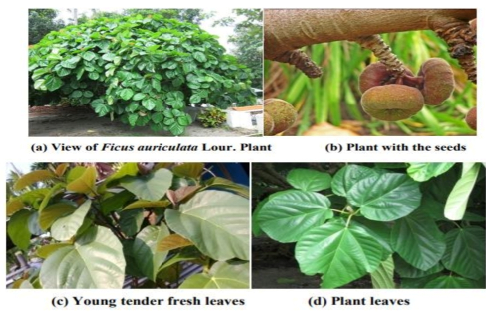
The leaves of Ficus auriculata Lour. and Sarcochlamys pulcherrima (Roxb). were collected in the month of June-July 2011, from Jonai, Dhemaji District, Assam and Rupnagar, Dist. Dibrugarh, Assam respectively. The plants were identified and authenticated at Botanical Survey of India (BSI), Eastern circle, Shillong, Meghalaya, India. The voucher specimens were deposited at the Department of Pharmaceutical Sciences, Dibrugarh University, Dibrugarh, Assam, for future references.
Drying and storage:
The collected leaves were cleaned, washed with water and dried under shade at room temperature with occasional shifting for three weeks. They were then powdered with a mechanical grinder. The powders were passed through sieve number 40 and stored in airtight container for further uses.
Successive solvent extraction:
The dried powdered materials (100 g) were defatted with petroleum ether (60-800C) and the defatted powdered materials thus obtained were further successively extracted using ethyl acetate and methanol as solvent in a Soxhlet apparatus by continuous hot percolation process. Each time, before extracting with next solvent of higher polarity, the marc was kept in hot air oven below 500C for 10 min. The extraction was carried out exhaustively and the solvents were recovered by distillation under reduced pressure using rotary vacuum evaporator. The concentrated extracts kept in refrigerator at 40C for further use. The extractive values of all the solvent extracts were recorded in terms of % w/w as per standard method. The percentage yields of ethyl acetate, methanol and water extract were found o be 0.5%, 0.85% and 3.15% respectively. The extracts were subjected to preliminary phytochemical analysis to identify the presence of phytoconstituents.
Phytochemical screening of extracts:
Qualitative phytochemical screening:
The qualitative phytochemical screening of the extracts, obtained by successive solvent extraction process was carried out to detect the presence of probable primary as well as secondary metabolites. An amount of 5 g of the extract was dissolved in 100mL alcohol/distilled water and the following tests were performed for identification of different chemical groups present in the extracts.
Preparation of reagents for qualitative phytochemical screening:
The reagents used for the different chemical group tests were prepared as per standard procedure and were as follows:
• Benedict’s reagent: 1.73g of cupric sulphate, 1.73g of sodium citrate and 10gm anhydrous sodium carbonate were dissolved in water and the volume was made up to 100mL with water.
• Dragendroff’s reagent:1.7 g basic bismuth nitrate and 20 g tartaric acid were dissolved in 80 ml of water. This solution was mixed with a solution containing 16 g potassium iodide in 40 mL of water
. • Fehling’s solution A:34.64 g copper sulphate was dissolved in a mixture of 0.5 mL of sulfuric acid and sufficient water to make up 500 mL.
• Fehling’s solution B: 176 g of sodium potassium tartarate and 77 g of sodium hydroxide were dissolved in sufficient water to produce 500 mL. An equal volume of above solutions (Fehling’s solution A and B) is mixed at the time of use.
• Hager’s reagent:1% solution of picric acid in water.
• Libermann-Burchard reagent: 5g of acetic anhydride was carefully mixed under cooling with 5mL of concentrated sulphuric acid and this mixture was added slowly to 50mL of absolute ethanol with cooling.
• Mayer’s reagent: Solution of 1.36 g of mercuric iodide in 60 mL of water was mixed with a solution containing 5 g of potassium iodide in 20 mL of water.
• Molish’s reagent:2.5 g of pure α-naphthol was dissolved in 25 mL of ethanol.
• Phloroglucinol solution: 2g of phloroglucin dissolved in 100 mL of 90% alcohol.
Test procedures for qualitative phytochemical screening:
a) Tests for Alkaloids:
• Dragendroff’s test: 0.1ml of dilute hydrochloric acid and 0.1 mL of Dragendroff’s reagent were added to 2mL solution of extract in a test tube. Development of orange brown colored precipitate suggested the presence of alkaloid.
• Mayer’s test: 1 mL of extract was taken in a test tube. 0.2 mL of dilute hydrochloric acid and 0.1 mL of Mayer’s reagent were added. Formation of yellowish buff colored precipitate gives positive test for alkaloid.
• Wagner’s test: 2mL of extract solution was treated with dilute hydrochloric acid and 0.1 mL of Wagner’s reagent. Formation of reddish-brown precipitate indicated the positive response for alkaloid.
• Hager’s test: 2mL of extract was mixed with 0.2mL of dilute hydrochloric acid and 0.1 mL of Hager’s reagent. A yellowish precipitate suggested the presence of alkaloid.
b) Tests for Amino acids:
• Millon’s test: To the test sample 2 mL of Millon’s reagent was added, a white precipitate formed which indicated the presence of amino acid.
• Ninhydrin test: Extract solution was treated with Ninhydrin (Triketohydrindene hydrate) solution at the pH range of 4-8. Development of purple color indicated the positive response for amino acids.
c) Tests for Carbohydrates:
• Molisch’s test: To 2-3 mL of filtrate few drops of α-naphthal (20 % in ethyl alcohol) was added shaken well and about 1ml of concentrated H2SO4 was added along the side of test tube. A reddish-violet colored ring at the junction of two layers was shown that the presence of carbohydrate.
• Fehling’s test: One mL of Fehling’s solution A and 1 ml of Fehling’s solution B were mixed properly and boiled for one minute. To this, equal amount of filtrate was added and heated on the boiling water bath for 5-10 minutes and observed for the formation of brick-red colored precipitate which indicated the presence of carbohydrate.
• Benedict’s test: Equal volume of Benedict’s reagent and filtrate was mixed. The mixture was heated in a water bath for 5 minutes and observed for the formation of yellowish green colored precipitate for indication the presence of carbohydrate.
• Keller-Kilian test (for Deoxy Sugars): Small amount of sugar was dissolved in glacial acetic acid and 2 drops of 5% ferric chloride was added. The solution was added to 2mL concentrated sulphuric acid by the side of the tube. Reddish brown color (which changed to bluish green to dark on standing) at the junction confirmed the presence of deoxy sugars in the sample.
d) Test for Fats and Oils:
• Saponification test: Few drops of 0.5N alcoholic potassium hydroxide was added to a few quantity of the extract along with a drop of phenolopthalein. The mixture was heated on a water bath for 1-2 hours. Not observed the formation of partial neutralization of alkali which indicated the absence of fixed oils. ?
• Spot test: A little quantity of the extract was separately pressed between two filter papers. No appearance of oil stain on the paper indicated the absence of fixed oils.
e) Test for Flavonoids:
• ml of extract solution was hydrolyzed with 10 % v/v H2SO4 and allowed to cooled. Then it was extracted with diethyl ether and separately divided into 3 portions in three test tubes. 1 ml of dilute ammonia, 1 mL of dilute sodium carbonate and 1 mL of 0.1 (N) sodium hydroxide were added to the first, second and third test tubes respectively. In each test tube found development of yellow color indicated the presence of flavonoids.
• Shinoda test: The extract was dissolved in alcohol. One piece of magnesium was added followed by concentrated hydrochloric acid drop wise and heated. Appearance of magenta color demonstrated the presence of flavonoids.
• Alkaline reagent test: Addition of increasing amount of sodium hydroxide to the residue showed yellow coloration which decolorized after addition of acid.
• To the small quantity of residue, lead acetate solution was added. A yellow-colored precipitate was formed.
• Ammonia test: Filter paper strip is dipped in alcoholic solution of the extract, ammoniated and observed for color change from white to orange.
f) Test for Glycosides:
Small portion of the extract was hydrolyzed with dilute hydrochloric acid for half an hour in water bath, filtrated and filtrate was then subjected to following tests.
• Keller-Killiani Test (for cardiac glycosides): To 2 mL of filtrate glacial acetic acid, one drop of 5% ferric chloride and concentrated sulphuric acid were added and there was no formation of reddish-brown color at the junction of two liquid layers and upper layer appeared bluish green in the absence of glycosides.
• Legal’s Test: To the filtrate, 1 ml of pyridine and 1 ml of sodium nitroprusside solution was added and there was no formation of pink to red color indicated the absence of cardiac glycoside.
• Test for Saponins glycosides:
Foam test: Small amount of residue is diluted with distilled water up to 20 ml and shaken vigorously
in a graduated cylinder for 15 minutes and observed for the formation of 1 cm layer of foam which is stable for 10 minutes.
2. To the alcoholic extract few drops of sodium bicarbonate was added and shaken well. The solution becomes honey comb like frothing confirmed the presence of saponins.
3. 1mL extract was treated with 1% lead acetate solution. Formation of white precipitate indicated the presence of saponins.
g) Test for Gums:
• Molisch’s test: 2ml of concentrated H2SO4 was added to 2ml of extract solution. Then it was treated with 15% α-naphthol in ethanol (Molisch’s reagent). Formation of a red violet ring at the junction of two layers indicated the presence of gums.
• Test solution was hydrolyzed using dilute HCL and Benedict’s or Fehling’s test was performed and observed for formation of red colour.
h) Test for Lignin:
A small quantity of the extract was treated with alcoholic solution ofphloroglucinol and hydrochloric acid, there was no appearance of red colour showed the absence of lignin.
i) Test for Mucilage:
• A small quantity of the extract was added separately to 25mL of absolute alcohol with constant stirring and filtered. The precipitate was dried in air and examined for its swelling properties.
j) Test for Proteins:
• Biuret test: One ml of 40% sodium hydroxide solution was added to two drops of 1% CuSO4 solution till a blue color produced, and then add to the 1 ml of the extract. Formation of pink or purple colour and violet color indicates the presence of proteins.
• Xanthoproteic test: To 1ml of the extract, 1 ml of concentrated nitric acid was added. A white precipitate was found and it was boiled and cooled. Then 20% of sodium hydroxide was added and observed the formation of orange color for presence of proteins.
k) Test for Steroids:
• Libermann-Burchard’s Test: 10mg of extract was dissolved in 1 ml solution of chloroform. To this 1ml of acetic anhydride was added following the addition of 2 ml of concentrated H2SO4. Formation of reddish violet color indicated the presence of steroids.
• Salkowski Test: 1mL of concentrated sulfuric acid was added to 10mg of extract dissolved in 1mL of chloroform. A reddish-blue color exhibited by chloroform layer and green fluorescence by the acid layer suggested the presence of steroids.
l) Tannins and Phenolicco Test:
• Ferric chloride test: About 5ml solution of extract was allowed to react with 1m of 5% ferric chloride solution. Greenish black color formed which indicated the presence of tannins.
• Test for Potassium dichromate: About 5m of the extract solution was treated with 1 ml of 10% aqueous potassium dichromate solution.Yellowish brown precipitate colour was formed which indicated the presence of tannins.
• Test for Lead acetate: About 5ml of the extract was treated with 1 ml of 10% lead acetate solution in water. Yellow color precipitation formed and indicated the positive test for tannins.
m) Test Volatile oil:
To the dried powder, sudan III solution was added, there was no formation of red colorization by globules indicated the absence of volatile oils. antitative estimation of phytochemicals
Estimation of total Phenolic Content:
By using Folin-Ciocalteu reagent (Singleton and Ross, 1965), the total amount of phenolic compounds cotent in the extracts was estimated. The extracts were diluted with the same solvent used for extraction, to a suitable concentration for analysis and 0.5 mL of commercial Folin-Ciocalteu reagent and 1.6 mL of aqueous sodium carbonate (20%) solution was added, mixed thoroughly, kept for 2 h and absorbance was measured at 760 nmUsing gallic acid monohydrate, a standard curve was prepared. The linearity obtained was in the range of 10-50 µg/mL. Using standard curve, the total phenolic compounds content was calculated and expressed as gallic acid equivalent in mg/g of extracts.
Estimation of total flavonoid Content:
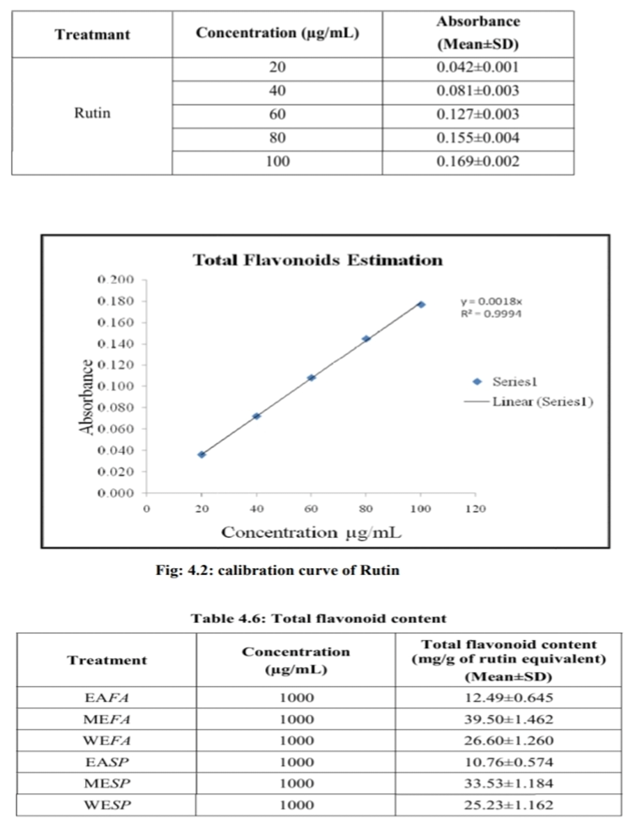
The sample was pipetted out in test tube and volume was made up to 0.5 mL with distilled water. Sodium nitrite (5%; 0.03 mL) was added to the tube and incubated for 5 min. at room temperature. Aluminium chloride solution (10%; 0.06 mL) was added and incubated for 5 min. at room temperature. Sodium Hydroxide solution (1 M; 0.2 mL) was added and total volume was made up to 1 mL with distilled water. Absorbance was measured at 510 nm against a reagent blank. Standard curve using different concentrations of rutin was prepared. From the standard curve, concentration of flavonoids in the test sample was determined and expressed as mg of rutin equivalent.
RESULTS OF PHYTOCHEMICAL SCREENING
Phytochemical screening
Qualitative phytochemical screening:
The preliminary phytochemical analysis of different successive leaves extracts of petroleum ether, ethyl acetate, methanoland water showed the presence of a number of different phytochemical constituents of the plants Ficus auriculata andSarcochlamys pulcherrima .
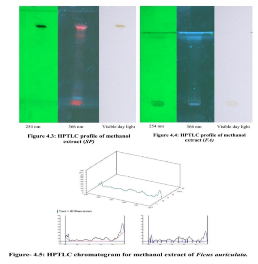
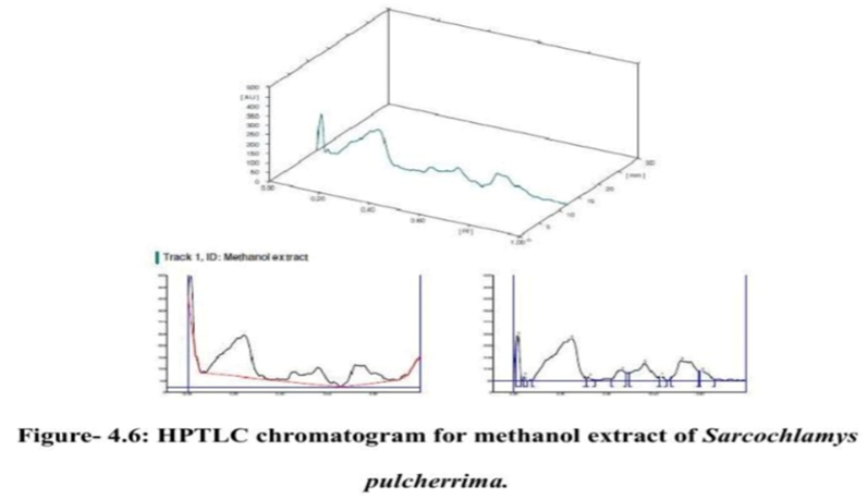
HPTLC fingerprint analysis:
HPTLC studies were carried out on successive methanol extract using Camag HPTLC system equipped with Linomat IV sample applicator, Camag TLC scanner 3 and CATS 4 software for interpretation of data. TLC plates (5 x 10 cm).precoated with silica gel 60 F254 (E. Merck) on aluminium were used. 10µL samples were applied through applicator and the plates were developed using solvent system, at the ratio of Chloroform: Methanol (9.5:0.5) for methanol extractof the plant leaves Ficus auriculata and Chloroform: Ethyl acetate: methanol (9:3:2) for the methanol extract of Sarcochlamys pulcherrima leaves extract.
The experimental result in HPTLC study (10µl sample volume) showed 12 spots of unknown phytoconstituents with Rf values values 0.03, 0.08, 0.15, 0.22, 0.39, 0.51, 0.54 ,0.62, 0.65 ,0.74 ,0.86, 0.93 in methanol extractsof Ficus auriculata and similarly the HPTLC result of Sarcochlamys pulcherrima methanoli leaves extracts showed 9 spots with Rf values 0.03, 0.06, 0.31, 0.35, 0.48, 0.62, 0.66, 0.8 and 0.87 of unknown phytoconstituents.
HPTLC fingerprint analysis:
HPTLC studies were carried out on successive methanol extract using Camag HPTLC system equipped with Linomat IV sample applicator, Camag TLC scanner 3 and CATS 4 software for interpretation of data. TLC plates (5 x 10 cm).precoated with silica gel 60 F254 (E. Merck) on aluminium were used. 10µL samples were applied through applicator and the plates were developed using solvent system, at the ratio of Chloroform: Methanol (9.5:0.5) for methanol extractof the plant leaves Ficus auriculata and Chloroform: Ethyl acetate: methanol (9:3:2) for the methanol extract of Sarcochlamys pulcherrima leaves extract. The experimental result in HPTLC study (10µl sample volume) showed 12 spots of unknown phytoconstituents with Rf values values 0.03, 0.08, 0.15, 0.22, 0.39, 0.51, 0.54 ,0.62, 0.65 ,0.74 ,0.86, 0.93 in methanol extractsof Ficus auriculata and similarly the HPTLC result of Sarcochlamys pulcherrima methanoli leaves extracts showed 9 spots with Rf values 0.03, 0.06, 0.31, 0.35, 0.48, 0.62, 0.66, 0.8 and 0.87 of unknown phytoconstituents.
MATERIAL AND METHODS:
Succesive extractions of plant leaves:
The dried plant leaves powdered materials of Ficus auriculata and Sarcochlamys pulcherrima were defatted with petroleum ether (60-80oC) and the defatted powdered materials thus obtained were further successively extracted using ethyl acetate, methanol and water as solvent in a soxhlet apparatus. The extraction was carried out exhaustively and the solvents were recovered by distillation under reduced pressure using rotary vacuum evaporator. The various concentrated leaves extract viz- ethyl acetate extract of Ficus auriculata (EAFA), water extracts of Ficus auriculata (WEFA) and methanolic extract of Ficus auriculata (MEFA); ethyl acetate extract of Sarcochlamys pulcherrima (EASP), water extracts of Sarcochlamys pulcherrima (WESP) and methanolic extract of Sarcochlamys pulcherrima (MESP) were dried in vacuum desiccators, preserved in well tight container. The concentrated extracts were kept in dessicator at 4oC for further use. The extracts were used for various pharmacological activities of the present study.
Acute oral toxicity study:
Preparation of the test materials: The dried plant crude extracts were powdered in mortar and suspension of required dose of all extracts prepared by using 0.5 % w/v Carboxymethylcellulose (CMC) as a vehicle and these preparations were used to determine acute toxicity and different pharmacological activities.
Test animals: Male Wistar albino rats of 160 – 200g were procured from registered breeder. They were grouped and housed in polyacrylic cages (38x23x10 cm), maintained under standard laboratory conditions (25±2°C) and humidity of 45-64 % with light/dark cycle (12/12 h). They were allowed free access to standard dry pellet diet (Hindustan Lever Limited, Kolkata, India) and water ad libitum. The animals were acclimatized to laboratory conditions for 10 days before commencement of the experiment. Approval of the Institutional Animal Ethics Committee (IAEC) of the Department of Pharmaceutical Sciences, Dibrugarh University, Dibrugarh (App. No-IAEC/DU/02, dated-04/05/2012) was obtainedprior to the study. The guidelines under ‘Principles of Laboratory animal care’ (NIH publication number 8523, revised, 1985) were followed.
Procedure of acute oral toxicity test: The acute oral toxicity study (LD50 determination) was evaluated in rats as per OECD-425 guidelines (OECD, Swapnil et al., 2011). Male Wistar albino rats were randomly selected for use in the study and marked to provide individual identification. The animals were divided into five groups and each group contained three rats. The animals were kept fasting for overnight providing only water, after which the extracts were administered orally at the dose level of 5 mg/kg of body weight and observed for 14 days. The control group received only 0.5 % w/v CMC. Each animal received at the dose of 10 ml/kg, p.o. of body weight.
In vitro antioxidant studies:
Preparation of test and standard solutions: All the plant leaves extracts were
dissolved in dimethyl sulphoxide (DMSO) separately and Ascorbic acid was used as a standard for the studies. Evaluation of in vitro antioxidant activities using the methods like DPPH scavenging activity, reducing power assay, Nitric oxide scavenging activity, Total antioxidant capacity and Metal chelating assay were performed as per the standard methods.
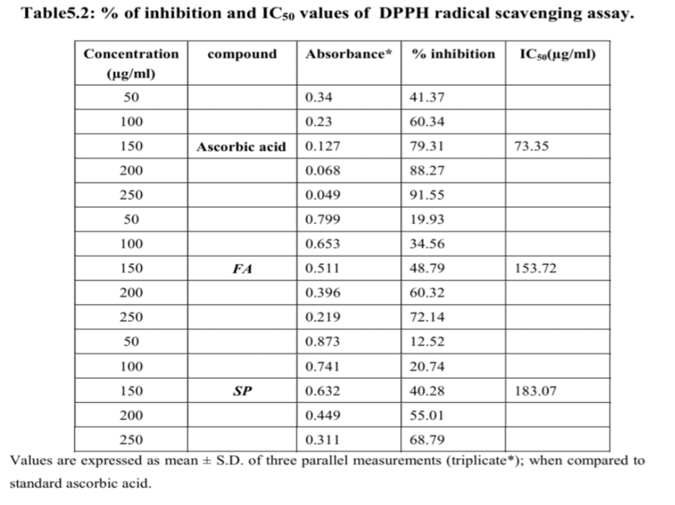
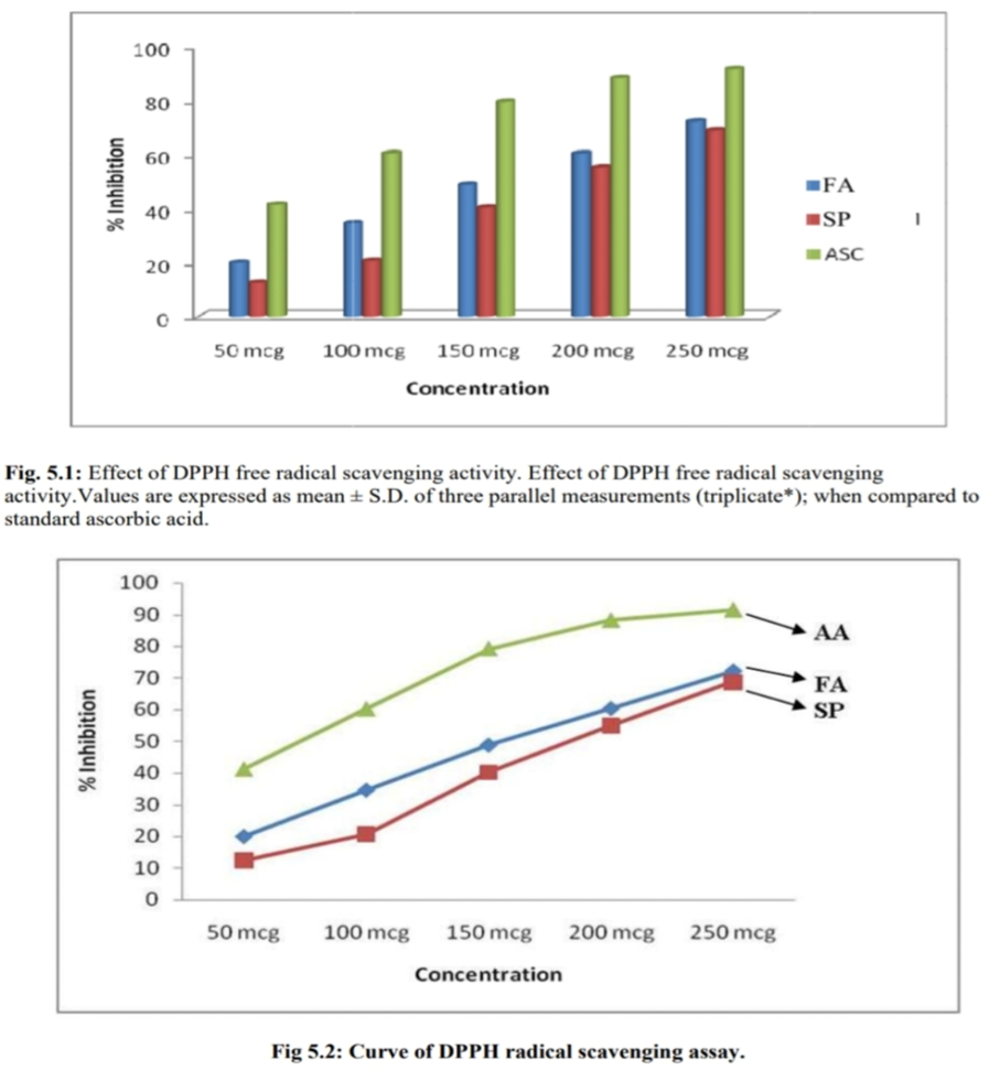
5 DPPH radical scavenging assay: The free radical scavenging activity of the methanolic extracts of both the plat leaf extracts were measured by using in vitro antioxidant activity 1, 1-diphenyl-2-picrylhydrazyl (DPPH) assay (Deoreet al., 2009). About 0.3 ml solution of DPPH in methanol was prepared and 1 ml of this solution was added to 3 ml of the extract solutions at different concentrations. The mixture was shaken and allowed to stand at room temperature for 30 minutes and the absorbance was measured in a spectrophotometer at 517 nm. Lower absorbance of the reaction mixture indicated higher free radical scavenging activity. The percentage of scavenging activity at different concentrations i,e (50, 100, 150, 200, 250 µ/ml) was determined and the IC50value of the fractions was compared with that of standard ascorbic acid (vitamin-C), which was used as the standard.
The IC50 value was defined as the concentration in (μg/ml) of extract that inhibits the formation of DPPH radicals by 50%. Radical scavenging activity was expressed as a percentage and Percentage of inhibition (I %) was calculated by using following
formula:
I % = 100 x (AC– AS)/AC
Reducing power assay: The reducing power of the extracts was assessed by the method of Oyaizu and Zhang. Various concentrations of the extracts (2.5 ml) were mixed with 2.5 ml of 200 mM sodium phosphate buffer (pH 6.6) and 2.5 ml of 1% potassium ferricyanide and the mixture was incubated at 50°C for 30 minutes. After that, 2.5 ml of 10% trichloro acetic acid was added to the mixtures .
RESULTS OF PHARMACOLOGICAL SCREENING:
In- vitro antioxidant activity:
DPPH radical scavenging activity assay of the plant extracts: Assessment of DPPH (1,1-dipheny l-2- picrylhydrazyl) scavenging activity of an extract or compound is another powerful in-vitro tool for evaluation of antioxidant activity. The DPPH radical is considered to be a model for a lipophilic radical. A chain in lipophilic radicals is initiated by the lipid autoxidation. DPPH is a stable free radical at room temperature and accepts an electron or hydrogen to become a stable diamagnetic molecule. The reduction capability of DPPH is determined by the decrease in its absorbance at 517 nm, which is induced by antioxidant. The scavenging effect of extracts and ascorbic acid on DPPH radical was compared. Table 5.2and Figure 5.1 and 5.2 illustrate a significant decrease in the concentration of DPPH radical due to the scavenging ability of the extracts of the plant.
The percentage scavenging activity at different concentrations was determined and the IC50 value of the fractions was compared with that of ascorbic acid (vitamin- C), which was used as the standard.
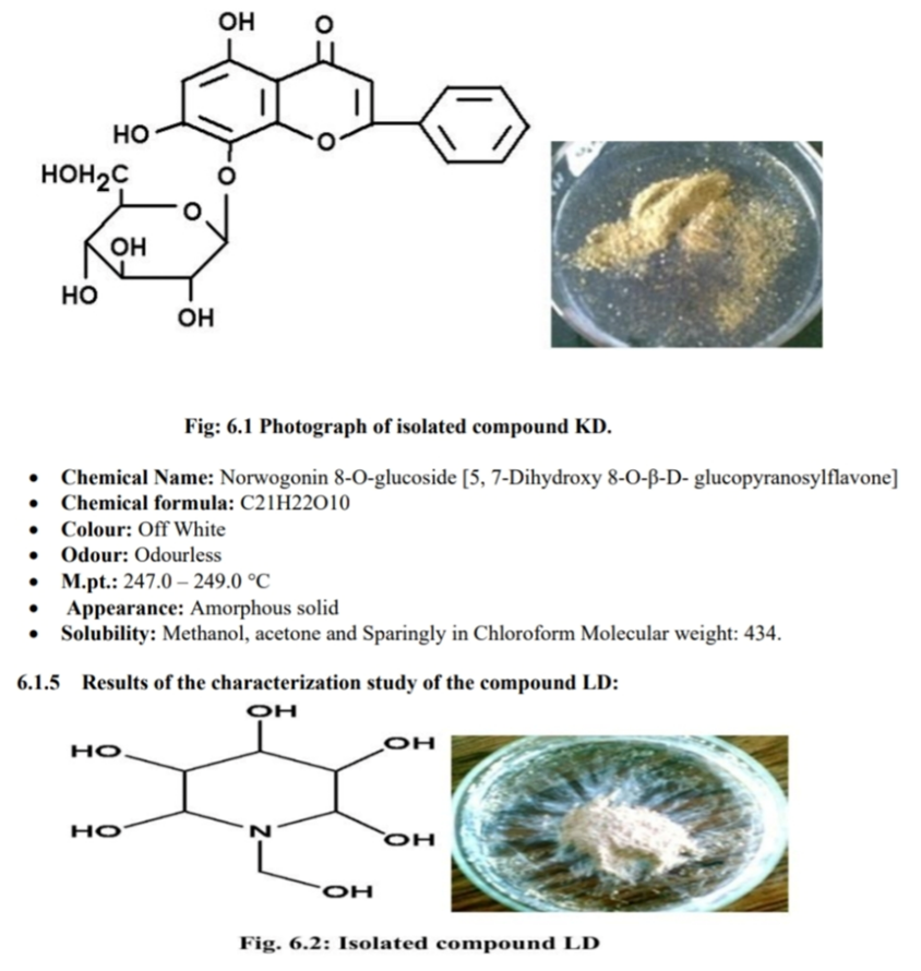
Results of the characterization study of the isolated compound, KD:
• Chemical structure:
REFERENCES

Jyoti Jadhav*, Jayashri Naphade, Phytochemical Screening and Antimicrobial Activity of Traditional Plants, Int. J. of Pharm. Sci., 2025, Vol 3, Issue 6, 2550-2568. https://doi.org/10.5281/zenodo.15652778
 10.5281/zenodo.15652778