1 Department of Pharmaceutical Sciences, Institute of Chemical Technology, Mumbai
2 R.C. Patel Institute of Pharmaceutical Education and Research, Shirpur
3 GES's Sir Dr. M. S. Gosavi College of Pharmaceutical Education and Research, Nashik
Herbal medicine has great potential for therapy, yet the many obstacles to clinical use include the poor solubility of phytoconstituents, the instability of herbal products, and the low oral bioavailability of the active ingredients. Phytosomes are a new type of vesicular drug delivery system that has been shown to enhance the absorption and therapeutic efficacy of plant-derived bioactive compounds by complexing them with phospholipids. This review will comprehensively address the concept of phytosomes; their structure; how phytosomes are prepared and the physicochemical and biological properties of phytosomes and how they can be formulated and evaluated. The review will focus on the physical and chemical characterization of phytosomes, bioavailability of phytosome product, commercial products containing phytosomes; and therapeutic uses of phytosomes for various diseases. Finally, the review will address the deficiencies, regulatory issues, and the future directions of phytosome technology and explain how phytosome technology may help to connect traditional herbal medicine to modern pharmaceutical drug delivery systems.
The combination of polar and hydrophobic properties found in herbs contributes to low bioavailability; as a result, the aqueous component is poorly absorbed, diminishing the effectiveness of herbal medicines1. The proper delivery of a herbal product to the gut, as well as its passage across lipid membranes, requires it to have good affinity for both aqueous and lipid environments2. The traditional use of plant-based medicinal treatments has continued to evolve into many forms with the development of New Drug Delivery Systems (NDDS), with considerable progress in improving the therapeutic efficacy of plant extracts through targeting and sustained/controlled release3.
When we talk about herbal medicine, which is made up of compounds like glycosides and flavonoids (Main ingredients), we find that it does not absorb well and that the therapeutic value of the active ingredients is lower when taken in their natural form. To help address this shortcoming, we propose that phyto-some and liposome technologies increase the bioavailability of herbal medicines compared to our traditional extraction methods4. Phytosomes (one of the proposed solutions) are a new structure that encapsulates herbal medicine within a vesicle, protecting it from breakdown and enabling it to pass through the cell membrane more easily into the bloodstream5. There is evidence that the development of phytosomes, while allowing the treatment of serious diseases, has also preserved the phytocompounds in herbal medicines, thereby enhancing their therapeutic effects. Phytosomes are created by combining phospholipids with selected plant materials, producing a new entity that delivers the active ingredients more effectively into the body6.
The use of phytosomes is an innovative approach that supports the utilisation of a wide range of polyphenolic flavonoids, terpenes and tannins7. As previously mentioned, the phytochemicals exhibit antioxidant and other beneficial health properties, but they are not significantly bioavailable when consumed orally8. Research has shown that the bioavailability and pharmacokinetics of phytosomal standard pharmacological products, such as silymarin, curcumin, and Ginkgo biloba extracts, can be significantly improved compared with their non-phytosomal counterparts. Additionally, through the use of phytosomal technologies, we are discovering how these phytochemicals can be combined with our traditional herbal medicine development approaches9.
This article provides a comprehensive overview of the key components of phytosomes, including methods of synthesis and characterisation, their advantages, and potential uses in optimising herbal medicine delivery systems and exploring opportunities to improve their effectiveness7. Finally, the article discusses the current and future directions of this new field, as well as the promise and potential benefits of phytosomes for the development of formulations containing natural bioactive compounds. Several different kinds of vesicles have been created as drug delivery vehicles. These include liposomes, niosomes, transfersomes and ethosomes10. The table below (Table 1) lists the references used in making these various vesicle types. Schematic representations of these vesicle constructions are shown in the image below (Figure 1).
Table I: Composition of vesicular systems
|
Vesicular System |
Primary Composition |
Structural Characteristics |
Key Differentiator |
References |
|
Liposome |
Phospholipids + Cholesterol |
Bilayered spherical vesicles (0.05–5.0 µm) |
The amphiphilic nature allows the entrapment of polar and non-polar solutes. |
11 12 |
|
Niosome |
Non-ionic surfactants + Cholesterol |
Non-biological bilayer; lamellar structure |
Higher chemical stability and lower production cost than phospholipids. |
13 |
|
Transfersome |
Phospholipids + Edge Activators (e.g., Sodium cholate) |
Ultra-deformable/elastic membrane |
High curvature elasticity allows penetration through pores smaller than the vesicle diameter. |
14 15 |
|
Ethosome |
Phospholipids + Ethanol (20–45%) + Water |
Soft, malleable vesicles with fluidised bilayers |
Ethanol-induced lipid perturbation enhances penetration of the stratum corneum. |
16 17 |
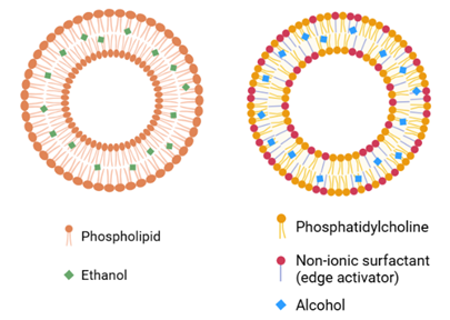
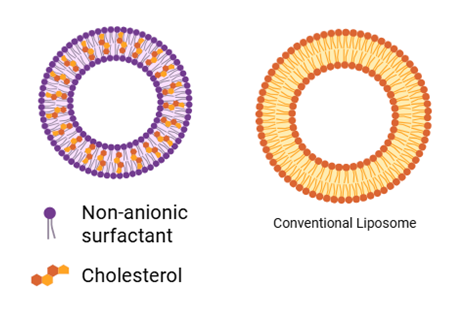
A) B) C) D)
Figure I: A) Ethosomes, B) Transferosomes, C) Niosomes, D) Liposomes
History
Phytosome technology has significantly improved the bioavailability of natural plant extracts since its development by Bombardelli in 19915, 18. Phytosome technology provides a means to overcome the challenges associated with the poor absorption of many phytochemicals, such as flavonoids19. Phytosome technology uses phospholipids to encapsulate plant extracts, enhancing the bioavailability of those compounds by increasing uptake through the gastrointestinal tract. The process of separating individual compounds from pure plant extracts often results in a loss of their therapeutic value1. Developing a standardised formulation of a plant extract is essential to ensuring its therapeutic effectiveness. The use of phospholipids to formulate phytosomes has been shown to significantly increase the absorption of herbal extracts and, consequently, their effectiveness. For instance, many of the most common herbal products, including Ginkgo biloba and grape seed, have been shown to have enhanced therapeutic effects when taken as phytosomes 19. Phytosomes are formed when phytomolecules associate with phosphatidylcholine via hydrogen bonds. This allows for more efficient passage across biological membranes than if they were taken individually6. Phosphatidylcholine is frequently used in laboratory preparation of phytosomes because it has an affinity for both polar and non-polar environments and therefore increases the solubility and stability of the phytomolecule. In addition to protecting the phytoconstituent from degradation in the gastrointestinal tract, phosphatidylcholine in phytosomes also has hepatoprotective properties, thereby contributing to overall health benefits20. Studies in both animals and humans have shown that phytosomes exhibit better pharmacokinetics and pharmacodynamics than traditional herbal extracts. The introduction of phytosome technology has enabled improved absorption of bioactive constituents from herbal products into the bloodstream21.
Table II: Intellectual Property status of Phytosomes.
|
Sr.No |
Title |
Novelty |
Patent No |
Ref |
|
1 |
Curcumin-containing phytosomes |
Curcumin, when combined with phospholipids, releases more parent agent into the bloodstream than curcumin that has not been complexed. |
WO2009/ 101551 (2009) |
22 |
|
2 |
olive fruits or leaves Phytosomes |
Oil-rich fruit and their extract bioavailability increased by forming a complex with phospholipids |
EP1844785 (2007) |
23 |
|
3 |
Ginkgo biloba containing phytosomes |
Treating allergy and inflammatory diseases |
EP1813280 (2007) |
24 |
|
4 |
thymosin-4, treats skin conditions and heals wounds |
This preparation promotes wound healing and contains thymosin-4. |
US/2007/ 0015698/ (2007) |
25 |
|
5 |
sorbityl furfural fatty acid monoesters and mixtures for cosmetic and medical applications |
For particular anti-hydroxyl radical action, sorbityl furfural's chosen fatty acid monoesters and lipophilic agents were used. |
EP1690862 (2006) |
26 |
|
6 |
Vitis vinifera extracts as an anti-atherosclerotic drug containing phytosomes |
Atherosclerosis is prevented and cure by Vitis vinifera with phospholipid complexes. |
US6297218(2001) |
27 |
Structure of Phytosomes
Phytosomes (Guggulosomes) are complexes of phyto-phospholipids in their chemical composition that are produced by the physical and chemical combination of the active phytoconstituents and the polar head (choline moiety) (Figure 2). Phospholipid head groups must be anchored in these complexes. The polar component is enclosed by fatty acid chains in complexes that produce a lipophilic surface. A constituent material is active, either in a space between the membrane's several layers28. The material that gives phytosomes their activity is a part of the membrane.
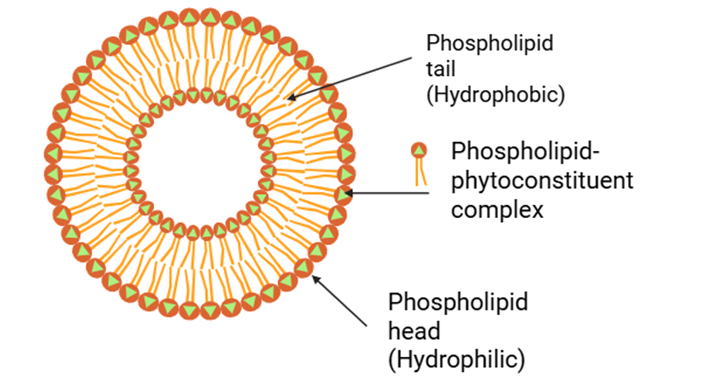
Figure II: Structure of Phytosomes
PROPERTIES OF PHYTOSOMES
properties and biological effects of phytosomes, complexes formed from natural phospholipids and herbal extracts, highlighting their potential therapeutic applications.
Table III: Physicochemical properties
|
Property |
Description |
Significance |
Reference |
|
Formation |
Complex structures are formed via hydrogen bonds between a natural compound and a phospholipid in a suitable solvent. |
forms a stable entity that is compatible with lipids and enhances bioavailability. |
6 5 |
|
Size |
Measuring a wide range of sizes, from 50 nm to several hundred micrometres |
Measuring a wide range of sizes, from 50 nm to several hundred micrometres. |
19 29 |
|
Solubility |
Soluble in non-polar solvents and fats, although they usually cannot dissolve in water. |
Enhances the absorption of water-soluble active compounds by making them lipophilic. |
7 |
|
Aqueous behaviour |
Comes into a micellar shape that resembles liposomes when exposed to water. |
This structure impacts absorption and efficacy in biological systems. |
30 |
Table IV: Biological properties
|
Health Area |
Phytosomes |
Effects |
Reference |
|
Cardiovascular Benefits |
Cardiovascular Benefits |
Reduces myocardial necrosis and increases antioxidant levels, protecting cardiac tissue from damage. |
31 |
|
Neuroprotective Effects |
Annona muricata phytosome |
Enhances blood-brain barrier permeability; may improve antidepressant activity by inhibiting MAO-B. |
7 32 |
|
Cognitive & Neurodegenerative Health |
Curcumin phytosome |
Improves brain bioavailability and neuroprotective efficacy compared to standard curcumin. |
21 33 |
|
Cerebral Ischemia Treatment |
Rutin, glycoside phytosome |
Enhances functional recovery after stroke, demonstrating strong therapeutic promise in ischemic models. |
34 |
|
Migraine Treatment |
Ginkgo biloba terpenes + Vitamin B2 + CoQ10 |
Reduced frequency and duration of migraine with aura over 4 months. |
35 36 |
|
Breast Cancer |
Green tea catechins |
Demonstrated tumour tissue penetration and potential therapeutic benefit |
37 38 |
|
Pancreatic Cancer |
Curcumin phytosome + Gemcitabine |
A Phase II trial demonstrated improved treatment response in patients with advanced pancreatic cancer. |
39 40 |
|
Urinary Tract Health |
Cranberry extract phytosome |
Equal antiadhesive effect vs Candida albicans despite lower proanthocyanidin levels. |
41 |
|
Wound Healing |
Ginkgo biloba, alpha-lipoic acid, grape seed phytosome |
Enhanced diabetic foot ulcer healing compared to standard care. |
42 |
|
Respiratory Issues |
Boswellia serrata phytosome |
Reduced inhaler dependence in asthma patients when combined with standard medication |
43 |
Formulation
Various types of Phytosome formulations can be prepared and administered via oral and topical routes using a range of encapsulation methods44. It is generally accepted that soft gelatin capsules are the most effective dosage form for a "Phytosome complex" because they enable the development of "dispersions" using liquid oil-based carriers45. To determine which oil-based carrier would yield the highest quality product, you must perform preliminary experiments with multiple carriers46. The volumetric filling procedures for hard gelatin capsules can also be used to encapsulate "Phytosome" complexes; however, the low density of Phytosomes makes it challenging to create a powder with sufficient volume, as the piston-tamping method for increasing the fill capacity of the hard gelatin capsule would require pre-compressing the fill amount, which negatively affects the time it takes for a hard gelatin capsule to disintegrate47. For tablet formulations, the dry granulation method is the most effective for producing tablets with extremely high active ingredient content and other favourable biopharmaceutical properties. Due to the low flowability and density of Phytosomes, the direct compression method is not recommended for producing Phytosome tablets; therefore, you will need to dilute the Phytosomes with other appropriate excipients to improve their flow characteristics48. Phytosome complexes can also be created in a way that is suitable for topical application, such as the incorporation into an emulsion at temperatures below 40°C to preserve their stability and effectiveness49. however, in these cases, Phytosomes can either be dispersed throughout the oil phase of an emulsion or incorporated into the aqueous phase, provided the process of making the emulsion is performed under strict temperature control to ensure stability50.
Material and Method of Preparation.
The phospholipids selected for phytosome preparation are derived from sources such as swine brain or dermis, as well as phosphatidylcholine, phosphatidylethanolamine, phosphatidylserine, or soy lecithin22. The acyl groups present in these phospholipids may be identical or different, primarily originating from stearic, palmitic, oleic, and linoleic acids51. The flavonoids used in phytosome formulation are chosen from compounds such as luteolin, 3-rhamnoside, luteolin-7-glucoside, ginkgonetine, isoginkgonetine, vitexine, diosmine, (+)-catechin, (–)-epicatechin, apigenin-7-glucoside, kaempferol, quercetin-3-rhamnoglucoside, and hyperoside52.
a) Antisolvent Precipitation Method:
The Antisolvent precipitation method for phytosomes is the process by which phospholipids and other plant metabolites are chemically combined to make larger molecules that dissolve better and are ingested more efficiently than those that do not53. The antisolvent precipitation procedure produces the phytosomes. A phytoconstituent solution, prepared from a good solvent (such as ethanol or methanol), is combined with a miscible (equal liquid) antisolvent (usually water)54. The phytoconstituents are insoluble in the antisolvent. They will precipitate from the solution upon addition of an antisolvent (by dropwise addition), altering the solubility order and causing the phytoconstituents to precipitate.
After precipitation, the mixture of phytoconstituents is stirred with a phospholipid to produce the final phytosome product. The mixing will continue until complete phytosome formation occurs. The phytosomes can then be filtered or centrifuged from the solution and washed with the antisolvent to remove any remaining solvent. Finally, the phytosomes can be vacuum dried, and the final phytosome product can be packaged and sold55.
b) Rotating Evaporator method
One way phytosomes may be produced is by using a rotary evaporator, which is not commonly used. In the rotary evaporator method, heat is used to separate volatile compounds (like ethanol or methanol)56. These two solvents are used to dissolve either phytoconstituents or phospholipids, and the resulting mixture is then placed in a round-bottom flask on the rotary evaporator 57. A water bath heated to an appropriate temperature, along with a vacuum, is used to facilitate evaporation. The final product of this method is the finished solid phytosomes. Dissolve the phytoconstituents and phospholipids in either ethanol or methanol by preparing a solution of these components in an organic solvent, using ethanol or methanol as the solvent58.Transfer the newly created solution to a round-bottom flask, secure the flask onto the rotovap, place the flask into the heated water bath, then lower the pressure after heating and allow the solvent to evaporate57. Once all of the solvent has been evaporated, collect the solid phytosomes, then wash them with an appropriate solvent to remove all traces of solvent from the solid. Then, after washing, dry the phytosomes in a vacuum59.
c) Solvent evaporation technique
The procedure that relies on the evaporating solvent to produce Phytosomes is the second technique available for manufacturing Phytosomal formulations. The procedure for producing Phytosomes using this method is superior to the rotary evaporator method mentioned previously. The final product from the evaporating solvent method consists of the solid Phytosome product (after completing the evaporation procedure) at the bottom of the evaporating solvent container60. To produce the Phytosome formulation through the evaporating solvent technique, the following steps are completed: Prepare a mixture (solution) of Phytoconstituents and Phospholipids with a suitable solvent (Ethanol or Methanol); then pour the mixture into a shallow container (Petri Dish) and allow the solvent to evaporate using room temperature and/or a gentle air flow to produce a solid residue at the bottom of the evaporating solvent container. The solid residue contains Phytosome and may be washed with a suitable solvent and dried in a vacuum.
d) Lyophilization (Freeze-Drying)
Freeze-drying, or "lyophilisation, is usually performed after preparation of the phytosomes to enhance their shelf-life. It also improves the formulation's physical characteristics61. In this technique, the phytosomal suspension, containing phosphate buffer, is first frozen. Then the water is removed or evaporated by a process called sublimation under vacuum, yielding a porous powder that is both stable and dry and can be easily reconstituted at the time of use62.
e) Sonication and Homogenisation
Both techniques were used to reduce aggregate size and improve dispersion uniformity. Sonication is achieved by applying ultrasonic energy, breaking larger aggregates into small, uniform particles. Homogenisation uses mechanical forces to accomplish the same. The use of either sonication or homogenisation can improve stability and increase the absorption capacity of the final phytosome product63.
f) Thin-Film Hydration Method:
Thin-film development is analogous to the evaporation of a liquid. Initially a thin film of lipids develops on the interface of a solution. When the solvent has been removed from the interface, the lipid film is then able to interact with water under controlled conditions so that phospholipid/phytocomponent vesicles develop spontaneously [57]. This method produces vesicles that are very uniform and works with both hydrophilic and lipophilic materials[58].
g) Complexation method
In the development of a molecular complex using the complexation method, both the phytoconstituent and phospholipid are solubilized in a specific solvent system before being combined at a pre-established ratio (1:1 or 1:2 molar ratio). Once all materials are combined, the two mixtures will be mixed until complete interaction has occurred between them before the solvent is removed so that a stable molecular complex results [59]. This method results in a stable molecular complexes via hydrogen bonding between the phytoconstituent and phospholipid, and this is beneficial for creating an effective encapsulated product with good bioavailability [60].
Bioavailability Enhancement
Phytosomes have significant antioxidant activity, as demonstrated by numerous studies. Pharmacokinetic studies show that phytosomes have a higher bioavailability than their parent molecules when administered at the same dose19. There have been countless successful applications of phytosome technology to well-known herbal extracts, such as ginseng, Ginkgo biloba, grape seed, hawthorn, milk thistle, and green tea64,5. Studies have shown that phytosomes can significantly increase absorption and bioavailability compared to traditional formulations. By adding flavonoids and polyphenolic compounds, phytosome-based formulations have produced standardised extracts with substantially higher bioavailability65. Silymarin is among the most extensively studied compounds and has been shown to improve the distribution of silybin by forming a silybin–phospholipid complex66. Yanyu and coworkers conducted a pharmacokinetic study demonstrating that the bioavailability of silybin was significantly increased when it was administered orally as a silybin-phospholipid complex (phytosome), due to the compound's improved lipophilicity67. Human studies have also been performed to assess the absorption of silybin when bound to phosphatidylcholine. In one study, plasma silybin concentrations after a single oral dose of silybin phytosome were about 7 times higher than those after a single oral dose of standard milk thistle extract68. Other studies have shown that Ginkgo biloba phytosomes outperformed conventional standardised extracts, which contain 6% terpene lactones and 24% ginkgo flavone glycosides, in terms of bioavailability. A phytosomal GBE produced a peak plasma concentration that occurred within three hours. It maintained plasma concentrations for at least 5 hours after dosing, achieving a peak terpene concentration 24 times higher than that of the non-phytosomal GBE. Clinically, ginkgo phytosomes have been shown to provide 30-60% improvement in patient symptoms of peripheral vascular disease compared with traditional GBE69. Phytosomes made from Green Tea Phytosome extracts (derived from Thea sinensis) are composed of (by means of per cent composition) 66.5% epigallocatechin, which is part of the polyphenol group of compounds. The polyphenolic group possesses many beneficial properties, including antioxidant, anticarcinogenic, antimutagenic, antiatherosclerotic, hypocholesterolaemic, cardioprotective, antibacterial, and anticariogenic effects, and phytosome types have been shown to play a significant role in maintaining homeostasis in chronic degenerative diseases such as cancer and atherosclerosis. Polyphenols have been shown to have limited oral bioavailability, i.e., poor absorption after ingestion8. However, complexing polyphenols with phospholipids significantly enhances oral bioavailability70. There have also been studies that have indicated that quercetin-phospholipid phytosomes were more effective than pure quercetin in preserving the liver of rats exposed to CCl4-induced liver damage71. Hesperetin complexed with hydrogenated phosphatidylcholine exhibited potent antioxidant and beneficial PK properties in rats treated with CCl472.
Table V: Commercial Phytosome Products and Their Biological Activity
|
Commercial product |
Plant origin |
Analysis |
Biological activity |
Ref |
|
18β-GLYCYRRHETINIC ACID PHYTOSOME® |
Glycyrrhiza glabra L. – Root |
≥27.0% ≤31.0% of 18β-glycyrrhetinic acid by HPLC |
Mitigative |
73 |
|
CASPEROME™ BOSWELLIA PHYTOSOME® |
Boswellia serrata Roxb. ex Colebr. – Resin |
≥25% boswellic acids by HPLC |
Anti-inflammatory, Mitigative |
74 |
|
CENTELLA ASIATICA TRITERPENE PHYTOSOME® |
Centella asiatica (L.) Urban – Leaf |
≥30.0% ≤35.0% of selected triterpenes by HPLC |
Collagen repair, Wrinkle removing |
75 76 |
|
ESCIN β-SITOSTEROL PHYTOSOME® |
Aesculus hippocastanum L. – Seed |
≥32.0% ≤40.0% of escin by TLC |
Capillary creating |
77 |
|
FRANKINCENSE PHYTOSOME® |
Boswellia serrata Roxb. ex Colebr. – Resin |
≥25% boswellic acids by HPLC |
Mitigative |
78 |
|
GINKGOSELECT® PHYTOSOME® |
Ginkgo biloba L. – Leaf |
≥7.0% of flavonglucosides, ≥0.8% bilobalide by HPLC |
Cognition, roaming increaser, vasokinetic |
79 |
|
GREENSELECT® PHYTOSOME® |
Camellia sinensis (L.) O. Kuntze – Young leaf |
≥19.0% ≤25.0% of polyphenols by HPLC |
Antioxidant |
80 |
|
LEUCOSELECT® PHYTOSOME® |
Vitis vinifera L. – Seed |
≥25% ≤30% of proanthocyanidins by GPC |
UV protectant, Antioxidant |
81 |
|
MERIVA® TURMERIC PHYTOSOME® |
Curcuma longa L. – Rhizome |
≥18.0% ≤22.0% of curcuminoids by HPLC |
Arthrosis health, Anti-inflammatory |
82 |
|
PROANTHOCYANIDIN A2 PHYTOSOME® |
Aesculus hippocastanum L. – Bark |
≥31.0% ≤37.0% of proanthocyanidin by HPLC |
Skin tightener, UV protectant |
83 |
|
SILIPHOS® SILYBIN PHYTOSOME® |
Silybum marianum (L.) Gaertn. – Fruit |
≥29.7% ≤36.3% of silybin by HPLC |
Liver protectant |
84 |
|
TERPENES PHYTOSOME® |
Ginkgo biloba L. – Leaf |
≥30.0% of total ginkgo terpenes by HPLC |
Anti-allergic, Mitigative |
22 |
|
VIRTIVA® |
Ginkgo biloba L. – Leaf |
≥5.0% of flavonglucoside phosphatidylserine by HPLC |
Cognition increaser |
85 |
Table VI: Benefits of Phytosomes
|
Sr.no |
Category |
Benefit |
Description |
Ref |
||||
|
1 |
Composition and Safety |
Biocompatible and Safe excipients |
Phytosomes consist of phospholipids and phytochemical constituents, which are safe for nutraceutical and medicinal use. |
18 |
||||
|
|
|
Phosphatidylcholine as a carrier |
Acts as a carrier for hepatoprotective and membrane-stabilising properties. |
19 |
||||
|
2 |
Structural Characteristics |
Chemical bonding |
Stable complexes are formed by phospholipids and phytoconstituents |
5 |
||||
|
|
|
Higher stability |
Chemical linkage gives higher stability |
86 |
||||
|
3 |
Manufacturing Advantage |
Simple preparation |
Straightforward and simple complexation enables easy scale-up for batch |
87 |
||||
|
4 |
Pharmacokinetic Advantage |
Enhanced bioavailability |
It significantly improves the absorption of phytochemicals. |
64 |
||||
|
|
|
Improved intestinal absorption |
It facilitates transport into the enterocyte membrane |
66 |
||||
|
|
|
Protection from gastric degradation |
Protects from gastric enzymes and bacterial degradation. |
88 |
||||
|
|
|
Protection during digestion |
Vesicle-like structures prevent the inactivation of phytoconstituent. |
67 |
||||
|
5 |
Therapeutic Advantage |
|
It provides nourishment and formulation stability. |
89 |
||||
|
|
|
Improved therapeutic efficiency |
Enhanced absorption and improved bioavailability. |
8 |
||||
|
6 |
Formulation Performance |
High entrapment efficiency |
Predictable high encapsulation efficiency of phytoconstituent |
90 |
||||
|
7 |
Drug Delivery Advantage |
Enhanced skin penetration |
Suitable for topical and transdermal delivery. |
55 |
||||
|
8 |
Biological compatibility |
Cell membrane affinity |
Improves cellular uptake and biocompatibility. |
91 |
||||
|
9 |
Adiitional advantage |
Skin nourishment and stability |
Provides nourishment and formulation stability. |
92 |
Evaluation of Phytosomes
A phytosome is formed by combining a plant-derived herbal active constituent with a phospholipid to produce a compound that is more readily absorbed into the body (bioavailable) and soluble in water. Several parameters can be used to assess the effectiveness of a phytosome-based herbal product, and each has specific acceptable ranges for the product's intended use7.
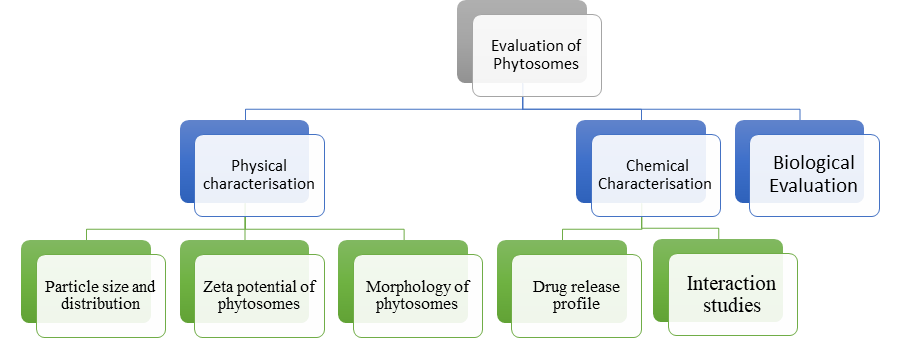
Physical Characterization
A crucial stage in physical characterisation that affects stability, bioavailability, and therapeutic efficacy is the evaluation of phytosome formulations. The behaviour of a formulation can be described by several parameters, such as morphology, zeta potential, particle size, and particle size distribution. All three of these elements define how phytosomes behave in vivo, how effective their drug encapsulation was and how stable they are when dispersed. Through rigorous physical characterisation, using state-of-the-art analytical tools, you can improve the reproducibility of formulations, as well as predict their potential for long-term stability while in storage...this makes it feasible and rational to develop phytosomal-based delivery systems that enhance absorption and produce consistent pharmacological effects.
The size and shape of phytosome particles were assessed using various analytical methods including Dynamic Light Scattering (DLS), Nanoparticle Tracking Analysis (NTA), Transmission Electron Microscopy (TEM), and Scanning Electron Microscopy (SEM). DLS provides an average particle size and PDI for a wide range of phytosome particles, while NTA allows you to measure the distribution of sizes and number of particles in suspension. To visualize the geometry of the phytosome particles, a SEM can be used, whereas a TEM demonstrates the nanoscale dimensions of phytosome particles. Most forms of Phytosomes measure between 100-300 nm and typically have low PDI values (<0.3), therefore making them an excellent candidate for consistent drug delivery and improved bioavailability.
a) Dynamic Light Scattering
Dynamic Light Scattering (DLS) analyses time-varying intensity changes in light scattered by small suspended particles (Brownian Motion) and allows particle size determination. To do this, phytosomes are suspended in a dilute solution of water or buffer, placed into a cuvette, inserted into a DLS unit, and the temperature and measurement time are set according to the instrument's instructions. The DLS unit uses the connected analysis software to determine size distribution profiles, average particle size, and the polydispersity index for the sample, yielding the most accurate results possible93. To obtain the best results, the sample must be well dispersed and free of aggregates.
b) Nanoparticle Tracking Analysis (NTA)
Nanoparticle tracking analysis (NTA) uses a laser to illuminate particles suspended in a fluid and collects data on their motion using scattered light. NTA can provide both particle size distribution and particle concentration data94.To perform an NTA analysis, the phyto-some sample must first be diluted to the appropriate concentration to allow accurate measurement with NTA, then loaded into the NTA instrument. The software associated with the NTA instrument will then track the motion of all the individual particles and, using the Stokes-Einstein equation, compute their respective sizes95. When performing NTA, ensure the sample has an appropriate concentration, as both high and low concentrations can yield inaccurate or unreliable results.
c) Scanning electron microscopy
High-Resolution Scanning Electron Microscopy (SEM) operates under the premise that high-resolution images of the Surface Morphology of the Phytosomes can be directly obtained 96. In this case you would take a small amount of Phytosomes and Place Them onto an Appropriate Substrate (In This Case a Silicon Wafer), Then let the phytosomes Dry, and If Necessary Coat the Sample With A Thin Conductive Coating to Allow For Better Imaging, Then Place The Dried Sample into The SEM and Capture Images of The Samples At Different Magnifications, From Which we be Able To Measure The Particle Sizes Directly from The Captured Images Using Image Analysis Software. It Should Be Noted That The Sample Preparation May Change The Morphology of The Particles; therefore, Minimal Efforts Should Be Made To Cause Structural Changes of The Particle During The Drying Process97.
d) Transmission Electron Microscopy
Using transmission electron microscopy (TEM), researchers can visualize detailed images of the internal structure and dimensions of phytosomes using highly magnified and high-resolution images[98]. Phytotype samples were diluted before examination by TEM; thus, a small amount was placed on a carbon-coated grid for later quantification. The area containing the phytosomes was located on the grid and examined by TEM, which provided researchers with sufficient information to quantify phytosome size.. As with Transmission electron microscopy (TEM), particular care must be taken to maintain sample integrity throughout all phases of sample preparation to prevent loss or contamination from mishandling.
e) Laser Diffraction
Laser diffraction uses the diffraction pattern produced when a laser passes through a medium containing phytosome particles to determine their size distribution. While preparing samples for testing, one must avoid forming hydrophobic agglomerates. It is beneficial to have large quantities of tiny particles (<1000 nm) so they can be absorbed easily and distributed evenly98. Additionally, narrow particle size distributions will give consistent drug delivery and improved bioavailability.
2. Zeta potential of phytosomes
The greater stability of any dispersion is considered based on high electrostatic repulsion between the particles. Zeta potential value > +20 mV or < -20 mV indicates the good physical stability of a dispersion99
a) Electrophoretic Light Scattering
Electrophoretic light scattering (ELS) measures how fast a particle travels in an electric field. Charged particles migrate toward the electrode of the opposite charge. The rate at which they do so depends on the zeta potential. A sample of phytosomes is placed in a conducting medium (typically a buffer) between two electrodes. A (determined by Henry’s equation), each system has a different set of parameters100. Each particle’s velocity of motion is monitored using a laser and recorded as data; then, you can evaluate either Henry's or Smoluchowski’s equations, and gather data at the same time as measuring zeta potential. The process of collecting data in this way provides a direct measurement of the zeta potential, in “real-time”97.
b) Electrophoresis
The methodology for calculating the electrophoretic mobility of single particles in an electric field relies on estimating the effect of each particle's electric force on the surrounding solution22. Once the electric field has been measured, the displacement can be converted to zeta potential using a mathematical formula. Therefore, by adding a dilute sample of phytosome to a cell with a known applied electric field, the motion (displacement) of each particle can be observed to measure the velocity at which they travel. The overall mobility of the particle can later be converted into a zeta potential based on the viscosity and dielectric constants defined by characteristics of the liquid medium containing the total particle volume, and the overall sample volume determination101. Thus, electrophoresis is much more effective than traditional methods, as it provides the same particle-volume analysis advantages for both concentrated and diluted suspended samples.
c) Conductivity Measurements
This method operates on the premise of an indirect relationship between conductivity and zeta potential, as well as on changes in the ionic strength of the solution surrounding the charged particle that affect their rate of movement. To obtain the zeta potential of a phytosome suspension, conductivity is measured with a conductivity meter, and the relationship between conductivity and zeta potential is established either through calibration or empirically. The analysis utilises a simple, straightforward procedure that is highly effective and can provide complementary analytical data when combined with other analytical methods.102.
d) Laser Doppler Electrophoresis
Laser Doppler Electrophoresis (LDE) is an advanced technique for measuring particle speed in a solution when an electric field is applied, using the Doppler principle22. The first step of this technique is to suspend a sample in a measurement chamber, apply an electric field (to produce particle motion), and then shine a laser beam at the sample to measure the frequency of the scattered light that has travelled along with or to the sample. The zeta potential can then be calculated from the speed of the particle moving through the electric field and the strength of the electric field that was applied to the solution. This method is extremely sensitive and can measure down to microscopic particles. The main drawback of the technique is that when the zeta potential is very high (above ±30 mV), it indicates that the dispersion is very stable and, consequently, there should be no particle interactions or particle aggregation103.
3. Morphology of phytosomes
To understand the structure of phytosomes and how it affects stability, drug release, and drug efficacy, it is essential to perform a morphological evaluation. The techniques that determine the morphological characteristics of phytosomes consist of Scanning Electron Microscopy (SEM) and Transmission Electron Microscopy (TEM)63. While SEM visualised the surface of the phytosomes in high resolution (surface morphology, particle size, shape, etc.). it helps to evaluate the aggregation and distribution of the phytosomes22. TEM offers even greater resolution and can reveal both internal and external structural details, including lipid bilayers and the materials within them. The ideal shape of a phytosome will be spherical or ovoid to promote efficient encapsulation and controlled drug release104.
Chemical Characterisation
The phytoformulation must contain the proper amount of drug to achieve sufficient therapeutic effect. The entrapped efficiency is the term used to describe how much of an active ingredient (i.e., drug/bioactive) is encapsulated within the phytoformulation and represents the percentage of active ingredient that was successfully entrapped relative to the total amount of active ingredient within the phytoformulation. Various methods are available to determine entrapped efficiency. For example, the centrifugation method separates both free and entrapped forms of the same active ingredient by centrifuging the formulation at high speed. The free active ingredient remains in the supernatant while the entrapped active ingredient settles to the bottom of the pellet. The total quantity of each active will be determined by methods such as HPLC. This is also done via dialysis105.The phytoformulation was placed in a dialysis bag and immersed in a buffer solution. The concentration of the active ingredient that has diffused into the buffer over time can be quantified106. If the active compound has an assigned absorbance (UV-Vis) wavelength, you could use UV-Vis spectroscopy to quantify the amount of active compound in the Phytopharmaceutical by measuring its absorbance and comparing this against a standard calibration curve for that active compound96.
a) Drug Release Profile
The drug release from a phytosome formulation into a release medium over different time periods provides an essential measure for poorly soluble drugs107, This is because it allows us to learn about the behaviour of phytosomes within the human body or any physiological setting where they will be found based on their composition(s). There are various types of evaluation methods and techniques available. One of the most frequently used evaluation techniques is an in situ release study100. In dissolution tests, the dissolution devices, such as the USP I and USP II apparatus, are used to simulate GI conditions108. The subsequent research consists of placing the phytosome in a buffer solution of specific(7-7.8) pH. Collection of samples at different time points for analysis of the drug amount using HPLC or UV-visible spectroscopy109.Dialysis is one way of obtaining drug release from a phytosome. To do this, place a phytosome into a dialysis bag (membrane), then place it into a larger volume of a release medium. Next, collect drug diffusion samples from the surrounding medium over time106. A two-chambered system that uses a semipermeable membrane to allow the exchange of substances between chambers, the Franz Diffusion Cell Method has one chamber (the donor chamber) containing the matrix for drug to be released into; the other (the receptor chamber) contains the medium for drug to be extracted from. The samples collected at different times and subjected to chemical or enzymatic analysis will then be utilized with one of several kinetic equations to relate sample times to amounts of drug that will be released 110. Zero-order kinetics indicates that the release of the drug is constant over time, whereas first-order kinetics suggests that the release of the drug will gradually decrease over time111. The Higuchi model describes release via diffusion through the matrix, whereas the Peppas model accounts for both diffusion and erosion mechanisms in a polymeric system112. The ideal drug delivery system should allow for the controlled and sustained release of the drug; therefore, a system that releases the drug too quickly may be potentially toxic or render the drug ineffective as a treatment modality97,113.
b) Cell interaction studies
MTT, XTT, and Alamar Blue cytotoxicity assays were used to assess the safety and biocompatibility of phytosomes by measuring cellular viability following exposure to various concentrations91. Cell uptake studies utilised fluorescence labelling and analysis using fluorescence microscopy or flow cytometry to evaluate the efficiency and level of phytosome internalisation114. Uptake mechanism studies using specific endocytosis inhibitors were conducted to determine which cellular pathways the phytosomes used. (i.e., endocytosis, phagocytosis, or passive diffusion).
c) Protein Binding Studies
Protein interactions are very important for the efficacy and bioavailability of therapeutic Phytosomes. Numerous experimental techniques may be used to examine binding affinities between proteins and phytosomes; these include Surface Plasmon Resonance (SPR) or Isothermal Titration Calorimetry (ITC), both of which allow for kinetically and quantitatively assessing protein-phytosome interactions115. In addition to these direct binding methods, competitive binding studies can be performed using radiolabeled or fluorescently labelled ligands to determine whether phytosomes can displace other ligands from their respective protein binding sites116.
d) Stability Studies in Biological Fluids:
Phytosomes were evaluated for stability in SGF and SIF, and for their ability to remain stable in contact with serum117. All three systems examine the release of the drug from phytosomes and changes in their morphology and/or size during exposure to these systems. Particle size, zeta potential, and the amount of drug contained within the phytosomes will be measured over time when exposed to serum proteins.
e) Interaction with Biological Membranes:
Phytosomes have been tested against a variety of model membranes (liposomes) to determine their diffusion in and out of lipid bilayers. This is done using lipid fusion and permeability testing methods. Hemolysis assays are also used to assess the compatibility of phytosomes with red blood cells by measuring haemoglobin release after incubation118. All of the above evaluations will provide confidence that there isn't a significant change in the interaction between the phytochemicals and phospholipids, ensuring the phytosome formulation will be safe, effective and biocompatible.
Biological Studies
Phytosomes' biological evaluation uses in vitro, in vivo, and stability studies to provide evidence of superior efficacy compared to free phytoconstituents. In vitro studies reveal enhanced cellular uptake and soluble and dissolution characteristics (increasing bioavailability without loss of biological activity). In vivo studies using appropriate animal models demonstrate that phytosomes have superior pharmacokinetic profiles (increased absorption, longer half-life, and decreased clearance) and, therefore, greater therapeutic efficacy. Stability studies will demonstrate that, regardless of storage conditions (temperature, humidity or light exposure), phytosomes will retain their physical properties (morphology and dispersion) and chemical integrity22.
Various new drug delivery systems
The main goal of new vesicular drug delivery systems is to deliver the active ingredient, or drug, to the site of action over time, in response to the body's needs. The goal of delivery technologies (including many different methods of drug administration) is to develop new and better targeted, controlled ways of delivering drugs. Targeted drug delivery utilises drug delivery.
Manufacturers are finding innovative ways of delivering therapeutic agents through targeted delivery systems to enhance recovery and minimise adverse effects; a small number of innovative drug delivery technologies are listed in Table 7. Targeted drug delivery is the delivery of a drug or active ingredient to a specific area of the human body, such as an organ, cell receptor, or tissue.
Table VII: Innovative drug delivery systems
|
Sr No |
Vesicular System |
Description |
Ref |
|
1 |
Aquasomes |
The particle's noncrystalline calcium phosphate core, which is covered in a polyhydroxyl oligomeric layer, is composed of ceramic diamond. |
119 |
|
2 |
Colloidosome |
Additionally, colloidal particles self-assemble at the interface of emulsion droplets to form hollow, elastic shells with carefully controlled permeability. |
120 |
|
3 |
Cryptosomes |
A lipid vesicle's surface coat composed of PC and a suitable derivative of polyoxyethylene phosphatidyl ethanolamine |
121 |
|
4 |
Cubosomes |
In bi-continuous cubic phases, two independent, continuous, but nonintersecting hydrophyllic areas are separated by a lipid layer that has been twisted into a periodic minimum surface with zero average curvature. |
122 |
|
5 |
Discosomes |
Niosomes combined with non-ionic surfactants (disc-like shape). |
123 |
|
6 |
Enzymosomes |
Covalently immobilising the enzyme on the liposome surface |
124 |
|
7 |
Erythrosomes |
Liposomal system that coats a lipid bilayer with the cytoskeletons of chemically cross-linked human erythrocytes. |
125 |
|
8 |
Virosomes |
Using liposomal bilayer lipids derived from retroviruses, viral glycoprotein-infused liposomes were integrated. |
126 |
|
9 |
Hemosomes |
Liposomes containing haemoglobin can be produced by immobilising haemoglobin with polymerisable phospholipids. |
127 |
Limitations
Phytosomes have many advantages but are associated with limitations to their use and the commercialisation of products containing them.
1. Formulation limitations: Developing phytosomes poses several challenges related to their formulation, including getting the "drug-to-phospholipid ratios" right; scaling up to manufacturing any significant quantity of phytosomes; consistently achieving reproducible results; and other difficulties such as high manufacturing costs and short stability or shelf-life. Because of these issues, phytosomes are not practical for large-scale use.
2. Phospholipid limitations: The variation of phospholipids causes an effect on the final quality of phytosomes. In addition, phospholipids are prone to degradation, leading to variability in their bioavailability and further affecting the consistency of phytosome formulations.
3. Phytoconstituent limitations: The variability of phytoconstituent composition in nature also impacts the reproducibility of phytosomes. Unfortunately, many phytoconstituents have poor solubility and/or low bioavailability, and others degrade over time under environmental and processing conditions.
4. Biological limitations: There is a lack of understanding regarding the interaction between phytoconstituents and biological systems. Therefore, some formulations may demonstrate adverse effects on different populations and the predictability of phytosome use as a therapeutic agent may vary from person to person.
5. Regulatory limitations: Another area limiting phytosome development is the absence of international standardisation and limited guidance for developing phytosome formulations, which creates challenges in securing regulatory approval for commercial applications.
6. Limitations of Technology: To produce phytosomes, advanced technologies are needed, but may not be accessible in many cases. Some significant obstacles are achieving uniform particle size and controlling drug release kinetics, which impede the reproducibility and scale-up of the technology.
CONCLUSION
The distribution of herbal medications has been revolutionised by phytosomes, which address issues with phytoconstituents' poor solubility, instability, and limited bioavailability. By increasing permeability to cell membranes and preventing the breakdown of bioactive substances that results from the complex formation between phospholipids and phytoconstituents, the use of phytosomes has improved the pharmacokinetic and pharmacodynamic profiles of phytoconstituents. Phytosomes are more effective than conventional herbal medications, according to a number of tests, including but not limited to physical, chemical, biological, and stability evaluations. Numerous preclinical, clinical, and commercial investigations provide evidence for the enhanced effectiveness of phytosomes, especially in the treatment of neurological, cardiovascular, inflammatory, and oncological conditions. Given the remaining difficulties with formulation, regulatory concerns, and scale-up, advances in formulation science and standardisation will further increase the clinical and commercial value of phytosomes, making them an extremely exciting prospect for the delivery of next-generation herbal and nutraceutical medicines.
REFERENCES


Nikhil Desale, Yash Bachhav, Dr. Rima Kuwar, Phytosomes: A Promising Approach to Overcome Limitations of Phytoconstituents, Int. J. of Pharm. Sci., 2026, Vol 4, Issue 2, 4641-4666. https://doi.org/10.5281/zenodo.18817131
 10.5281/zenodo.18817131
